Abstract
Introduction
Rhododendron is the largest genus of woody plants in the Northern Hemisphere and the most species-rich genus in Ericaceae (Fang et al. 2005; Shrestha et al. 2018). Rhododendron decorum is widely distributed across northwestern Yunnan, western Sichuan, and eastern Xizang, occurring at elevations of 1,800–4,000 m (Tian et al. 2011). As a dominant evergreen shrub or small tree at the edges of montane forests and in alpine shrublands, R. decorum plays an important role in maintaining habitat structure, promoting soil stability, and supporting biodiversity (Zhang et al. 2021). In addition, its large and fragrant flowers make it a valuable ornamental species, while its traditional use as both a medicinal and edible plant underscores its cultural and economic importance in southwestern China (Wang et al. 2013; Zhu et al. 2019).
Given the ecological, ornamental, and ethnobotanical importance of Rhododendron (Ericaceae), it is crucial to study and document the fungi associated with this host. Among plant-associated microorganisms, endophytic fungi are of particular interest because they inhabit healthy plant tissues without causing apparent disease symptoms and typically establish mutualistic associations with their hosts (Petrini & Fisher 1990; Saikkonen et al. 1998, 2004). These fungi play important ecological roles by promoting host growth, enhancing resistance to biotic and abiotic stresses, and protecting against pathogen invasion (Gao et al. 2010; Fontana et al. 2021; Poveda et al. 2021). In addition, many endophytic fungi are capable of synthesizing bioactive secondary metabolites, including compounds that are identical or structurally similar to those of their host, which have been demonstrated to possess antibacterial, antitumor, and antiviral activities (Zhao et al. 2011; Kusari et al. 2013; Meena et al. 2019). This remarkable metabolic capacity renders endophytic fungi a promising microbial resource for drug discovery and the sustainable utilization of medicinal plants. Moreover, endophytic fungi exhibit considerable ecological plasticity, being able to shift among mutualistic, saprotrophic, and pathogenic lifestyles depending on host conditions and environmental factors (Promputtha et al. 2007; De Silva et al. 2017), thereby contributing to plant health and nutrient cycling.
Northwestern Yunnan, located on the southeastern margin of the Qinghai–Xizang Plateau and within the core area of the “Three Parallel Rivers” region of the Hengduan Mountains, it is characterized by unique geology, complex topography, and diverse climatic conditions, and is widely recognized as one of the most biodiversity-rich regions in the world (Sun et al. 2017; UNESCO World Heritage Centre 2025). These conditions have fostered highly heterogeneous habitats that support exceptionally high plant diversity, with Ericaceae being particularly well represented. Among them, R. decorum is a dominant evergreen shrub or small tree in subalpine coniferous forests and rhododendron thickets, playing an important role in maintaining ecosystem functions, while also serving as a traditional medicinal and edible resource for local communities (Tian et al. 2011; Georgian & Emshwiller 2016). In these high-altitude environments, endophytic fungi associated with R. decorum may have co-evolved with their host, forming unique interactions that enhance host nutrient acquisition, stress tolerance, and pathogen defense (Schardl et al. 2008; Tian et al. 2011). However, systematic studies on the diversity of endophytic fungi associated with R. decorum in northwestern Yunnan are still very limited.
As part of the investigation into the diversity of endophytic fungi associated with R. decorum in northwestern Yunnan, China, 16 fungal strains were isolated and identified from fresh healthy leaves. These strains belong to the families Dermateaceae, Glomerellaceae, Myrotheciomycetaceae, and Pestalotiopsidaceae. Based on morphological characteristics and multigene phylogenetic analyses, three novel species, Pestalotiopsis decori, P. jianchuanensis, and Pezicula gaoligongensis are described herein, along with one new host record Colletotrichum godetiae, and one known species, Trichothecium roseum. Detailed descriptions and illustrations of these species are provided. These findings not only enrich our understanding of the endophytic fungal diversity associated with R. decorum in northwestern Yunnan, but also provide valuable references for the conservation and utilization of these fungi. Furthermore, all isolated strains have been preserved as resources for future studies on their secondary metabolites and functional properties.
Materials and methods
Sample collection, isolation and morphology
After recording important field information (Rathnayaka et al. 2024), fresh and healthy leaves of R. decorum were collected from the northwest of Yunnan Province, China (Table 1). The leaves were placed in a sterile polyethylene bag and stored at 4 ℃. The symptomless and mature leaves of R. decorum were rinsed with gently running tap water to remove the surface debris. Surface sterilization and inoculation were performed on a clean laminar flow bench. They were surface-sterilized using 75% ethanol for 1 min, followed by treatment with 0.1% HgCl2 for 3 min, rinsed five times with sterilized distilled water, and subsequently dried on sterile filter paper (Tao et al. 2013; Gu et al. 2022). To confirm sterilization efficiency, aliquots of the final rinse water were plated onto potato dextrose agar (PDA) as negative controls. Leaf discs (5 mm in diameter) treated as above were placed on PDA plates with antibiotics. The PDA plates were incubated in ambient light at 25 °C. Once colonies appeared, they were transferred onto fresh PDA plates for further incubation at 25 ℃ in ambient light for morphological examination.
Macromorphological characters of conidiomata produced on PDA were observed using an Optec SZ 760 stereomicroscope. A Nikon ECLIPSE Ni-U compound microscope was used to observe microscope slides mounted with spore suspension and to capture micromorphological characters. The sporulated culture was placed onto a water-agar medium (WA) containing glycerol and air-dried at room temperature (De Silva et al. 2019). Specimens were deposited in the Herbarium of Cryptogams Kunming Institute of Botany, Academia Sinica (KUN-HKAS), Kunming, China. Living cultures were deposited in the China General Microbiological Culture Collection Center (CGMCC), Beijing, China, and the Kunming Institute of Botany Culture Collection Center (KUNCC), Kunming, China. The Fungal Name number (FN) of the new species was registered (Fungal Names). New species were established following the recommendations outlined in Chethana et al. (2021).
| Sample sites | Longitude (°E) | Latitude (°N) | Elevation (m) | Date |
| Jianchuan County | 99°54′15.23″ | 26°31′53.02″ | 2713 | 2023.10.22 |
| Yulong Snow Mountain | 100°10′59.75″ | 26°57′21.75″ | 3037 | 2024.09.26 |
| Weixi County | 99°21′35.24″ | 27°00′45.96″ | 2475 | 2024.09.26 |
| Gongshan County | 98°36′22.80″ | 28°01′08.22″ | 1794 | 2024.09.28 |
| Gaoligong Mountain | 98°44′42.03″ | 25°58′2.95″ | 2792 | 2024.04.04 |
DNA extraction, PCR amplification and sequencing
The TreliefTM Plant Genomic DNA Kit (TSP101-50) was used to extract DNA from fresh mycelium growing on PDA at 25 ℃ (Beijing Tsingke Biological Engineering Technology and Services Co., Ltd, Beijing, P.R. China). Nine gene regions (ITS, LSU, tef1-α, act, chs1, gapdh, rpb2, his3, and tub2) were amplified using the following primer pairs: ITS5/ITS4 (White et al. 1990), LR0R/LR5 (Vilgalys & Hester 1990), EF1-728F/EF2 (Carbone & Kohn 1999), Act-512F/Act-783R (Carbone & Kohn 1999), CHS-79F/CHS-345R (Carbone & Kohn 1999), GDR1/GDF1 (Templeton et al. 1992), fRPB2-5F2/fRPB2-7cR (Liu et al. 1999), CYLH3F/CYLH3R (Crous et al. 2004), and Bt2a/Bt2b (Glass & Donaldson 1995).The PCR reaction mixture had a total volume of 25 μL, containing 12.5 μL 2× Taq PCR Master Mix with blue dye (Sangon Biotech, China), 1 μL of each primer (10 μM), 1 μL of genomic DNA, and 9.5 μL deionized water (Wang et al. 2019). PCR amplification conditions followed the protocols described by Damm et al. (2012) and Gu et al. (2022). Amplifications were verified on 1% agarose electrophoresis gel stained with ethidium bromide. The resulting PCR amplicons were purified and sequenced at Tsingke Biological Engineering Technology and Services Company, Yunnan, China.
Phylogenetic Analyses
Sequences were subjected to BLAST searches in NCBI (NCBI, accessed on 7 June 2025) using the BLASTn algorithm. Consensus and reference sequences were automatically aligned using MAFFT v.7 (MAFFT Server, accessed on 7 June 2025) (Kuraku et al. 2013; Katoh et al. 2019), and trimmed using trimAl v1.2 with default settings (trimAl, Capella-Gutiérrez et al. 2009). Aligned sequences of each gene region were combined using BioEdit v.7.0.5.2 (Hall 1999). FASTA alignment formats were converted to PHYLIP and NEXUS formats using the ALignment Transformation EnviRonment (ALTER) (ALTER, accessed on 7 June 2025, Glez-Peña et al. 2010). Maximum likelihood (ML) and Bayesian inference (BI) methods were employed to conduct phylogenetic analyses.
Maximum likelihood analysis was performed in the CIPRES Science Gateway v.3.3 (Miller et al. 2010) using RAxML-HPC2 on ACCESS (Stamatakis et al. 2006, 2008). The optimal ML tree search was conducted with 1,000 independent runs using the default algorithm of the program, starting from a random tree for each run. The final tree was selected from the suboptimal trees generated during each run by comparing their likelihood scores using the GTRGAMMA+I substitution model. Bootstrap support values equal to or greater than 75% were given as the first set of numbers above the nodes in the resulting ML tree.
Bayesian inference analysis was performed using MrBayes v.3.1.2 (Ronquist & Huelsenbeck 2003). The model for each gene was estimated using MrModeltest 2.3 (Nylander 2004). Posterior probabilities (PP) (Rannala & Yang 1996) were estimated through Markov chain Monte Carlo (MCMC) sampling in MrBayes v.3.1.2 (Liu et al. 2012). Phylogenetic trees were visualized using FigTree v.1.4.0 (http://tree.bio.ed.ac.uk/software/figtree/), with editing and typesetting performed in Adobe Illustrator (AI) (Adobe Systems Inc., San Jose, CA, USA). New sequences have been deposited in GenBank, and their accession numbers are provided in Supplementary Tables S1–S4.
Results
Phylogenetic analyses
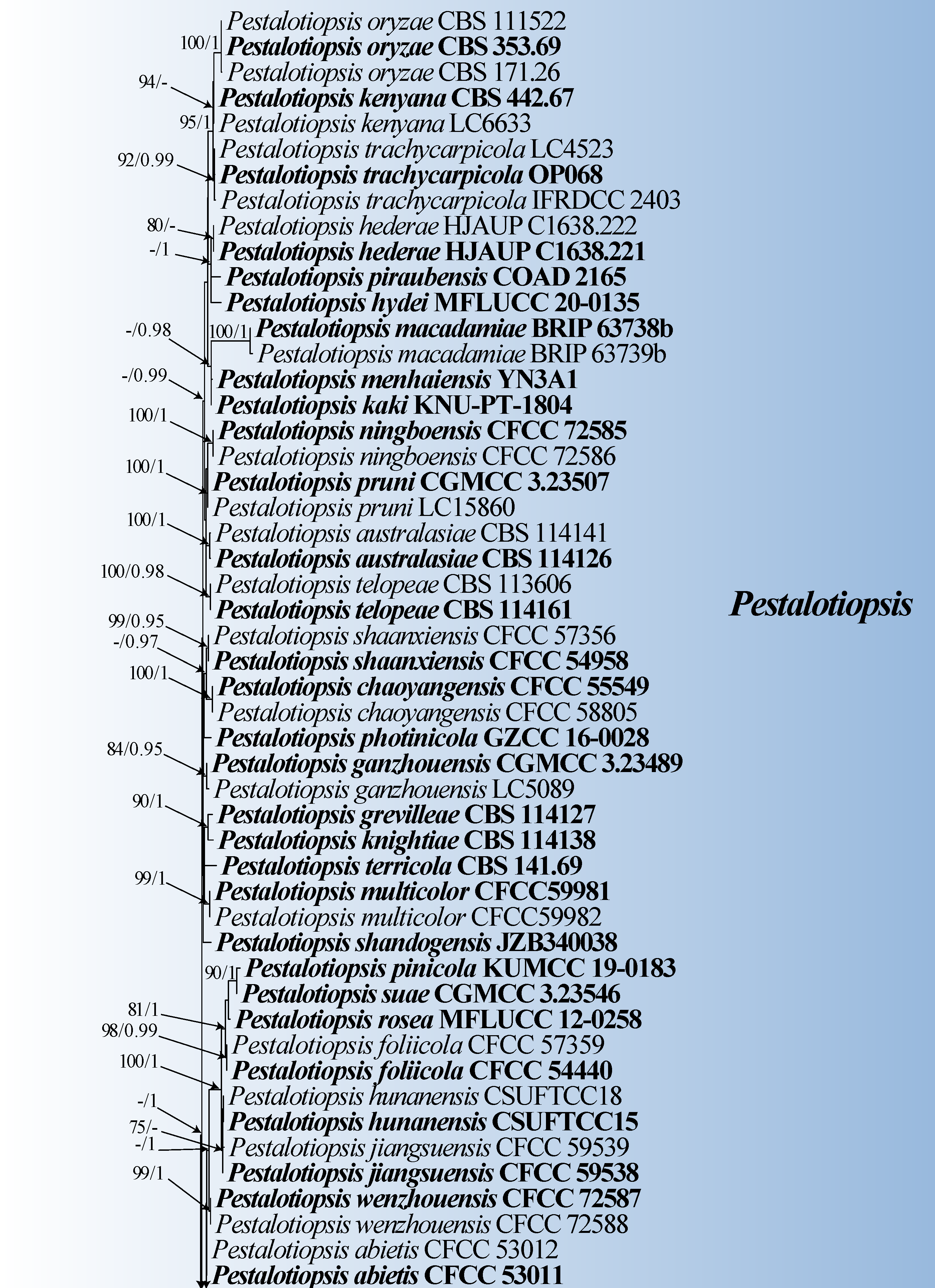
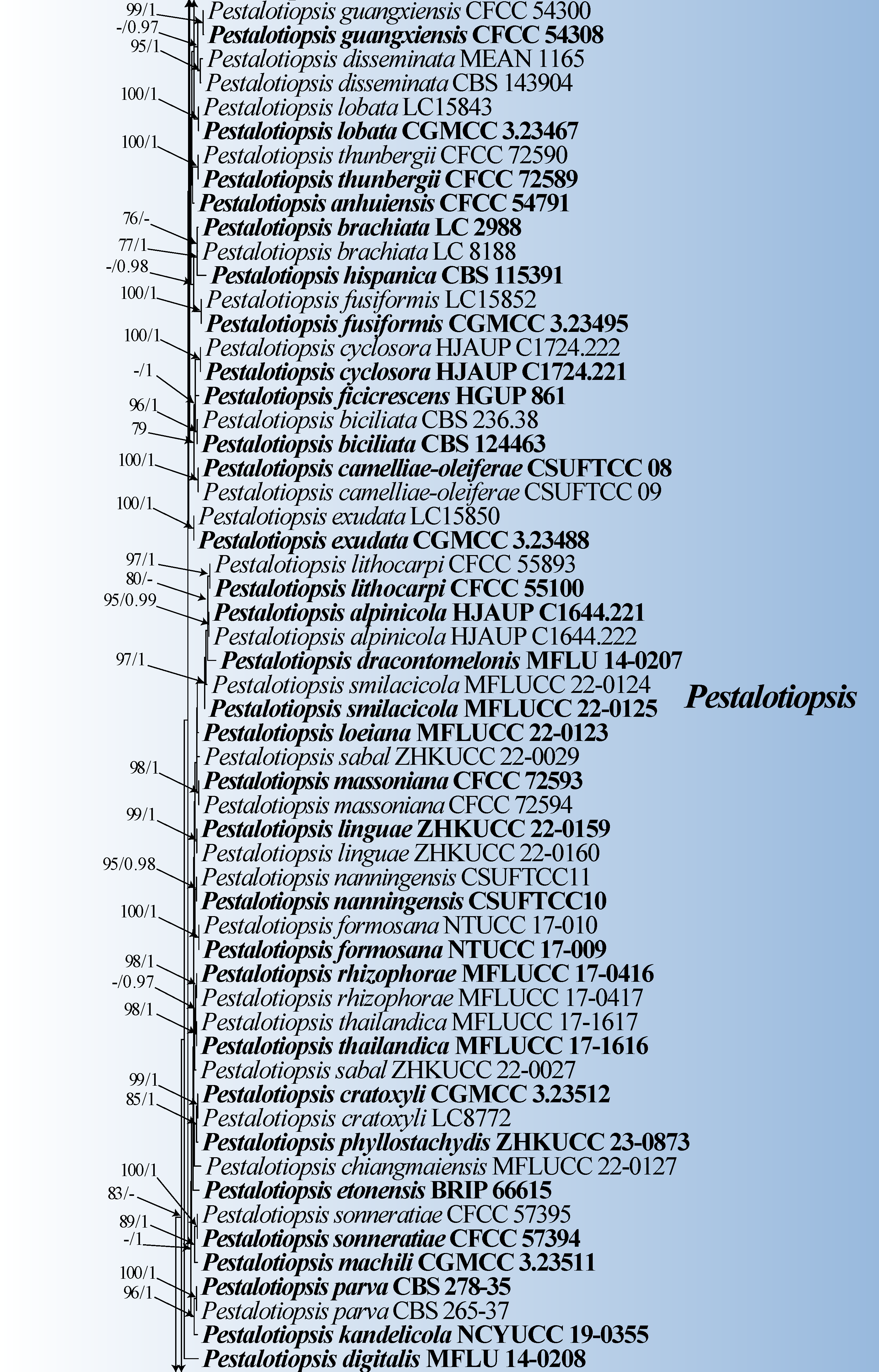
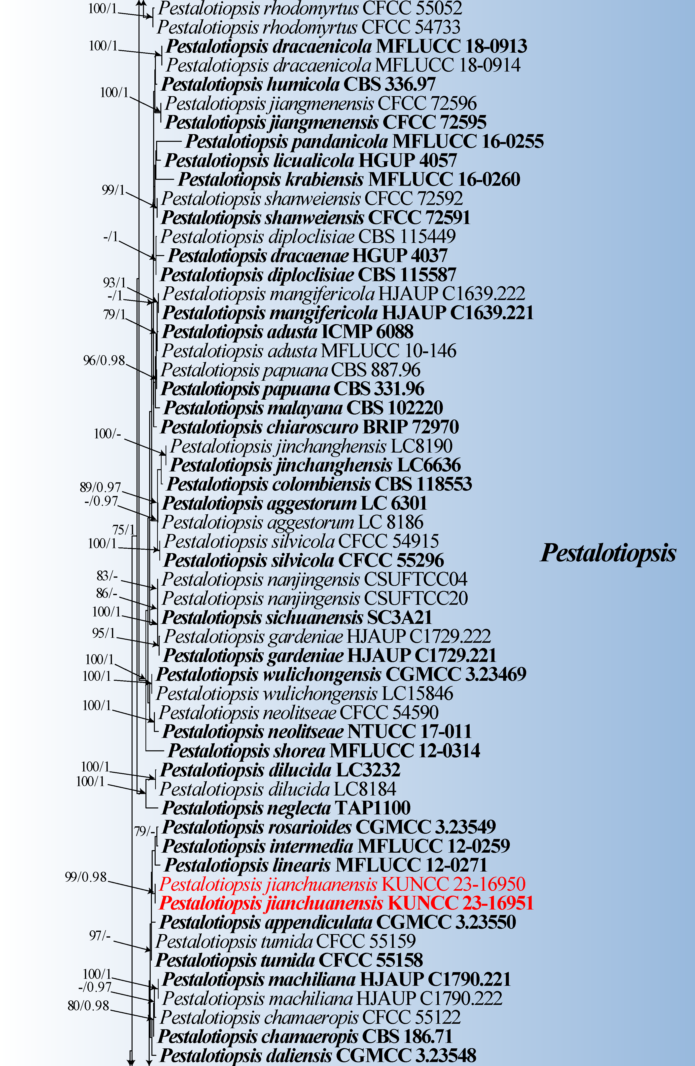
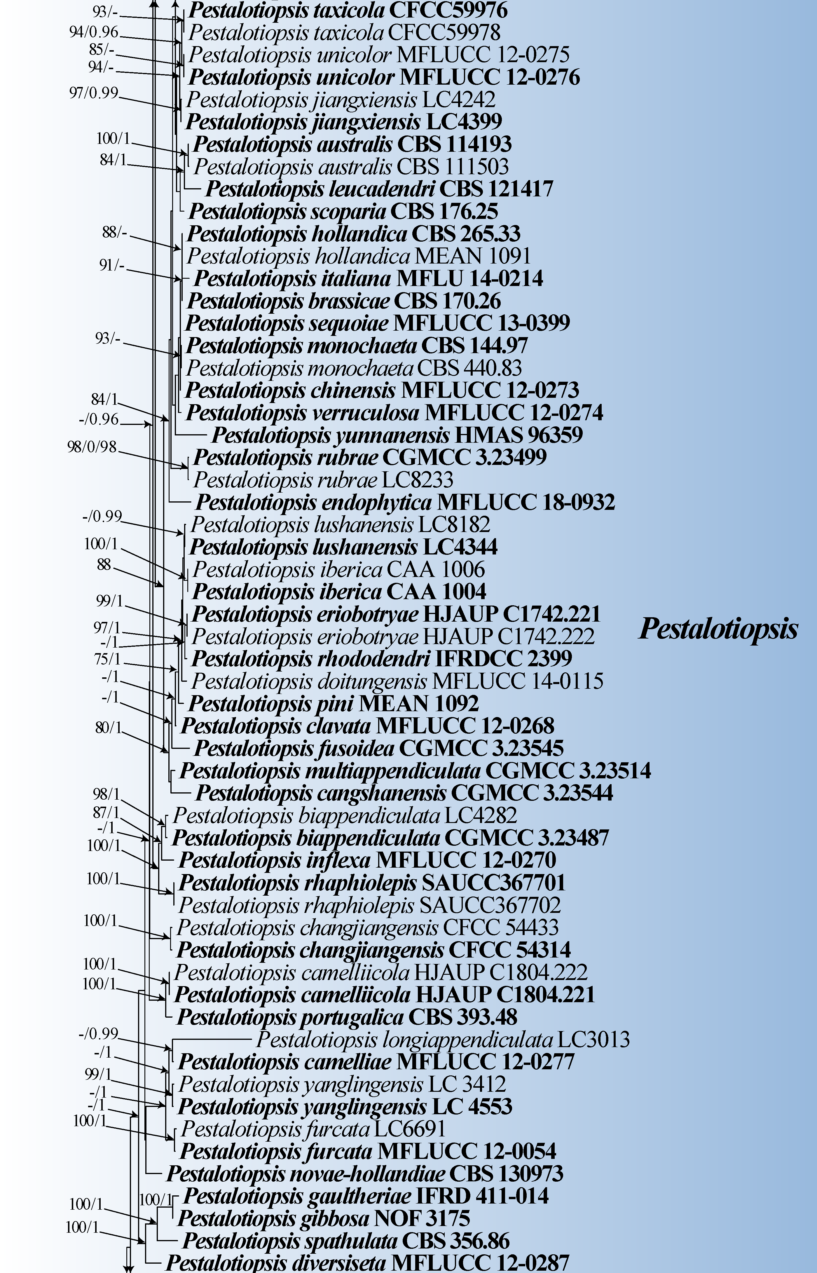
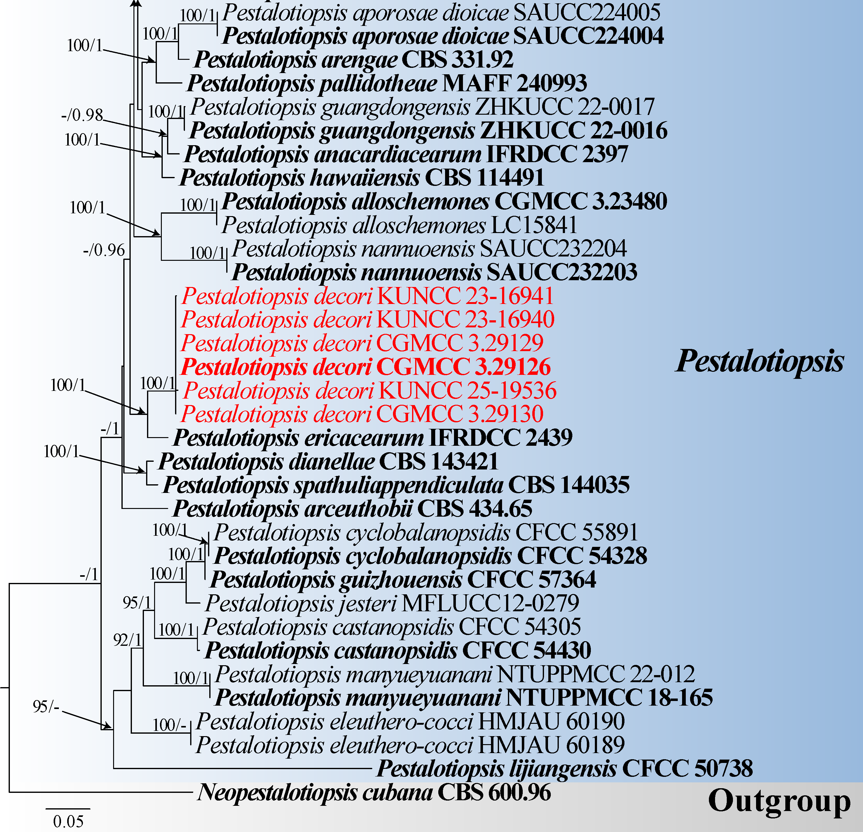
The phylogenetic topology of Pestalotiopsis was inferred from a combined dataset of ITS, tef1-α, and tub2 sequences, comprising 253 Pestalotiopsis isolates and one outgroup taxa (Neopestalotiopsis cubana CBS 600.96), with a total of 2,137 characters including gaps (ITS: 1–528 bp; tef1-α: 529–1452 bp; tub2: 1453–2137 bp). Phylogenetic analyses showed that the six newly obtained strains (CGMCC 3.29126, KUNCC 23-16940, KUNCC 23-16941, CGMCC 3.29129, CGMCC 3.29130 and KUNCC 25-19536) clustered with P. ericacearum (IFRDCC 2439) with 100% ML and 1.00 PP support (Fig. 1). In addition, two strains (KUNCC 23-16950 and KUNCC 23-16961) clustered with P. rosarioides (CGMCC 3.23549), P. intermedia (MFLUCC 12-0259), and P. linearis (MFLUCC 12-0271) to form an independent clade (Fig. 1).
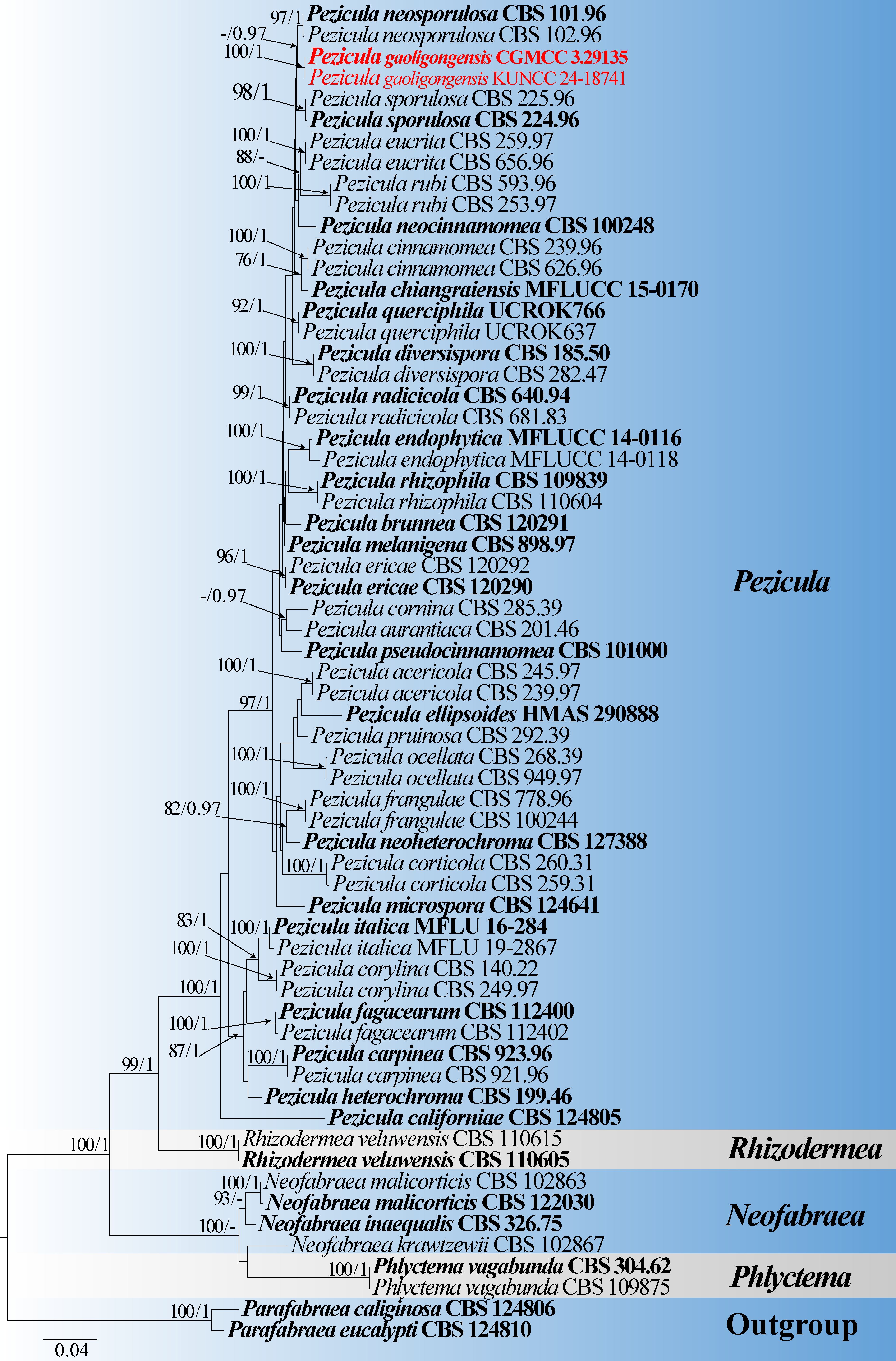
The phylogenetic topology of Pezicula was inferred from a combined dataset of ITS, LSU, and rpb2 sequences, comprising 53 Pezicula strains and two outgroup taxa (Parafabraea caliginosa CBS 124806 and P. eucalypti CBS 124810), with 2,340 characters including gaps (ITS: 1–547 bp; LSU: 548–1294 bp; rpb2: 1295–2340 bp). Phylogenetic analyses showed that our strains (CGMCC 3.29135 and KUNCC 24-18741) clustered with P. neosporulosa (CBS 101.96 and CBS 102.96) with 0.97 PP support (Fig. 2).
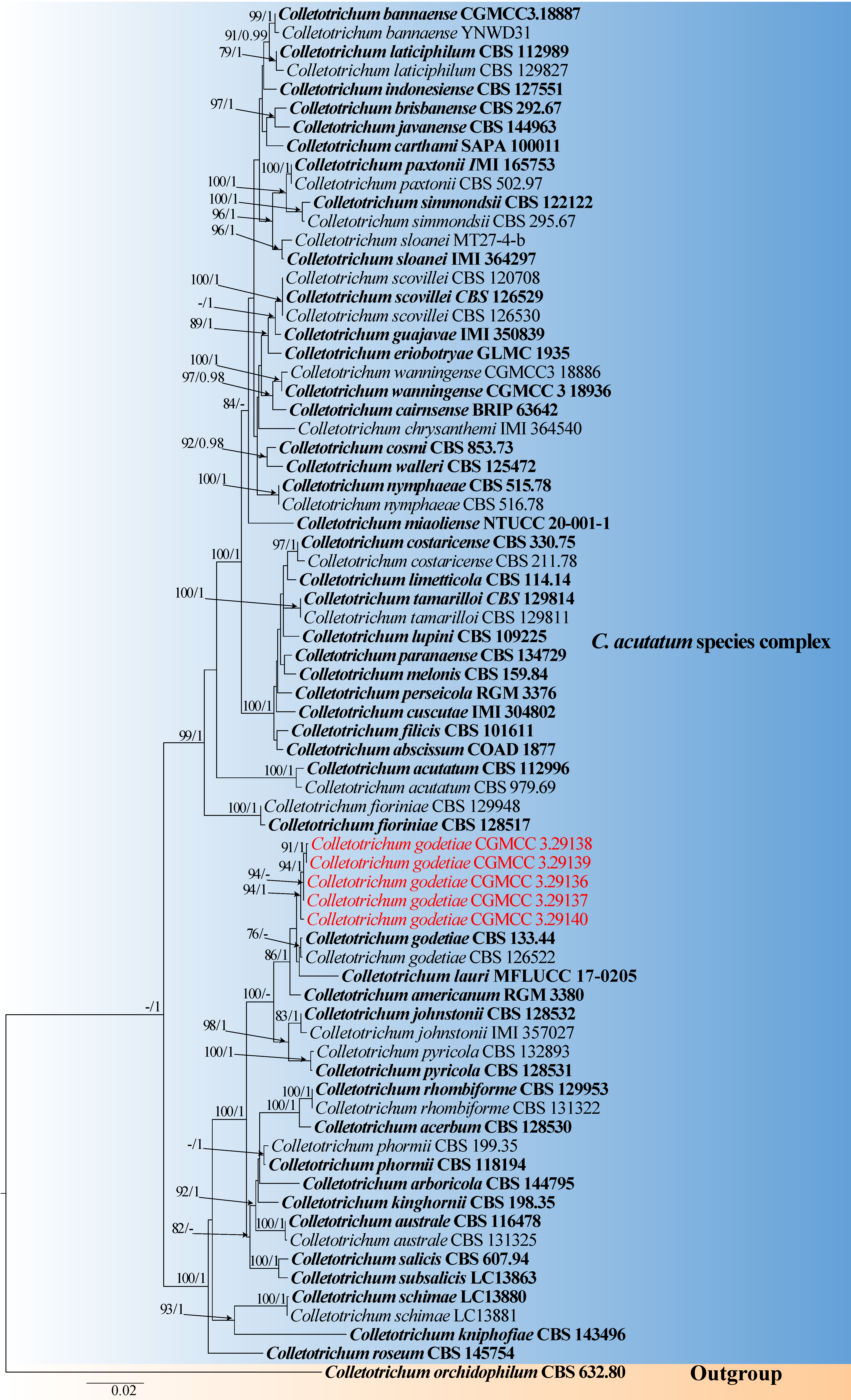
The phylogenetic topology of Colletotrichum was inferred from a combined dataset of ITS, act, chs1, gapdh, his3, and tub2 sequences, comprising 72 Colletotrichum strains and one outgroup taxa (Colletotrichum orchidophilum CBS 632.80), with 2,181 characters including gaps (ITS: 1–537 bp; act: 538–783 bp; chs1: 784–1063 bp; gapdh: 1064–1310 bp; his3: 1311–1692 bp; tub2: 1693–2181 bp). The RAxML and BI analyses of the combined dataset resulted in similar topologies. Phylogenetic analyses showed that the five newly obtained strains of Colletotrichum godetiae (CGMCC 3.29136, CGMCC 3.29137, CGMCC 3.29138, CGMCC 3.29139 and CGMCC 3.29140) clustered with its ex-type strain with 94% ML and 1.00 PP support and are located in the C. acutatum species complex (Fig. 3).
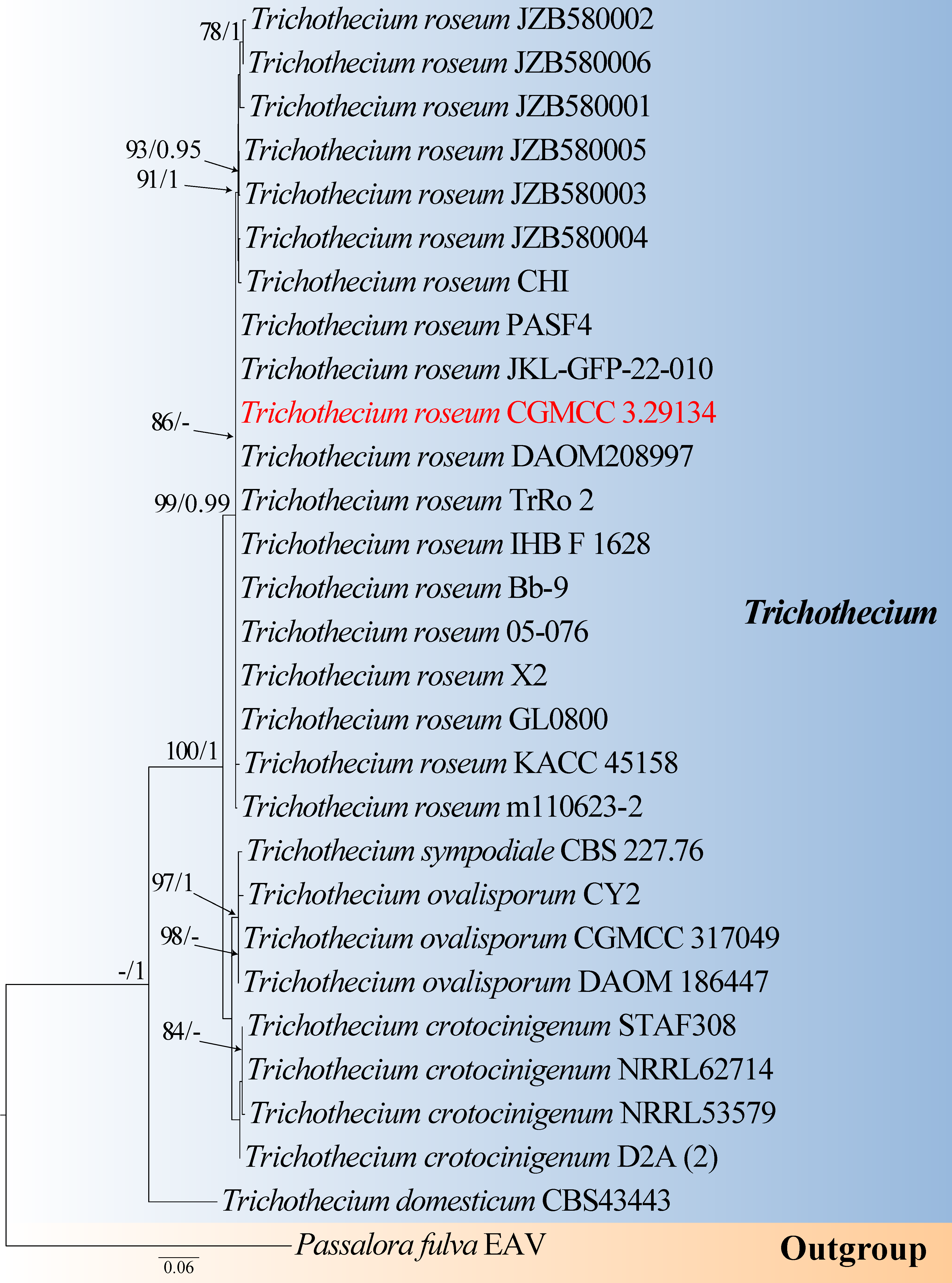
The phylogenetic topology of Trichothecium was inferred from a combined dataset of ITS and LSU sequences, comprising 28 Trichothecium strains and one outgroup taxa (Passalora fulva EAV), with 1,066 characters including gaps (ITS: 1–509 bp; LSU: 510–1066 bp). Phylogenetic analyses showed that our Trichothecium roseum strain (CGMCC 3.29134) clustered with its type strain with 86% ML support (Fig. 4).
Taxonomy
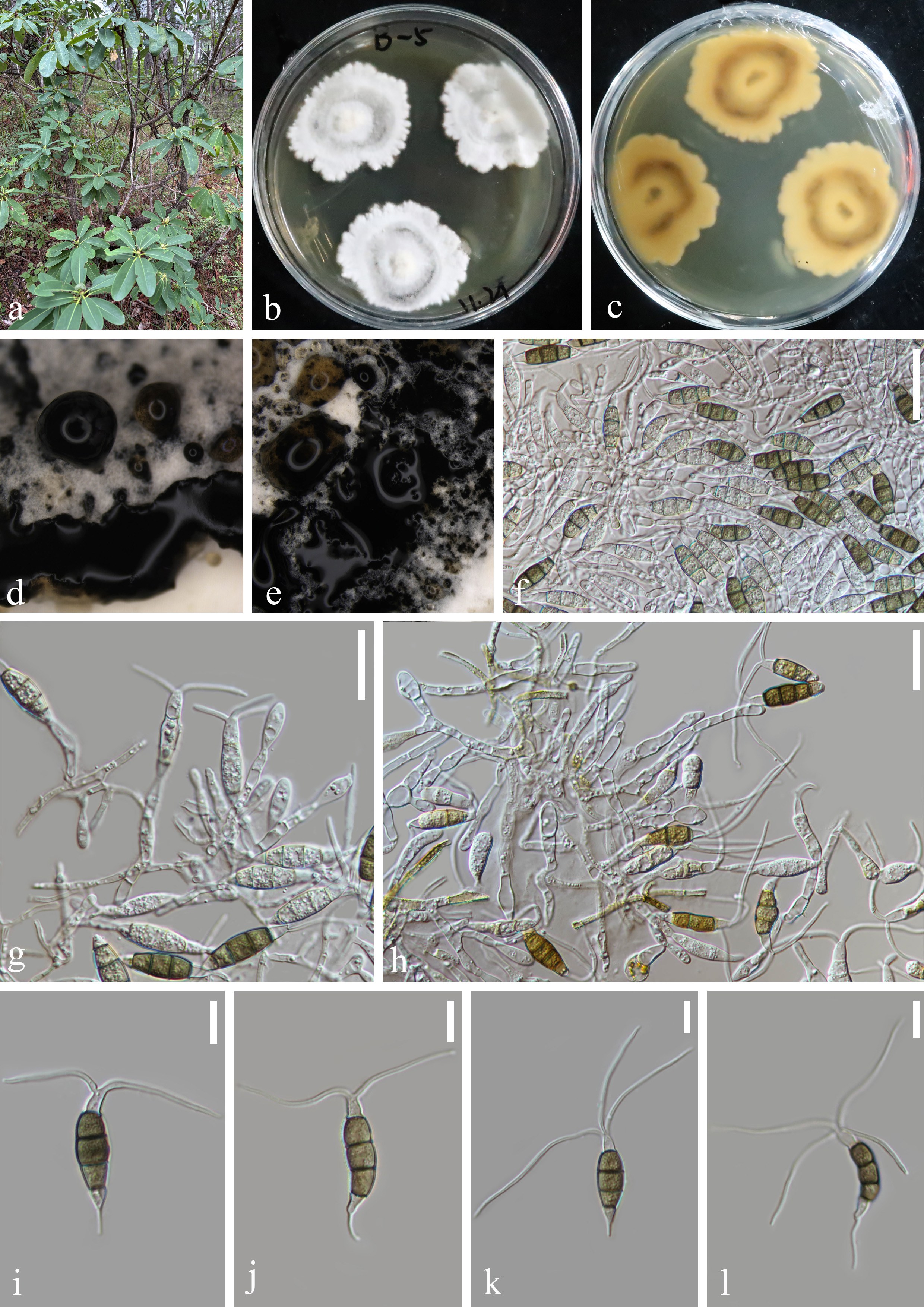
Pestalotiopsis decori Z.Y. Huang, H.W. Shen & Z.L. Luo, sp. nov., Figure 5.
Fungal Names number: FN 572994.
Holotype: —HKAS 136937
Etymology: — “decori” refers to the host plant Rhododendron decorum, which the fungus was isolated.
Endophytic in healthy Rhododendron decorum leaves. Sexual morph: Undetermined. Asexual morph: Conidiomata in culture sporodochial, saucer-shaped, scattered or gregarious, superficial to immersed, shining, releasing black conidial masses on the surface. Conidiophores branched, subcylindrical, hyaline to light brown, indistinct, often reduced to conidiogenous cells. Conidiogenous cells cylindrical or ampulliform, hyaline, smooth-walled, solitary to aggregated, (12–22 × 3.5–5.5 μm) ( = 17 × 4.5 μm, n = 30). Conidia 23–29 × 7–9 µm (= 26 × 8 µm, n = 30), fusoid, ellipsoid, rosary, straight to slightly curved, four-septate; basal cell conic with a truncated base, hyaline or light brown, and thin-walled, 4–6 µm long ( = 5 µm, n = 30); three-median cells dark,17–19 µm long ( = 18 µm, n = 30), smooth wall, concolourous, septa darker than the rest of the cells (second cell from the base pale brown and enlarged, 5–6 µm long; third cell 5–7 µm long; fourth cell expands to 6–7 µm long); apical cell 5–7 µm long ( = 6 µm, n = 30), hyaline, subcylindrical, thin and smooth-walled, with 2–4 tubular apical appendages 30–52 µm long ( = 41 µm, n = 30) arising from the apical crest, unbranched, filiform; basal appendage
8–20 µm long ( = 14 µm, n = 30), single, tubular, unbranched, centric, straight, or slightly curved.
Culture characteristics: —Colonies on PDA reaching 30–40 mm diameter after 14 days at 25 ℃ in darkness. The surface of the colony was white, the aerial hyphae were flocculent, the back was light orange, and the edges were irregular. Black conidiomata are formed after 14 days on PDA.
Material examined: —China, Yunnan Province, Dali City, Jianchuan County, 26°31′53.02″ N, 99°54′15.23″ E (2713 m), isolated from healthy leaves of R. decorum, October 2023, Z.Q. Zhang, B-5 (HKAS 136937, holotype), ex-holotype living culture, CGMCC 3.29126 = KUNCC 23-16938; ibid., isolated from healthy leaves of R. decorum, October 2023, Z.Q. Zhang, B-7 (HKAS 136938), living culture, KUNCC 23-16940; ibid., isolated from healthy leaves of R. decorum, October 2023, Z.Q. Zhang, B-8 (HKAS 136939), living culture, KUNCC 23-16941; ibid., isolated from healthy leaves of R. decorum, October 2023, Z.Q. Zhang, B-32 (HKAS 136931), living culture, CGMCC 3.29129 = KUNCC 23-16959; ibid., isolated from healthy leaves of R. decorum, October 2023, Z.Q. Zhang, B-33 (HKAS 136936), living culture, CGMCC 3.29130 = KUNCC 23-16960; China, Yunnan Province, Nujiang City, Gongshan County, 28°01′08.22″ N, 98°36′22.80″ E (1794 m), isolated from healthy leaves of R. decorum, September 2024, Z.Y. Huang, B-766 (HKAS 148956, paratype), ex-paratype living culture, KUNCC 25-19536.
Notes: —Six newly obtained strains (CGMCC 3.29126, KUNCC 23-16940, KUNCC 23-16941, CGMCC 3.29129, CGMCC 3.29130, and KUNCC 25-19536) of Pestalotiopsis decori clustered with P. ericacearum (IFRDCC 2439) and formed a well-supported sister clade (100% ML/1.00 PP, Fig. 1). Sequence comparison revealed 57 nucleotide differences between CGMCC 3.29126 and P. ericacearum including 9 bp differences in ITS (523 bp), 40 bp differences in tef1-α (450 bp), and 8 bp differences in tub2 (417 bp). Morphologically, Pestalotiopsis decori (HKAS 136937) differs from P. ericacearum by its larger conidia (23–29 × 7–9 μm vs. 16–20 × 5–9 μm), longer apical appendages (30–52 μm vs. 20–43 μm) and basal appendages (8–20 μm vs. 2–9 μm) (Zhang et al. 2013). In addition, P. decori possesses 2–4 apical appendages, whereas P. ericacearum bears 3–4 (Zhang et al. 2013). Notably, both species were isolated from the leaves of Rhododendron species, however, P. ericacearum was obtained from R. delavayi, whereas our collections were recovered from R. decorum.
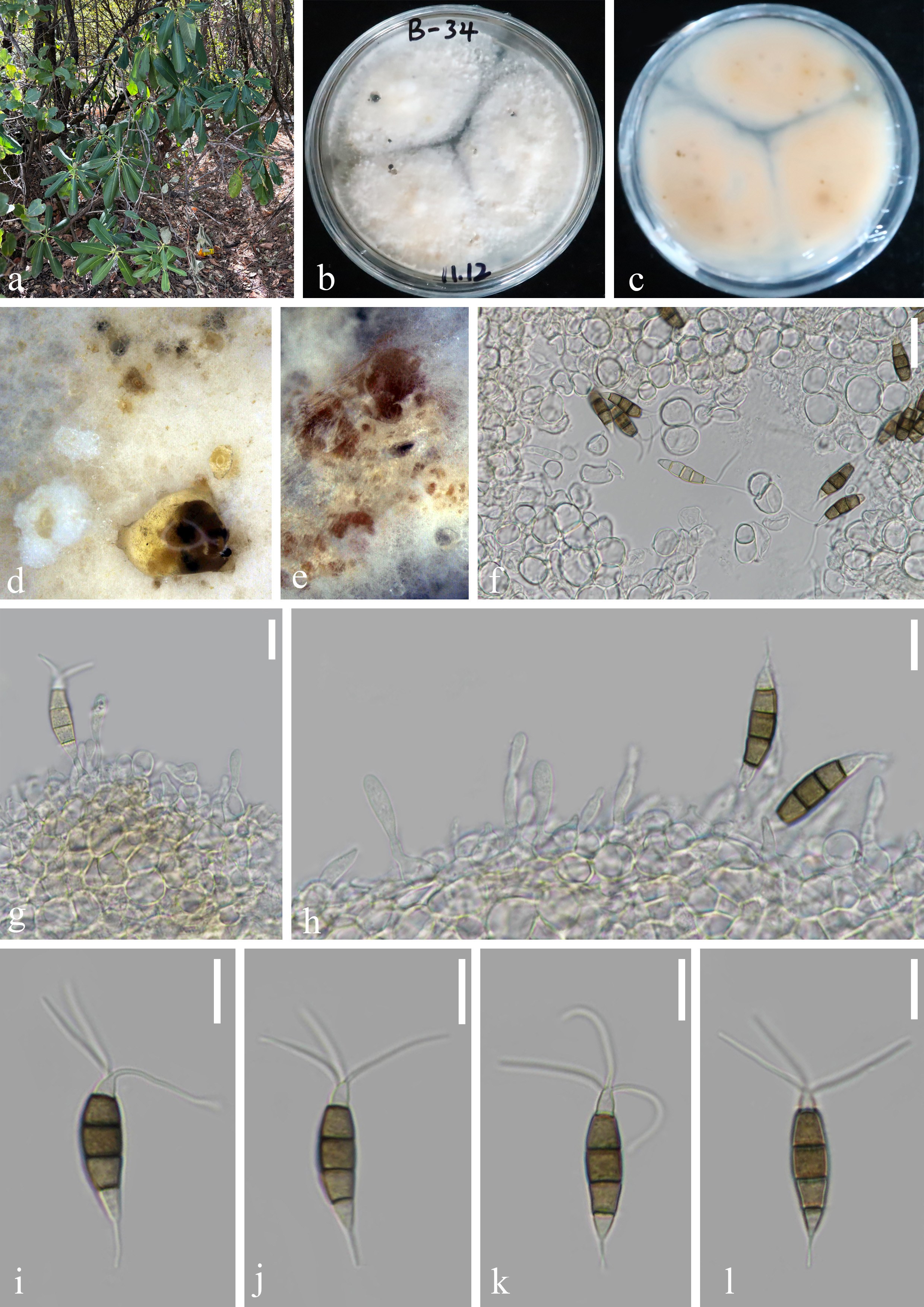
Pestalotiopsis jianchuanensis Z.Y. Huang, H.W. Shen & Z.L. Luo, sp. nov., Figure 6.
Fungal Names number: FN 572995.
Holotype: —HKAS 148954
Etymology: — “jianchuanensis” refers to Jianchuan County, Yunnan Province, China, where the holotype was collected.
Endophytic in healthy Rhododendron decorum leaves. Sexual morph: Undetermined. Asexual morph: Conidiomata (on PDA) pycnidial, globose to clavate, solitary, exuding globose, dark-brown to black conidial masses. Conidiophores indistinct and typically reduced to conidiogenous cells. Conidiogenous cells cylindrical or ampulliform, hyaline, smooth-walled, solitary to aggregated, (8–14 × 4–12 μm) ( = 11 × 8 μm, n = 30). Conidia 22–27 × 6–8 µm ( = 23 × 7 µm, n = 30), fusoid, ellipsoid, rosary, straight to slightly curved,four-septate; basal cell conic with a truncated base, hyaline or light brown, and thin-walled, 4–6 µm long ( = 5 µm, n = 30); three-median cells dark, 14–18 µm long ( = 16 µm, n = 30), smooth wall, concolourous, septa darker than the rest of the cells (second cell from the base pale brown and enlarged, 4–5 µm long; third cell 5–6 µm long; fourth cell expands to 5–6 µm long); apical cell 3–5 µm long ( = 4 µm, n = 30), hyaline, subcylindrical, thin-walled, and smooth-walled, with 2–3 tubular apical appendages 12–24 µm long ( = 18 µm, n = 30) arising from the apical crest, unbranched, filiform; basal appendage 4–8 µm long ( = 6 µm, n = 30), single, tubular, unbranched, centric, straight, or slightly curved.
Culture characteristics: —Colonies on PDA reaching 40–60 mm diameter after 14 days at 25 ℃ in darkness. The surface of the colony was white, the aerial hyphae were flocculent, the back was light orange, and the edges were regular. Black conidiomata are formed after 14 days on PDA.
Material examined: —China, Yunnan Province, Dali City, Jianchuan County, 26°31′53.02″ N, 99°54′15.23″ E (2713 m), isolated from healthy leaves of R. decorum, October 2023, Z.Q. Zhang, B-34 (HKAS 148954, holotype), ex-holotype living culture, KUNCC 23-16961; ibid., isolated from healthy leaves of R. decorum, October 2023, Z.Q. Zhang, B-18 (HKAS 148959), living culture, KUNCC 23-16950.
Notes: —In the phylogenetic analyses, Pestalotiopsis jianchuanensis sp. nov. is closely related to P. intermedia, P. linearis, and P. rosarioides (Fig. 1). Sequence comparisons between P. jianchuanensis (KUNCC 23-16961) and related taxa revealed notable nucleotide differences: 19 bp in tub2 (342 bp) relative to P. linearis (MFLUCC 12-0271); 1 bp, 3 bp, and 8 bp differences in ITS (523 bp), tef-1α (549 bp), and tub2 (362 bp), respectively, relative to P. intermedia (MFLUCC 12-0259); and 4 bp and 3 bp differences in tef1-α (406 bp) and tub2 (324 bp), respectively, relative to P. rosarioides (CGMCC 3.23549). Morphologically, P. jianchuanensis can be distinguished from P. intermedia and P. linearis by its conidial size and appendages characteristics (Table 2). In comparison with P. rosarioides, which was also isolated from R. decorum, P. jianchuanensis can be distinguished by its distinctly longer apical appendages (12–24 μm vs. 5–9 μm), longer basal appendages (4–8 μm vs. 4–5 μm), and shorter apical cells (3–5 μm vs. 4–7 μm) (Gu et al. 2022).
Pezicula gaoligongensis Z.Y. Huang, H.W. Shen & Z.L. Luo, sp. nov., Figure 7.
Fungal Names number: FN 572996.
Holotype: —HKAS 148950
Etymology: — “gaoligongensis” refers to the Gaoligong Mountain, where the holotype was collected.
Endophytic in healthy Rhododendron decorum leaves. Sexual morph: Undetermined. Asexual morph: Conidiomata erumpent on surface, aggregated or scattered, with brown to black conidial masses, globose to irregular. Conidiophores hyaline, cylindrical, straight to flexuous, septate, unbranched or branched, smooth-walled, reduced to conidiogenous cells. Conidiogenous cells hyaline, enteroblastic, integrated, smooth-walled. Conidia 31–38 × 7–9 µm ( = 33 × 8 µm, n = 30), hyaline when young, becoming to pale yellow and eventually to orange-brown with age, elongated ellipsoid to cylindrical, straight or slightly curved, rounded at the apex, narrow and slightly truncate at the base, and with a protruding scar, 2–4-septate, with septa becoming inconspicuous at maturity, granular to guttulate, thin-walled.
Culture characteristics: —Colonies on PDA reaching 40–60 mm diameter after 14 days at 25 ℃ in darkness. The surface of the colony was white to pale brown, the aerial hyphae were flocculent, the back was brown to white, and the edges were irregular. Brown to black conidiomata developed on PDA after 14 days.
| Species | Conidia | Apical cell | Apical appendage | Basal appendage | Host | Country | References |
| P. jianchuanensis | 22–27 × 6–8 µm | 3–5 µm | 2–3 tubular apical appendages, 12–24 µm | single basal appendage, 4–8 µm | Rhododendron decorum | China | This study |
| P. intermedia | 24–28 × 5.5–6.5 μm | 4–5 μm | 2–3 (rarely 4) tubular apical appendages, 10–28 μm | single basal appendage, 6–10 μm | dead leaf of tree | China | Maharachchikumbura et al. 2012 |
| P. linearis | 24–33×4.7–6 μm | 4–5 μm | 2–3 (rarely 1) tubular apical appendages, 10–20 μm | single (rarely 2) basal appendages, 4–7 µm | Trachelospermum sp. | China | Maharachchikumbura et al. 2012 |
| P. rosarioides | 22–25 × 6–7 µm | 4–7 μm | 1–3 tubular apical appendages, 5–9 µm | single basal appendage, 4–5 µm | Rhododendron decorum | China | Gu et al. 2022 |
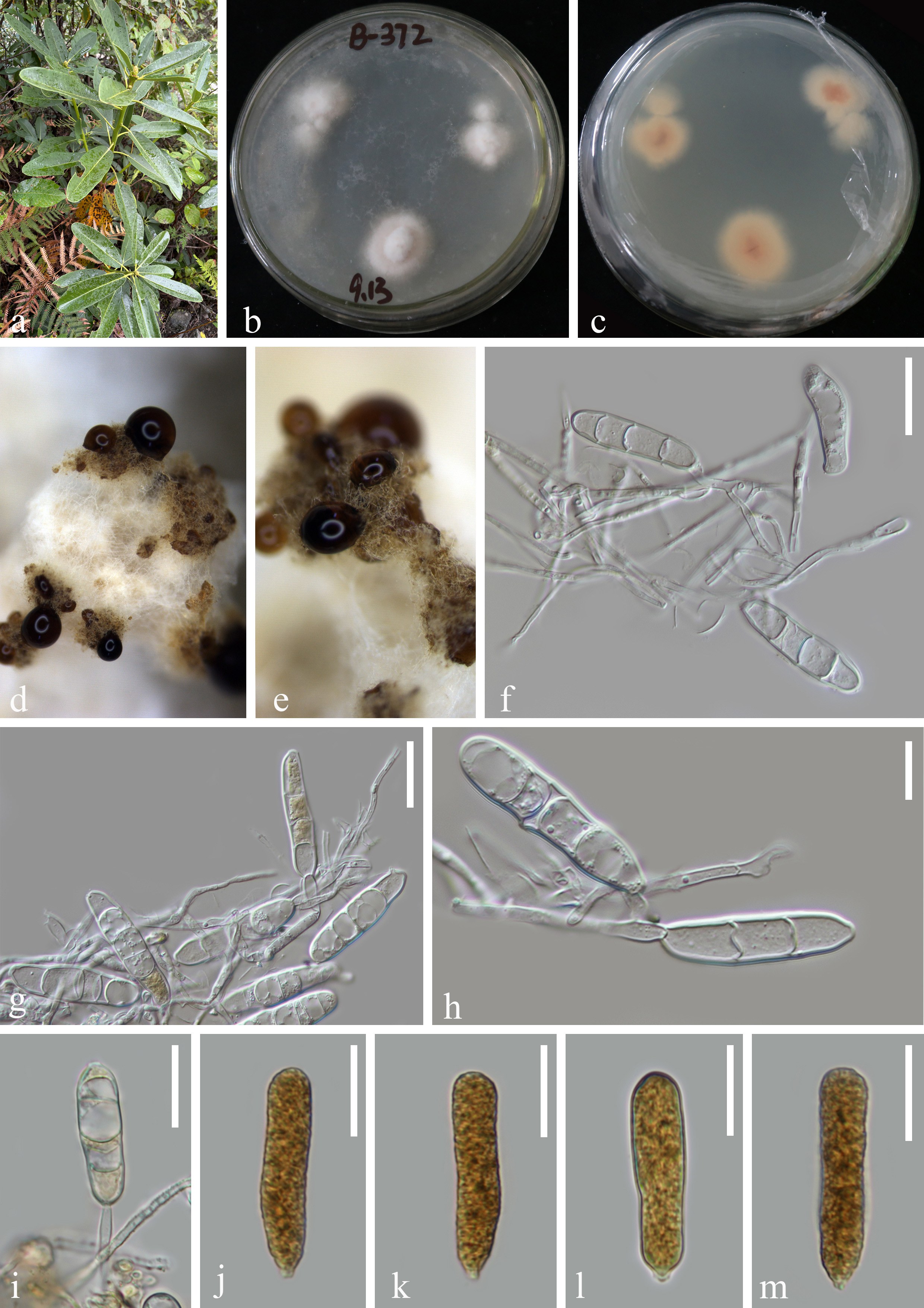
Material examined: —China, Yunnan Province, Nujiang City, Gaoligong Mountain, 25°58′2.95″ N, 98°44′42.03″ E (2792 m), isolated from healthy leaves of R. decorum, April 2024, R.Z. Ji, B-372 (HKAS 148950, holotype), ex-holotype living culture, CGMCC 3.29135 = KUNCC 24-18742; ibid., isolated from healthy leaves of R. decorum, April 2024, R.Z. Ji, B-371 (HKAS 148952), living culture, KUNCC 24-18741.
Notes: —Our two strains of Pezicula gaoligongensis (CGMCC 3.29135 and KUNCC 24-18741) clustered together and formed a sister clade to P. neosporulosa (CBS 101.96), with 0.97 PP support (Fig. 2). Sequence comparisons revealed 18 bp differences between P. gaoligongensis (CGMCC 3.29135) and P. neosporulosa (CBS 101.96), including 4 bp in ITS (506 bp), 1 bp in LSU (523 bp), and 13 bp in rpb2 (875 bp). Morphologically, P. gaoligongensis differs from P. neosporulosa by its longer but narrower conidia (31–38 × 7–9 μm vs. 20–36.5 × 9–12 μm), and its mature conidia become orange-brown, whereas those of P. neosporulosa remain hyaline (Yuan & Verkley 2015). In addition, P. neosporulosa produces both macroconidia and microconidia (Yuan & Verkley 2015), whereas this characteristic was not observed in P. gaoligongensis (CGMCC 3.29135). Notably, P. gaoligongensis was isolated as an endophyte from healthy leaves of R. decorum, while P. neosporulosa was obtained from Abies alba (Yuan & Verkley 2015).
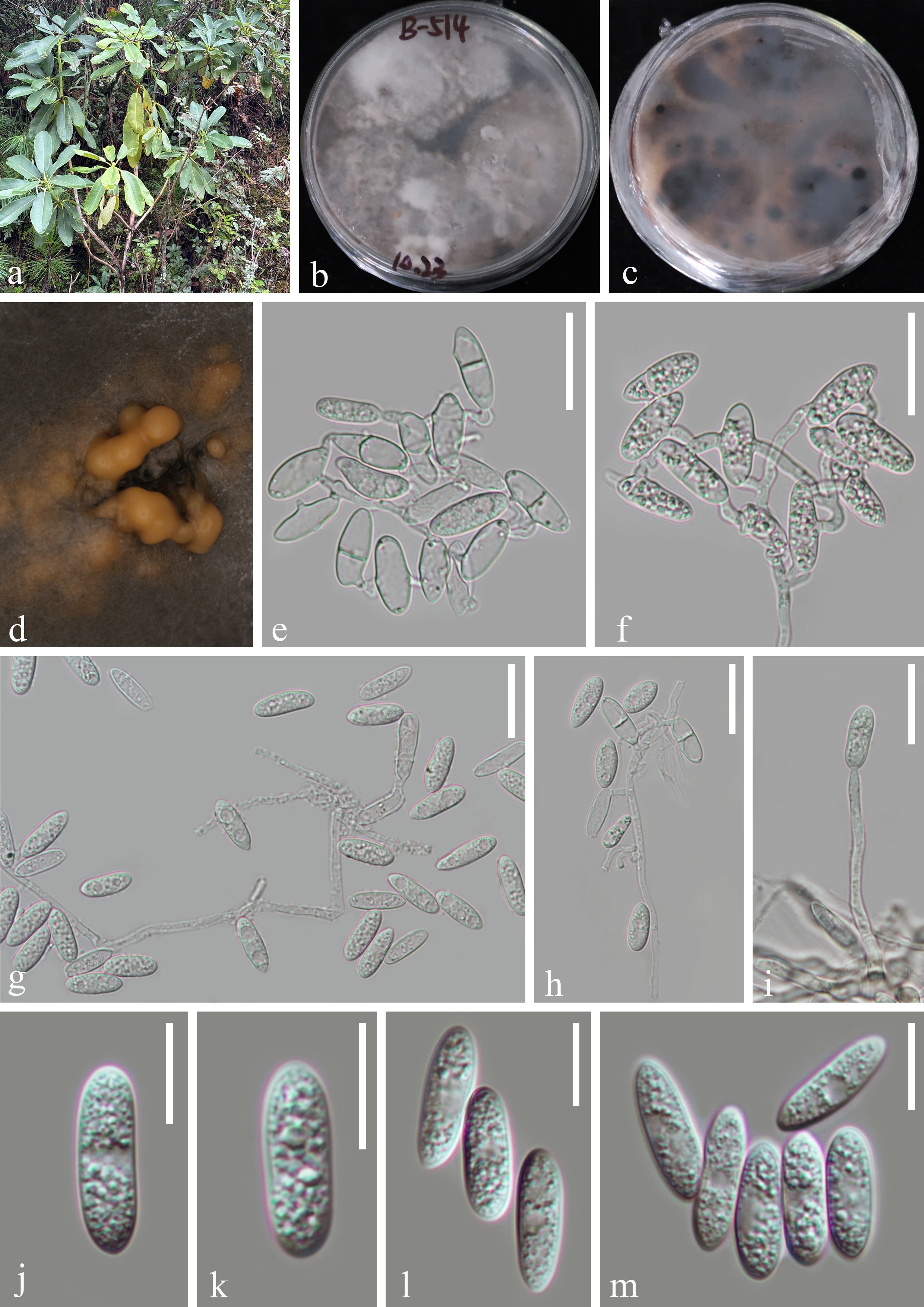
Colletotrichum godetiae Neerg., Friesia 4(1-2): 72 (1950), Figure 8.
Endophytic in healthy Rhododendron decorum leaves. Sexual morph: Undetermined. Asexual morph: Conidiomata acervular, gregarious, dark brown. Appressoria and Setae not observed on PDA. Conidiophores hyaline, unbranched, approximately cylindrical. Conidiogenous cells hyaline, smooth-walled, cylindrical. Conidia (15–17 × 4–6 μm) ( = 16 × 5 μm, n = 30), hyaline, elongated ellipsoid, straight, aseptate, obtuse at the base, rounded at the apex, smooth-walled, guttulate.
Culture characteristics: —Colonies on PDA reaching 40–70 mm diameter after 14 days at 25 ℃ in darkness. The surface of the colony was white or gray, the aerial hyphae were flocculent, the back was dark grey to greyish pink, and the edges were irregular. Dark brown conidiomata are formed after 14 days on PDA.
Material examined: —China, Yunnan Province, Lijiang City, Yulong Snow Mountain, 26°57′21.75″ N, 100°10′59.75″ E (3037 m), isolated from healthy leaves of R. decorum, September 2024, Z.Y. Huang, B-514 (HKAS 148951), living culture, CGMCC 3.29136 = KUNCC25-19544; ibid., isolated from healthy leaves of R. decorum, September 2024, Z.Y. Huang, B-520 (HKAS 148953), living culture, CGMCC 3.29137 = KUNCC 25-19550; ibid., isolated from healthy leaves of R. decorum, September 2024, Z.Y. Huang, B-521 (HKAS 148955), living culture, CGMCC 3.29138 = KUNCC 25-19551; ibid., isolated from healthy leaves of R. decorum, September 2024, Z.Y. Huang, B-522 (HKAS 148957), living culture, CGMCC 3.29139 = KUNCC 25-19552; China, Yunnan Province, Nujiang City Gongshan County, 28°01′08.22″ N, 98°36′22.80″ E (1794 m), isolated from healthy leaves of R. decorum, September 2024, Z.Y. Huang, B-797 (HKAS 148958), living culture, CGMCC 3.29140 = KUNCC 25-19537.
Notes: —Our five strains (CGMCC 3.29136, CGMCC 3.29137, CGMCC 3.29138, CGMCC 3.29139, and CGMCC 3.29140) clustered within the same clade as Colletotrichum godetiae and C. lauri in the C. acutatum species complex (Fig. 3). Sequence comparisons of CGMCC 3.29136 with C. godetiae (CBS 133.44) across the ITS, act, chs1, gapdh, his3, and tub2 regions revealed only minor nucleotide differences (2 bp in act, 2 bp in gapdh, and 1 bp in tub2), whereas comparisons with C. lauri (MFLUCC 17-0205) showed substantially greater sequence divergence (1 bp in ITS, 4 bp in act, 1 bp in gapdh, 6 bp in chs1, and 10 bp in tub2). In contrast, the morphological characteristics of our isolates are consistent with those of C. godetiae (CBS 133.44) (Damm et al. 2012; Hosking et al. 2024). Consequently, we temporarily identified the five strains as C. godetiae. This study represents the first report of C. godetiae isolated from Rhododendron decorum.
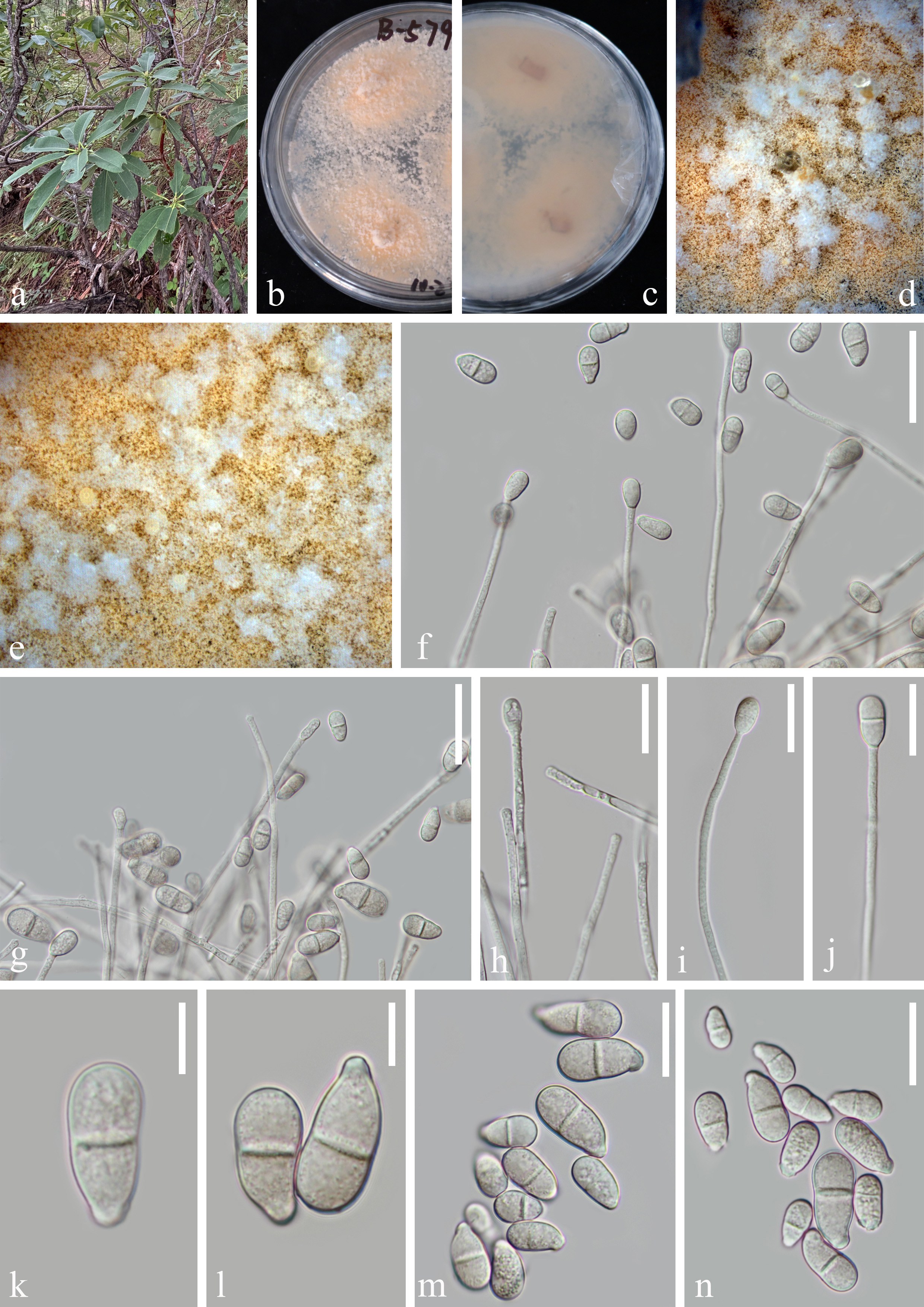
Trichothecium roseum (Pers.) Link, Mag. Gesell. Naturf. Freunde, Berlin 3(1-2): 18 (1809). Figure 9.
Endophytic in healthy Rhododendron decorum leaves. Sexual morph: Undetermined. Asexual morph: Colonies on PDA, superficial, effuse, gregarious, hyaline. Conidiophores (124–198 × 2.5–3 μm) ( = 161 × 3 μm, n = 15), micronematous, mononematous, cylindrical, septate, hyaline, smooth. Conidiogenous cells arising directly from mycelia, monoblastic, cylindrical, hyaline to pale brown. Conidia (17–21 × 8–10 μm) ( = 19 × 9 μm, n = 30), ellipsoidal to pyriform, hyaline, with an obliquely prominent truncate basal scar, 2-celled, constricted at the septum, smooth, thick-walled.
Culture characteristics: —Colonies on PDA reaching 40–70 mm diameter after 14 days at 25 ℃ in darkness. The surface of the colony was pale pinkish-brown, abundant aerial mycelium, grayish pinkish-brown on the reverse. Pale yellow conidiomata are formed after 14 days on PDA.
Material examined: —China, Yunnan Province, Diqing City, Weixi County, 27°00′45.96″ N, 99°21′35.24″ E (2475 m), isolated from healthy leaves of R. decorum, September 2024, Z.Y. Huang, B-579 (HKAS 148949), living culture, CGMCC 3.29134 = KUNCC25-19535.
Notes: —Phylogenetic analyses showed that our isolate clustered together with Trichothecium roseum (Fig. 4). Morphologically, the isolate closely resembles T. roseum (JKL-GFP-22-010) characterized by hyaline, septate, branched conidiophores, and 2-celled, ellipsoidal to pyriform conidia with an obliquely prominent truncate basal scar (Monika & Benjarong 2023). JKL-GFP-22-010 was selected for sequence comparison as it represents a well-documented reference strain of T. roseum in a recent taxonomic study (Monika & Benjarong 2023). The ITS and LSU sequences of our strain show 100% similarity with JKL-GFP-22-010.
Discussion
High-altitude plants such as Rhododendron decorum are known to harbor a rich diversity of endophytic fungi (Nair & Padmavathy 2014; Rojas-Jiménez et al. 2016; Huang et al. 2024). However, the isolation and morphological identification of these fungi remain particularly challenging due to their inherently slow growth, low sporulation rates, and frequent contamination by epiphytic microorganisms. Following the methods of Tao et al. (2013) and Gu et al. (2022), the effectiveness of surface sterilization was verified by plating aliquots of the final rinse water onto potato dextrose agar (PDA) as negative controls. To minimize contamination by epiphytic microorganisms, leaf discs were subsequently inoculated onto PDA plates supplemented with antibiotics. These results demonstrate that appropriate surface sterilization and the use of antibiotic-supplemented media are effective in reducing contamination during the isolation of endophytic fungi.
Species delimitation of endophytic fungi presents additional challenges, particularly in genera such as Pestalotiopsis, Pezicula, and Colletotrichum, which often exhibit highly similar morphological characteristics, making identification based solely on traditional morphology difficult (Damm et al. 2012; Maharachchikumbura et al. 2014; Bao et al. 2025). In this study, we applied integrative taxonomy by combining detailed morphological observations with multi-locus phylogenetic analyses (Bao et al. 2025), achieving substantial progress in resolving species boundaries. We successfully identified two novel Pestalotiopsis species (P. decori and P. jianchuanensis), one new Pezicula species (P. gaoligongensis), and reported C. godetiae from R. decorum for the first time. These findings highlight the remarkable potential of high-altitude plants as valuable reservoirs of fungal diversity. Overall, this study not only reveals the diversity of foliar endophytic fungi associated with R. decorum, but also emphasizes the complexity and necessity of microbial resource surveys in extreme ecological environments. Such documentation not only enriches our understanding of fungal taxonomy and systematics but also lays the groundwork for future investigations into plant, microbe interactions and the potential exploitation of these fungi in biotechnology.
Conclusions
Based on multigene phylogenetic analyses and morphological characteristics, five endophytic fungal species associated with Rhododendron decorum were identified, including three new species (Pestalotiopsis decori, P. jianchuanensis, and Pezicul gaoligongensis), one new host record (Colletotrichum godetiae), and one known species (Trichothecium roseum). While the number of species documented in this study is limited, these findings provide preliminary insights into the diversity of foliar endophytic fungi associated with R. decorum in high-altitude ecosystems. Further studies with broader sampling are warranted to comprehensively assess species diversity, elucidate ecological functions, and explore the potential roles of these fungi in plant–microbe interactions and high-altitude ecosystem processes.
Acknowledgments
We thank the High-Level Talents Program of Yunnan Province (YNQR-QNRC-2020-113) for the financial support. Zi-Yang Huang thanks Zheng-Quan Zhang, Wen-Peng Wang, and Kai-Yang Niu for sharing morphological and phylogenetic knowledge. Sha Luan, Liang Zhang, and Ying Wang are acknowledged for their help in DNA extraction and PCR amplification. Rong-Ze Ji and Li-Heng Mu are also acknowledged for providing valuable support during sample collection.
Author contributions
Conceptualization, Huang ZY and Luo ZL; methodology, Huang ZY and Shen HW; formal analysis, Huang ZY; resources, Luo ZL; writing—original draft preparation, Huang ZY; writing—review and editing, Shen HW, Huang ZY and Luo ZL; supervision, Luo ZL; funding acquisition, Luo ZL. All authors have read and agreed to the published version of the manuscript.
ORCID
Zi-Yang Huang: https://orcid.org/0009-0007-1284-3995
Zong-Long Luo: https://orcid.org/0000-0001-7307-4885
Conflict of interest statement
The author list includes members of the Editorial Board of PhytoMycology. They were not involved in the journal’s review of, or decisions related to, this manuscript. The authors declare no competing interests.
Supplementary Information
The online version contains supplemental information available at https://doi.org/10.65390/phytomyc.2026.2004. Table S1-S4. Strains used in this study and their accession numbers. Newly generated sequences are in red and type strains are in bold.
Rights and permissions
The Author(s) 2026. Published by BioAcademic Press on behalf of School of Life Science and Technology, University of Electronic Science and Technology of China and Faculty of Agriculture, Chiang Mai University. This is an open access article under the Creative Commons Attribution license (http://creativecommons.org/licenses/by/4.0), which permits use, distribution and reproduction in any medium, provided the original work is properly cited.
References
- Bao DF, Zhang JY, Lu YZ, Tian XG, Hyde K, Liu N, Xiao Y, Luo Z, Karunarathna S, Samarakoon M et al (2025) Endophytic fungi associated with medicinal ferns in Guizhou Province, China I: Morpho-molecular characterization of culturable endophytic fungi associated with Dicranopteris spp. Mycosphere 16(1):2259–2455.
https://doi.org/10.5943/mycosphere/16/1/13 - Bhat DJ, McKenzie EHC, Bahkali AH, Hyde KD (2012) A multi-locus backbone tree for Pestalotiopsis, with a polyphasic characterization of 14 new species. Fungal Diversity 56(1):95–129.
https://doi.org/10.1007/s13225-012-0198-1 - Capella-Gutiérrez S, Silla-Martínez JM, Gabaldón T (2009) trimAl: A tool for automated alignment trimming in large-scale phylogenetic analyses. Bioinformatics 25(15):1972–1973.
https://doi.org/10.1093/bioinformatics/btp348 - Carbone I, Kohn LM (1999) A method for designing primer sets for speciation studies in filamentous ascomycetes. Mycologia 91:553–556.
https://doi.org/10.1080/00275514.1999.12061051 - Chethana KWT, Manawasinghe IS, Hurdeal VG, Bhunjun CS, Appadoo MA, Gentekaki E, Raspé O, Promputtha I, Hyde KD (2021) What are fungal species and how to delineate them? Fungal Diversity 109:1–25.
https://doi.org/10.1007/s13225-021-00483-9 - Crous PW, Groenewald JZ, Risède JM, Simoneau P, Hyde KD (2004) Calonectria species and their Cylindrocladium anamorphs, species with sphaeropedunculate vesicles. Studies in Mycology 50:415–430.
https://doi.org/10.3114/sim.55.1.213 - Damm U, Cannon PF, Woudenberg JH, Crous PW (2012) The Colletotrichum acutatum species complex. Studies in Mycology 73(1):37–113.
https://doi.org/10.3114/sim0010 - De Silva DD, Crous PW, Ades PK, Hyde KD, Taylor PWJ (2017) Life styles of Colletotrichum species and implications for plant biosecurity. Fungal Biology Reviews 31:155–168. https://doi.org/10.1016/j.fbr.2017.05.001
- De Silva NI, Brooks S, Lumyong S, Hyde KD (2019) Use of endophytes as biocontrol agents. Fungal Biology Reviews 33:133–148.
https://doi.org/10.1016/j.fbr.2018.10.001 - Deepak H, Virk V (2022) Optimization of surface sterilization method for the isolation of endophytic fungi associated with Curcuma longa L. and their antibacterial activity. Journal of Advanced Biotechnology and Experimental Therapeutics 5(2):334-346.
https://doi.org/10.5455/jabet.2022.d119 - Fang MY, Fang RC, He MY, Hu LZ et al (2005) Rhododendron. Flora China 14:260–455.
- Fontana DC, de Paula S, Torres AG, de Souza VHM, Pascholati SF, Schmidt D, Dourado Neto D (2021) Endophytic Fungi: Biological Control and Induced Resistance to Phytopathogens and Abiotic Stresses. Pathogens 10(5):570.
https://doi.org/10.3390/pathogens10050570 - Gao FK, Dai CC, Liu XZ (2010) Mechanisms of fungal endophytes in plant protection against pathogens. African Journal of Microbiology Research 4(13):1346–1351.
- Georgian E, Emshwiller E (2016) Rhododendron Uses and Distribution of this Knowledge within Ethnic Groups in Northwest Yunnan Province, China. Open J Soc Sci 4:138-150.
https://doi.org/10.4236/jss.2016.41018 - Glez-Peña D, Gómez-Blanco D, Reboiro-Jato M, Fdez-Riverola F, Posada D (2010) ALTER: program-oriented conversion of DNA and protein alignments. Nucleic Acids Research 38:W14-W18. https://doi.org/10.1093/nar/gkq321
- Gu R, Bao DF, Shen HW, Su XJ, Li YX, Luo ZL (2022) Endophytic Pestalotiopsis species associated with Rhododendron in Cangshan Mountain, Yunnan Province, China. Frontiers in Microbiology 13:1016782.
https://doi.org/10.3389/fmicb.2022.1016782 - Hall TA (1999) BioEdit: a user-friendly biological sequence alignment editor and analysis program for Windows 95/98/NT. Nucleic Acids Symposium Series 41:95–98.
- Hosking JE, Mesarich CH, Tarallo M, de la Rosa S, Rivera SA (2024) First Report of Colletotrichum fioriniae and Colletotrichum godetiae causing anthracnose disease of Blueberry (Vaccinium corymbosum L.) Fruit in New Zealand. Australasian Plant Pathology 53:503–506.
https://doi.org/10.1007/s13313-024-01002-5 - Huang ZY, Shen HW, Su XJ, Luo ZL (2024) Phyllosticta cangshanensis sp. nov., an endophytic fungus from Rhododendron decorum in Cangshan Mountain, Yunnan Province, China. Phytotaxa 678(3):146-166.
https://doi.org/10.11646/phytotaxa.678.3.1 - Katoh K, Rozewicki J, Yamada KD (2019) MAFFT online service: multiple sequence alignment, interactive sequence choice and visualization. Briefings in Bioinformatics 20:1160–1166.
https://doi.org/10.1093/bib/bbx108 - Kuraku S, Zmasek CM, Nishimura O, Katoh K (2013) A leaves facilitates on-demand exploration of metazoan gene family trees on MAFFT sequence alignment server with enhanced interactivity. Nucleic Acids Research 41:W22–W28.
https://doi.org/10.1093/nar/gkt389 - Kusari S, Pandey SP, Spiteller M (2013) Untapped mutualistic paradigms linking host plant and endophytic fungal production of similar bioactive secondary metabolites. Phytochemistry 91:81–87.
https://doi.org/10.1016/j.phytochem.2012.07.021 - Liu JK, Phookamsak R, Doilom M, Wikee S, Li YM, Ariyawansha H, Boonmee S, Chomnunti P, Dai DQ, Bhat JD et al (2012) Towards a natural classification of Botryosphaeriales. Fungal Diversity 57:149–210.
https://doi.org/10.1007/s13225-012-0207-4 - Liu YJ, Whelen S, Hall BD (1999) Phylogenetic relationships among ascomycetes: evidence from an RNA polymerse II subunit. Molecular Biology and Evolution 16(12):1799–1808.
https://doi.org/10.1093/oxfordjournals.molbev.a026092 - Maharachchikumbura SSN, Guo LD, Cai L, Chukeatirote E, Wu WP, Sun X, Crous PW, Bhat DJ, McKenzie EHC, Bahkali AH et al (2012) A multi-locus backbone tree for Pestalotiopsis, with a polyphasic characterization of 14 new species. Fungal Diversity 56:95–129.
https://doi.org/10.1007/s13225-012-0198-1 - Maharachchikumbura SSN, Hyde KD, Groenewald JZ, Xu J, Crous PW (2014) Pestalotiopsis revisited. Studies in Mycology 79(1):121–186.
https://doi.org/10.1016/j.simyco.2014.09.005 - Meena H, Hnamte S, Siddhardha B (2019) Secondary metabolites from endophytic fungi: chemical diversity and application. In: Singh BP (ed) Advances in Endophytic Fungal Research: Present Status and Future Challenges. Springer, Cham, pp 145–169.
https://doi.org/10.1007/978-3-030-03589-1_7 - Miller MA, Pfeiffer W, Schwartz T (2010) Creating the CIPRES science gateway for inference of large phylogenetic trees. In: Proceedings of the 2010 Gateway Computing Environments Workshop (GCE), November 2010. New Orleans, Louisiana, pp 1–8.
https://doi.org/10.1109/GCE.2010.5676129 - Monika G, Benjarong KT (2023) First Detection of Trichothecium roseum Causing Leaf Spots on Tomato in Germany. Plant Dis 107(4):1233.
https://doi.org/10.1094/PDIS-07-22-1588-PDN - Nair DN, Padmavathy S (2014) Impact of endophytic microorganisms on plants, environment and humans. The Scientific World Journal 2014:250693.
https://doi.org/10.1155/2014/250693 - Nylander JAA (2004) MrModeltest v2 Program distributed by the author. Evolutionary Biology Centre, Uppsala University: Uppsala, Sweden.
- Petrini O, Fisher P (1990) Occurrence of fungal endophytes in twigs of Salix fragilis and Quercus robur. Mycological Research 94:1077–1080.
https://doi.org/10.1016/s0953-7562(09)81336-1 - Poveda J, Eugui D, Abril-Urías P, Velasco P (2021) Endophytic fungi as direct plant growth promoters for sustainable agricultural production. Symbiosis 85(1):1–19.
https://doi.org/10.1007/s13199-021-00789-x - Promputtha I, Lumyong S, Dhanasekaran V, McKenzie EH, Hyde KD, Jeewon R (2007) A phylogenetic evaluation of whether endophytes become saprotrophs at host senescence. Microbial Ecology 53(4):579–590.
https://doi.org/10.1007/s00248-006-9117-x - Rannala B, Yang Z (1996) Probability distribution of molecular evolutionary trees: a new method of phylogenetic inference. Journal of Molecular Evolution 43:304–311.
https://doi.org/10.1007/BF02338839 - Rathnayaka AR, Tennakoon DS, Jones GE, Wanasinghe DN, Bhat DJ, Priyashantha AKH, Stephenson SL, Tibpromma S, Karunarathna SC (2024) Significance of precise documentation of hosts and geospatial data of fungal collections, with an emphasis on plant-associated fungi. New Zealand Journal of Botany 63(2–3):462–489.
https://doi.org/10.1080/0028825X.2024.2381734 - Rojas-Jiménez K, Hernández M, Blanco J, Vargas LD, Acosta-Vargas LG, Tamayo G (2016) Richness of cultivable endophytic fungi along an altitudinal gradient in wet forests of Costa Rica. Fungal Ecology 20:124–131.
https://doi.org/10.1016/j.funeco.2015.12.006 - Ronquist F, Huelsenbeck JP (2003) MrBayes3: bayesian phylogenetic inference under mixed models. Bioinformatics 19:1572–1574.
https://doi.org/10.1093/bioinformatics/btg180 - Saikkonen K, Faeth SH, Helander M, Sullivan TJ (1998) Fungal endophytes: a continuum of interactions with host plants. Annual Review of Ecology and Systematics 29(1):319–343. https://doi.org/10.1146/annurev.ecolsys.29.1.319
- Saikkonen K, Helander M, Faeth SH (2004) Fungal endophytes: hitchhikers of the green world. In: Mukerji KG (ed) Plant Microbiology. Taylor & Francis, London, pp 81–101.
- Schardl CL, Craven KD, Speakman S, Stromberg A, Lindstrom A, Yoshida R (2008) A novel test for host-symbiont codivergence indicates ancient origin of fungal endophytes in grasses. Systematic Biology 57(3):483–498.
https://doi.org/10.1080/10635150802172184 - Shrestha N, Wang Z, Su X, Xu X, Lyu L, Liu Y, Dimitrov D, Kennedy JD, Wang Q, Tang Z et al (2018) Global patterns of Rhododendron diversity: The role of evolutionary time and diversification rates. Global Ecology and Biogeography 27:913–924.
https://doi.org/10.1111/geb.12750 - Stamatakis A (2006) RAxML-VI-HPC: maximum likelihood-based phylogenetic analyses with thousands of taxa and mixed models. Bioinformatics 22:2688–2690.
https://doi.org/10.1093/bioinformatics/btl446 - Stamatakis A, Hoover P, Rougemont J (2008) A rapid bootstrap algorithm for the RAxML web-servers. Systematic Biology 57:758–771.
https://doi.org/10.1080/10635150802429642 - Sun H, Zhang J, Deng T, Boufford DE (2017) Origins and evolution of plant diversity in the Hengduan Mountains, China. Plant Diversity 39(4):161–166.
https://doi.org/10.1016/j.pld.2017.09.004 - Tao G, Liu ZY, Liu F, Gao YH, Cai L (2013) Endophytic Colletotrichum species from Bletilla ochracea (Orchidaceae), with descriptions of seven new speices. Fungal Diversity 61:139–164.
https://doi.org/10.1007/s13225-013-0254-5 - Templeton AR, Crandall KA, Sing CF (1992) A cladistic analysis of phenotypic associations with haplotypes inferred from restriction endonuclease mapping and DNA sequence data. III. Cladogram estimation. Genetics 132(2):619–633.
https://doi.org/10.1093/genetics/132.2.619 - Tian W, Zhang CQ, Qiao P, Milne R (2011) Diversity of culturable ericoid mycorrhizal fungi of Rhododendron decorum in Yunnan, China. Mycologia 103(4):703–709.
https://doi.org/10.3852/10-296 - UNESCO World Heritage Centre (2025) Three Parallel Rivers of Yunnan Protected Areas. UNESCO World Heritage Centre.
https://whc.unesco.org/en/list/1083/. - Vilgalys R, Hester M (1990) Rapid genetic identification and mapping of enzymatically amplified ribosomal DNA from several Cryptococcus species. Journal of Bacteriology 172(8):4238–4246.
https://doi.org/10.1128/jb.172.8.4238-4246.1990 - Wang GN, Yu XD, Dong W, Bhat DJ, Boonmee S, Zhang D, Zhang H (2019) Freshwater hyphomycetes in Eurotiomycetes: a new species of Minimelanolocus and a new collection of Thysanorea papuana (Herpotrichiellaceae). Mycological Progress 18:511–522.
https://doi.org/10.1007/s11557-019-01473-7 - Wang XQ, Huang Y, Long CL (2013) Assessing the genetic consequences of flower-harvesting in Rhododendron decorum Franchet (Ericaceae) using microsatellite markers. Biochemical Systematics and Ecology 50:296–303.
https://doi.org/10.1016/j.bse.2013.04.009 - White TJ, Bruns T, Lee SJWT, Taylor J (1990) Amplification and direct sequencing of fungal ribosomal RNA genes for phylogenetics. In: Innis MA, Gelfand DH, Sninsky JJ, White TJ (eds) PCR Protocols: A Guide to Methods and Applications. Academic Press, San Diego, pp 315–322.
https://doi.org/10.1016/b978-0-12-372180-8.50042-1 - Yuan ZL, Verkley GJM (2015) Pezicula neosporulosa sp. nov. (Helotiales, Ascomycota), an endophytic fungus associated with Abies spp. in China and Europe. Mycoscience 56:205-213.
https://doi.org/10.1016/j.myc.2014.06.004 - Zhang JH, Li KJ, Liu XF, Yang L, Shen SK (2021) Interspecific Variance of Suitable Habitat Changes for Four Alpine Rhododendron Species under Climate Change: Implications for Their Reintroductions. Forests 12(11):1520.
https://doi.org/10.3390/f12111520 - Zhang YM, Maharachchikumbura SSN, Tian Q, Hyde KD (2013) Pestalotiopsis species on ornamental plants in Yunnan province, China. Sydowia 65(1):113–128.
https://doi.org/10.1016/j.riam.2013.01.005 - Zhao J, Shan T, Mou Y, Zhou L (2011) Plant-derived bioactive compounds produced by endophytic fungi. Mini Reviews in Medicinal Chemistry 11(2):159–168.
https://doi.org/10.2174/138955711794519492 - Zhu YX, Zhang ZX, Zhang HP, Chai LS, Li L, Ma SG, Li Y (2019) A new ascorbic acid derivative and two new terpenoids from the leaves and twigs of Rhododendron decorum. Journal of Asian Natural Products Research 21(6):579–586.
https://doi.org/10.1080/10286020.2018.1561669