Abstract
Introduction
The ultimate aim of modern taxonomy is to develop a “natural system” of living organisms that mirrors their evolutionary history. The trend in recent decades has been shifting from relying solely on phenotypic characters (mainly morphological ones) toward an integrative approach combining multiple independent lines of evidence (Keating et al. 2023). Special significance is usually given to evolutionary relationships inferred using molecular phylogenetics, since orthologous nucleotide and amino acid sequences provide rich information for the formal analysis of evolution. In the phylogenetic taxonomic framework, only monophyletic groups of organisms should be recognized as valid taxa, that is, taxa that include a common ancestor and all its descendants. Hence, a taxonomic hierarchy should mirror the branching patterns of phylogenetic trees, with deeper clades corresponding to higher-ranking taxa (Rasnitsyn 2002; Wiley & Lieberman 2011; Aubert 2015; Borkent 2018; Kluge 2020; Dubois et al. 2021).
Unlike many other major eukaryotic groups that have largely transitioned to phylogeny-based classifications, plasmodial slime molds (class Myxomycetes) remain one of the few groups whose taxonomy is predominantly based on morphological characters. Myxomycetes are the most species-rich group in the Amoebozoa, with more than 1100 described species (Lado 2005–2026) traditionally classified into five orders (Martin & Alexopoulos 1969) and with an increasing rate of new species descriptions per year (Schnittler et al. 2025). Displaying a fungus-like dispersal strategy by releasing airborne spores from fruiting bodies (sporophores) and being recognized as protists relatively late in history (de Bary 1862), these organisms were historically studied mostly by mycologists. Due to this, the nomenclature of myxomycetes is governed by the International Code of Nomenclature for algae, fungi, and plants (ICN), while the majority of protists, including the Amoebozoa, are under the International Code of Zoological Nomenclature (ICZN). This causes problems in high-level taxonomic treatments, which list myxomycetes as a group within the Amoebozoa (Fiore-Donno et al. 2013; Kang et al. 2017; Adl et al. 2019). However, a transition of myxomycetes to the zoological nomenclature would cause serious nomenclatural instability (Ronikier & Halamski 2018).
The history of myxomycete systematics can be divided into three main periods (Ing & Stephenson 2022; Moroz et al. 2024). The first period led to the establishment of the system used for myxomycetes based on morphological characters of sporophores and spores. This approach was used by Józef Tomasz Rostafiński in his monograph “Śluzowce Monografia” (Rostafiński 1874, 1875, 1876). In this classification, the group Endosporeae (Myxogastraceae) included species with spores formed under a common sporophore sheath (peridium). He distinguished two subgroups: Lamprosporeae with yellow or brown spores and Amaurosporeae with purple or brown-purple spores. This classification became the background for several further treatments in the 20th century (Cooke 1877; Massee 1892; Macbride 1922; Lister 1925).
The most important milestone of this period was the monograph of G.W. Martin and C.J. Alexopoulos (Martin & Alexopoulos 1969), in which the myxomycetes were ranked as a class (Myxomycetes) with two subclasses: Ceratiomyxomycetidae with a single genus Ceratiomyxa and Myxogastromycetidae with five orders (Echinosteliales, Liceales, Trichiales, Physarales, and Stemonitidales). This classification remained stable for nearly 50 years, undergoing only minor modifications in subsequent treatments (Nannenga-Bremekamp 1974; Farr 1979; Neubert et al. 1993, 1995, 2000; Poulain et al. 2011; Nannenga-Bremekamp 2022). It was based exclusively on morphological characters of sporophores and spores.
The beginning of the second period was marked by the first attempts to reconstruct a phylogeny from differences in morphological characters and to determine the major lineages (Collins 1979; Ramírez-Ortega et al. 2009). However, only with the onset of molecular investigations did major transformations in the classification of myxomycetes take place. The research in this period relied primarily on single-gene data or sometimes two-gene data, to infer phylogenetic relationships among the major lineages of myxomycetes and their position in the tree of life (Baldauf & Doolittle 1997; Fiore-Donno et al. 2005, 2008, 2010b, 2012, 2013; Kretzschmar et al. 2016). A major result was that molecular phylogenies supported the basal split between the bright-spored and dark-spored myxomycetes, which was essentially proposed by Rostafinski as the Lamprosporeae and the Amaurosporeae. Exactly fifty years after Martin & Alexopoulos’ monograph, a new system for the class Myxomycetes was proposed, which attempted to reconcile the morphology-based system with phylogenetic results accumulated within this period based on single- or two-gene sequencing data (Leontyev et al. 2019). The exosporous genus Ceratiomyxa, which was long considered in a separate subclass within Myxomycetes (Martin & Alexopoulos 1969), was transferred to a new class Ceratiomyxomycetes, together with the protosteloid genera Clastostelium and Protosporangium. The deep division of myxomycetes into bright- and dark-spored species was recognized with two subclasses, the Lucisporomycetidae and the Columellomycetidae. Instead of the five-order system used in all monographs since 1945, a system of nine orders and 13 families was proposed.
The increasing availability of affordable DNA sequencing and the development of new primers marked the third period since ca. 2020, when taxonomic revisions based on species-rich phylogenies constructed with alignments of 3–4 genes began to appear. Prominent examples include revisions of the bright-spored order Trichiales (García-Cunchillos et al. 2022; Yatsiuk et al. 2024; Zamora et al. 2025) based on two- or three-gene phylogenies (nrSSU, EF1A and mtSSU), revisions of the dark-spored family Didymiaceae (Ronikier et al. 2022; Prikhodko et al. 2023a; Li et al. 2024c) and of the order Physarales (García-Martín et al. 2023) based on two- (nrSSU and EF1A), three- (nrSSU, EF1A and COI) or four-gene phylogenies (nrSSU, EF1A, tubA and mtSSU). All of these studies revealed further discrepancies between morphology-based classifications and phylogenies and partially revised generic delimitations within the Trichiales and Physarales. Studies during this period indicated the need for further revisions of the classification, especially at the levels of family and genus. Seemingly, even the phylogenetic signal from several independent genetic loci obtained by Sanger sequencing is insufficient to resolve some relationships in the phylogenies. This lack of phylogenetic resolution is evidenced by unresolved deep branching within the orders Physarales, Stemonitidales and Trichiales, including unclear boundaries between a number of families within these orders (García-Cunchillos et al. 2022; Ronikier et al. 2022; García-Martín et al. 2023; Gmoshinskiy et al. 2023; Lloyd et al. 2023; Novozhilov et al. 2023; Prikhodko et al. 2023a). In addition, many species and some genera of myxomycetes have not yet been included in phylogenetic analyses. Due to this, some lineages are completely absent from phylogenies and some are undersampled, resulting in a number of long singleton branches with unresolved positions, often represented by rare species.
A significant increase in the number of studied genetic loci can potentially improve phylogenetic resolution. We thus need a new period in myxomycete phylogenetics, which can be started by utilizing next generation sequencing technologies to expand the number of loci available for the analysis, as demonstrated for various groups of protists, animals, fungi and plants. Phylogenomics has already changed our understanding of deep eukaryote evolution (Parfrey et al. 2011; Burki 2014; Cerón-Romero et al. 2022). Depending on the research question, diversity of the targeted taxonomic group, prior knowledge of genome sequences and their size and variability, the proportion of repetitive sequences, the amount of available DNA and funding, several approaches can be applied to obtain sequencing data for phylogenomics. The options include (i) whole-genome sequencing (Clark et al. 2007; Clarke et al. 2013; Schaap et al. 2016); (ii) transcriptome analysis by RNA sequencing (Wang et al. 2009; Kang et al. 2017; Fry et al. 2024; Porfirio-Sousa et al. 2024) and (iii) genome sequencing with reduced representation, or target enrichment. Target enrichment can be achieved by anchoring target genomic regions using specific DNA probes (McCormack et al. 2011; Faircloth et al. 2012; Homziak et al. 2019; Karín et al. 2020). Although this set of methods is cost-efficient and well-established for many large taxa (Lemmon et al. 2012; Ješovnik et al. 2017; Grewe et al. 2020; Reichelt et al. 2021), it is limited by the availability of DNA probes, which have to be designed based on annotated reference genomes or transcriptomes. For myxomycetes, nuclear and mitochondrial genome assembly of only one model species, Badhamia polycephala (Schwein.) J.M. García-Martín, J.C. Zamora and Lado (≡Physarum polycephalum Schwein.), has been published to date (Takano et al. 2001; Schaap et al. 2016). The knowledge of one genome is insufficient for developing universal probes for the class Myxomycetes since genetic divergence within this group is higher than in vascular plants, higher animals, and fungi (Fiore-Donno et al. 2012; Adl et al. 2014; Kretzschmar et al. 2016).
One solution could be to use sequencing methods that do not require any prior knowledge on genome sequences, for instance, transcriptome sequencing or shotgun genome sequencing. Transcriptome sequencing is problematic for myxomycetes since the active trophic stages (myxamoebae and plasmodia) are difficult to access; less than 20% of described species have been cultured in the laboratory (Haskins & Basanta 2008). However, a recent transcriptomic analysis of spores of the slime mold Ceratiomyxa spp. (Fry et al. 2024) has yielded sequences of ribosomal RNA and more than a hundred housekeeping genes. This result makes the spores of myxomycetes a promising source of RNA for transcriptomic investigations.
In this study we relied on a variant of the shotgun genome sequencing approach called “genome skimming” (Straub et al. 2012). The idea of this approach is to “skim off” the high-copy fraction of the genome (mitochondrial, plastid, and ribosomal DNA as well as satellite DNA, dispersed repeats, etc.) by shallow sequencing of the eukaryotic genome. Since shotgun methods sequence the genome at random, multi-copy genomic regions have a higher chance of being sequenced compared to single-copy regions and are more likely to appear even in low-coverage genome sequencing data. Such data have yielded highly resolved phylogenies for several different groups of organisms (Dodsworth 2015; Richter et al. 2015; Vanhove et al. 2018; Zeng et al. 2018; Trevisan et al. 2019; Muggia et al. 2020; Vakati & Dodsworth 2020; Young & Gillung 2020; Tan et al. 2021) or allowed for metagenomic analysis, as demonstrated for lichens (Greshake et al. 2016; Meiser et al. 2017) and insects (Linard et al. 2015). Even the abundance data for different types of repetitive elements obtained with genome skimming contain a phylogenetic signal and have been used in biodiversity studies for plants and insects (Dodsworth 2015). Since dry herbarium specimens still remain our main source of information on myxomycete diversity (Stephenson et al. 2008), it is important to note that genome skimming has proven successful for herbarium specimens in plants (Nevill et al. 2020) and fungi (Besnard et al. 2014; Bakker 2017; Zeng et al. 2018), including a century-old specimen of a fungus (Nilsen et al. 2020). The present study represents the first attempt to use genome skimming to produce a robust and detailed phylogeny of the dark-spored myxomycetes (subclass Columellomycetidae).
Materials and methods
Material studied
We used a selection of herbarium specimens (Supplementary Material 1) representing species from different phylogenetic clades of dark-spored myxomycetes (subclass Columellomycetidae). The primary focus was on type species of genera, on representatives of the major phylogenetic clades and on taxa with an uncertain taxonomic position, taking into account the results of previous phylogenetic studies listed in the introduction. Most specimens were drawn from the mycological herbarium of the Komarov Botanical Institute RAS (LE), including duplicates of specimens from the private collections of M. Meyer (MM) and M. Schnittler (sc) stored in LE, and the collection of myxomycetes at the Department of Mycology and Algology of the Faculty of Biology of Moscow State University (MYX). It should be noted that specimen numbers beginning with the lowercase “myx” followed by three digits do not belong to the herbarium MYX but represent nucleotide sequences from GenBank submitted by Thomas Hoppe. Specimens were chosen according to several criteria. These were (1) well-developed, displaying the typical morphology of the species, without traces of fungal contamination; (2) collected not longer than 10 years ago, with some exceptions for rare species; and (3) the amount of material had to be sufficient for morphological analyses and for low-pass genome sequencing. This required a specimen to include 4–6 sporocarps 0.5–1.5 mm in diameter, 6–10 sporocarps 0.2–0.5 mm in diameter, or a similar amount of spores contained in plasmodiocarps or aethalia. Whenever possible, priority was given to type specimens and to specimens for which taxonomical identification was previously confirmed by DNA barcodes or phylogenetic analyses.
In addition to the Columellomycetidae, we included six bright-spored species from the order Trichiales (subclass Lucisporomycetidae) as an outgroup for the phylogenetic analyses.
After a thorough morphological investigation of the selected specimens, 112 accessions were used for genome skimming (low-pass genome sequencing) and 71 for Sanger sequencing of 1–4 loci.
Morphological investigations
Morphological identification was carried out using Poulain et al. (2011) and several recent descriptions and revisions (Feng et al. 2016; Janik & Ronikier 2016; Nannenga-Bremekamp 2022; Novozhilov et al. 2022a, 2022b, 2023; Ronikier et al. 2022; Gmoshinskiy et al. 2023; Prikhodko et al. 2023a; Yatsiuk et al. 2023). Identification of air-dried fruiting bodies was confirmed with macro- and micromorphology and by using DNA sequencing. Nomenclature follows Lado (2005–2026). Light microscopy was carried out with a Zeiss Axio Imager A1 light microscope (LM) with differential interference contrast (DIC), a Zeiss Stemi 2000 dissecting microscope (DM) and a Zeiss Axio Zoom.V16 (Carl Zeiss Microscopy Deutschland GmbH, Oberkochen, Germany) motorized stereomicroscope at the Core Facility Center of the Komarov Botanical Institute of the Russian Academy of Sciences, and a Micromed var. 3LED light microscope equipped with a E3CMOS06300 digital camera and epi illumination at the Faculty of Biology, MSU. For microscopy, sporocarps were preserved as permanent slides in polyvinyl-lactophenol or mounted on a temporary slide with 4% KOH. Microscopic measurements were made using Zeiss Zen 3.2, ImageJ 1.52a or TopView 3.7 software. Scanning electron micrographs were obtained with a JSM-6390LA (Jeol, Tokyo, Japan) scanning electron microscope (SEM) at the Core Facility Center of the Komarov Botanical Institute of the Russian Academy of Sciences and a Jeol JSM-6380 LA and Thermo Scientific Quattro S (Brno, Czech Republic) scanning electron microscopes in the shared research facility “Electron microscopy in life sciences” at the Moscow State University. Specimens for SEM, without critical point drying, were mounted on copper stubs with a double-sided adhesive tape or a thin layer of nail polish and then sputter-coated with gold. The resulting illustrations of the sequenced specimens are provided in Supplementary Material 3.
Genome skimming
DNA isolation and purification from 112 specimens for genome skimming were carried out using the ExtractDNA Blood & Cells spin column kit (Evrogen, Moscow, Russia) or MagnoPlus-NK-Bio magnetic bead-based kit (Alkor-Bio, Saint Petersburg, Russia) and the Auto-Pure 96 nucleic acid extraction station (Hangzhou Allsheng Instruments, Hangzhou, China). For this, several sporocarps were placed into sterile 2 ml screw-cap plastic tubes containing glass beads one mm in diameter. The tubes were frozen at −20 °C for at least 30 min and the samples were homogenized in a Bioprep-24 homogenizer (Hangzhou Allsheng Instruments, Hangzhou, China) with three cycles of 10 s at a speed of 6 m/s and intervals of 5 s. After that, homogenized samples were resuspended in 100–200 μl phosphate-buffered saline (pH 7.4) depending on the amount of starting material (Supplementary Material 1) and incubated for 10–15 minutes at room temperature. Downstream cell lysis and DNA extraction steps were performed according to the manufacturer's protocol. The final elution volume was adjusted depending on the amount of spore material used and the initial volume of the sample, ranging from 50 to 100 µl (for exact values, see Supplementary Material 1).
Spectral characteristics of DNA extracts were checked with an Implen P300 nanophotometer (Implen GmbH, Munich, Germany) or Nano-500 spectrophotometer (Hangzhou Allsheng Instruments, Hangzhou, China) and DNA content was measured with a Qubit Flex fluorometer (Thermo Fisher Scientific, Waltham, MA, USA) using a QuDye dsDNA HS Assay Kit (Lumiprobe RUS, Moscow, Russia). Further DNA quality control, library preparation and low-pass shotgun whole-genome sequencing were performed by third-party organizations (Institute of Genomic Analysis, Moscow, Russia) and using the equipment of the Smorodintsev Research Institute of Influenza (St. Petersburg, Russia). A total of 50–100 ng DNA per sample was fragmented using a Covaris ME220 ultrasonicator (Covaris, Woburn, MA, USA) to achieve a fragment length distribution peak in the range 250–320 bp, then a DNA library was prepared with the MGIEasy Universal DNA Library Prep Kit (MGI Tech, Shenzhen, China). For 83 samples, sequencing with a target average coverage of the nuclear genome 2X and paired-end reads 150 bp long was performed on DNBSEQ-G400RS (MGI Tech, Shenzhen, China) using the DNBSEQ-G400RS High-throughput Sequencing Set (FCL PE150) according to the manufacturer’s protocol.
Genomic libraries for another 23 samples were prepared using MGIEasy Fast PCR-free FS DNA Library Prep Set (MGI Tech, Shenzhen, China) according to the manufacturer’s protocol with minor optimizing adjustments. A total of 70 ng DNA per sample underwent fragmentation with the objective of achieving 100 bp DNA fragments. After a fragmentation product clean-up, dual-barcode adapter ligation and adapter-ligated DNA clean up, DNA libraries were measured with a Qubit Flex fluorometer using QuDye dsDNA HS Assay Kit (Lumiprobe RUS, Moscow, Russia). After equal-mass pooling of DNA libraries, a dual-barcode circularization was performed using the MGIEasy Dual Barcode Circularization Kit (MGI Tech, Shenzhen, China). The circularized library was subjected to DNA nanoball preparation, after which quality control was performed with a Qubit Flex fluorometer (Thermo Fisher Scientific, Waltham, MA, USA) using QuDye ssDNA Assay Kit (Lumiprobe RUS, Moscow, Russia).
Quality-approved DNA nanoballs were sequenced in a single-end 100-bp sequencing mode (~3× nuclear genome coverage) on a DNBSEQ-G400RS instrument using the High-Throughput Sequencing Set FCL SE100 (MGI Tech, Shenzhen, China) in accordance with the manufacturer’s protocol. Alternatively, genomic libraries for eight samples were prepared using the Nextera XT DNA Library Preparation Kit (Illumina, San Diego, CA, USA) according to the manufacturer’s protocol. Quality control was performed by measuring DNA concentration with a Qubit Flex fluorometer using the QuDye dsDNA HS Assay Kit. After equal-mass pooling of DNA libraries, they were sequenced with a higher depth on the NextSeq 2000 platform (Illumina, San Diego, CA, USA) with 116 bp single-end reads. Among these, two samples were sequenced in the single-end mode both on NextSeq 2000 and DNBSEQ-G400RS and their data were combined for downstream analyses.
Raw sequence reads were deposited in Sequence Read Archive under BioProject PRJNA1456511.
Nextera transposase sequences were trimmed from the Illumina reads using fastp version 0.23.4 (Chen 2023). Adapter-trimmed and demultiplexed paired-end and single-end DNBSEQ reads and single-end Illumina reads were assembled in contigs using two approaches in parallel: (1) a de novo genome assembly using SPAdes 3.15.4 (Prjibelski et al. 2020) in a default error correction and assembly mode with a “--careful” flag for a more thorough error correction and (2) a de novo assembly of nuclear ribosomal genes, mitochondrial genome and a selection of single-copy nuclear genes using GetOrganelle version 1.7.7.0 (Jin et al. 2020). GetOrganelle uses reference sequences of organellar genomes (“seed database”) as a bait to select matching reads from shotgun genome sequencing data and use them as a seed for the de novo assembly of the organellar genomes. Another reference database (“label database”) is then recruited to distinguish among sequences of different origin (plastome, mitogenome, and nuclear) during the assembly graph filtering and disentangling. Since these two databases are specific for taxonomic groups, custom reference databases for myxomycetes had to be created following the required format. For this, we created a seed and a label database for three types of genomic regions: (1) mitochondrial loci, based on annotated mitogenomes of Didymium iridis (Ditmar) Fr. and various Dictyosteliomycetes, as well as on previously published partial sequences of mitochondrial cytochrome c oxidase I gene (COI) and mitochondrial 16S rDNA (mtSSU) of various myxomycete species; (2) nuclear ribosomal DNA, based on available full-length and partial nuclear 18S rDNA (nrSSU), 28S rDNA (nrLSU) and 5.8S rDNA sequences of myxomycetes; (3) nuclear single-copy genes, based on the annotated genome of Badhamia polycephala (the only myxomycete with a published nuclear genome thus far) and the Dictyosteliomycetes (the closest relatives of Myxomycetes with several sequenced genomes). The parameter “--target-genome-size” was set at 70000 for the mitogenome, 25000 for the nuclear rDNA and 15000 for the nuclear single-copy genes. All genomic assemblies were run on the High-Performance Computing cluster of the University of Greifswald.
Without any additional filtering, all contigs obtained for all samples with both assembly methods were pooled together into a single FASTA file that was turned into a binary BLAST database using the “makeblastdb” command in BLAST version 2.14.0+ (Camacho et al. 2009). Since reference orthologous sequences for a large number of different taxa of myxomycetes are available only for a very few loci that are commonly used in phylogenetic studies (nrSSU, translation elongation factor 1-alpha [EF1A], COI, and mtSSU), the lack of reference sequences for other loci complicated the search of orthologs. To solve this problem, candidate sequences for orthologs of nuclear and mitochondrial loci were identified in an iterative process. In the first step, reference sequences from the label databases (the same selection that was used for the GetOrganelle assemblies) were searched against the contigs database using the discontinuous MegaBLAST algorithm. Second, a custom python script was used to extract the fragments of contigs matching reference sequences, to adjust orientation of extracted fragments according to the reference sequences, and to sort them into separate FASTA files for each gene (Shchepin 2026). Third, these candidate ortholog sequences were searched with the online BLAST tool against the GenBank Nucleotide database to filter out obviously unrelated sequences and contaminants. Fourth, a sequence alignment with MAFFT version 7.505 (Katoh & Standley 2013) using either E-INS-I or an automatically selected alignment strategy was prepared for each gene, including selected reference sequences. Furthermore, a phylogenetic tree was constructed, employing IQ-Tree multicore version 2.3.5 (Nguyen et al. 2015) with automatic model selection using ModelFinder (Kalyaanamoorthy et al. 2017) and one thousand replicates of ultrafast bootstrap (Hoang et al. 2018). These gene trees allowed us to exclude obvious paralogs, contaminants, and excessively long branches. As the last step, identified orthologous sequences were added to the label database as new references for the new iteration of the ortholog search. In many cases, the extracted orthologous sequence of a particular species was represented by two or more fragments (overlapping or not) that had to be manually assembled into a single longer sequence based on the alignment with reference sequences.
Sequencing depth was estimated for each extracted orthologous sequence of each analyzed sample. For this, individual FASTA files with sequences extracted for each sample were indexed and raw FASTQ reads were aligned to them using bwa version 0.7.17 (Li 2013). The resulting SAM files (Sequence Alignment Map format) were converted to BAM (Binary Alignment Map format), sorted, and indexed using samtools version 1.12 (Danecek et al. 2021). Finally, the original FASTA files were converted to BED (Browser Extensible Data format) with arbitrary chromosome coordinates using a custom awk command and the sequencing depth was estimated for each position of each locus using bedtools version 2.30.0 (Quinlan & Hall 2010) by summarizing the alignment data from BAM files over the loci in BED files with the command “bedtools coverage” and flags “-d” and “-sorted”. Per-locus average sequencing depth was extracted for each sample into a summary table using a custom python script. When calculating the average sequencing depth for a locus, outlier sequence positions were excluded, defined as positions with depth values outside the lower and upper boundaries of the interquartile range in a sorted dataset, or “Tukey’s fences”.
In total, after some loci with an extensive paralogy were excluded, putative orthologs of 22 loci were extracted from the genome skimming data. The list includes ten nuclear loci—nrSSU, nrLSU, nuclear 5.8S rDNA, EF1A, alpha-tubulin gene (tubA), 90-kilodalton heat shock protein gene (hsp90), myosin B gene (MyoB), DNA-directed RNA polymerase II largest subunit gene (rpb1), myosin II heavy chain gene (mhcA), and mannitol dehydrogenase gene (mdh). Another 12 loci were of mitochondrial localization—mtSSU, mitochondrial large subunit rDNA (mtLSU), COI, cytochrome c oxidase III gene (COIII), ATP synthase subunit alpha gene (atpA), apocytochrome B gene (cob), five genes for NADH dehydrogenase subunits (nad1, 2, 4, 5, 7), and the gene for the mitochondrial ribosomal protein L2 (rPL2).
Additional high-throughput sequencing data
Multilocus data obtained in this study were supplemented by the analysis of publicly available high-throughput sequencing data of three cultivable species of myxomycete. Transcriptomic data of Echinostelium bisporum (L.S. Olive & Stoian.) K.D. Whitney & L.S. Olive (isolate Nx14-A1-A, Sequence Read Archive accession SRX2691245) and Echinosteliopsis oligospora D.J. Reinh. & L.S. Olive (isolate HH14-A2-1A, Sequence Read Archive accession SRX2691244) from Kang et al. (2017) were downloaded from the Sequence Read Archive. Clipped reads were assembled into contigs using rnaSPAdes version 3.15.4 with default parameters. In addition, the nuclear genome assembly of Badhamia polycephala isolate LU352 (Larue et al. 2020) and mitogenome assembly of B. polycephala isolates CH934 × CH938 of the Colonia Leicester strain (Takano et al. 2001) were downloaded. All contigs obtained for these three species were pooled together with the contigs obtained from the genome skimming data and orthologs of the same 22 loci were extracted using the same approach. Combined together, these sequences represent the 22-loci sequence dataset of 115 accessions, further referred to as main dataset.
To provide an outgroup to the class Myxomycetes for the coalescent species tree analysis, the same set of loci was searched in the annotated reference nuclear and mitochondrial genomes of two species in the Dictyosteliomycetes—Dictyostelium discoideum Raper (strains AX3 and AX4, BioProjects PRJNA927338 and PRJNA13925; Eichinger et al. 2005; Ogawa et al. 2000) and Cavenderia fasciculata (F. Traub, H.R. Hohl & Cavender) S. Baldauf, S. Sheikh & Thulin (strain SH3, BioProject PRJNA193617; Heidel et al. 2011).
Expanded sequence data set
To extend the selection of species from the traditional orders Echinosteliales, Physarales, and Stemonitidales, numerous specimens were investigated for 1–4 loci by Sanger sequencing. For this purpose, approximately 2–5 sporophores of each specimen were placed in 2 ml plastic tubes with screw caps. Ceramic beads of 3 mm diameter were added, the tubes were frozen at –20 °C for at least 30 min, and the samples were finally homogenized in a Bioprep-24 homogenizer (Hangzhou Allsheng Instruments, Hangzhou, China) with three cycles of 10 s at a speed of 6 m/s at 5 s intervals. DNA was extracted using the PhytoSorb kit (Syntol, Moscow, Russia) according to the manufacturer's protocol with minor modifications. These were that the spore homogenate was eluted with 450 μl of extraction buffer; lysis buffer was added without prior precipitation and the supernatant was transferred to a new sterile tube; the final elution volume was 60–100 μl, depending on the amount of material.
A fragment of approximately 550–800 base pairs from the 5’ end of nrSSU that is free of introns was obtained with forward primer S1 or S2 and reverse primer SU19R or SSU_rev (Fiore-Donno et al. 2008, 2011; Prikhodko et al. 2023a). Fragments of the protein elongation factor 1-alpha (EF1A) were obtained using the primer pair PB1F/PB1R (Novozhilov et al. 2013) and, in some cases, expanded with fragments obtained using a set of primers for a semi-nested PCR EF03(EF04)/KEF_R3 (Wrigley de Basanta et al. 2017; Ronikier et al. 2020). Fragments of mtSSU were obtained using the primer pair Kmit_F/Kmit_R (Lado et al. 2022). Partial sequences of the cytochrome oxidase I gene (COI) were produced using the primers COIF1/COIR1 (Feng & Schnittler 2015) or COMF/COMRs (Liu et al. 2015; Novozhilov et al. 2019). For the Echinosteliales, a reverse primer SR4Ech (Kretzschmar et al. 2016) and a forward primer 314F (this study) were used for nrSSU and EF1A, respectively. A list of primers, their sequences and amplification protocols for different primer combinations are provided in Table 1.
PCR reactions were prepared with 2 × BioMaster HS-Taq PCR-Color reaction mix (Biolabmix, Novosibirsk, Russia) containing 100 mM KCl, 0.4 mM dNTPs, 4 mM MgCl2, 0.06 U/μl TaqDNA polymerase, 0.2% Tween20, several dyes (xylene cyanol, bromphenol blue, OrangeG, and tartrazine) with the addition of 3–5 nmol of each primer, 2 μl of template DNA and diluted with diH2O to obtain a total volume of 20 μl. The amplification was carried out via thermal cycler C1000 Touch (Bio-Rad, Hercules, CA, USA). Products of amplification were stained with dsGreen (Lumiprobe RUS, Moscow, Russia), separated by 1.2% agarose gel electrophoresis, observed in GelDoc Go (Bio-Rad, Hercules, CA, USA), and then purified using the Evrogen CleanMag DNA purification kit before semi-nested PCR or sequencing with the BrilliantDye Terminator v3.1 Cycle Sequencing Kit (Nimagen, Nijmegen, the Netherlands). Sequencing products were purified with the Nimagen D-Pure DyeTerminator Cleanup kit, and then analyzed on ABI 3500 automated DNA sequencer (Applied Biosystems, Foster City, CA, USA) with a standard 50 cm capillary array.
Forward and reverse sequences were manually edited, assembled, and quality-checked using BioEdit 7.2.5 (Hall 1999) or Unipro UGENE (Okonechnikov et al. 2012). All sequences were deposited in the GenBank Nucleotide database (Supplementary Material 1). In total, 41 new partial sequences of nrSSU, 27 EF1A, 29 mtSSU and 33 COI were generated for various species in the Echinosteliales, Physarales, and Stemonitidales.
In addition to the newly obtained Sanger sequences, the main dataset was further expanded with specimens of dark-spored myxomycetes, for which sequences of 2–4 loci were previously published in various phylogenetic studies (Fiore-Donno et al. 2010a, 2010b, 2011, 2018; Gao et al. 2018; Zhang et al. 2018; Strelow et al. 2020; Wijayawardene et al. 2020; Zhao et al. 2021; Novozhilov et al. 2022a, 2022b, 2023; García-Martín et al. 2023; Gmoshinskiy et al. 2023, 2024; Lloyd et al. 2023; Prikhodko et al. 2023a, 2023b; Gøtzsche et al. 2024; Li et al. 2024a, 2024b, 2024c; Shchepin et al. 2024). For a few species, exceptions were made by including specimens sequenced only for nrSSU sequences in case of important material (type specimens) or to support occurrences in the phylogenies that would otherwise appear as singletons. The resulting dataset is from this point on referred to as the expanded dataset.
Sequence alignment
Due to differences in sequence length, structure, and variability between studied loci, as well as the number of available sequences, different alignment strategies had to be applied. The nrSSU gene sequences in myxomycetes have a complex structure with several conserved parts and hypervariable helices of extremely different length (see fig. 4 in Fiore-Donno et al. 2012 for a visualization of the first part), which would lead to many obvious alignment errors if a straightforward approach was applied. The size and complexity of the nrSSU dataset also caused MAFFT to crush with an "out of memory" error if slow and thorough alignment
| Name | F/R | Sequence (5’–3’) | Gene | Protocol |
| S1 | F | AACCTGGTTGATCCTGCC | nrSSU | 95 °C: 5 min, 36 cycles (95 °C: 30 s, 56 °C: 20 s, 72 °C: 50 s), 72 °C: 5 min. |
| S2 | F | TGGTTGATCCTGCCAGTAGTGT | ||
| SU19R | R | GACTTGTCCTCTAATTGTTACTCG | ||
| SR4Ech | R | TGAGCAATTTTTAAGGCCCAC | ||
| SSU_rev | R | AGACTTGTCCTCYAATTGTTAC | ||
| 314F | F | GAGTTCGARGCHGGTATYGCHAA | EF1A | 95 °C: 5 min, 32–35 cycles (95 °C: 30 s, 65.4 °C: 20 s, 72 °C: 60 s), 72 °C: 10 min. |
| PB1F | F | TGATCTACAAGTGCGGTG | ||
| PB1R | R | CCGTTCTTGATGTTCTTGG | ||
| EF03 | F | TGATCTACAAGTGCGGTG | EF1A | 95 °C: 5 min, 30 cycles (95 °C: 30 s, 60 °C: 30 s, 72 °C: 90 s), 72 °C: 10 min. |
| KEF_R3 | R | CCGTTCTTGATGTTCTTGG | ||
| EF04 | F | TGGGTGTTGGACAAACTC | EF1A | 95 °C: 5 min, 35 cycles (95 °C: 30 s, 60 °C: 30 s, 72 °C: 90 s), 72 °C: 10 min. |
| KEF_R3 | R | CCGTTCTTGATGTTCTTGG | ||
| Kmit_F | F | AGTGTTATTCGTGATGACTGG | mtSSU | 95 °C: 5 min, 32 cycles (95 °C: 30 s, 52 °C: 20 s, 72 °C: 60 s), 72 °C: 10 min. |
| Kmit_R | R | CGAATTAAACCACATCTCCACC | ||
| COIF1 | F | CTGCWTTAATTGGTGGBTTTGG | COI | 95 °C: 5 min, 35 cycles (95 °C: 30 s, 50.7 °C: 20 s, 72 °C: 60 sec), 72 °C: 10 min. |
| COIR1 | R | ACGTCCATTCCKACWGTRTAC | ||
| COMF | F | GCTCCTGATATGGCWTTTC | COI | 95 °C: 5 min, 35 cycles (95 °C: 30 s, 52 °C: 20 s, 72 °C: 60 s), 72 °C: 10 min. |
| COMRs | R | CATGRAAWGCATATCWARACC |
methods were applied. To solve this problem, the nrSSU alignment from Strelow et al. (2020) was used as template and consecutively expanded with several batches of sequences using “--add” option in MAFFT version 7.505 with default gap penalties. Longer sequences were added first, followed by shorter sequences. After manual correction, several well-aligned blocks of columns were manually selected and assigned as anchors for a thorough re-alignment of more variable parts using “regionalrealignment.rb” script version 0.2 (https://mafft.cbrc.jp/alignment/software/regionalrealignment.html). For re-alignment, “--maxiterate 100” and “--localpair” options of MAFFT with default gap penalties were applied. The resulting alignment was trimmed using ClipKIT version 2.3.0 (Steenwyk et al. 2020) in a gappy mode with gaps threshold set to 0.8.
The even longer nrLSU gene sequences contain huge unalignable variable helices and no template alignment is available for myxomycetes, so the alignment had to be carried out de novo in two steps. First, a batch of 27 full-length sequences from different taxonomic groups of myxomycetes was selected and aligned with PRANK version 170427, which tends to avoid over-aligning (Löytynoja 2014). Then the alignment was manually corrected and expanded with the remaining sequences using the --add option in MAFFT with default gap penalties. The resulting alignment was manually corrected and trimmed using trimAl version 1.5.rev0 (Capella-Gutiérrez et al. 2009) with a flag “-strictplus”.
Full-length sequences of nuclear protein-coding genes were aligned using the E-INS-i strategy in MAFFT. The resulting alignment was manually corrected and then expanded with the remaining sequences using the “--add” option in MAFFT with default gap penalties. Exons and introns were determined according to the published sequences of Badhamia polycephala. Introns were manually trimmed for downstream analyses.
Mitochondrial genes in myxomycetes contain many short deletions due to MICOTREM (Mitochondrial Insertional Cotranscriptional RNA Editing in Myxomycetes; Miller et al. 2021). These deletions occur on average every 25 nucleotides and their location varies among species, making it difficult to unambiguously align sequences of distantly related taxa without additionally obtaining transcriptomic data. To accommodate the alignment uncertainty for mitochondrial genes, an alignment ensemble was generated for each locus using MAFFT with four different alignment modes: (1) E-INS-i and default gap penalties, (2) E-INS-i and gap opening penalty 0.7, (3) L-INS-i and default gap penalties, and (4) L-INS-i and gap opening penalty 0.7. These modes were applied to the sequences in the direct and in the reverse complement orientation ("heads or tails" approach). The resulting eight alignments were compared to each other using trimAl version 1.5.rev0 with a "-compareset" option and consistency value threshold set to 0.9. The alignment with the highest consistency score was automatically selected and columns with low consistency were trimmed. The same alignment approach was used for the nuclear 5.8S rDNA because of a high uncertainty of the alignment.
Specimens that showed in total fewer than 500 non-ambiguous nucleotide positions in the concatenated alignment after trimming were removed from the expanded dataset, resulting in a concatenated alignment of 22 genes for 1046 specimens.
Species tree inference
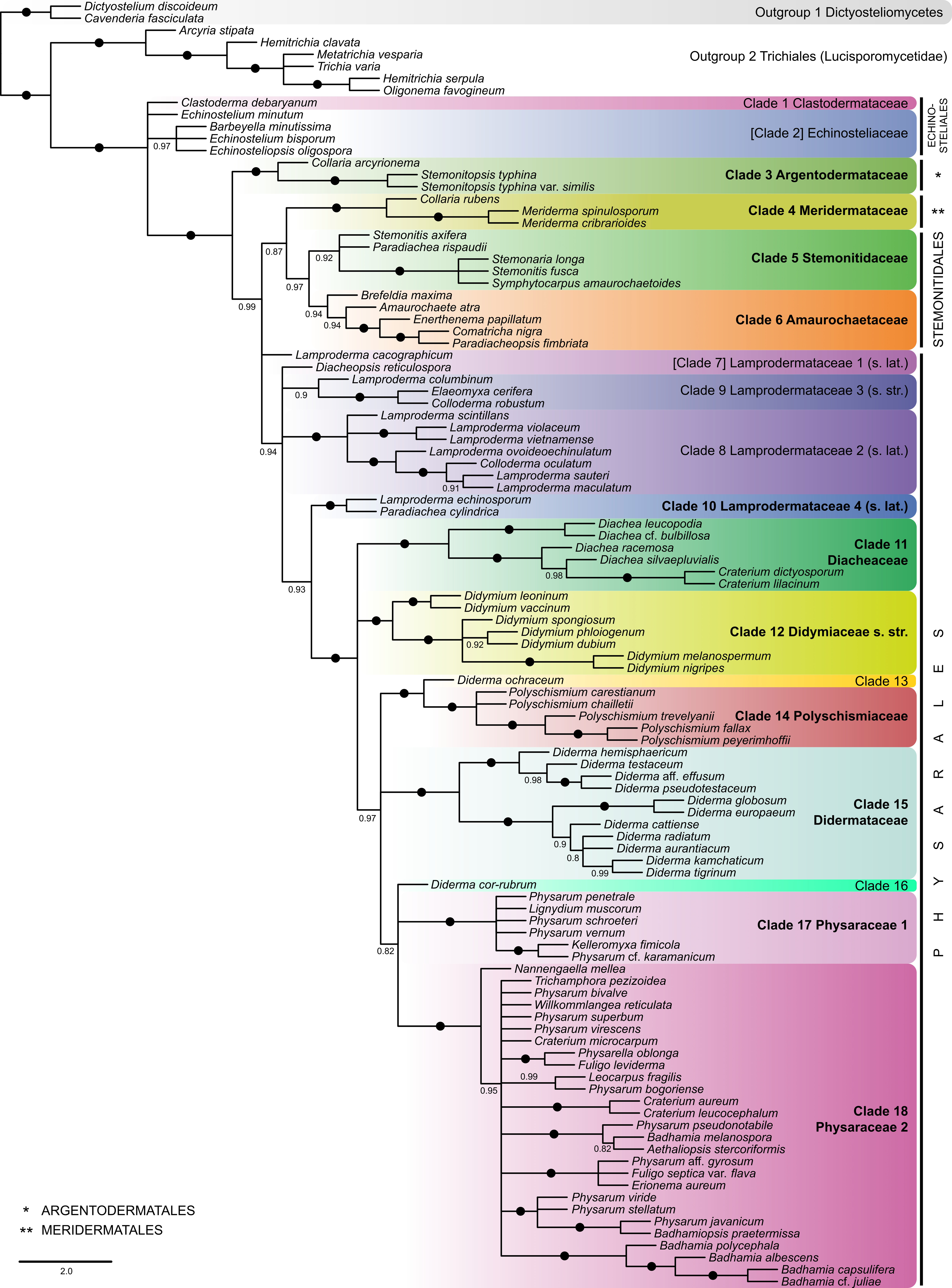
A two-step approach of species tree inference under the multi-species coalescent model was employed (Pamilo & Nei 1988; Rannala & Yang 2003) to take into account the discordance among the evolutionary histories of different genes. First, separate gene trees were generated for each gene alignment from the main dataset using IQ-Tree version 2.3.5 (Supplementary Material 2). Models for unpartitioned analyses were selected using ModelFinder according to BIC. Tree inferences were carried out with default tree search parameters, random seed, and one thousand replicates of ultrafast bootstrap. Second, a species tree was constructed from all gene trees using wASTRAL (weighted ASTRAL) version v1.20.3.7 (Zhang & Mirarab 2022). This method reimplements ASTRAL-III (Zhang et al. 2018) and takes into account phylogenetic uncertainty by integrating signals from branch length and branch support in gene trees without introducing thresholds. The wASTRAL analysis was done with the default weighting mode (“hybrid”), 30 initial rounds of placements, 30 rounds of subsampling per exploration step, and branch local posterior probability values (Sayyari & Mirarab 2016) written to the final species tree. Multiple individuals from the same species, if they were confirmed as forming a clade in the concatenated analyses, were forced to be considered as one species, resulting in a tree of 105 species of myxomycetes rooted with two species from the Dictyosteliomycetes (Fig. 1).
Phylogenetic analyses on concatenated alignments
Maximum likelihood tree inference with IQ-Tree version 2.3.5 was carried out on four sets of concatenated alignments: (1) alignments of ten nuclear genes from the main dataset (115 specimens, Fig. 2), (2) alignments of twelve mitochondrial genes from the main dataset (114 specimens, Fig. 2), (3) alignments of 22 genes from the expanded dataset (1046 specimens, Figs. 3–5), and (4) alignments of 21 genes from the expanded dataset, which were reduced to include only members of the family Stemonitidaceae, with the Meridermataceae as outgroup and additional nrSSU sequences of some members of Stemonitidaceae that were not included into the expanded dataset due to insufficient length in trimmed alignments (Supplementary Fig. S1). Tree inference was performed in five independent IQ-Tree runs for the three sets of alignments (1, 2, 4) and in fifteen independent runs for the expanded dataset (3). In each tree inference run, the concatenated alignments were partitioned by genes; nuclear protein-coding genes were additionally partitioned by codon positions. Edge-linked partition models (Chernomor et al. 2016) with proportional branch lengths, separate substitution models, and separate rates across partitions were selected using ModelFinder according to BIC, with partition merging allowed. Branch supports were obtained with 1000 ultrafast bootstrap (UFB) replicates and 1000 SH-like approximate likelihood ratio test (SH-aLRT) replicates (Guindon et al. 2010). For each set of alignments, the maximum likelihood tree with the highest likelihood among several independent IQ-Tree runs was chosen for visualization.
Results
Genome skimming
Low-pass genome sequencing was successfully carried out for 112 herbarium specimens of myxomycetes, including six bright-spored species from the order Trichiales. Sequencing depth per sample varied from 0.034 Gb in Diderma cor-rubrum T. Macbr. to 22.1 Gb in Diderma kamchaticum Novozh., Schnittler, Shchepin & Prikhodko; the average figure was 1.4±2.3 Gb (Supplementary Material 1). This average value would correspond to an approximately 5.6× coverage for a genome the size of that of Badhamia polycephala.
With the low-pass genome sequencing, mostly multicopy genome regions have a chance of receiving sufficient coverage for sequence assembly from short reads. We aimed at extracting mostly nuclear ribosomal genes (nrSSU, nrLSU, 5.8S rDNA), which are located in numerous minichromosomes per nucleus in myxomycetes, and mitochondrial genes, present in multiple copies of the mitochondrial genome per mitochondrion. We were able to retrieve full or partial orthologous sequences of all three nuclear ribosomal genes for 91.1% of analyzed samples (overall missingness: 3.3%) and all twelve selected mitochondrial genes for 71.4% of samples (overall missingness: 7.1%) (Supplementary Material 1). For Stemonitis axifera (Bull.) T. Macbr. (LE344639, Fig. S23), we had to exclude all mitochondrial genes from the analyses because they were very deviating and formed extremely long branches in our phylogenies.
In addition to these multicopy loci, we were able to extract partial or full ortholog sequences of seven single- or low-copy nuclear genes, excluding those that showed extensive paralogy. We found all seven selected genes in only 16.1% of the samples, but for 89.3% we found at least some of these genes (overall missingness 34.2%).
For nuclear single-copy genes that we were able to retrieve, each sequence position was on average covered by 8.5 reads (1.0–71.1, SD 9.2), for nuclear ribosomal genes by 397.5 reads (1.4–5503.0, SD 708.9), and for mitochondrial genes by 1725.5 reads (2.2–45606.7, SD 4815.7). The copy
number of mitochondrial genes and nuclear ribosomal genes per nuclear haploid genome was approximated by dividing the coverage of each mitochondrial and nuclear ribosomal gene by the average coverage of nuclear single-copy genes for each sample. This resulted in an average estimated copy number of 204.8 for mitochondrial genes (1.2–3847.9, SD 364.8) and 53.6 for nuclear ribosomal genes (2.0–858.7, SD 83.3). Notably, the highest copy numbers for both groups of loci were estimated for Brefeldia maxima (Fr.) Rostaf., the species known to produce the largest fruiting bodies among myxomycetes (Supplementary Material 1, Supplementary Fig. S2).
We did not find a linear dependence between sample sequencing depth and the total number of recovered loci (Pearson r 0.139, p = 0.145). However, we found a statistically significant moderate monotonic trend with the Spearman rank-based correlation coefficient: samples with greater sequencing depth tended to have more loci recovered (0.401, p = 1.19×10⁻⁵). For nuclear single-copy loci, this dependence was more prominent. The linear dependence was small but significant (Pearson r 0.199, p = 0.035), and the Spearman correlation coefficient revealed a highly significant moderate-strong monotonic association (0.495, p = 2.8 × 10⁻⁸).
To characterize how locus recovery saturates with sequencing depth, we fitted hyperbolic curves to two separate data sets: Expected loci(x) = where x =
sequencing depth, Vmax = the asymptote (maximum loci recoverable), KM = the depth at which half the loci are recovered. The asymptote was fixed at seven nuclear single-copy genes and 15 multi-copy (mitochondrial + nuclear-ribosomal) loci (Supplementary Fig. S3a–b). For the single-copy genes, the half-saturation constant Km was at a sequencing depth 0.39 Gb, and the curve reached 90% of its asymptote (≈ 6.3 loci) at ~3.5 Gb and 95% (≈ 6.7 loci) at ~7.4 Gb. Marginal gains fell below 0.001 loci per Mb beyond ~1.26 Gb (Supplementary Fig. S3c).
Multi-copy loci saturated much earlier: Km ≈ 0.014 Gb, with 90% completeness (13.5 of 15 loci) already at 0.129 Gb and 95% (14.3 loci) at 0.272 Gb (Supplementary Fig. S3b, d). At the 90% level the instantaneous discovery rate had dropped to < 1% of its initial value, making further sequencing increasingly inefficient.
Phylogenetic analyses
Four phylogenetic trees represent the main phylogenetic results of the present study.
(1) Species tree (Fig. 1): obtained with wASTRAL by summarizing 22 separately inferred maximum likelihood gene trees (Supplementary Material 2), which were based on the alignments from the main dataset (Supplementary Material 4). This shows relationships among 105 species of myxomycetes and is based on sequences of 115 specimens, with sequencing data coming from genome skimming, transcriptomics or whole genome assembly, and takes into account topological conflicts between individual gene trees by utilizing a multispecies coalescent model. Two species in the Dictyosteliomycetes were added as an outgroup to infer relationships between the Lucisporomycetidae, Echinosteliales, and other members of the Columellomycetidae.
(2) Maximum likelihood nuclear gene tree (Fig. 2, left): inferred from a concatenated alignment of 10 nuclear genes from the main dataset, which included 115 specimens with 16258 columns, 7636 of them parsimony-informative and 6812 constant (Supplementary Material 5), with sequencing data coming from genome skimming, transcriptomics or whole genome assembly.
(3) Maximum likelihood mitochondrial gene tree (Fig. 2, right): inferred from a concatenated alignment of 12 mitochondrial genes from the main dataset, which included 114 specimens with 15034 columns, 10981 of them parsimony-informative and 2681 constant (Supplementary Material 6), with sequencing data coming from genome skimming, transcriptomics or whole genome assembly.
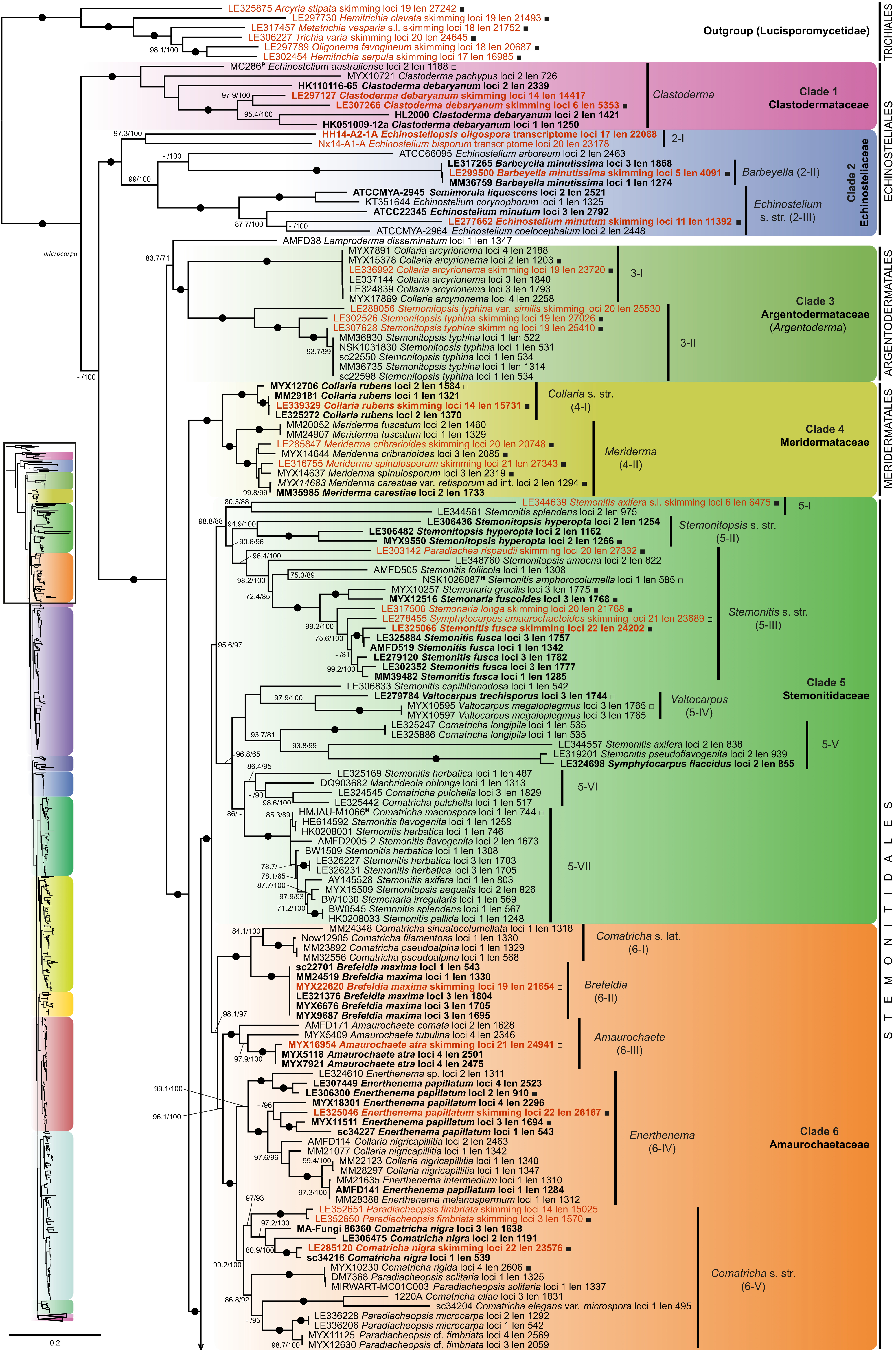
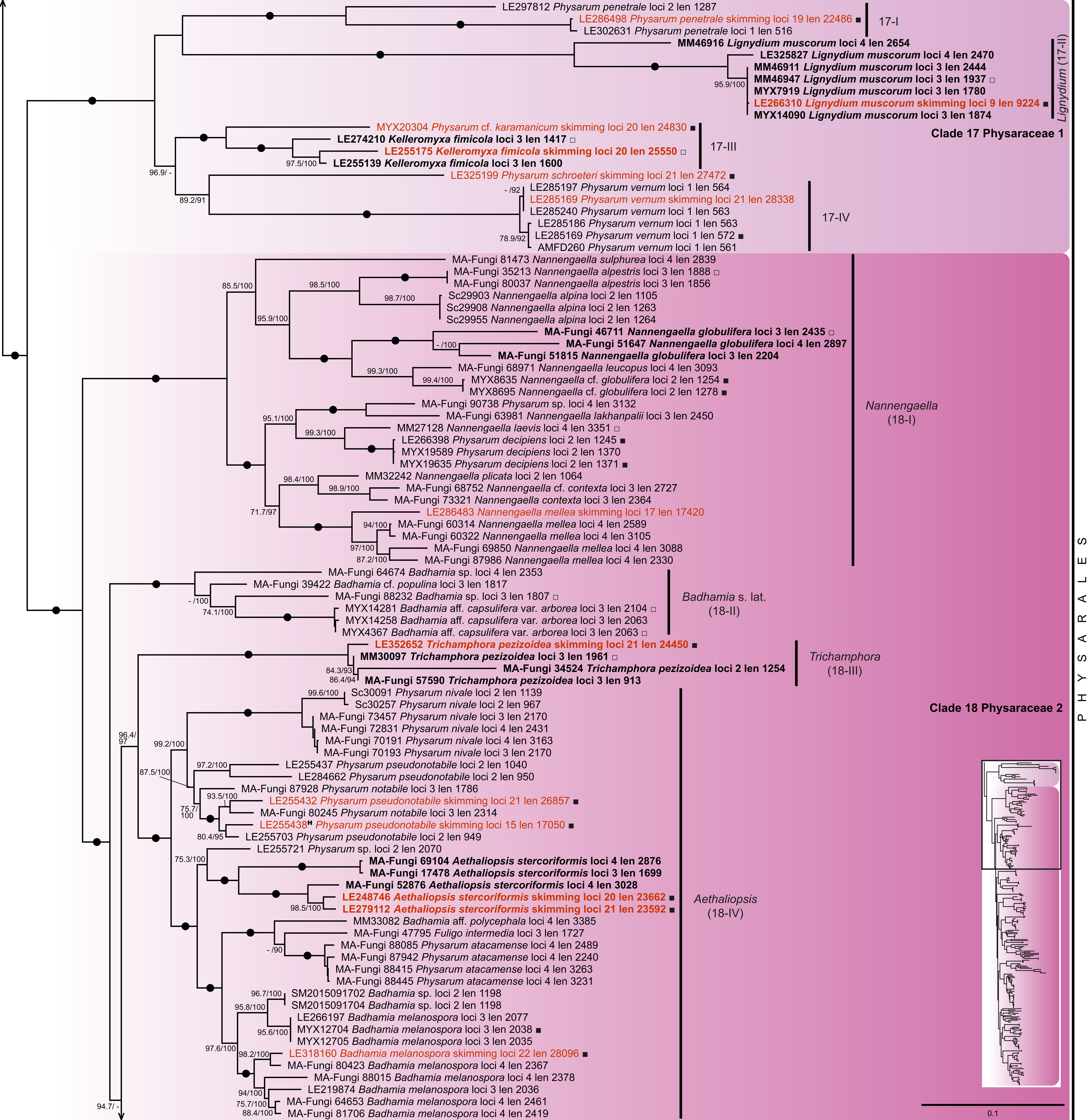
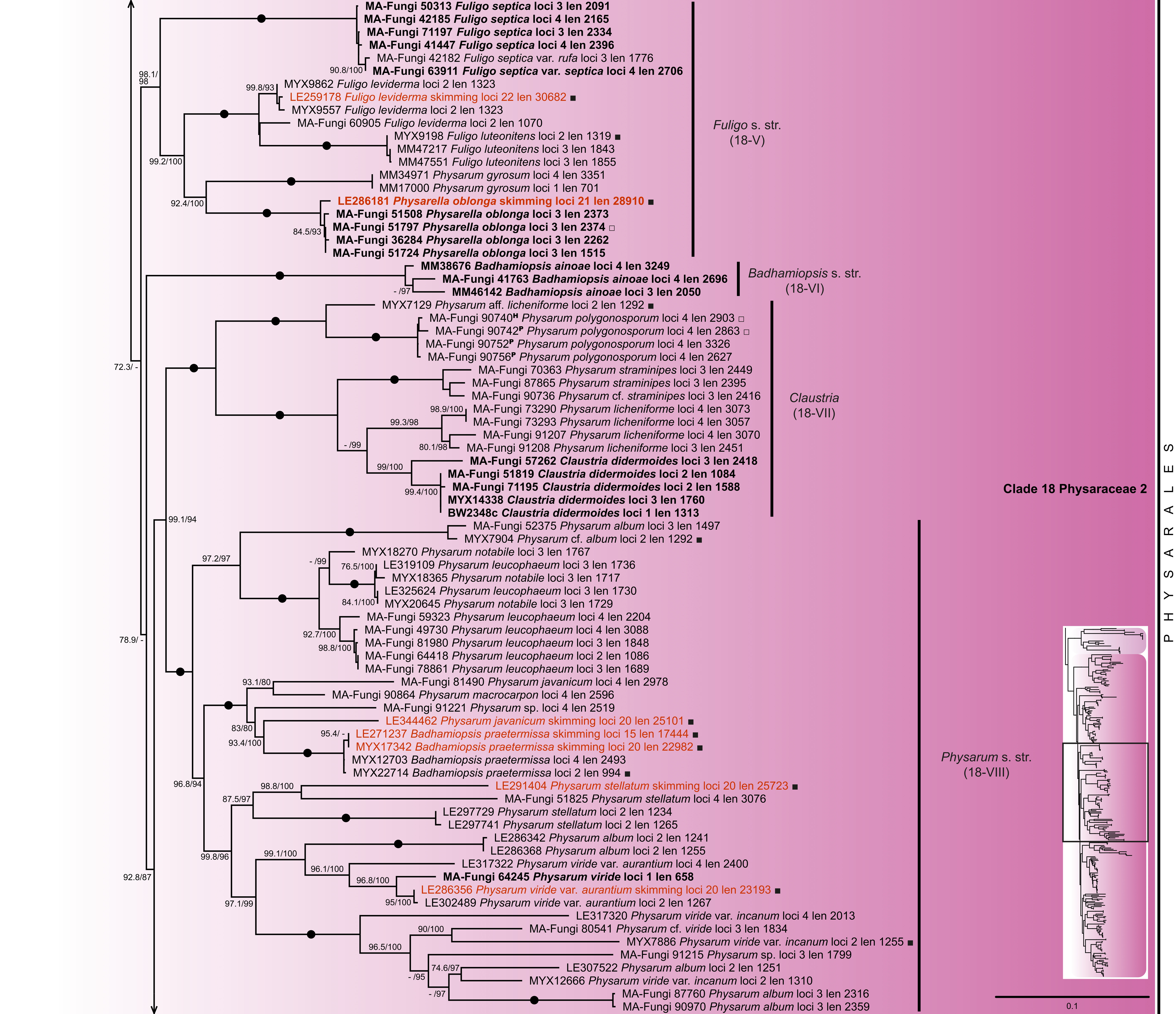
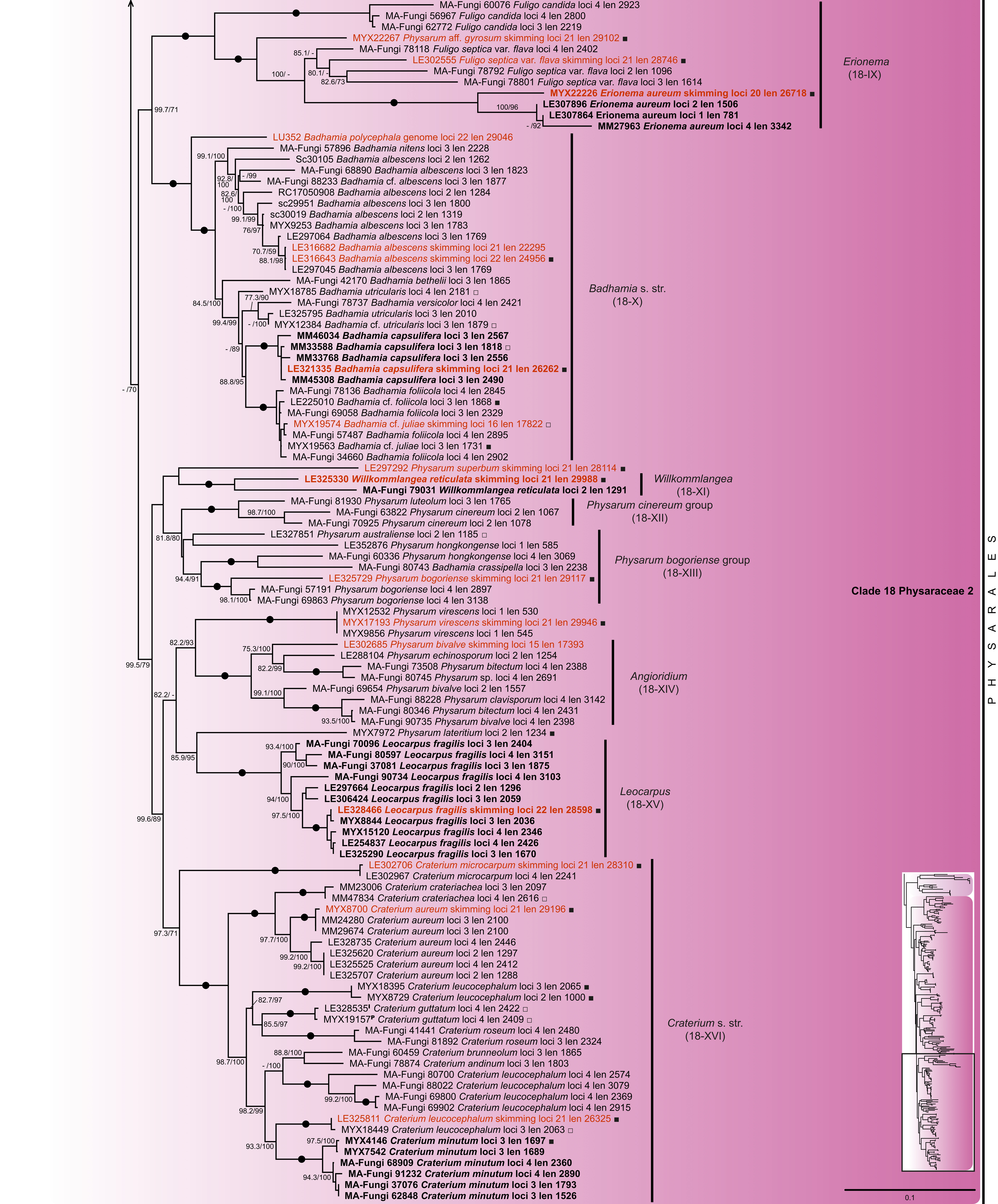
(4) Maximum likelihood tree of the expanded sequence dataset (Figs. 3–5): Constructed from a concatenated alignment of 22 genes, which included 1046 specimens with 31557 columns, 19243 of them parsimony-informative, 3218 singleton and 9099 constant (Supplementary Materials 7, 8). The sequencing data originated from genome skimming, transcriptomics or whole genome assembly for 115 samples and the remaining sequences were obtained with Sanger sequencing (by us or from GenBank). This tree uses the 22-gene backbone to show the relationships within the Columellomycetidae with greatly expanded sampling of species for which only a few loci are available. The barcode region established for dark-spored myxomycetes (the first part of the nrSSU; Schnittler et al. 2017; Inoue et al. 2024) is given in Supplementary Material 7, nrSSU alignment for 1008 accessions, pos. 128–1943.
In addition to that, one supplementary tree (Fig. S1) shows phylogenetic relationships within the Stemonitidaceae in more detail. This maximum likelihood phylogeny is based on a concatenated alignment of 21 genes from the expanded dataset for the members of the Stemonitidaceae (the mdh gene was mostly missing in the Stemonitidaceae), with Sanger sequences included for additional samples. The alignment comprised 99 specimens with 31998 columns, 6590 of them were parsimony-informative and 19743 were constant (Supplementary Material 9).
In the species tree obtained by summarizing 22 gene trees within the multispecies coalescent framework and rooted with the two members of the Dictyosteliomycetes (Fig. 1), class Myxomycetes splits into two major lineages. These two lineages correspond to subclasses Lucisporomycetidae, or bright-spored myxomycetes (represented here by the Trichiales), and the Columellomycetidae, or dark-spored myxomycetes, the target group of this study. These are represented by members of all orders and families of this subclass in the circumscription of Leontyev et al. (2019). Members of the genera Barbeyella Meylan, Clastoderma A. Blytt, Echinostelium de Bary, and Echinosteliopsis D.J. Reinh. & L.S. Olive form a basal unresolved group of branches within the Columellomycetidae, supporting their belonging to this subclass.
The coalescent species tree (Fig. 1) and phylogenies obtained using the alignment concatenation approach within the maximum likelihood phylogenetic framework (Figs. 2–5, Supplementary Fig. S1) grouped species into 18 clades with different levels of statistical support. The clades are numbered according to the maximum likelihood phylogeny based on the expanded sequence data set (Figs. 3–5). Most of these clades did not differ between phylogenies obtained separately for nuclear and mitochondrial genes (left and right parts in Fig. 2). Clades 3, 4, 10–12, 14, 15, 17 and 18 were reproduced in all phylogenies with nearly maximum to
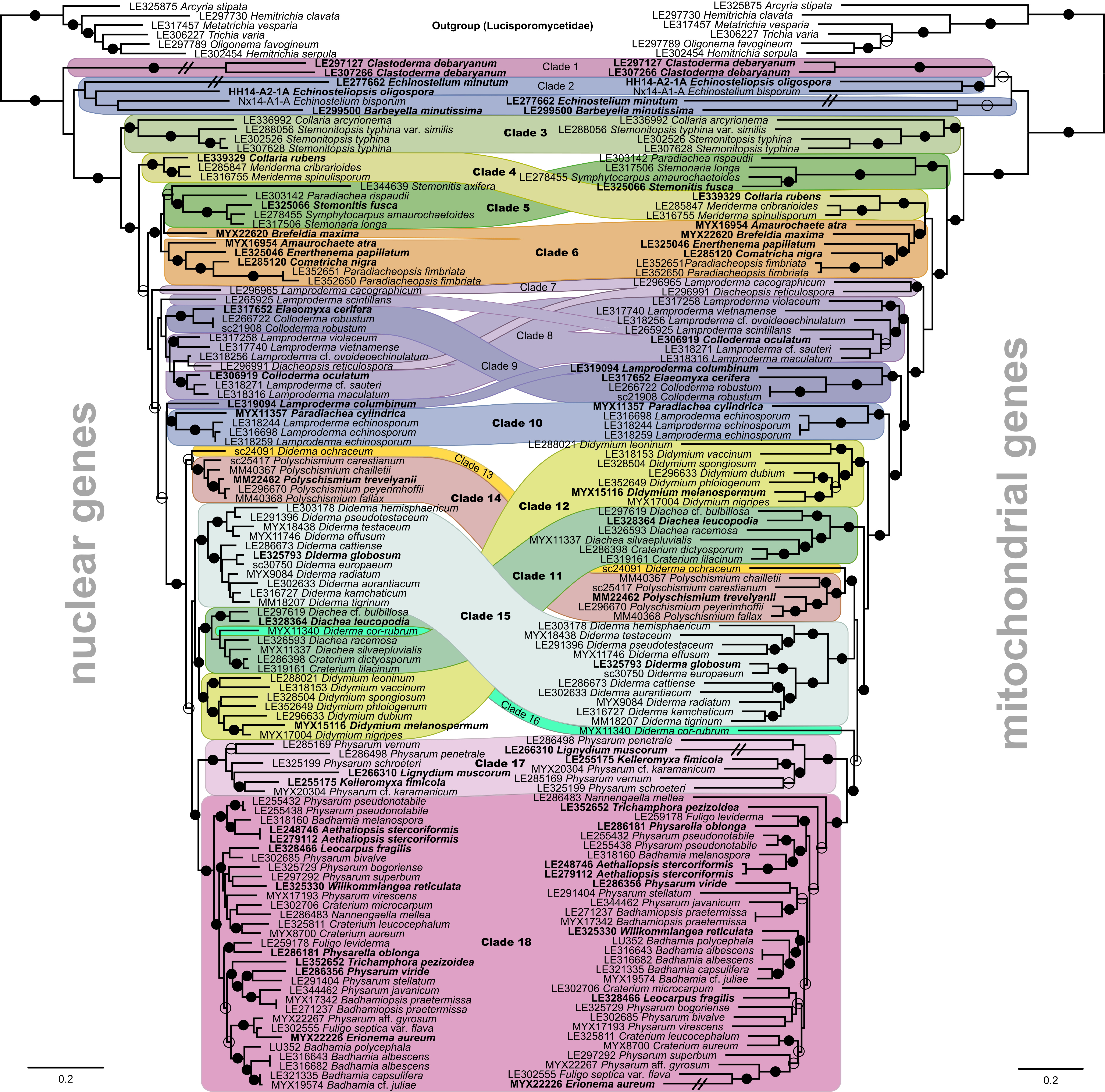
maximum statistical support (local posterior probabilities = 1.00, SH-aLRT ≥ 99, UFB ≥ 99). Two more clades, Clades 5 and 6, received moderately high to high statistical support in all our phylogenies (LPP ≥ 0.92, SH-aLRT ≥ 95.6, UFB ≥ 97), except for the nuclear gene concatenation analysis, where the relationships between Brefeldia maxima and other species of Clade 6 were not resolved (Fig. 2, left).
Five of the eleven robust clades partially or fully correspond to the described families Meridermataceae (Clade 4), Stemonitidaceae (Clade 5), Amaurochaetaceae (Clade 6), and Physaraceae (Clades 17 + 18) (Leontyev et al. 2019). Clade 3 unites Collaria arcyrionema (Rostaf.) Nann.-Bremek. ex Lado, Stemonitopsis typhina (F.H. Wigg.) Nann.-Bremek. and S. typhina var. similis (G. Lister) Nann.-Bremek. & Y. Yamam., forming the earliest diverging lineage after the Echinosteliales in the Columellomycetidae. Clades 11, 12, 14 and 15 mostly represent the four largest genera of the Didymiaceae sensu Leontyev et al. (2019) – Diachea Fr., Didymium Schrad., Polyschismium Corda and Diderma Pers., respectively. The remaining robust clade, Clade 10, includes Lamproderma echinosporum Meyl. and Paradiachea cylindrica (Bilgram) Hertel ex H. Neubert, Nowotny & K. Baumann sensu Novozhilov et al. (2023).
Three other clades were singletons in the main dataset, represented by one or two accessions of a single species analyzed with genome skimming. These include Clades 1, 13
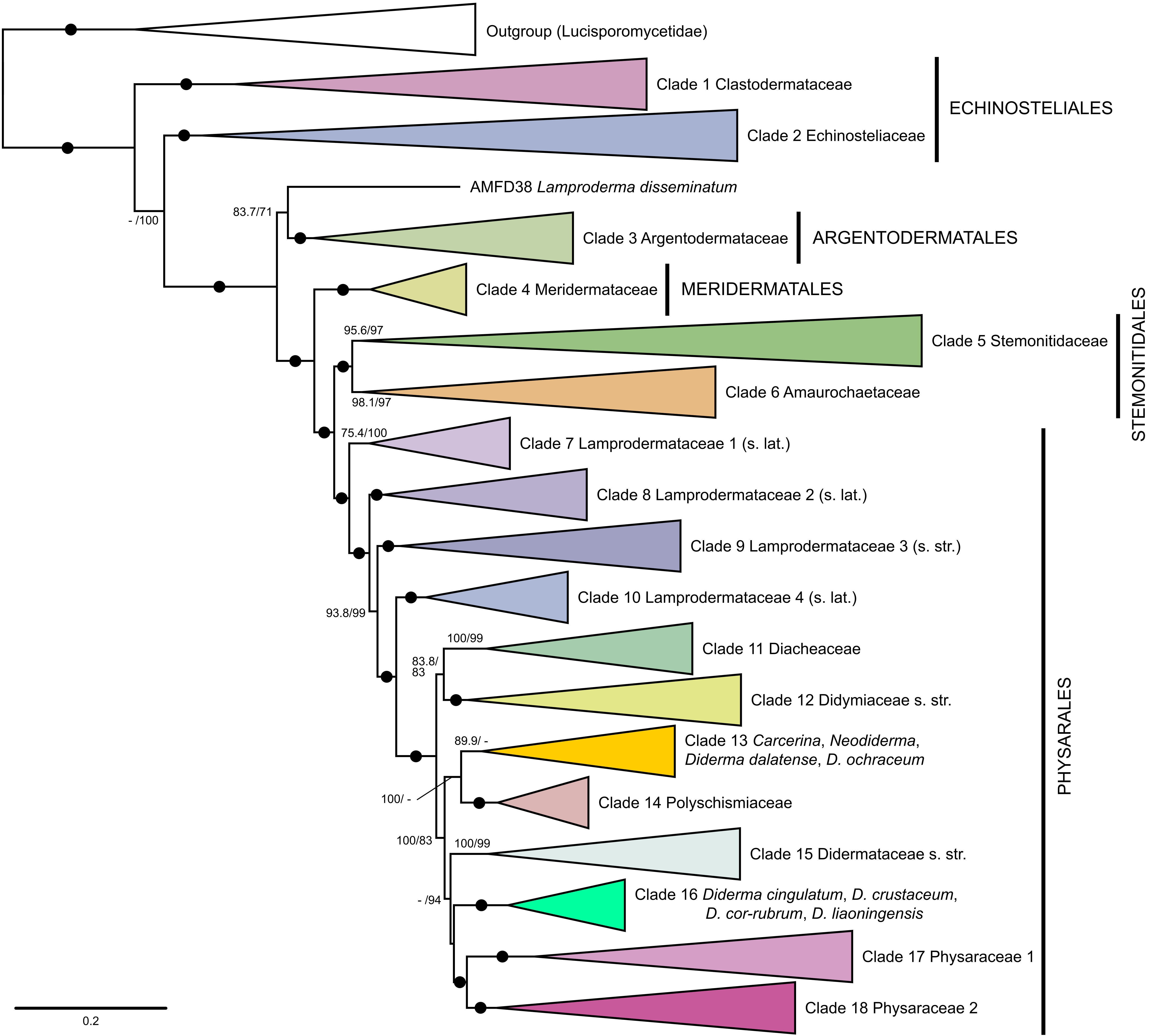
and 16 (Figs. 1, 2). However, all of these are represented by multiple species in the expanded dataset (Figs. 3, 4). Clade 1 is fully supported and unites two species of Clastoderma and Echinostelium australiense K.J. Knight, S.L. Stephenson & Novozh. within the family Echinosteliaceae (Fig. 4). Two other clades concern divergent species of Diderma Pers. and Neodiderma X.F. Li, B. Zhang & Yu Li that do not group with Diderma s. str. Clade 13 is only weakly supported (SH-aLRT 89.9, UFB ≤ 50) and occupies a sister position to Polyschismium spp. in all phylogenies. It splits into two subclades: the first one (13-I; SH-aLRT 99.8, UFB 70) unites Diderma dalatense Novozh., Prikhodko & Shchepin, D. ochraceum Hoffm. and Neodiderma rufum X.F. Li, B. Zhang & Yu Li; the second subclade (13-II and III; SH-aLRT 85.4, UFB 86) unites Diderma spumarioides (Fr. & Palmquist) Fr. and the remaining five species of the genus Neodiderma, including its type Neodiderma macrosporum X.F. Li, B. Zhang & Yu Li (Fig. 4). Clade 16 has a maximum statistical support and unites Diderma cingulatum Nann.-Bremek., D. cor-rubrum T. Macbr., D. crustaceum Peck, and D. liaoningensis H.N. Zhao, B. Zhang & Yu. Li. Nuclear gene concatenation analysis (Fig. 2, left) places D. cor-rubrum inside Clade 11 (SH-aLRT 100, UFB 100), but it appears as a sister to clades 17 + 18 in the mitochondrial gene tree (Fig. 2, right; SH-aLRT 90, UFB 96), while in the other phylogenies this species occupies unresolved position among major clades of the Physarales (Figs. 1, 3, 4).
The remaining four clades were not robust, as different phylogeny reconstruction methods and genes of different localization produced highly contradicting or insufficiently resolved results. Clade 2 was obtained as a fully supported monophyletic group only in the expanded concatenation analysis (Figs. 3, 4; SH-aLRT 100, UFB 100), but it appeared paraphyletic in the mitochondrial gene tree, with Clade 1 nested inside (Fig. 2, right; SH-aLRT 93, UFB 98), and completely unsupported in the other trees (Figs. 1, 2). This group includes species of the Echinosteliaceae sensu Leontyev et al. (2019) and Echinosteliopsis oligospora.
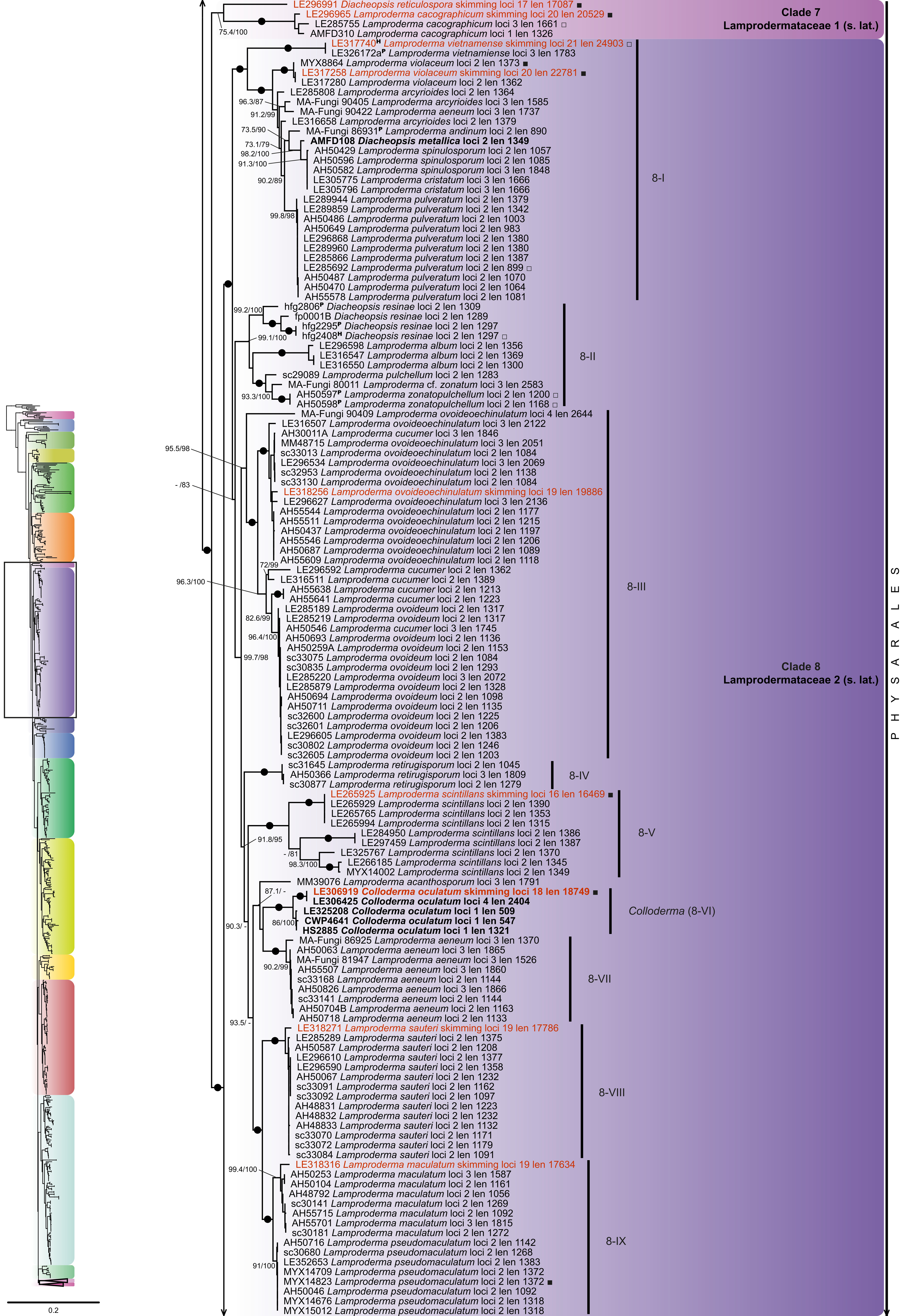
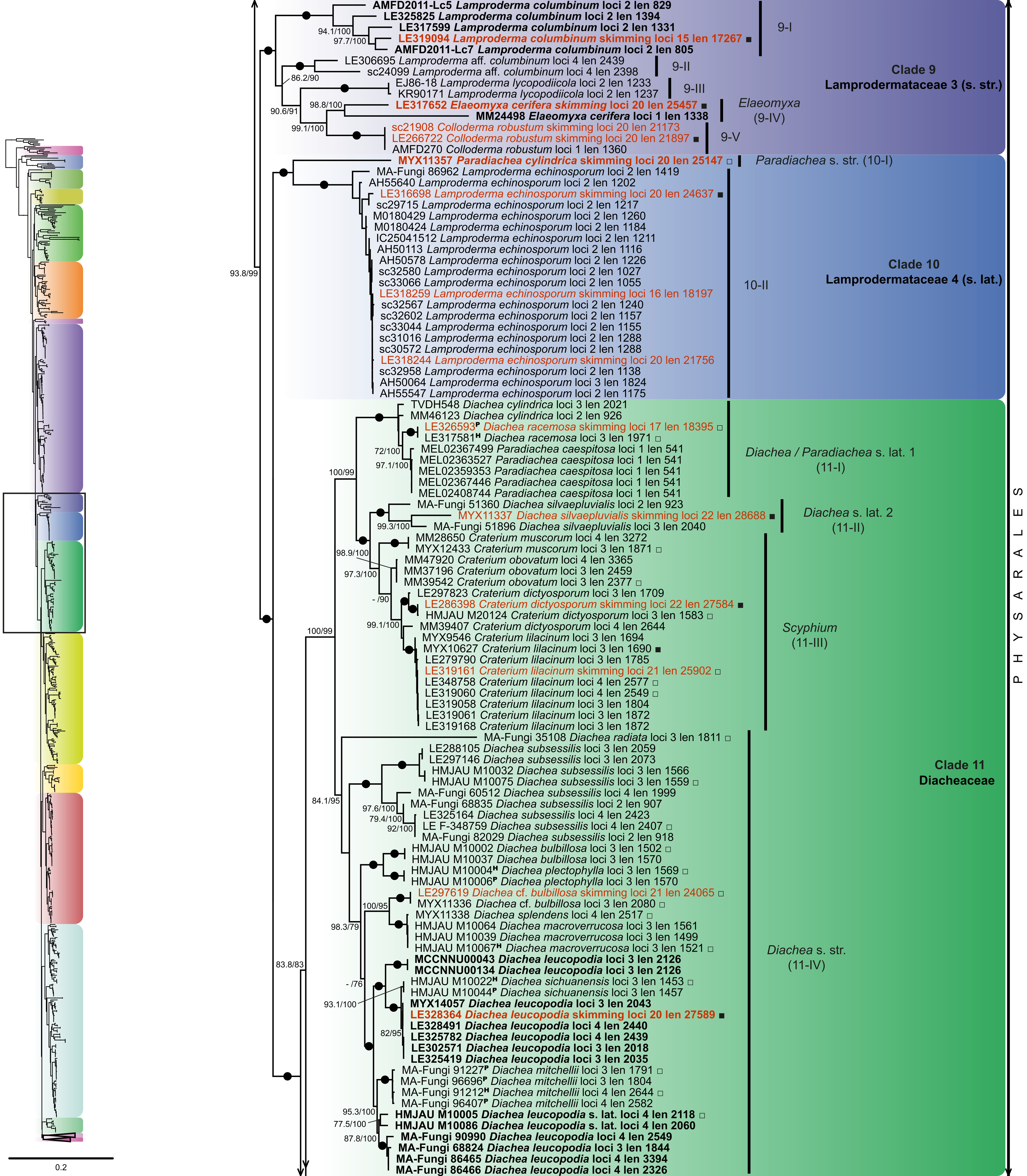
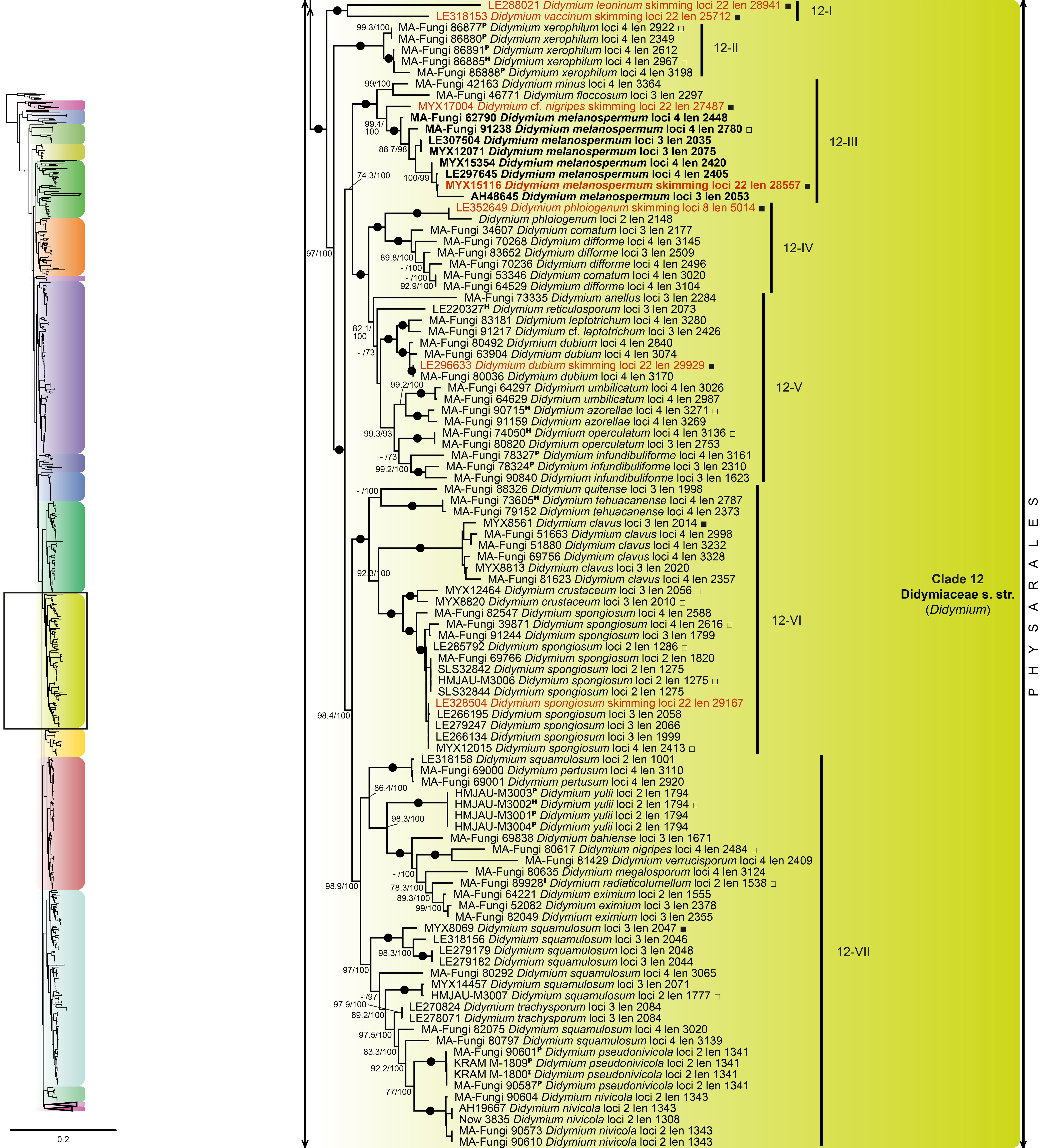
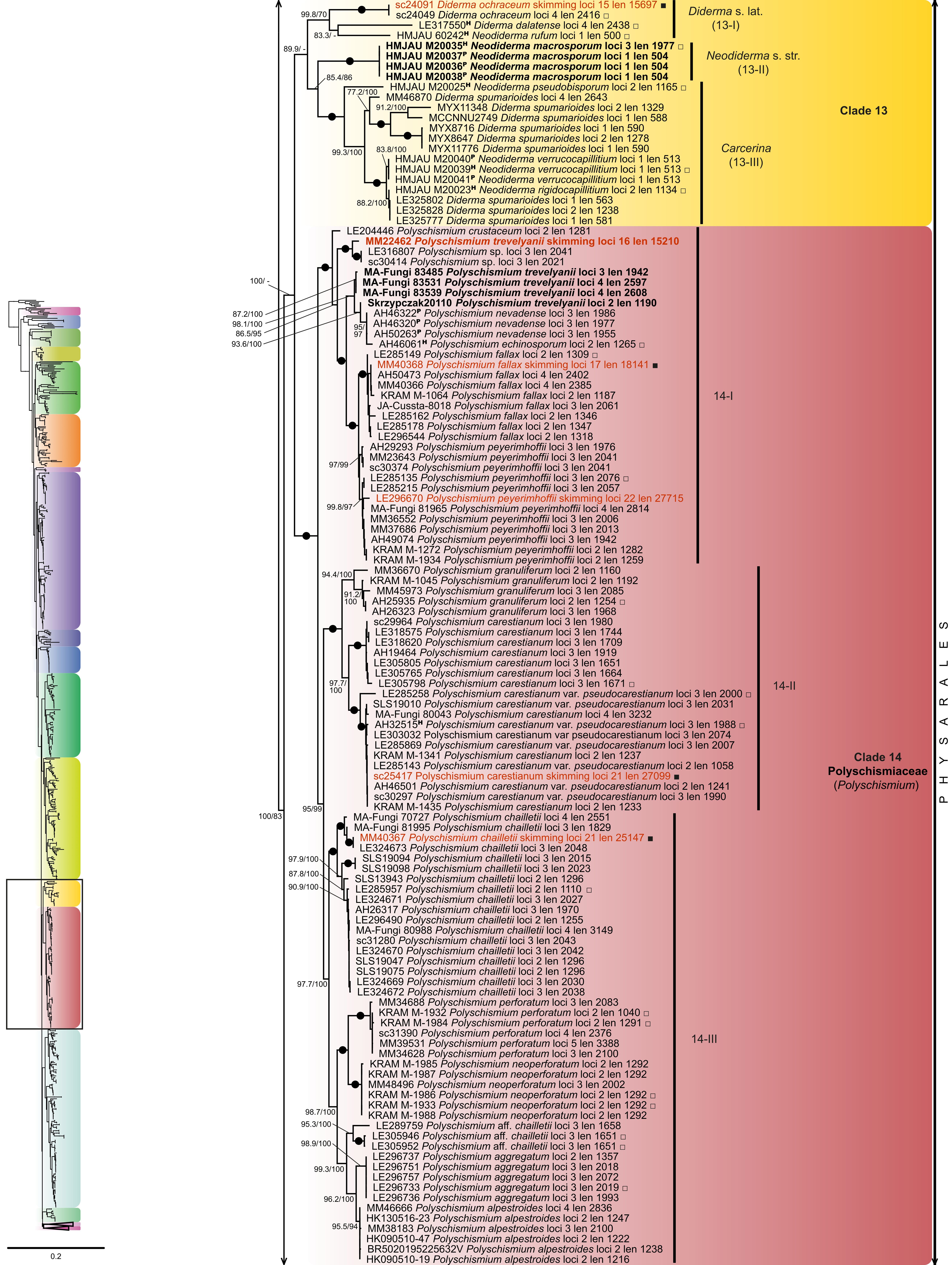
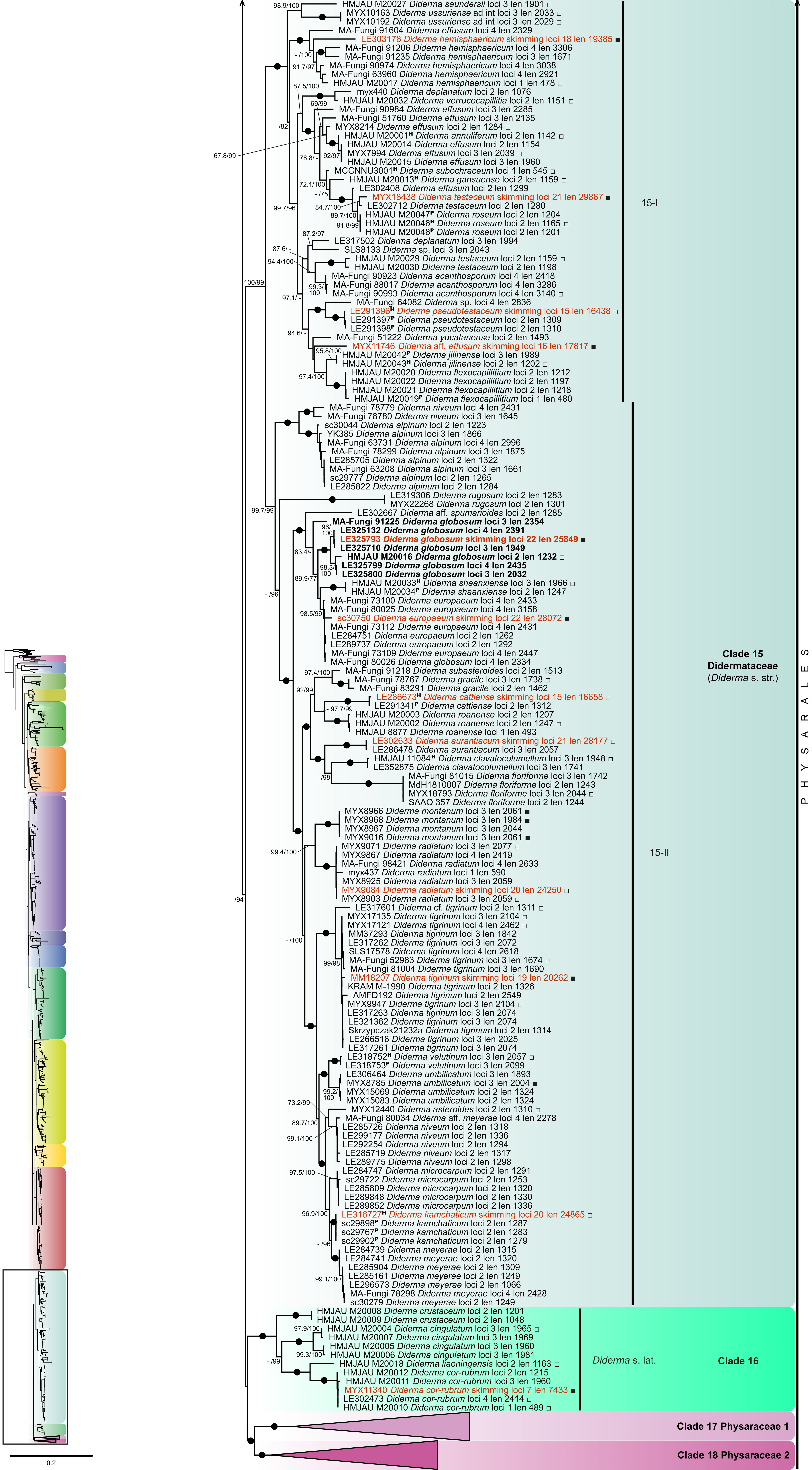
Clades 7–9 received moderate to maximum statistical support in the expanded concatenation phylogeny (Fig. 3; SH-aLRT ≥ 75.4, UFB 100) and high to maximum support in the mitochondrial gene concatenation tree (Fig. 2, right; SH-aLRT ≥ 98, UFB ≥ 99). However, these three clades were not reproduced in the nuclear gene concatenation tree (Fig. 2, left) and only Clades 8 and 9 were supported in the species tree (Fig. 1; LPP 1.00 and 0.90, respectively). These three clades, together with Clade 10, mostly include species of the Lamprodermataceae sensu Leontyev et al. (2019).
Some of the relationships between the 18 clades are resolved differently in our phylogenies. The strongest discordance is observed between the nuclear and mitochondrial gene trees (Fig. 2). Clades 5 (Stemonitidaceae) and 6 (Amaurochaetaceae) do not form a monophyletic group in the mitochondrial gene tree, in contrast to the other phylogenies. Instead, Clade 5 branches more basally, and Clade 4 (Meridermataceae) is in a poorly supported sister position to Clade 6 (SH-aLRT 78, UFB 90). In the species tree, these three clades (4, 5 and 6) group together with a moderate support (Fig. 1; LPP 0.87). The nuclear gene tree gives a high support to the monophyly of Clade 11 (mostly Diachea) + Clade 12 (Didymium) (SH-aLRT 98, UFB 99), which is also partially supported in the expanded concatenation tree (SH-aLRT 83.8, UFB 83; Figs. 2, 3). However, the species tree and the mitochondrial gene tree do not support these relationships. Instead, they show mostly unresolved relationships between the Clades 11–15 of the family Didymiaceae sensu Leontyev et al. (2019).
The results of the phylogenetic analyses were used to make formal taxonomic conclusions. For this purpose, we adopted the following criteria: (1) only a monophyletic group can be considered as a taxon; (2) the respective clade has to receive high statistical support throughout all phylogenetic analyses; (3) the respective clade has to be reproduced in phylogenies constructed with different methods and loci of different localization (nuclear and mitochondrial); (4) sister clades should receive the same taxonomic rank (if their relationships are sufficiently resolved). Because morphological convergence is common, we did not require a unique suite of morphological characters to justify taxonomic recognition, but such characters are noted where present.
When introducing taxonomic novelties, we strictly followed criteria 1 and 2. Criterion 3 was applicable mostly for the order and family level, but only partially for genera, as many species were still represented by only a few genetic markers and were not included in Figs. 1 and 2. For criterion 4, we tried to be as conservative as possible by preserving the already existing higher-level taxa and proposing new taxa of the same ranks when formally necessary to avoid non-monophyly.
This led us to select ten out of the eleven robust clades (excluding Clade 10) to define family-level taxonomic units in the updated system of the Columellomycetidae proposed below. This is intended to stabilize the system at the family level because the robustness of these clades to different phylogeny reconstruction methods, species and gene sampling makes it unlikely that they will change significantly in the future revisions.
For some species with highly supported phylogenetic relationships within Clades 1, 2, and 13 obtained in the expanded concatenation analysis (Figs. 3, 4), we propose several new combinations. These include a transfer of Semimorula liquescens E.F. Haskins, McGuinn. & C.S. Berry to Echinostelium within Clade 2, a transfer of Echinostelium australiense to Clastoderma, and a transfer of four species of Neodiderma to Carcerina Fr. For the other four non-robust clades, we remain conservative and do not propose any formal taxonomic changes.
Detailed descriptions of the obtained topologies and comparisons of our phylogenies with each other and with the ones published elsewhere are provided in the notes on taxa and clades in the taxonomic section below.
Taxonomy
Subclass Columellomycetidae Leontyev, Schnittler, S.L. Stephenson, Novozhilov & Shchepin, Phytotaxa 399(3): 220 (2019).
Taxa below are presented in the same order as they appear in Figs. 3–5.
• Order Echinosteliales G.W. Martin, Mycologia 52(1): 127 (1961) [Clades 1–2].
Notes: In the recently proposed classification of myxomycetes (Leontyev et al. 2019), the genus Echinosteliopsis was listed as Columellomycetidae incertae sedis, while in a later update (Wijayawardene et al. 2020), the five early branching genera within the subclass Columellomycetidae were placed in three different orders: Echinosteliopsidales (Echinosteliopsis), Echinosteliales (Echinostelium, Barbeyella and Semimorula E.F. Haskins, McGuinn. & C.S. Berry) and Clastodermatales (Clastoderma). This separation of the traditional order Echinosteliales into two orders and description of a separate order and family for Echinosteliopsis (Wijayawardene et al. 2020) was based on nrSSU and two-gene (nrSSU + EF1A) phylogenies. In contrast to that, our multigene phylogenies do not separate these five genera into three reciprocally monophyletic groups that would correspond to three orders (Figs. 1–4).
Clastoderma debaryanum A. Blytt, C. pachypus Nann.-Bremek., and Echinostelium australiense K.J. Knight, S.L. Stephenson & Novozh. form a fully supported clear-cut clade (Clade 1), suggesting a need to transfer E. australiense to Clastoderma (Fig. 4). Echinosteliopsis oligospora groups with low to high support into a Clade 2 with species of Echinostelium, Barbeyella, and Semimorula (Figs. 1–4). Clade 2 is fully supported only in the expanded concatenation tree (Figs. 3, 4), while in the species tree, the nuclear gene tree and the mitochondrial gene tree, the relationships between the members of this clade are mostly unresolved (Figs. 1, 2). Relationships between Clade 1 and Clade 2 are unresolved in all our phylogenies. Because of this, we decided to take a conservative approach. We recognize two families, Clastodermataceae (Clade 1) and Echinosteliaceae (Clade 2), within the single order Echinosteliales, and we consider Echinosteliopsis as a genus in Echinosteliaceae.
The results of our phylogenetic analyses and previous morphological studies of the peridium and the capillitium system of Clastoderma debaryanum and Barbeyella minutissima Meyl. show that there is no close relationship between Clastoderma and Barbeyella (Frederick et al. 1986; Haskins & McGuinness 1989; Schnittler et al. 2000; Haskins & Clark 2016). In contrast, Barbeyella minutissima groups in a clade with Echinostelium spp. and forms a partially supported (-/100) subclade with Echinostelium arboreum H.W. Keller & T.E. Brooks within the Echinosteliaceae (Fig. 4). Moreover, B. minutissima and E. arboreum have a similar peridium morphology (Schnittler et al. 2000; Fiore-Donno et al. 2012; Leontyev et al. 2019). For this reason, we support the previously proposed transfer of Barbeyella to the Echinosteliaceae (Kretzschmar et al. 2015; Leontyev et al. 2019).
Our phylogeny (Fig. 4) confirms the long-known close phylogenetic relatedness between the stalkless monotypic genus Semimorula and Echinostelium corynophorum K.D. Whitney (Fiore-Donno et al. 2009, 2018; Kretzschmar et al. 2016). We thus combine Semimorula liquescens with Echinostelium. This is one of several cases where the stalk is lost in some taxa of an otherwise stalked group, and some of these stalkless taxa have been traditionally recognized as separate genera (e.g., Diacheopsis Meyl. vs. Lamproderma Rostaf.).
•• Family Clastodermataceae Alexop. & T.E. Brooks, Mycologia 63(4):926 (1971) [Clade 1].
Type: Clastoderma A. Blytt, Bot. Zeitung (Berlin) 38: 343 (1880).
Notes: Previously, the phylogenetic analyses based on nrSSU sequences showed that the family Clastodermataceae in its traditional circumscription was polyphyletic due to the affinity of Barbeyella with species in the genus Echinostelium, and thus Barbeyella was transferred to the Echinosteliaceae (Kretzschmar et al. 2016; Leontyev et al. 2019). Our multigene phylogenies confirmed this taxonomic conclusion by grouping Barbeyella minutissima with species of Echinostelium and never with Clastoderma (Figs. 1–4).
In our expanded concatenated phylogeny (Fig. 4), the species Clastoderma debaryanum, C. pachypus, and Echinostelium australiense form a fully supported monophyletic group, separate from Echinostelium and allied genera. Previously, the family Clastodermataceae was raised to the order level based on its separation from the Echinosteliales in a nrSSU phylogeny (Leontyev et al. 2019), with only one sequenced species of the family, Clastoderma debaryanum. Our phylogenies show poorly supported and contradicting relationships among the Clastodermataceae, Echinosteliaceae, and Echinosteliopsis and do not provide enough evidence for the separation of the order Clastodermatales.
When the family Clastodermataceae included only members of the genus Clastoderma (Leontyev et al. 2019), the diagnosis of the family was based on the presence of morphological structures such as (1) a stalk filled with refuse material in the basal part; (2) a columella gradually transforming into a capillitium; (3) a branching and anastomosing capillitium, whose threads at the periphery merge and form plate-like swellings; (4) a fugacious peridium; (5) the absence of lime in sporophore structures; and (6) the pale-brown spores. However, the recent discovery of Clastoderma confusum K.J. Knight & Lado (Knight & Lado 2020) and the position of Echinostelium australiense (Knight et al. 2023) in our phylogeny, whose sporophore morphology does not fit the accepted concept of the genera, require clarification and extension of the diagnosis of the Clastodermataceae.
••• Clastoderma A. Blytt, Bot. Zeitung (Berlin), 38: 343. 1880 [Clade 1].
Type: Clastoderma debaryanum A. Blytt, Bot. Zeitung (Berlin) 38: 343 (1880).
Species investigated here: Clastoderma australiense (K.J. Knight, S.L. Stephenson & Novozh.) Shchepin, Prikhodko, K.J. Knight, S.L. Stephenson, Schnittler & Novozh., Clastoderma debaryanum A. Blytt (Fig. S10), Clastoderma pachypus Nann.-Bremek.
Notes: Our phylogeny of the genus Clastoderma includes only two of the four currently accepted species (C. debaryanum and C. pachypus), and the monophyly of the genus remains uncertain (Fig. 4). Clade 1 of our phylogeny includes as well Echinostelium australiense, a species characterized by the dark coloration of the sporotheca, stalk and spores. These characters are unusual for the genus Echinostelium, members of which more typically have a pale sporotheca, a hyaline to pale brown stalk of uniform color and spores pale pink or hyaline in mass (Whitney 1980; Haskins & Clark 2016). It should be emphasized that when Echinostelium australiense was described (Knight et al. 2023), no data on its molecular phylogeny were available. Its assignment to the genus Echinostelium, despite the pronounced dark pigmentation of sporophore structures, was based on the absence of capillitium and columella, as well as the presence of spore-like bodies at the top of the stalk, which is characteristic of some species of the genus Echinostelium (Haskins & Clark 2016). However, the phylogeny we obtained shows that these morphological characters may have appeared independently in Echinostelium and Clastoderma. Besides, an evanescent peridium persisting only as a collar at the base of sporotheca is found in other unrelated genera, for example, Collaria Nann.-Bremek. (Meridermatales) and Arcyria F.H. Wigg. (Trichiales). Therefore, taking into account the phylogenetic affinity of Echinostelium australiense and the two species of Clastoderma, as well as morphological characters indicated above, we transfer E. australiense to the genus Clastoderma, further expanding the morphological borders of this genus in addition to the recently described Clastoderma confusum (Knight & Lado 2020).
All our studied specimens of C. debaryanum have been identified as the nominal variety C. debaryanum var. debaryanum A. Blytt. According to our results, C. debaryanum shows a high genetic variability and is represented by several highly genetically divergent lineages. This observation corroborates previous results (Kretzschmar et al. 2016) and suggests that we may deal with a species complex showing high genetic and morphological variability (Eliasson & Keller 1996).
Taxonomic novelties:
Clastoderma australiense (K.J. Knight, S.L. Stephenson & Novozh.) Shchepin, Prikhodko, K.J. Knight, S.L. Stephenson, Schnittler & Novozh., comb. nov.
MycoBank: MB860541
Basionym: Echinostelium australiense K.J. Knight, S.L. Stephenson & Novozh., Nuytsia 34:180 (2023).
•• Family Echinosteliaceae Rostaf. ex Cooke, Contr. Mycol. Brit.: 53. 1877 [Clade 2].
Type: Echinostelium de Bary, in Rostafiński, Vers. Syst. Mycetozoen 7 (1873).
••• Echinosteliopsis D.J. Reinh. & L.S. Olive, Mycologia 58(6):967 (1967) [Subclade 2-I, partially].
Type: Echinosteliopsis oligospora Reinhardt & L.S. Olive Mycologia. 58(6):967 (1966).
Species investigated here: Echinosteliopsis oligospora Reinhardt & L.S. Olive.
Notes: This monospecific genus represents one of the extreme cases of reductive evolution in Myxomycetes. Echinosteliopsis oligospora forms very small stalked sporophores bearing only 1–8 spores and completely lacking a columella and capillitium. In contrast to all other myxomycetes with a known life cycle, E. oligospora has no flagellate stage (Reinhardt & Olive 1966). In a transcriptome-based gene-rich phylogeny of Amoebozoa by Kang et al. (2017), this species forms a fully supported clade with Echinostelium bisporum and E. minutum. Our expanded concatenation phylogeny unites E. oligospora with Echinostelium bisporum, another species with highly a reduced morphology, into a highly supported subclade 2-I (SH-aLRT 97.3 and UFB = 100; Fig. 4). Both species are characterized by sporophores forming very few spores (1–8) and a complete absence of peridium and capillitium. However, this grouping is only supported by mitochondrial genes, while in the nuclear gene tree and in the species tree their relationships remain unresolved (Figs. 1, 2). The relationships between E. oligospora and E. bisporum within the Echinosteliales also remained unresolved in the two-gene phylogeny of Fiore-Donno et al. (2018). In contrast to nrSSU-based and two-gene phylogenies (Fiore-Donno et al. 2018; Shchepin et al. 2019), E. oligospora does not form a separate, most basal clade of the Columellomycetidae, but groups with species of Echinostelium and Barbeyella with low to high support in our multigene trees (Figs. 1–4). These results do not support a separate family and order for Echinosteliopsis, as was proposed previously (Wijayawardene et al. 2020). We therefore accommodate Echinosteliopsis as a genus in the Echinosteliaceae.
••• Barbeyella Meylan, Bull. Soc. Bot. Geneve, Set. 2, 6:89. (1914) [Subclade 2-II].
Type: Barbeyella minutissima Meylan. Bull. Soc. Bot. Geneve, Set. 2, 6:89 (1914).
Species investigated here: Barbeyella minutissima Meylan (Fig. S11).
Notes: With the transfer of Echinostelium australiense to Clastoderma, Barbeyella minutissima remains the only species in the Echinosteliaceae with dark spores. It has been noted several times that B. minutissima is morphologically and phylogenetically close to Echinostelium arboreum (Schnittler et al. 2000; Kretzschmar et al. 2016; Leontyev et al. 2019), and peridial platelets seem to be their synapomorphy. However, this grouping is only partially supported (-/100) in our phylogeny (Fig. 4), and we therefore refrain from recombining E. arboreum to Barbeyella.
••• Echinostelium de Bary, in Rostafinski, Vers. Syst. Mycetozoen 7 (1873) [Subclade 2-III].
= Semimorula E.F. Haskins, McGuinn. & C.S. Berry, Mycologia 75(1):153 (1983).
Type: Echinostelium minutum de Bary, in Rostafinski, Sluzowce monogr. 215 (1874).
Species investigated here: Echinostelium arboreum H.W. Keller & T.E. Brooks, Echinostelium bisporum (L.S. Olive & Stoian.) K.D. Whitney & L.S. Olive, Echinostelium coelocephalum T.E. Brooks & H.W. Keller, Echinostelium corynophorum K.D. Whitney, Echinostelium liquescens (E.F. Haskins, McGuinn. & C.S. Berry) Shchepin, Schnittler & Novozh., Echinostelium minutum de Bary (Fig. S12).
Notes: The relationships between the species of Echinostelium, Echinosteliopsis, and Barbeyella remained mostly unresolved in our phylogenies (Figs. 1–4) and none of the trees recovered Echinostelium as a monophyletic genus.
As it was shown in previous studies, the stalkless genus Semimorula E.F. Haskins, McGuinn. & C.S. Berry branches within the genus Echinostelium (Fiore-Donno et al. 2009, 2018; Kretzschmar et al. 2016). Our results confirm these data. Therefore, we transfer the only species of the genus Semimorula to the genus Echinostelium and consider the absence of a stalk in Semimorula as a secondary loss of this morphological structure.
Most species of Echinostelium have weakly pigmented spores with a pronounced thickened region of the wall (Whitney 1980; Haskins & Clark 2016). Echinostelium apitectum is an exception from the latter rule, but it has not been studied molecularly and its phylogenetic affinities remain unknown. This character may be apomorphic for the core subclade or the genus Echinostelium (2-III, Fig. 4), but due to the lack of species sequenced for multiple genes and the contradicting, low-supported topologies within the Echinosteliales (Figs. 1, 2), we cannot yet include this character in the diagnosis of the subclade and the genus Echinostelium. In addition, the genus also includes species with traces of dark pigmentation of the columella (Echinostelium brooksii) or spore mass (Echinostelium microsporum), and their phylogenetic affinities remain unknown. Another species, Echinostelium novozhilovii (Vlasenko et al. 2018), has very large spores (20–30 µm in diam.) and is most likely not a myxomycete, but a species of Sorogena (Ciliophora), the only known ciliate genus which forms stalked fructifications (Bardele et al. 1991). These findings indicate the need for additional studies and further taxonomic revision of the genus Echinostelium.
Taxonomic novelties:
Echinostelium liquescens (E.F. Haskins, McGuinn. & C.S. Berry) Shchepin, Schnittler & Novozh., comb. nov.
MycoBank: MB860485
Basionym: Semimorula liquescens E.F. Haskins, McGuinn. & C.S. Berry, Mycologia 75(1):156 (1983).
• Order Argentodermatales Shchepin, Prikhodko, Gmos-hinskiy, López-Vill., Schnittler & Novozh., ord. nov. [Clade 3].
MycoBank: MB860486
Description: Sporophores sporocarpic, stalked. Stalks epihypothallic. Sporotheca globose to cylindrical. Peridium thin, with metallic iridescence, usually persistent, rarely very thin and almost completely evanescent, usually remaining as a collar at the base of the sporotheca. Columella conspicuous. Capillitium conspicuous, arising from the top of the columella or along its entire length, but then with extensions at the points of attachment. Spores brown to almost black in mass, rather small, (5–) 6–8 (–9) µm in diam., warted with several groups of warts.
Notes: In previously published nrSSU-based phylogenies (Feng & Schnittler 2017; Strelow et al. 2020), Stemonitopsis typhina appeared as a highly divergent long branch (solitary or with Collaria arcyrionema), one of the deepest branches among the dark-spored myxomycetes, and its position was assumed to be an artifact. An isolated position of Collaria arcyrionema was also shown in Novozhilov et al. (2022a). However, the grouping of Stemonitopsis typhina and Collaria arcyrionema and their separate deep positions within the Columellomycetidae were reproduced in the majority of our gene trees, in the concatenated analyses and in the species tree, thus suggesting that it is not an artifact (Figs. 1–4, Supplementary Material 2). The position of this group within the Columellomycetidae is even more basal than the order Meridermatales. In addition to the morphological characters indicated in the description, the spores of both Stemonitopsis typhina and Collaria arcyrionema were reported to germinate via a V-shaped split (Dai et al. 2018, 2019), while germination via a pore is typical for the various Stemonitidales for which germination has been described (Alexopoulos 1959; Wollman & Alexopoulos 1968; Yang 1968; Indira 1969; Mims 1973; Bratteng 1975; Farr 1982; Dai et al. 2017).
Some other described species have morphological characters fitting the description of Argentoderma and may belong to this genus, but their affinities have not yet been ascertained with multigene phylogenies. These include Stemonaria argentella Y. Yamam, Collaria biasperospora (Kowalski) Dhillon & Nann.-Bremek. ex Ing, and C. collinsii (T.N. Lakh. & K.G. Mukerji) Nann.-Bremek. In the original description of S. argentella, there is no explicit indication that the spores have groups of warts (Yamamoto 1990). However, in the schematic drawing, it is visible that some warts are either grouped or even fused into short ridges (l. c., fig. 2c), which, together with the persistent peridium and the structure of the capillitium, fit the description of Argentoderma.
In a nrSSU phylogeny by Strelow et al. (2020), Lamproderma disseminatum Kowalski groups with Stemonitopsis typhina and Collaria arcyrionema with a rather high support (Felsenstein bootstrap 89, Bayesian posterior probability 1). The same nrSSU sequence of L. disseminatum forms a sister branch to Argentoderma in our expanded phylogeny (Figs. 3, 4) but with a weak support, which is not sufficient to assign it to the genus (SH-aLRT 83.7, UFB 71). According to the original description by Kowalski (1970) and later investigations of the holotype (Singer et al. 2005), this species has a relatively thick, long-persistent peridium, brown or bronze, dull to slightly iridescent, breaking into large pieces, and a capillitium forming a dense rigid net with numerous short free ends, having a tendency to form a weak surface net. These characters fit well into the description of Argentoderma. However, the spores differ from Argentoderma both in ornamentation (minutely but densely and evenly spinulose vs. groups of warts) and diameter (11–13 µm vs. 6–8). As noted by Kowalski (1970), L. disseminatum is morphologically closest to Comatricha fusiformis (Kowalski) Kowalski, which is another candidate for this genus.
•• Family Argentodermataceae Shchepin, Prikhodko, Gmoshinskiy, López-Vill., Schnittler & Novozh., fam. nov. [Clade 3].
MycoBank: MB860487
Type: Argentoderma Shchepin, Prikhodko, Gmoshinskiy, Bortnikov, López-Vill., Schnittler & Novozh.
Description: With the characters of the order.
••• Argentoderma Shchepin, Prikhodko, Gmoshinskiy, Bortnikov, López-Vill., Schnittler & Novozh., gen. nov. [Clade 3].
MycoBank: MB860488
Etymology: From Latin argentum (silver) and Greek derma (δέρμα, skin), referring to the distinctive dark metallic or silvery iridescence of the peridium characteristic of species in this genus.
Type: Argentoderma typhinum (F.H. Wigg.) Shchepin, Gmoshinskiy, López-Vill., Schnittler & Novozh.
Description: With the characters of the order and the family.
Species investigated here: Argentoderma arcyrionema (Rostaf.) Shchepin, Prikhodko, Gmoshinskiy, López-Vill., Schnittler & Novozh. (Figs. S13, S14), A. simile (G. Lister) Shchepin, Gmoshinskiy, López-Vill., Schnittler & Novozh., A. typhinum (F.H. Wigg.) Shchepin, Gmoshinskiy, López-Vill., Schnittler & Novozh. (Figs. S15, S16).
Taxonomic novelties:
Argentoderma arcyrionema (Rostaf.) Shchepin, Prikhodko, Gmoshinskiy, López-Vill., Schnittler & Novozh., comb. nov.
MycoBank: MB860489
Basionym: Lamproderma arcyrionema Rostaf., Sluzowce monogr. 208 (1874).
≡ Collaria arcyrionema (Rostaf.) Nann.-Bremek. ex Lado, Ruizia 9:26 (1991).
= Lamproderma arcyrionema var. japonicum Meyl., Bull. Soc. Vaud. Sci. Nat. 58:323 (1935).
= Comatricha shimekiana T. Macbr., Bull. Iowa Univ. Lab. Nat. Hist. 2(4):380 (1893).
Notes: The distinguishing features of L. arcyrionema var. japonica were the large spores, 8–9 μm in diam. vs. 6–8 μm in diam. in var. arcyrionema, as well as smaller sporophores and loose capillitium (Meylan 1935). More recently, Kowalski (1975) studied Meylan's type specimen and concluded that these taxa were conspecific. However, this opinion was not accepted by some authors (Yamamoto & Nannenga-Bremekamp 1995).
Argentoderma simile (G. Lister) Shchepin, Gmoshinskiy, López-Vill., Schnittler & Novozh., comb. and stat. nov.
MycoBank: MB860490
Basionym: Comatricha typhoides var. similis G. Lister, in Lister, Monogr. Mycetozoa, ed. 2, 158 (1911).
≡ Stemonitopsis typhina var. similis (G. Lister) Nann.-Bremek. & Y. Yamam., Proc. Kon. Ned. Akad. Wetensch., C. 90(3):348 (1987).
Description: Stalked sporocarps, gregarious but not heaped, 0.7–3.2 mm tall, brown. Sporotheca cylindrical, slightly narrowed at the apex. Hypothallus inconspicuous, translucent, brown in transmitted light. Stalk 1/3–1/4 of total height, black, opaque, smooth, shining. Peridium fugacious, membranous, translucent, remaining as a small collar at the base of the sporotheca. Columella reaching the top of sporotheca, attenuate above. Capillitium threads wide near the columella and gradually thin out at the periphery, forming an internal net with 3–4 meshes across radius, with a very fragmentary surface net, which is denser at the base of the sporotheca. Spores brown in mass, pale lilac-brown in transmitted light, 6–8 μm in diam., densely warted with very minute pale warts and with small, conspicuous groups of dark wartlets. Plasmodium white.
Notes: The basionym of Argentoderma simile was recently lectotypified (Lado & Wrigley de Basanta 2018). Argentoderma simile was previously considered a variety of A. typhinum. The main distinguishing features of this species include the absence of a pronounced silvery sheath on the surface of the stalk, a highly reduced peridium represented only by a thin, easily destructible translucent film, and a denser capillitium that does not point downward-outward from the columella and which forms a very fragmentary surface net, which is denser at the base of the sporotheca (Martin & Alexopoulos 1969; Nannenga-Bremekamp 2022). Since the sequenced specimen forms an independent clade with strong support and possesses well-defined morphological characteristics, we consider it possible to elevate this variety to a species rank.
Argentoderma typhinum (F.H. Wigg.) Shchepin, Gmoshinskiy, López-Vill., Schnittler & Novozh., comb. nov.
MycoBank: MB860491
Basionym: Stemonitis typhina F.H. Wigg., Prim. fl. holsat. 110 (1780).
≡ Stemonitopsis typhina (F.H. Wigg.) Nann.-Bremek., Nederlandse Myxomyceten (Zutphen) 209 (1975).
Comatricha typhina (F.H. Wigg.) Rostaf., Sluzowce monogr. 197 (1874).
= Comatricha typhoides (Bull.) Rostaf., in Lister, Monogr. Mycetozoa, ed. 1, 120 (1894).
• Order Meridermatales Leontyev, Schnittler, S.L. Stephenson, Novozhilov & Shchepin, Phytotaxa 399(3): 222 (2019) [Clade 4].
•• Family Meridermataceae Leontyev, Schnittler, S.L. Stephenson, Novozhilov & Shchepin, Phytotaxa 399(3): 222 (2019) [Clade 4].
Type: Meriderma Mar. Mey. & Poulain, in Poulain, Meyer & Bozonnet, Myxomycètes: 551 (2011).
••• Collaria Nann.-Bremek., Proc. Kon. Ned. Akad. Wetensch., C. 70(2):208 (1967) [Subclade 4-I].
Type: Collaria rubens (Lister) Nann.-Bremek., Nederlandse Myxomyceten (Zutphen) 236 (1975).
Species investigated here: Collaria rubens (Lister) Nann.-Bremek. (Fig. S17).
Notes: Our phylogenies (Figs. 1–4) and other published studies (Fiore-Donno et al. 2012; Leontyev et al. 2019; Novozhilov et al. 2022a) leave no doubt that Collaria rubens groups with Meriderma. With C. rubens being the type species of the genus, species of Collaria that are scattered among other clades, such as C. arcyrionema and C. nigricapillitia (Nann.-Bremek. & Bozonnet) Lado, have to be transferred to different genera. Collaria biasperospora and C. collinsii have morphological characters uniting them with the new genus Argentoderma and may be transferred to this genus if molecular phylogenies confirm this affinity. Collaria nigricapillitia groups with species of Enerthenema (subclade 6-IV) and should be recombined to this genus, although this would render a unique character of this genus, the disk-like flattened tip of the columella, to lose diagnostic value for Enerthenema. The position of Collaria lurida and C. rigidireta remains unknown.
••• Meriderma Mar. Mey. & Poulain, in Poulain, Meyer & Bozonnet, Myxomycètes: 551 (2011) [Subclade 4-II].
Type: Stemonitis carestiae Ces. & De Not., Erb. Critt. Ital. Series II, fasc. 18, no. 888 (1879).
Species investigated here: Meriderma carestiae (Ces. & De Not.) Mar. Mey. & Poulain, M. carestiae var. retisporum ad int. (Poulain et al. 2011) (Fig. S22), M. cribrarioides (Fr.) Mar. Mey. & Poulain (Fig. S18, S19), M. spinulosporum ad. int. (Poulain et al. 2011) (Fig. S20, S21), M. fuscatum (Meyl.) Mar. Mey. & Poulain.
Notes: A genetically diverse genus showing diverse spore ornamentation patterns ranging from shallow warts (M. verrucosporum ad. int.) or spines (M. spinulosporum ad int.) to highly elevated ridges forming a reticulum (M. cribrarioides). There are a number of tentatively described species (e.g., M. aggregatum ad int.), and spore ornamentation does not seem to exactly coincide with clades in phylogenies based on partial nrSSU sequences (Feng et al. 2016; Janik & Ronikier 2016). It is certainly a monophyletic genus, but the delimitation of species boundaries requires extensive further research.
• Order Stemonitidales T. Macbr., N. Amer. Slime-moulds, ed. 2, 22, 148 (1922), as “Stemonitales”, excl. Colloderma G. Lister, Diacheopsis Meyl., Elaeomyxa Hagelst., Lamproderma Rostaf. [Clades 5, 6].
Notes: Our analyses produced the first decently resolved and species-rich phylogenies of the order Stemonitidales, allowing for a partial revision of the taxonomic composition of its families (Figs. 1–4, S1). With Stemonitopsis typhina and S. typhina var. similis excluded, the monophyly of the order Stemonitidales was reproduced in the species tree, in the nuclear concatenation analysis and in the expanded concatenation analysis. However, in the mitochondrial concatenation analysis the Amaurochaetaceae was more closely related to the Meridermataceae than to the Stemonitidaceae (Figs. 1–4). In the species tree, the Amaurochaetaceae, Meridermataceae, and Stemonitidaceae together also form a clade with moderate support (Fig. 1; LPP 0.87), suggesting that these three families might belong to one evolutionary lineage.
It has been shown before that many species of the genera Comatricha Preuss and Stemonaria Nann.-Bremek., R. Sharma & Y. Yamam. belong to the family Stemonitidaceae (Figs. 4, S1; Gmoshinskiy et al. 2023), which makes it impossible to distinguish the two families based on a solid fibrous stalk in the Amaurochaetaceae and a hollow corneous stalk in the Stemonitidaceae (Leontyev et al. 2019). The sporophores of species in these two families in the new circumscription can be distinguished surprisingly well by the shape of sporotheca (more or less globose in the Amaurochaetaceae vs. elongated in the Stemonitidaceae), although with some exceptions. Comatricha sinuatocolumellata G. Moreno, H. Singer, A. Sánchez & Illana with an elongated sporotheca is placed in the Amaurochaetaceae in our phylogenies (Figs. 4, S1), while C. spinispora Novozh. & D.W. Mitch. with a globose sporotheca is placed in the Stemonitidaceae. For now, we refrain from providing emended descriptions for the Amaurochaetaceae and Stemonitidaceae until reliable diagnostic characters are found and more species are investigated.
•• Family Stemonitidaceae Fr., Syst. Mycol. 3(1): 75 (1829), excl. Amaurochaete Rostaf., Brefeldia Rostaf., Colloderma G. Lister, Comatricha Preuss, Diacheopsis Meyl., Elaeomyxa Hagelst., Enerthenema Bowman, Lamproderma Rostaf. and Paradiacheopsis Hertel [Clade 5].
Type: Stemonitis Gled., Meth. fung. 140 (1753).
Notes: Our phylogenies show that, with the exception of the recently described genus Valtocarpus Gmoshinskiy, Prikhodko, Bortnikov, Shchepin & Novozh. (Gmoshinskiy et al. 2023), the remainder of the family is highly heterogeneous, with representatives of the genera Stemonaria, Stemonitis, Stemonitopsis and Symphytocarpus Ing & Nann.-Bremek. intermixed. The observed topology confirms that the delineation of these genera based on the structure of the surface capillitium net and the type of sporophores, as proposed by Nannenga-Bremekamp (1967), does not reflect true evolutionary relationships among these genera. As seen from the obtained phylogenies, spore ornamentation becomes the most informative character for generic delimitations in the Stemonitidaceae. To avoid nomenclatural instability, we propose recognizing three monophyletic clades at the generic level: Valtocarpus (for wetland species with pseudoaethalia and banded-reticulate spores, subclade 5-IV), Stemonitopsis (for species with banded-reticulate spores with numerous meshes of variable sizes, subclade 5-II), and Stemonitis s. str. (for species with warted-reticulate spores, subclade 5-III). We refrain from proposing any taxonomic novelties for the remaining representatives of the family. Some species (but not types) of the genera Comatricha and Macbrideola also fall into the clade corresponding to Stemonitidaceae—see the notes to these genera below.
••• Stemonitopsis (Nann.-Bremek.) Nann.-Bremek., Nederlandse Myxomyceten (Zutphen) 203 (1975) [Subclade 5-II].
Type: Stemonitopsis hyperopta (Meyl.) Nann.-Bremek., Nederlandse Myxomyceten (Zutphen) 206 (1975).
Species investigated here: Stemonitopsis hyperopta (Meyl.) Nann.-Bremek. (Fig. S24), S. microspora (Lister) Nann.-Bremek. (≡ Comatricha typhoides var. microspora Lister; Stemonitis hyperopta var. microspora (Lister) G. Lister; Comatricha microspora (Lister) G. Lister).
Notes: In our phylogenies of the Stemonitidaceae, multiple accessions of Stemonitopsis hyperopta and S. microspora (Fig. S1) group together in a highly supported clade close to Stemonitis s. str. (94.9/100 and 99/100 in Figs. 4 and S1, respectively). The distinctive features of Stemonitopsis s. str. are the fugacious peridium and the reticulate spores, in which the reticulum consists of solid ridges, usually low, and the meshes vary greatly in size. In addition to the type species, S. hyperopta, we also recognize here a second species, S. microspora, which has spores with ornamentation similar to S. hyperopta but smaller in diameter, on average 4 µm (Moreno et al. 2018). Our data indicate that these species represent a complex, which requires a separate revision. Two members of Stemonitopsis with warted spores forming groups of warts are transferred to the genus Argentoderma based on their phylogenetic position.
••• Stemonitis Gled., Meth. fung. 140 (1753), s. str. [Subclade 5-III].
= Stemonaria Nann.-Bremek., R. Sharma & Y. Yamam., in Nannenga-Bremekamp, Yamamoto & Sharma, Proc. Kon. Ned. Akad. Wetensch., C. 87(4):450 (1984).
Type: Stemonitis fusca Roth, Bot. Mag. (Römer & Usteri) 1(2):26 (1787).
Species investigated here: Stemonitis amaurochaetoides (Nann.-Bremek.) Shchepin, Gmoshinskiy, Schnittler & Novozh., S. amphorocolumella A. Vlasenko, G. Moreno & V. Vlasenko, S. foliicola Ing, S. fusca Roth (Fig. S29), S. fuscoides (Nann.-Bremek. & Y. Yamam.) Shchepin, Prikhodko, Gmoshinskiy, Schnittler & Novozh. (Fig. S27), S. gracilis (Nann.-Bremek. & Y. Yamam.) Shchepin, Prikhodko, Gmoshinskiy, Schnittler & Novozh. (Fig. S26), S. longa (Peck) Massee (Fig. S28), S. marjana Y. Yamam., S. pinicola Shchepin, Prikhodko, Gmoshinskiy, Schnittler & Novozh., S. rispaudii (Hagelst.) Shchepin, Bortnikov, S.L. Stephenson, Schnittler & Novozh. (Fig. S25), S. virginiensis Rex.
Notes: In our analysis, the species Paradiachea rispaudii, Stemonaria fuscoides (type species of Stemonaria), S. gracilis, S. longa, Stemonitis amphorocolumella, S. foliicola, S. fusca (type species of Stemonitis), S. marjana, S. virginiensis, Stemonitopsis amoena, and Symphytocarpus amaurochaetoides form a clade with high support (96.4/100 and 90.3/96 in Figs. 4 and S1, respectively). Most of these species have warted-reticulate spores: the tops of the warts are connected by bridges resembling aqueducts, forming a network. According to Rammeloo (1975), this is ornamentation type No. 7: simple reticulate type with perforated muri (l.c., fig. 15–22).
One species with banded-reticulate spores (Fig. S25), Paradiachea rispaudii, forms a basal branch in this clade (96.4/100; Fig. 4) or is located in a poorly resolved near-basal position within the clade (Fig. S1). Another specimen in this clade, Stemonitopsis sp. LE326277 from Vietnam, is closely related with Stemonitopsis amoena (Fig. S1) but possesses spores with warts that form a partial network virtually identical to that of Comatricha anomala Rammeloo, probably resulting from a secondary reduction.
We accept these species within the genus Stemonitis s. str. The name Stemonaria was published later and cannot be used for this clade, despite the fact that the type species of this genus, S. fuscoides, also falls within this clade.
Most of the sequenced species of Stemonitis with warted or banded-reticulate spores have an uncertain position within the family, getting mixed with each other and with members of other genera. These species remain in Stemonitis s. lat. only temporarily and in the future should be revised and transferred to other genera. Stemonitis axifera (Bull.) T. Macbr. appears to be a complex of unrelated species and requires a revision, since specimens morphologically identified as this species are scattered among three different subclades (5-I, 5-V, 5-VII). Other species in the Stemonitidaceae with warted-reticulate spores that have not yet been sequenced may belong to Stemonitis s. str., such as Comatricha reticulospora Ing & P.C. Holland, Stemonaria liaoningensis B. Zhang & Yu Li, S. minuta Nann.-Bremek. & Y. Yamam., S. pilosa Nann.-Bremek., S. reticulospora Nann.-Bremek., R. Sharma & K.S. Thind, Stemonitis emotoi Nann.-Bremek. & Y. Yamam., S. rossii Ejale, and Stemonitopsis curiosa Nann.-Bremek. & Y. Yamam.
Taxonomic novelties:
Stemonitis amaurochaetoides (Nann.-Bremek.) Shchepin, Gmoshinskiy, Schnittler & Novozh., comb. nov.
MycoBank: MB860492
Basionym: Symphytocarpus amaurochaetoides Nann.-Bremek., in Ing & Nannenga-Bremekamp, Proc. Kon. Ned. Akad. Wetensch., C. 70(2):220 (1967).
Stemonitis gracilis (Nann.-Bremek. & Y. Yamam.) Shchepin, Prikhodko, Gmoshinskiy, Schnittler & Novozh., comb. nov.
MycoBank: MB860493
Basionym: Stemonaria gracilis Nann.-Bremek. & Y. Yamam., in Nannenga-Bremekamp, Yamamoto & Sharma, Proc. Kon. Ned. Akad. Wetensch., C. 87(4):461 (1984).
Stemonitis fuscoides (Nann.-Bremek. & Y. Yamam.) Shchepin, Prikhodko, Gmoshinskiy, Schnittler & Novozh., comb. nov.
MycoBank: MB860494
Basionym: Stemonaria fuscoides Nann.-Bremek. & Y. Yamam., in Nannenga-Bremekamp, Yamamoto & Sharma, Proc. Kon. Ned. Akad. Wetensch., C. 87(4):460 (1984).
Stemonitis pinicola Shchepin, Prikhodko, Gmoshinskiy, Schnittler & Novozh., nom. nov.
MycoBank: MB860495
Replaced synonym: Comatricha amoena Nann.-Bremek., Proc. Kon. Ned. Akad. Wetensch., C. 71(1):45 (1968).
≡ Stemonitopsis amoena (Nann.-Bremek.) Nann.-Bremek., Nederlandse Myxomyceten (Zutphen) 205 (1975).
non. Stemonitis amoena Trentep., in Roth, Catal. bot. 1:222 (1797) (= Arcyria obvelata (Oeder) Onsberg).
Etymology: The name is associated with the fact that the type specimen was discovered on a dead pine tree (Nannenga-Bremekamp 1968).
Stemonitis rispaudii (Hagelst.) Shchepin, Bortnikov, S.L. Stephenson, Schnittler & Novozh., comb. nov.
MycoBank: MB860496
Basionym: Comatricha rispaudii Hagelst., Mycologia 21(5):297 (1929).
≡ Paradiachea rispaudii (Hagelst.) Hertel ex H. Neubert, Nowotny & K. Baumann, Myxomyceten 3:250 (2000).
Notes: The isosyntype of Comatricha rispaudii was recently investigated morphologically, and its banded-reticulate spores were shown to occasionally have small perforations in the ridges under SEM (Bortnikov et al. 2026, figs. 4 & 5).
••• Valtocarpus Gmoshinskiy, Prikhodko, Bortnikov, Shchepin et Novozh., Protistology 17(4): 229 (2023) [Subclade 5-IV].
Type: Valtocarpus trechisporus (Berk. ex Torrend) Gmoshinskiy, Prikhodko, Bortnikov, Shchepin & Novozh., Protistology 17(4):229 (2023).
Species investigated here: Valtocarpus megaloplegmus Gmoshinskiy, Prikhodko, Bortnikov, Shchepin & Novozh., V. trechisporus (Berk. ex Torrend) Gmoshinskiy, Prikhodko, Bortnikov, Shchepin & Novozh.
Notes: In our phylogenies, this recently described pseudoaethalioid genus with banded-reticulate spores remains the only monophyletic genus of the Stemonitidaceae (Figs. 4, S1).
••• Symphytocarpus Ing & Nann.-Bremek., Proc. Kon. Ned. Akad. Wetensch., C. 70(2): 218 (1967) [Subclade 5-V partially].
Type: Symphytocarpus flaccidus (Lister) Ing & Nann.-Bremek., Proc. Kon. Ned. Akad. Wetensch., C. 70(2):217 (1967).
Species investigated here: Symphytocarpus flaccidus (Lister) Ing & Nann.-Bremek.
Notes: The type species of the genus, Symphytocarpus flaccidus, appears closely related to the isotype of Stemonitis pseudoflavogenita in a fully supported clade and is grouped with a long-branch accession of S. axifera with high support (93.8/99 and 96.6/99 in Figs. 4 and S1, respectively). Comatricha longipila Nann.-Bremek. groups with these species with moderate support in the expanded concatenation tree (93.7/81; Fig. 4), but not in Fig. S1. Herein we transfer another studied species of the genus, Symphytocarpus amaurochaetoides, to Stemonitis s. str., while Symphytocarpus trechisporus (Berk. ex Torrend) Nann.-Bremek. has been recently combined with Valtocarpus. We are not proposing any taxonomic novelties for this subclade due to the scarcity of studied material, limited genetic data (one or two loci only) and long branches.
••• Subclade 5-VII (Comatricha s. lat., Stemonaria s. lat., Stemonitis s. lat., Stemonitopsis s. lat.).
Notes: This fully supported clade includes a number of genetically closely related specimens identified as Comatricha macrospora B. Zhang & Yu. Li, Stemonaria irregularis (Rex) Nann.-Bremek., R. Sharma & Y. Yamam., Stemonitis axifera, S. flavogenita E. Jahn, S. herbatica Peck, S. splendens Rostaf., S. pallida Wingate and Stemonitopsis aequalis (Peck) Y. Yamam. (Figs. 4, S1). Other specimens morphologically identified as two of these species, Stemonitis axifera (Fig. S23) and S. herbatica, occur as well in other subclades. This subclade does not contain the type species of any genera and is a good candidate for describing a new genus. However, it first requires a more comprehensive revision with more specimens studied genetically and morphologically.
•• Family Amaurochaetaceae Rostaf. ex Cooke, Contr. Mycol. Brit. 52 (Jan.-Jul. 1877) [Clade 6].
Type: Amaurochaete Rostaf., Vers. Syst. Mycetozoen 8 (1873).
Notes: The clade corresponding to the Amaurochaetaceae is highly supported in most of our phylogenies (Figs. 1–4). Only in the nuclear gene concatenation analysis do Brefeldia maxima (Fr.) Rostaf. and Amaurochaete atra (Alb. & Schwein.) Rostaf. have unresolved relationships with other members of this family (Fig. 2, left side).
••• Subclade 6-I (Comatricha s. lat).
Notes: This subclade (84.1/100, Fig. 4) includes Comatricha filamentosa Meyl. (= C. anastomosans Kowalski), C. pseudoalpina G. Moreno, H. Singer, A. Sánchez & Illana, and C. sinuatocolumellata. All three species belong to the nivicolous ecological guild. This subclade may be a candidate for a new genus, but it requires a more in-depth study since all accessions have been sequenced only for nrSSU. It is in a fully supported sister position to Brefeldia maxima, but this species does not have any apparent morphological or ecological similarity to the members of this subclade.
••• Brefeldia Rostaf., Vers. Syst. Mycetozoen 8 (1873) [Subclade 6-II].
Type: Brefeldia maxima (Fr.) Rostaf., in Fuckel, Jahrb. Nassauischen Vereins Naturk. 27-28:70 (1873).
Species investigated here: Brefeldia maxima (Fr.) Rostaf.
Notes: Specimens of the aethalioid species Brefeldia maxima, the only species of its genus, group together and are placed within Amaurochaetaceae in our phylogenies (Figs. 1–4). This confirms the results of previous studies (Fiore-Donno et al. 2012; Leontyev et al. 2019; Gmoshinskiy et al. 2023). The only exception is the nuclear concatenation analysis, where it has an unresolved position within Stemonitidales (Fig. 2, left side).
••• Amaurochaete Rostaf., Vers. Syst. Mycetozoen 8 (1873) [Subclade 6-III].
Type: Amaurochaete atra (Alb. & Schwein.) Rostaf., Sluzowce monogr. 211 (1874).
Species investigated here: Amaurochaete atra (Alb. & Schwein.) Rostaf., A. comata G. Lister & Brândza, A. tubulina (Alb. & Schwein.) T. Macbr.
Notes: The genus Amaurochaete forms a fully supported clade (Fig. 4), confirming the conclusions of Gmoshinskiy et al. (2023). The only missing species of Amaurochaete in our analysis is A. fusiformis (Nann.-Bremek. & Härk.) H. Marx & A. Kuhnt due to its rarity.
••• Enerthenema Bowman, Trans. Linn. Soc. London 16:152 (1830) [Subclade 6-IV].
Type: Enerthenema elegans Bowman (= Enerthenema papillatum (Pers.) Rostaf.).
Species investigated here: Enerthenema intermedium Nann.-Bremek. & R.L. Critchf., E. melanospermum T. Macbr. & G.W. Martin, E. nigricapillitium (Nann.-Bremek. & Bozonnet) Shchepin, Prikhodko, Gmoshinskiy, Bortnikov, López-Vill., Schnittler & Novozh., E. papillatum (Pers.) Rostaf. (Figs. S30–32).
Notes: In our analysis, a highly supported clade (99.1/100, Fig. 4) includes three of the four previously recognized species of Enerthenema, with the type species of the genus, E. papillatum, among them. This clade also accommodates four accessions of Collaria nigricapillitia and one unidentified specimen (LE324610, Vietnam) initially misidentified as Paradiacheopsis solitaria, which was obtained in a moist chamber culture prepared with a bark of a living Pinus sp. tree (pH = 4.11). Both taxa lack the characteristic expansion at the top of the columella, and the columella does not reach the top of the sporotheca, contradicting the existing diagnosis for Enerthenema. However, Nannenga-Bremekamp (1991) notes that sometimes poorly developed sporophores of Enerthenema spp. have capillitium that arises from the whole length of the columella instead of the apical disc.
Therefore, we propose using the structure of the capillitium, rather than the expansion at the top of the columella, as a diagnostic feature of the genus. The threads of the capillitium are not entirely straight and smooth but are almost always irregular and bear numerous outgrowths (see Johannesen & Vetlesen 2020, fig. 8I; Ronikier 2022, fig. 2B for C. nigricapillitia; Poulain et al. 2011, Pl. 490 for E. papillatum; Neubert et al. 2000, p. 95 for E. melanospermum). In this interpretation, the species Paradiacheopsis cribrata Nann.-Bremek. may also belong to the genus Enerthenema, as it also has characteristic outgrowths on the capillitium (Nannenga-Bremekamp 1968, fig. 5C). However, additional studies of specimens with the typical morphology of this species are desirable to confirm its position, and for now, we consider this species within the genus Comatricha (see below regarding the dissolution of Paradiacheopsis).
It is worth noting that the genus Enerthenema requires a separate detailed revision, as E. papillatum and C. nigricapillitia are not reciprocally monophyletic in our tree, and three different specimens (MM21635, AMFD141 and MM28388) have identical nrSSU sequences but are identified as three different species of Enerthenema. During the revision of the genus, attention should also be paid to four described varieties of E. papillatum, which may represent independent species. These are E. papillatum var. ancyrophorus (Raunk.) Torrend, E. papillatum var. carneogriseum Meyl. (see detailed description in: Kowalski 1975), E. papillatum var. polytricha L.F. Čelak., and E. papillatum var. sparsa L.F. Čelak.
Taxonomic novelties:
Enerthenema nigricapillitium (Nann.-Bremek. & Bozonnet) Shchepin, Prikhodko, Gmoshinskiy, Bortnikov, López-Vill., Schnittler & Novozh., comb. nov.
MycoBank: MB860497
Basionym: Lamproderma nigricapillitium Nann.-Bremek. & Bozonnet, in Nannenga-Bremekamp, Proc. Kon. Ned. Akad. Wetensch., C. 92(4):510 (1989).
≡ Collaria nigricapillitia (Nann.-Bremek. & Bozonnet) Lado, Cuad. Trab. Fl. Micol. Iber. 16:19 (2001).
Comatricha nigricapillitia (Nann.-Bremek. & Bozonnet) A. Castillo, G. Moreno & Illana, in Castillo, Moreno, Illana & Lago, Mycol. Res. 101(11):1331 (1997).
••• Comatricha Preuss, Linnaea 24:140 (1851), s. str. [Subclade 6-V].
Type: Comatricha nigra (Pers. ex J.F. Gmel.) J. Schröt., in Cohn, Krypt.-Fl. Schlesien 3(1):118 (1885).
Species investigated here: Comatricha elegans (Racib.) G. Lister, C. ellae Härk., C. fimbriata G. Lister & Cran (≡ Paradiacheopsis fimbriata (G. Lister & Cran) Hertel ex Nann.-Bremek.) (Fig. S33), C. microcarpa (Meyl.) Kowalski (≡ Paradiacheopsis microcarpa (Meyl.) D.W. Mitch. ex Ing), C. nigra (Pers. ex J.F. Gmel.) J. Schröt., G. Moreno & Illana (Figs. S34, S35), C. rigida (Brândza) G. Moreno, A. Sánchez & A. Castillo, C. solitaria Nann.-Bremek. (≡ Paradiacheopsis solitaria (Nann.-Bremek.) Nann.-Bremek.).
Notes: Comatricha includes around 40 species and is the most species-rich genus in the Stemonitidales. The positions of the eleven sequenced species show that this genus is not monophyletic (Figs. 4, S1). The type species Comatricha nigra groups with C. ellae, C. elegans, C. rigida, and the analyzed accessions of Paradiacheopsis with high support (99.2/100). The type species of Paradiacheopsis, Paradiacheopsis curitibana (Hertel 1954), cannot be studied, since it is known only from its type material that was lost (Bezerra & Cavalcanti 2009). Moreover, Farr (1976) considered P. curitibana a dubious taxon and a possible synonym of Comatricha elegans. Morphologically, species of Paradiacheopsis can be characterized as small species of Comatricha with a very straight, dichotomously branching capillitium. Taking all this into account, we find it likely that Paradiacheopsis should be considered a synonym of Comatricha. For now, we support the treatment of the three sequenced species of Paradiacheopsis in Comatricha.
Nevertheless, Comatricha still remains a non-monophyletic genus and requires further revision. Three nivicolous species, Comatricha sinuatocolumellata, C. filamentosa, and C. pseudoalpina, are placed in a basal subclade within the Amaurochaetaceae, together with Brefeldia maxima. Four other species, Comatricha longipila, C. macrospora, C. pulchella, and C. spinispora, were placed in the family Stemonitidaceae. Moreover, the majority of the species of Comatricha have not been sequenced thus far, leaving their phylogenetic relationships unknown.
••• [Paradiacheopsis Hertel., Dusenia 5(3–4):191 (1954).]
Notes: The type species, Paradiacheopsis curitibana Hertel, Dusenia 5(3–4):192 (1954), cannot be studied, since it is only known from its type material (Hertel 1954), which was lost (Bezerra & Cavalcanti 2009). The three analyzed species do not form a clade but instead group with the type species of Comatricha (Fig. 4).
• Order Physarales T. Macbr., N. Amer. Slime-moulds, ed. 2, 22 (1922), incl. Colloderma, Diacheopsis, Elaeomyxa, Lamproderma [Clades 7–17].
Type: Physarum Pers., Neues Mag. Bot. 1:88 (1794).
Notes: The monophyly of the order Physarales in the circumscription of Leontyev et al. (2019) receives maximal statistical support (SH-aLRT 100, UFB 100) in the expanded concatenation tree (Fig. 4); it also has a high support in the species tree (LPP 94; Fig. 1), with only Lamproderma cacographicum having an unresolved position. However, it shows very low support values when nuclear and mitochondrial genes are analyzed separately (SH-aLRT 39 and 30, UFB 72 and 70, respectively; Fig. 2).
The families Lamprodermataceae T. Macbr. and Didymiaceae Rostaf. ex Cooke are not monophyletic in any of our phylogenies (Figs. 1–4). The Lamprodermataceae splits into four clades (Clades 7–10), whose relationships are partially or fully resolved in some of our phylogenies (Figs. 2 right, 3, 4) but not resolved in the others (Figs. 1, 2 left; see the notes on Lamprodermataceae). The Didymiaceae does not form a single clade in any of our analyses but always splits into four large clades, which mostly correspond to its four main genera with some allied species (Clades 11, 12, 14, and 15) and two smaller clades, which consist of some deviating species of Diderma Pers. and the recently described genus Neodiderma X.F. Li, B. Zhang & Yu Li (Clades 13 and 16). The relationships among these clades are partially resolved in concatenation analyses (Figs. 2–4), each of which shows different relationships among the six clades. In the species tree, the four larger clades are reproduced, but the relationships among them are almost completely unresolved.
While we were unable to find any morphological characters to support the four clades we observed in the Lamprodermataceae, the family in the traditional view remains morphologically well circumscribed. Because of this, and the insufficient phylogenetic resolution discussed above, we refrain from making any taxonomic changes to this family in the current study. The situation is somewhat different for the Didymiaceae. Several previous studies have shown the paraphyly of the Didymiaceae and transferred some species from the Physaraceae to the Didymiaceae based on their phylogenetic position (Erastova et al. 2013; Kretzschmar et al. 2016; Fiore-Donno et al. 2018; García-Martín et al. 2023; Prikhodko et al. 2023a, 2023b). Due to this, the previously accepted delimitation of the Physaraceae and Didymiaceae, which was based on the presence of lime in the capillitium and the type of lime deposits, was rendered obsolete. The four large clades of the Didymiaceae obtained in this study are robust to different phylogenetic reconstruction methods and changes in the datasets, as they were reproduced in all our phylogenies (Figs. 1–4) and in several published studies (García-Martín et al. 2023; Prikhodko et al. 2023a, 2023b). Furthermore, they are relatively well defined morphologically. It seems unlikely that future studies will demonstrate the monophyly of the Didymiaceae in its current circumscription. Therefore, we propose to divide the Didymiaceae into four monophyletic families corresponding to Clades 11, 12, 14, and 15. In contrast to them, the smaller Clades 13 and 16 both include only one accession studied with genome skimming and are thus represented by single species in Figs. 1 and 2, making any conclusions on their stability and circumscription difficult. Moreover, in the expanded concatenation analysis, Clade 13 is only partially supported (89.9/-; Figs. 3 and 4), while Clade 16 is fully supported there, but in the nuclear concatenation analysis Diderma cor-rubrum is nested inside of Clade 11 (Fig. 2). Due to this, we consider taxa from Clades 13 and 16 as incertae sedis.
The family Physaraceae Chevall. (Clades 17+18) appears monophyletic in all our concatenation-based phylogenies with maximal statistical support (Figs. 2–5) if a group of species of Craterium with a true calcareous columella is excluded, confirming the results of previous studies (García-Martín et al. 2023; Prikhodko et al. 2023b). In the species tree, relationships among the Clades 16–18 appear unresolved (Fig. 1). Clade 17 is fully supported in all our phylogenies (Figs. 1–5) and is separated from Clade 18 (the main clade of the Physaraceae) by rather long branches. It corresponds to “clade 5” in García-Martín et al. (2023), which included Kelleromyxa fimicola and Lignydium muscorum, but our Clade 17 additionally includes four species of Physarum. With this expanded species composition and the high stability of Clade 17, it could deserve recognition at the family level. However, we decided to keep both clades within the Physaraceae due to several reasons: (1) in this circumscription (Clades 17 and 18), the Physaraceae remains monophyletic and does not require splitting; (2) there is no existing family name that could be applied to Clade 17 because the Kelleromyxaceae (Erastova et al. 2013) is a nom. inval., Art. F.5.1.; (3) the species grouped in Clade 17 are extremely different in their morphology and ecology, making a new family difficult to diagnose in this circumscription.
In total, we recognize six families within the Physarales, making all families within the Physarales monophyletic except for the Lamprodermataceae. Three of these families are monogeneric.
•• Family Lamprodermataceae T. Macbr., N. Amer. Slime-moulds, ed. 1, 136 (1899) [Clades 7–10].
Type: Lamproderma Rostaf., Vers. Syst. Mycetozoen 7 (1873).
Notes: There are four genera currently accepted in the family Lamprodermataceae (Leontyev et al. 2019). These are Colloderma, Elaeomyxa, Diacheopsis, and Lamproderma itself. For Colloderma, the two studied species, C. oculatum and C. robustum, appear in Clade 8 and Clade 9, respectively. Elaeomyxa cerifera, as the only studied species for this genus (Figs. 1–4), is nested within Lamproderma in Clade 9; the genus is also likely polyphyletic, since the barcode of Elaeomyxa reticulospora has a very low similarity to E. cerifera (M. Schnittler, unpublished). The polyphyly of Diacheopsis was already indicated in the two-gene phylogeny of Gøtzsche et al. (2024) and is confirmed for the three species studied by us. Therefore, all genera within the Lamprodermataceae have to be considered non-monophyletic, and the need for their revision has been mentioned before (Fiore-Donno et al. 2012; Leontyev et al. 2019; Novozhilov et al. 2022a; Gøtzsche et al. 2024). The genus Collaria is no longer associated with the Lamprodermataceae since none of the sequenced species falls into Clades 7–10: its type species Collaria rubens falls into the Meridermataceae and we transfer Collaria arcyrionema to the Argentodermataceae.
This family remains non-monophyletic. In our study, sequences from species of Lamprodermataceae form four moderately to fully supported clades in the expanded concatenation analysis (Figs. 3, 4). The same clades are highly supported and their relationships are well-resolved in the mitochondrial concatenation analysis, while in the nuclear concatenation analysis and in the species tree these clades are only partially reproduced and their relationships are mostly unresolved (Figs. 1, 2). Therefore, we only comment on these clades and refrain from drawing any taxonomic conclusions within the Lamprodermataceae until a higher level of resolution is achieved.
[Clade 7]
The clade uniting Lamproderma cacographicum (Fig. S37) and Diacheopsis reticulospora (Fig. S36), which appears in the main tree as a partially supported (75.4/100) basal clade within the Physarales (Figs. 3, 4), is also highly supported by mitochondrial loci but not by nuclear loci (Fig. 2). These two species occupy different unresolved positions in the species tree and nuclear concatenation tree (Figs. 1, 2). A highly divergent position outside the majority of the species of Lamproderma was shown multiple times for L. cacographicum (Fiore-Donno et al. 2012; Leontyev et al. 2019; Novozhilov et al. 2022a; Gøtzsche et al. 2024; Moreno et al. 2024). The same specimen of D. reticulospora, LE296991, occupied an unresolved divergent position near L. cacographicum (but not grouping in a clade), basal to the remaining members of the Lamprodermataceae in a recent two-gene phylogeny (Gøtzsche et al. 2024). All these contradictions in the positions of these two species, as well as the apparent lack of morphological support for this grouping, probably means that this grouping is an artifact. We consider these two species incertae sedis.
[Clade 8]
This large clade includes the majority of the species of Lamproderma sequenced to the moment, together with the type species of two other genera of the Lamprodermataceae, Diacheopsis metallica and Colloderma oculatum (Fig. S41), but not the type species of Lamproderma (L. columbinum). Lamproderma scintillans (Fig. S40) also falls into this clade, not supporting previous phylogenies where this species appeared as a basal branch in the Physarales (Leontyev et al. 2019). This fully supported clade (100/100 in Figs. 3, 4) is supported by mitochondrial loci but not by nuclear loci and not in the species tree (Figs. 1, 2), where its relationships with Clade 9 are not resolved. We consider the separation of the Clades 7 and 8 to be an artifact and we lack any morphological characters to distinguish between these two clades.
[Clade 9]
Similar to Clade 8, this clade is not supported by the nuclear gene tree and species tree (Figs. 1, 2 left). It unites Colloderma robustum (Fig. S45), Elaeomyxa cerifera (type species; Fig. S44), Lamproderma columbinum (type species; Fig. S43) and L. lycopodiicola. While the separation of the Clades 8 and 9 is probably artefactual and the position of L. columbinum remains unclear, at least the close relationships between C. robustum and E. cerifera are consistently reproduced in different phylogenies.
[Clade 10]
A separate clade uniting Paradiachea cylindrica and Lamproderma echinosporum (Fig. S46) outside other members of the Lamprodermataceae is fully supported in our phylogenies (Figs. 1–4). In contrast to Clades 8 and 9, this grouping is also fully supported in the nuclear gene tree (Fig. 2 left), but the relationships with other clades of the Lamprodermataceae remain unresolved there.
Lamproderma echinosporum was first shown to occupy a sister position relative to the Didymiaceae + Physaraceae in a nrSSU-based phylogeny by Fiore-Donno et al. (2012). A similar position of L. echinosporum was shown in a three-gene phylogeny, where it was placed outside other species of Lamproderma and in a separate clade with L. gulielmae (Lloyd et al. 2023). A similar position was also shown previously for the same accession of Paradiachea cylindrica MYX11357 in a three-gene analysis without L. echinosporum (Novozhilov et al. 2023). The grouping of these two species together has not been shown before. Except for the iridescent peridium, we could not find any morphological characters uniting them. It should be noted that our understanding of P. cylindrica differs from that of Lado et al. (2022) and García-Martín et al. (2023); please see the notes on the genus Paradiachea given below.
••• Lamproderma Rostaf., Vers. Syst. Mycetozoen 7 (1873) [Clades 7–10 partially].
Type: Lamproderma columbinum (Pers.) Rostaf., in Fuckel, Jahrb. Nassauischen Vereins Naturk. 27-28:69 (1873).
Species investigated here: Lamproderma acanthosporum Kowalski, L. aeneum Mar. Mey. & Poulain, L. andinum A. Ronikier & Lado, L. arcyrioides (Sommerf.) Rostaf., L. cacographicum Bozonnet, Mar. Mey. & Poulain (Fig. S37), L. columbinum (Pers.) Rostaf. (Fig. S43), L. cristatum Meyl., L. cucumer (Meyl.) Nowotny & H. Neubert, L. echinosporum Meyl. (Fig. S46), L. lycopodiicola A. Kuhnt, L. maculatum Kowalski, L. ovoideoechinulatum Mar. Mey. & Poulain, L. ovoideum Meyl., L. pseudomaculatum Mar. Mey. & Poulain (Fig. S42), L. pulchellum Meyl., L. pulveratum Mar. Mey. & Poulain, L. puncticulatum Härk., L. retirugisporum G. Moreno, H. Singer, Illana & A. Sánchez, L. sauteri Rostaf., L. scintillans (Berk. & Broome) Morgan (Fig. S40), L. spinulosporum Mar. Mey., Nowotny & Poulain, L. vietnamense Novozh., Prikhodko, Fedorova, Shchepin & Schnittler, L. violaceum Fr. ex Rostaf. (Figs. S38, S39), L. zonatopulchellum Yatsiuk, Leontyev, López-Vill. & Schnittler, L. zonatum Mar. Mey. & Poulain.
Notes: The species of this genus fall into four clades with dubious relationships (see the commentary on Clades 7–10). The type species, L. columbinum, does not appear monophyletic and splits into two subclades (9-I and 9-II) within the Clade 9, close to Elaeomyxa cerifera, Colloderma robustum and Lamproderma lycopodiicola (Fig. 4) but in the nuclear gene tree, the position of this species in the Lamprodermataceae is unresolved (Fig. 2). Further studies on Lamproderma and allied genera are needed to bring them to monophyletic taxonomic units.
••• Diacheopsis Meyl., Bull. Soc. Vaud. Sci. Nat. 57:149 (1930) [Subclade 8-I partially].
Type: Diacheopsis metallica Meyl.
Species investigated here: Diacheopsis metallica Meyl., D. reticulospora Mar. Mey. & Poulain (Fig. S36), D. resinae Woerly & Gøtzsche.
Notes: This genus seems to constitute an artificial group of sessile forms of various species of Lamproderma and possibly other sessile limeless members of the Physarales. At least two species of Diacheopsis (D. metallica and D. pauxilla) have nrSSU sequences identical or nearly identical to different species of Lamproderma (Fiore-Donno et al. 2012; Gøtzsche et al. 2024). Three species included in our analysis, D. metallica, D. resinae and D. reticulospora, fall into Subclades 8-I, 8-II and Clade 7, respectively (Fig. 4), making the genus polyphyletic.
••• Colloderma G. Lister, J. Bot. 48:312 (1910) [Subclade 8-VI].
Type: Colloderma oculatum (C. Lippert) G. Lister, J. Bot. 48:312 (1910).
Species investigated here: Colloderma oculatum (C. Lippert) G. Lister (Fig. S41), C. robustum (G. Lister ex Meyl.) Meyl. (Fig. S45).
Notes: The type species of the genus, Colloderma oculatum, occupies an unresolved position within the Clade 8 among various species of Lamproderma. The second sequenced species, C. robustum, is a sister clade to Elaeomyxa cerifera in Clade 9, confirming the relationships shown in Fiore-Donno et al. (2012). Even with only two sequenced species, Colloderma appears polyphyletic and requires revision.
••• Elaeomyxa Hagelst., Mycologia 34(5):593 (1942) [Subclade 9-IV].
Type: Elaeomyxa cerifera (G. Lister) Hagelst., Mycologia 34(5):593 (1942).
Species investigated here: Elaeomyxa cerifera (G. Lister) Hagelst. (Fig. S44).
Notes: Elaeomyxa cerifera, the type species of the genus, is placed in Clade 9 as a sister branch to Colloderma robustum with the highest or nearly highest support in all of our phylogenies (Figs. 1–4). Colloderma robustum may be included in the genus Elaeomyxa if enough evidence for this is accumulated in the future. A DNA barcode of at least one other species of Elaeomyxa was recently sequenced (E. reticulospora), with its high genetic divergence pointing towards the non-monophyly of the genus (M. Schnittler, unpublished).
••• Paradiachea Hertel, Dusenia 7:349 (1956) [Subclade 10-I].
Type: Diachea cylindrica Bilgram, Proc. Acad. Nat. Sci. Philadelphia 57:524 (1905).
Species investigated here: Paradiachea caespitosa (Sturgis) Hertel ex H. Neubert, Nowotny & K. Baumann, P. cylindrica (Bilgram) Hertel ex H. Neubert, Nowotny & K. Baumann.
Notes: The examined specimen of Paradiachea cylindrica (MYX11357 = LE328189), which is placed in Clade 10 together with Lamproderma echinosporum, has a completely limeless columella and corresponds to the original description and morphology of the authentic material (Bilgram 1905; Farr 1979; Kuhnt 2017; see discussion and illustrations in Novozhilov et al. 2023). Two other specimens, TVDH548 and MM46123 from Lado et al. (2022) and García-Martín et al. (2023), possess a whitish and partially calcareous columella and are placed among the species of the genus Diachea s. lat. (Subclade 11-I), probably representing an undescribed species that differs from P. cylindrica by calcification of the columella (Fig. 3; Novozhilov et al. 2023). Five accessions of P. caespitosa from Australia, a species without columella, are also placed among Diachea spp., close to TVDH548, MM46123, and the recently described species Diachea racemosa with a limeless columella (Novozhilov et al. 2023, fig. 6). The studied accession of Paradiachea rispaudii (LE303142) falls into Stemonitis s. str. within the Stemonitidaceae. For this reason, we combine P. rispaudii with Stemonitis. Phylogenetic relationships of the other two species of Paradiachea remain unclear due to the rarity of all species of Paradiachea. This creates an outstanding example of convergent evolution in myxomycetes, as diagnostic characters of a genus reoccur in several unrelated lineages: the three studied species of Paradiachea occur in three different families and two orders of the Columellomycetidae.
There are two alternative views on what Paradiachea cylindrica is, differing in the absence or presence of lime in the columella. The resolution of this question can lead to different taxonomic consequences. If the specimen MYX11357 (= LE328189) with a limeless columella represents the “true” P. cylindrica, then a new genus has to be described for P. caespitosa, Diachea racemosa and the specimens TVDH548 and MM46123 (Subclade 11-I) within the family Diacheaceae (Clade 11). If the specimens TVDH548 and MM46123 with a partially calcareous columella represent the “true” P. cylindrica, then D. racemosa has to be combined to Paradiachea and the specimen MYX11357 (= LE328189) is an undescribed species in the Lamprodermataceae (Novozhilov et al. 2023). Unfortunately, we have only one specimen of what we consider P. cylindrica s. str. available for examination, and more material of this species, especially from North America, has to be studied to make taxonomic conclusions.
•• Family Diacheaceae Shchepin, Prikhodko, Gmoshinskiy, López-Vill., Schnittler & Novozh., fam. nov. [Clade 11].
MycoBank: MB860498
Type: Diachea Fr., Syst. orb. veg. 143 (1825).
Description: Sporocarps stalked, less frequently sessile, scattered across the substrate or occasionally forming dense clusters. The peridium is single-layered, thin and membranous, often exhibiting a metallic iridescence, which may sometimes be obscured by abundant deposits of granular lime evenly distributed over the surface. The hypothallus is usually well developed, extensive, calcareous and white or thin, membranous, although it can be strongly reduced. The stalk is typically well defined, containing granular and/or crystalline lime deposits, but may be absent; when present, it extends into the sporotheca as a columella. The columella is white, heavily calcareous, and usually reaches the middle of the sporotheca. The capillitium consists of coarse tubes, either colored or glassy, sometimes with nodular thickenings filled with granular lime. Spores are dark brown to black in mass, free or occasionally grouped in small clusters, with pronounced ornamentation. Most species in the family are associated either with leaf litter or mosses and moss-covered bark; occurrence on decaying wood is a rare exception or represents an incidental presence.
Notes: A clade with a similar composition was first obtained by García-Martín et al. (2023) and was further expanded by Novozhilov et al. (2023). It has received a high support (100/99) in our expanded concatenation analysis and a full support in other phylogenies (Figs. 1–4). With the expanded taxon and specimen sampling, it includes all sequenced species of Diachea, as well as Paradiachea caespitosa, P. cylindrica sensu Lado et al. (2022) (see notes on Paradiachea) and four species of Craterium from the traditionally circumscribed family Physaraceae: Craterium dictyosporum, C. lilacinum (≡ Badhamia lilacina), C. muscorum, and C. obovatum. Members of this clade are arranged in four subclades. Two of these correspond to the genera Diachea s. str. and Scyphium, while the taxonomic status and composition of the two others remain to be clarified.
••• Subclade 11-I (Diachea s. lat. / Paradiachea s. lat.).
Notes: This fully supported subclade includes Paradiachea cylindrica (≡ Diachea cylindrica) sensu García-Martín et al. (2023) together with P. caespitosa and Diachea racemosa. Depending on the interpretation of the type species of Paradiachea, P. cylindrica, this subclade either represents Paradiachea s. str. or requires a description of a new genus (see the notes on Paradiachea Subclade 10-I).
••• Subclade 11-II (Diachea silvaepluvialis).
Notes: Diachea silvaepluvialis M.L. Farr (Fig. S47) forms a separate fully supported clade sister to Scyphium. It does not fit into the description of Scyphium due to its iridescent peridium without lime deposits and spinulose spores. We leave this species as Diachea s. lat. temporarily, but this subclade may deserve its own genus in the future. Some species of Diachea that are often compared in the literature with D. silvaepluvialis but lack sequence data, such as D. megalospora K.S. Thind & Manocha and D. thomasii Rex, might belong to the same subclade.
••• Scyphium Rostaf., Sluzowce monogr. 148 (1874) [Subclade 11-III].
Type: Scyphium rubiginosum Rostaf. Sluzowce monogr. 148 (1874) (designated here by Bortnikov & Shchepin; MycoBank: MBT 10032782) (= Scyphium obovatum).
Species investigated here: Scyphium dictyosporum (Rostaf.) Shchepin, Prikhodko, Gmoshinskiy, Bortnikov, Schnittler & Novozh. (Fig. S48), S. lilacinum (Fr.) Shchepin, Prikhodko, Gmoshinskiy, Bortnikov, Schnittler & Novozh. (Fig. S49), S. muscorum (Ing) Shchepin, Prikhodko, Gmoshinskiy, Bortnikov, Schnittler & Novozh., S. obovatum (Peck) Shchepin, Prikhodko, Gmoshinskiy, Bortnikov, Schnittler & Novozh.
Notes: We designate Scyphium rubiginosum Rostaf. as the type species of the resurrected genus Scyphium, since Rostafiński (1874) did not assign a type when the genus was originally described, and the only typification known to us, proposed by García-Martín et al. (2023), does not comply with the requirements of Art. 7.11 of the ICN. The name Scyphium rubiginosum Rostaf. in itself can be interpreted as either a “sp. nov.” or a “nom. nov.” for Physarum rubiginosum Chevall, and this will be resolved after the lectotypification of Scyphium rubiginosum Rostaf. (Art. 6.13).
It should also be noted that, in order to stabilize the taxonomy of Scyphium and to avoid the proliferation of redundant names within this small genus, it would be beneficial to conduct a modern, integrative revision of the types of Craterium obovatum Peck, Didymium curtisii Berk., and Scyphium rubiginosum Rostaf., with the goal of confirming or refuting their conspecificity. Until such a revision is carried out and while the names continue to be regarded as synonyms, the correct name for this taxon is Scyphium obovatum (Peck) Shchepin, Prikhodko, Gmoshinskiy, Bortnikov, Schnittler & Novozh.
Taxonomic novelties:
Scyphium dictyosporum (Rostaf.) Shchepin, Prikhodko, Gmoshinskiy, Bortnikov, Schnittler & Novozh., comb. nov.
MycoBank: MB860499
Basionym: Badhamia dictyospora Rostaf., Sluzowce monogr. suppl. 4 (1876).
≡ Craterium dictyosporum (Rostaf.) H. Neubert, Nowotny & K. Baumann, Myxomyceten 2:194 (1995).
≡ Diachea dictyospora (Rostaf.) J.M. García-Martín, J.C. Zamora & Lado, Persoonia 51:106 (2023).
Scyphium lilacinum (Fr.) Shchepin, Prikhodko, Gmoshinskiy, Bortnikov, Schnittler & Novozh., comb. nov.
MycoBank: MB860500
Basionym: Physarum lilacinum Fr., Syst. mycol. 3(1):141 (1829).
≡ Badhamia lilacina (Fr.) Rostaf., Sluzowce monogr. 145 (1874).
≡ Craterium lilacinum (Fr.) Massee, Monogr. Myxogastr. 271 (1892).
Scyphium muscorum (Ing) Shchepin, Prikhodko, Gmoshinskiy, Bortnikov, Schnittler & Novozh., comb. nov.
MycoBank: MB860501
Basionym: Craterium muscorum Ing, Trans. Brit. Mycol. Soc. 78(3):443 (1982) [Ing proposed this as a nom. nov., based on Badhamia rubiginosa var. globosa Lister & G. Lister, 1904, non Craterium globosum (Schwein.) Fr., 1829; see Lado (2005–2026)].
≡ Diachea muscorum (Ing) J.M. García-Martín, J.C. Zamora & Lado, Persoonia 51:106 (2023).
Scyphium obovatum (Peck) Shchepin, Prikhodko, Gmoshinskiy, Bortnikov, Schnittler & Novozh., comb. nov.
MycoBank: MB860502
Basionym: Craterium obovatum Peck, Bull. Buffalo Soc. Nat. Sci. 1:64 (1873).
≡ Badhamia obovata (Peck) S.J. Sm., in Martin, Brittonia 13:112 (1961).
≡ Diachea obovata (Peck) J.M. García-Martín, J.C. Zamora & Lado, Persoonia 51:106 (2023).
= Didymium curtisii Berk., Grevillea 2:65 (1873).
≡ Scyphium curtisii (Berk.) Rostaf., Sluzowce monogr. 149 (1874).
≡ Badhamia curtisii (Berk.) Rostaf., Sluzowce Monografia, Appendix: 5 (1876).
≡ Craterium curtisii (Berk.) Massee, Monogr. Myxogastr. 272 (1892).
= Scyphium rubiginosum Rostaf., Sluzowce monogr. 148 (1874).
≡ or = Physarum rubiginosum Chevall, Fl. gén. env. Paris 1:338 (1826), nom. illeg., non P. rubiginosum Fr., 1817, nec P. rubiginosum Berk., 1836 [the type of synonymy of Physarum rubiginosum Chevall. will be clarified after lectotypification of Scyphium rubiginosum Rostaf.].
≡ Badhamia rubiginosa (Rostaf.) Rostaf., Sluzowce Monografia, Appendix: 5 (1876).
≡ Craterium rubiginosum (Rostaf.) Massee, Monogr. Myxogastr. 270 (1892).
••• Diachea Fr., Syst. orb. veg. 143 (1825) [Subclade 11-IV].
Type: Diachea leucopodia (Bull.) Rostaf., Sluzowce monogr. 190 (1874).
Species investigated here: Diachea bulbillosa (Berk. & Broome) Lister, D. leucopodia (Bull.) Rostaf. (Fig. S50), D. macroverrucosa D. Dai & B. Zhang, D. mitchellii Lado & Treviño, D. plectophylla X.F. Li, D. Dai, B. Zhang & Yu Li, D. radiata G. Lister & Petch, D. sichuanensis X.F. Li, D. Dai, B. Zhang & Yu Li, D. splendens Peck, D. subsessilis Peck.
Notes: The type species, D. leucopodia, appears non-monophyletic, forming three groups of specimens within this subclade, with D. mitchellii and D. sichuanensis in between. This species complex requires a separate taxonomic revision.
The recently described D. macroverrucosa (Liu et al. 2024) belongs to a monophyletic clade with D. splendens (Fig. 4). Since D. macroverrucosa is also morphologically virtually indistinguishable from D. splendens, Bortnikov (2025) concluded that D. macroverrucosa should be regarded as a heterotypic synonym of D. splendens.
The grouping of D. radiata with Diachea s. str. has only a moderate support (84.1/95) in our phylogeny (Fig. 4) but is congruent with the topology obtained in a four-gene phylogeny by García-Martín et al. (2023), so we include it in the list of studied species of this genus. Six species of Diachea (D. arboricola H. W. Keller & Skrabal, D. aurantipes Nann.-Bremek. & Y. Yamam., D. koazei Y. Yamam. (=Diachea synspora H.Z. Li), D. megalospora K.S. Thind & Manocha, D. muscicola Y. Yamam., and D. thomasii Rex) have not yet been sequenced and their position remains unknown. D. aurantipes has been recently considered as a synonym of D. leucopodia by one of the authors of this taxon (Yamamoto 2021), probably as its non-calcified form. D. koazei is morphologically very similar to D. leucopodia but differs in having spores clustered in groups. Based on this, we can speculate that these two species will likely retain their position in the genus Diachea. Four other species, D. arboricola, D. megalospora, D. muscicola, and D. thomasii, share some characters with D. silvaepluvialis such as a yellow or orange calcareous stalk and grouped or irregularly distributed spore ornamentation elements (warts or spinules). These characters might indicate their affinity with Subclade 11-II.
•• Didymiaceae Rostaf. ex Cooke, Contr. Mycol. brit. 29 (1877), excl. Diachea, Diderma, Polyschismium [Clade 12].
Type: Didymium Schrad., Nov. gen. pl. 20 (1797).
Notes: All species of the genus Didymium sequenced thus far form a monophyletic group (Figs. 1–4). We narrow the circumscription of the family Didymiaceae to include only Clade 12 containing the genus Didymium because of the apparent paraphyly of the family in its traditional circumscription (see the notes on the Physarales), thus making it monophyletic. This circumscription of the Didymiaceae is closest to Krzemieniewska (1960), who included in this family only Didymium Schrad. and Spumaria Pers., and the latter is now considered as a heterotypic synonym of Didymium. Since the genera Mucilago and Protophysarum have been recently dissolved and their species transferred to Didymium (García-Martín et al. 2023), the family Didymiaceae in our circumscription also becomes monogeneric. This may change in the future, as the sequenced species of Didymium group into seven moderately to highly supported subclades (82.1/100 – 100/100), reproducing the subclades from García-Martín et al. (2023) and adding one more basal subclade (12-I), which includes Didymium leoninum and D. vaccinum. At least some of the subclades are diagnosable, as outlined in detail by García-Martín et al. (2023), and may deserve recognition at the generic level in the future. For Subclade 12-I, the old generic name Lepidodermopsis Höhn. could be invoked, since its type species is D. leoninum. However, only 35 out of more than 90 species of Didymium (Lado 2005–2026) were included in the analysis, and splitting Didymium into smaller genera seems premature. Moreover, detailed taxonomic revisions at the species level are needed, since many species do not appear monophyletic in the trees referenced above.
••• Didymium Schrad., Nov. Gen. Pl. 20 (1797).
Type: Didymium melanospermum (Pers.) T. Macbr., N. Amer. Slime-moulds, ed. 1, 88 (1899) [typ. cons., ICN: App. III].
≡ Physarum melanospermum Pers., Neues Mag. Bot. 1:88 (1794).
Investigated species: Didymium anellus Morgan, D. azorellae D. Wrigley, Lado & Estrada, D. bahiense Gottsb., D. clavus (Alb. & Schwein.) Rabenh. (Fig. S57), D. comatum (Lister) Nann.-Bremek., D. difforme (Pers.) Gray, D. crustaceum Fr., D. dubium Rostaf. (Fig. S56), D. eximium Peck, D. floccosum G.W. Martin, K.S. Thind & Rehill, D. iridis (Ditmar) Fr., D. infundibuliforme D. Wrigley, Lado & Estrada, D. leoninum Berk. & Broome (Fig. S51), D. leptotrichum (Racib.) Massee, D. megalosporum Berk. & M.A. Curtis, D. melanospermum (Pers.) T. Macbr. (Fig. S54), D. minus (Lister) Morgan, D. nigripes (Link) Fr. (Fig. S53), D. nivicola Meyl., D. operculatum D. Wrigley, Lado & Estrada, D. pertusum Berk., D. phloiogenum (M. Blackw. & Alexop.) J.M. García-Martín & Lado (Fig. S55), D. pseudonivicola Janik, A. Ronikier & Lado, D. quitense (Pat.) Torrend, D. radiaticolumellum Bellido, G. Moreno, Mar. Mey. & J.F. Moreno, D. reticulosporum Novozh. & Zemly., D. spongiosum (Leyss.) J.M. García-Martín, J.C. Zamora & Lado, D. squamulosum (Alb. & Schwein.) Fr. & Palmquist (Fig. S58), D. tehuacanense Estrada, D. Wrigley & Lado, D. trachysporum G. Lister, D. umbilicatum D. Wrigley, Lado & Estrada, D. vaccinum (Durieu & Mont.) Buchet (Fig. S52), D. verrucisporum A.L. Welden, D. xerophilum Lado, Estrada & D. Wrigley, D. yulii S.-Y. Liu & F.-Y. Zhao.
Notes: In its 24th Report, the Nomenclature Committee for Fungi recommended the conservation of the generic name Didymium Pers. against the competing earlier names Mucilago and Spumaria. Additionally, the Committee conserved the type species as D. melanospermum (Pers.) T. Macbr., because the previously designated type (D. farinaceum Schrad.) is illegitimate (Zamora et al. 2023, May 2024).
•• Clade 13 (Carcerina, Diderma s. lat., Neodiderma).
Notes: The clade uniting Diderma dalatense, D. ochraceum (Fig. S59), D. spumarioides, and all species of Neodiderma has a weak support in the expanded concatenation phylogeny (89.9/-; Figs. 3, 4) and an unresolved position within the Physarales. It splits into three subclades with moderately supported or unresolved relationships among them.
••• Subclade 13-I.
Notes: The grouping of Diderma dalatense, D. ochraceum, and Neodiderma rufum (Subclade 13-I) has received a partial support (99.8/70) in our expanded concatenation phylogeny (Fig. 4). For D. ochraceum, the position outside Diderma s. str. (Clade 15), in a sister position to Polyschismium (Clade 14), is highly supported in the species tree, in the nuclear and mitochondrial concatenation analyses (Figs. 1–2). A similar but unsupported position of D. dalatense and D. ochraceum was first shown in three-gene and two-gene phylogenies by Prikhodko et al. (2023a, 2023b). D. dalatense and N. rufum were not subject to low-pass genome sequencing and thus were not included in the main dataset. Interestingly, Poulain et al. (2011) noted the morphological similarity of D. ochraceum and Lepidoderma (now Polyschismium). The grouping of these three species (D. dalatense, D. ochraceum, and N. rufum) is supported morphologically by a thick ochraceous or reddish-brown outer peridial layer. The same character also unites these with the species of Polyschismium from Subclade 14-I. If phylogenetic affinity of these three species with Polyschismium is supported in future studies, they might be included in this genus.
In the three-gene phylogeny of Li et al. (2024b), the same two accessions of D. ochraceum are included as Neodiderma ochraceum in the clade corresponding to the genus Neodiderma described therein, but a new taxonomic combination for this species is not provided in their work. Moreover, their analysis does not include Diderma dalatense or any species of Polyschismium, making their taxonomic conclusions unreliable. We consider Diderma dalatense, D. ochraceum, and Neodiderma rufum as species incertae sedis until more phylogenetic data are available.
••• Neodiderma X.F. Li, B. Zhang & Yu Li, in Li, Hu, Tuo et al., Mycology 16 (1): 134 (2024) [Subclade 13-II].
Type: Neodiderma macrosporum X.F. Li, B. Zhang & Yu Li, in Li, Hu, Tuo et al., Mycology 16 (1): 134 (2024).
Species investigated here: Neodiderma macrosporum X.F. Li, B. Zhang & Yu Li.
Notes: This subclade includes only the type species of the recently described genus Neodiderma (Li et al. 2024b). It forms a sister branch to Subclade 13-III, where the majority of species of Neodiderma are placed, with a moderate support (85.4/86; Fig. 4). In the three-gene phylogeny of Li et al. (2024b), the clade corresponding to Neodiderma formed a highly supported group (UFB 95.2, Bayesian posterior probability 1) with an unresolved position among the Physarales. The composition of their Neodiderma clade generally corresponds to our Clade 13, but Diderma dalatense and Polyschismium spp. were not included in their analysis, making any conclusions on the monophyly of this genus doubtful. Moreover, if the monophyly of Neodiderma in the original circumscription is confirmed in future phylogenetic studies, according to ICN, all species of Neodiderma will have to be combined with Carcerina under the principle of priority (see notes to the Subclade 13-III). Based on our current phylogenetic results, we consider Neodiderma macrosporum as the only species in the genus Neodiderma, Neodiderma rufum as a species incertae sedis and the other species of Neodiderma as members of Carcerina.
••• Carcerina Fr., Summa Veg. Scand. 451 (1849) [Subclade 13-III].
Type: Carcerina spumarioides (Fr. & Palmquist) Fr., Summa Veg. Scand. 451 (1849).
Species investigated here: Carcerina pseudobispora (X.F. Li, B. Zhang & Yu Li) Shchepin, Gmoshinskiy, Schnittler & Novozh., C. rigidocapillitia (X.F. Li, B. Zhang & Yu Li) Shchepin, Gmoshinskiy, Schnittler & Novozh., C. spumarioides (Fr. & Palmquist) Fr., C. verrucocapillitia (X.F. Li, B. Zhang & Yu Li) Shchepin, Gmoshinskiy, Schnittler & Novozh.
Notes: In García-Martín et al. (2023), two accessions of D. spumarioides, MM46870 and MCCNNU2749, occupied a sister position to Diachea spp., but another accession, MM45939 (only mtSSU available), occupied an unresolved position between the Physaraceae and the Didymiaceae in their four-gene phylogeny. However, in their mtSSU phylogeny, the same specimens MM46870 and MM45939 grouped together as a sister clade to Diachea spp., although without significant statistical support. We consider the apparent non-monophyly of this species in their main phylogeny to be an artifact. In our expanded concatenation phylogeny, nine sequenced accessions of D. spumarioides from different regions fall into the Subclade 13-III (Fig. 3).
In a recent revision of the species of the Didymiaceae in China, Li et al. (2024b) described a new genus, Neodiderma, based on morphological and molecular evidence, with the type species Neodiderma macrosporum. The genus included several newly described species as well as a recombination of Diderma spumarioides into Neodiderma spumarioides. The authors indicated Diderma spumarioides (Fr. & Palmquist) Fr. as the basionym for the new combination. However, Didymium spumarioides Fr. & Palmquist (1818) is the correct basionym for this species, and it has historically served as the type species for the validly and effectively published genus Carcerina Fr. (1849). Li et al. (2024b) also attempted to provide a neotype for this species, but ICN Article 7.11 was violated (no indication “designated here”).
Our phylogeny (Fig. 4) groups Neodiderma pseudobisporum, N. rigidocapillitium, and N. verrucocapillitium into a fully supported Subclade 13-III together with Diderma spumarioides (≡ Carcerina spumarioides). The type species of Neodiderma, N. macrosporum, is in a moderately supported sister position to this subclade (85.4/86). For the species of Neodiderma that fall into the same subclade as Carcerina spumarioides we must adopt the earlier generic name Carcerina. We formally propose the necessary combinations here and recommend that future taxonomic and nomenclatural treatments of this subclade use Carcerina as the correct genus name.
If the monophyly of Neodiderma in the original circumscription is confirmed in future phylogenetic studies, Neodiderma X.F. Li, B. Zhang & Yu Li will become a legitimate, but incorrect name for the genus that includes Carcerina spumarioides according to the ICN Article 11.3 (Turland et al. 2018). The remaining species published under Neodiderma will have to be transferred to Carcerina to maintain compliance with the ICN, unless formally conserved under ICN Article 14.4.
Taxonomic novelties:
Carcerina pseudobispora (X.F. Li, B. Zhang & Yu Li) Shchepin, Gmoshinskiy, Schnittler & Novozh., comb. nov.
MycoBank: MB860503
Basionym: Neodiderma pseudobisporum X.F. Li, B. Zhang & Yu Li, in Li, Hu, Tuo et al., Mycology 16 (1): 134 (2024).
Carcerina rigidocapillitia (X.F. Li, B. Zhang & Yu Li) Shchepin, Gmoshinskiy, Schnittler & Novozh., comb. nov.
MycoBank: MB860504
Basionym: Neodiderma rigidocapillitium X.F. Li, B. Zhang & Yu Li, in Li, Hu, Tuo et al., Mycology 16 (1): 134 (2024).
Carcerina verrucocapillitia (X.F. Li, B. Zhang & Yu Li) Shchepin, Gmoshinskiy, Schnittler & Novozh., comb. nov.
MycoBank: MB860505
Basionym: Neodiderma verrucocapillitium X.F. Li, B. Zhang & Yu Li, in Li, Hu, Tuo et al., Mycology 16 (1): 134 (2024).
•• Polyschismiaceae Shchepin, Prikhodko, Gmoshinskiy, López-Vill., Schnittler & Novozh., fam. nov. [Clade 14].
MycoBank: MB860506
Type: Polyschismium Corda, Icon. fung. 5:20 (1842).
Description: Sporophores sporocarpic or plasmodiocarpic, short-stalked or sessile. Peridium either single and coated with often large lime scales, or double, the outer layer coated with usually small packed lime scales, or triple, the outer layer cartilaginous, ochraceous or brownish orange, closely appressed to the white calcareous middle layer; the inner layer membranous, translucent or light-brown, sometimes firmly adhered to the outer layers. Capillitium well developed, consisting of abundantly branching colored threads, occasionally with large nodules containing crystalline lime. Spores warted or spinulose, dark brown or black in mass.
Notes: The genus Polyschismium in its recently revised circumscription (Ronikier et al. 2022; Prikhodko et al. 2023a) appears monophyletic in all our phylogenies (Figs. 1–4), confirming the results of previous studies. As one of the steps towards resolving the paraphyly of the Didymiaceae in its traditional circumscription, we describe a new monophyletic and monogeneric family for Clade 14, which may become non-monogeneric in the future (see the notes on Polyschismium). Also, all or some members of Clade 13 may be included in this family if sister relationships between them and Clade 14 are supported in future studies.
••• Polyschismium Corda, Icon. fung. 5:20 (1842) [emend. A. Ronikier, Janik, M. de Haan, A. Kuhnt., Mycologia 114(6):1027. 2022] [Clade 14].
Type: Leangium trevelyanii Grev., Scott. Crypt. Fl. 3(27): pl. 132 (1824).
Notes: The same three major subclades within Polyschismium as in the three-gene phylogeny of Prikhodko et al. (2023a) and four-gene phylogeny of García-Martín et al. (2023) are reproduced. In contrast to their phylogenies, now the relationships among these subclades are resolved with a high statistical support (Fig. 4). The “carestianum” group (Subclade 14-II) and “chailletii” group (Subclade 14-III) are united in a larger subclade with high support (95/99) and are characterized by plasmodiocarps or sessile, usually aggregated sporocarps of irregular shape. The “trevelyanii” group (Subclade 13-II), which contains the type species of the genus, forms a fully supported sister subclade and is morphologically quite distinct from the other two subclades: it is characterized by short-stalked to sessile sporocarps, globose to obovoid, a peridium breaking into polygonal plates along the lines and a well-developed pseudocolumella. In the future, it may be worthwhile to describe the subclade uniting the “chailletii” and “carestianum” groups (14-II + 14-III) as a separate genus.
Species investigated here: Polyschismium aggregatum (Kowalski) Prikhodko, Shchepin, Novozh., G. Moreno, López-Vill. & Schnittler, P. alpestroides (Mar. Mey. & Poulain) A. Ronikier, J.M. García-Martín, A. Kuhnt, J.C. Zamora, M. de Haan, Janik & Lado, P. carestianum (Rabenh.) A. Ronikier, J.M. García-Martín, A. Kuhnt, J.C. Zamora, M. de Haan, Janik & Lado (Fig. S61), P. carestianum var. pseudocarestianum (G. Moreno, A. Sánchez, Mar. Mey., López-Vill. & A. Castillo) Prikhodko, Shchepin, Novozh., G. Moreno, López-Vill. & Schnittler, P. chailletii (Rostaf.) A. Ronikier, J.M. García-Martín, A. Kuhnt, J.C. Zamora, M. de Haan, Janik & Lado (Fig. S62), P. cristatosporum (G. Moreno, López-Vill., S.L. Stephenson & A. Castillo) Prikhodko, Shchepin, Novozh., G. Moreno, López-Vill. & Schnittler, P. crustaceum (Kowalski) A. Ronikier, A. Kuhnt, M. de Haan & Janik, P. echinosporum (G. Moreno, López-Vill. & S.L. Stephenson) Prikhodko, Shchepin, Novozh., G. Moreno, López-Vill. & Schnittler, P. fallax (Rostaf.) A. Ronikier, J.M. García-Martín, A. Kuhnt, J.C. Zamora, M. de Haan, Janik & Lado (Fig. S60), P. granuliferum (W. Phillips) A. Ronikier, J.M. García-Martín, A. Kuhnt, J.C. Zamora, M. de Haan, Janik & Lado, P. neoperforatum (A. Kuhnt) A. Ronikier, A. Kuhnt, M. de Haan & Janik, P. nevadense (G. Moreno, A. Sánchez, Mar. Mey., López-Vill& A. Castillo) Prikhodko, Shchepin, Novozh., G. Moreno, López-Vill. & Schnittler, P. perforatum (Mar. Mey. & Poulain) A. Ronikier, J.M. García-Martín, A. Kuhnt, J.C. Zamora, M. de Haan, Janik & Lado, P. peyerimhoffii (Maire & Pinoy) A. Ronikier, J.M. García-Martín, A. Kuhnt, J.C. Zamora, M. de Haan, Janik & Lado, P. trevelyanii (Grev.) Corda ex Rostaf.
•• Didermataceae Shchepin, Prikhodko, Gmoshinskiy, López-Vill., Schnittler & Novozh., fam. nov. [Clade 15].
≡ Didermataceae Krzem., Sluzowce, 132 (1960), as “Didermaceae”, nom. inval., Art. 39.1.
≡ Didermataceae Locq., Syn. Gen. Fung. (1972), as “Didermaceae”, nom. inval., Art. 39.1.
MycoBank: MB860507
Type: Diderma Pers., Neues Mag. Bot. 1:89 (1794).
Description: Sporophores sporocarpic or plasmodiocarpic, stalked or sessile. Peridium typically double, but sometimes apparently single or triple. The outer layer, if present, cartilaginous, robust, smooth, often shining, closely applied to the inner and middle layers. The middle layer calcareous, with amorphous, globose, or crystalline, but not stellate lime deposits. The inner layer membranous, delicate, free or closely adhering to the outer layer. Columella usually conspicuous, globose, calcareous, sometimes reduced to thickened base of sporocarp. Capillitium consisting of slender, branched and anastomosed limeless tubules. Spores dark brown or black in mass.
Notes: The majority of analyzed species of Diderma group into Clade 15 with a high support in all our phylogenies, which we describe here as a family-level taxon (Figs. 1–4). A few species of Diderma are placed in two other clades. Diderma dalatense, D. ochraceum, and D. spumarioides (≡ Carcerina spumarioides) group with Neodiderma spp. in Clade 13 in a partially supported sister position to the Polyschismiaceae (Fig. 4). Diderma cingulatum, D. crustaceum, D. cor-rubrum, and D. liaoningensis form Clade 16 with an unresolved position within the Physarales (Fig. 4).
Helena Krzemieniewska was the first to describe the family Didermataceae (as “Didermaceae”), but the description was provided only in Polish, violating Art. 39.1 of ICN (Krzemieniewska 1960). She included the genera Diderma, Wilczekia Meyl., Lepidoderma de Bary ex Rostaf., and Leptoderma G. Lister into this family, which mostly complies with the circumscription we propose herein. Kowalski (1975) combined the only species of Wilczekia, W. evelinae Meyl., with Diderma (although phylogenetic relationships of Diderma evelinae (Meyl.) Kowalski remain unknown), and the type species of Lepidoderma was also recently transferred to Diderma (Prikhodko et al. 2023a). In contrast to Krzemieniewska (1960), we do not include Leptoderma in the Didermataceae and consider it as Physarales incertae sedis.
••• Diderma Pers., Neues Mag. Bot. 1:89 (1794), s. str. [Clade 15].
Type: Diderma globosum Pers., Neues Mag. Bot. 1:89 (1794).
Species investigated here: Diderma acanthosporum Estrada & Lado, D. alpinum (Meyl.) Meyl., D. annuliferum X.F. Li, B. Zhang & Yu Li, D. asteroides (Lister & G. Lister) G. Lister, D. aurantiacum Y. Yamam. & Nann.-Bremek., D. cattiense Novozh. & D.W. Mitch., D. clavatocolumellum X.F. Li, B. Zhang and Y. Li, D. deplanatum Fr., D. effusum (Schwein.) Morgan (Fig. S65), D. europaeum (Buyck) A. Kuhnt (Fig. S67), D. flexocapillitium X.F. Li, B. Zhang & Yu Li, D. floriforme (Bull.) Pers., D. gansuense X.F. Li, B. Zhang & Yu Li, D. globosum Pers. (Fig. S66), D. gracile Aramb., D. hemisphaericum (Bull.) Hornem. (Fig. S63), D. jilinense X.F. Li, B. Zhang & Yu Li, D. kamchaticum Novozh., Schnittler, Shchepin, Prikhodko, D. meyerae H. Singer, G. Moreno, Illana & A. Sánchez, D. microcarpum Meyl., D. montanum (Meyl.) Meyl. (Fig. S68), D. niveum (Rostaf.) E. Sheld., D. pseudotestaceum Novozh. & D.W. Mitch., D. radiatum (L.) Morgan, D. roanense (Rex) T. Macbr., D. roseum X.F. Li, B. Zhang & Yu Li, D. rugosum (Rex) T. Macbr., D. saundersii (Berk. & Broome ex Massee) E. Sheld., D. shaanxiense X.F. Li, B. Zhang & Y. Li, D. subasteroides M.L. Farr, D. subochraceum Shuang L. Chen & Yang Gao, D. testaceum (Schrad.) Pers. (Fig. S64), D. tigrinum (Schrad.) Prikhodko, Shchepin, Novozh., López-Vill., G. Moreno & Schnittler (Fig. S69), D. umbilicatum Pers. (Fig. S70), D. velutinum Bornikov, D. verrucocapillitia H.N. Zhao, B. Zhang & Yu. Li, D. yucatanense Estrada, Lado & S.L. Stephenson.
Notes: Similar to topologies in García-Martín et al. (2023), Prikhodko et al. (2023a), and Li et al. (2024b,c), Clade 15 splits into two highly supported subclades in all our phylogenies (Figs. 1, 2, 4), which may prove useful to be recognized at the generic level in the future. However, for now only 46 species of Diderma (39 of them Diderma s. str.) out of about 90 recognized species (Lado 2005–2026) had enough sequence data to be included in the analysis, and any putative future taxonomic changes within Diderma require a broader sampling of species, together with a thorough morphological investigation.
•• Clade 16 (Diderma s. lat.).
Notes: This fully supported clade includes four species of Diderma s. lat. These are Diderma cingulatum, D. crustaceum, D. cor-rubrum (Fig. S71) and D. liaoningensis (Figs. 3, 4). However, only one of these, D. cor-rubrum, has been analyzed with the genome skimming approach, and it yielded only seven loci out of 22 due to insufficient sequencing depth (lowest among all samples).
This clade occupies an unresolved position among the Physarales, outside of Diderma s. str. (Clade 15), in the expanded concatenation tree and species tree (Figs. 1, 3, 4). There is an apparent discordance between loci of different localization. Diderma cor-rubrum is nested in Clade 11 (Diacheaceae) in the nuclear gene tree, but it forms a moderately supported (SH-aLRT 90, UFB 96) sister branch to the Physaraceae (Clades 17 + 18) in the mitochondrial gene tree (Fig. 2).
Due to these contradictions and insufficient sequencing data, we conclude that the phylogenetic affinities of this clade remain unknown and refrain from drawing any taxonomic conclusions. If a separate position of this species group outside Diderma s. str. is confirmed in the future, a new genus should be erected.
•• Family Physaraceae Chevall., Fl. gén. env. Paris 1:332 (1826) [Clades 17 + 18].
Type: Physarum Pers., Neues Mag. Bot. 1:88 (1794).
Notes: With Craterium dictyosporum, C. lilacinum (≡ Badhamia lilacina), C. muscorum, and C. obovatum excluded, the remaining sequenced species of the family Physaraceae constitute a highly supported clade in our phylogenies (Figs. 2–5), except for the species tree, where the order of branching for Clades 16–18 is unresolved (Fig. 1). The monophyly of the Physaraceae in this circumscription was shown in other species-rich phylogenies in recent publications (García-Martín et al. 2023; Novozhilov et al. 2023; Prikhodko et al. 2023b). Many of the deeper relationships among the subclades in the Physaraceae remain unresolved even with the broader gene and species sampling than in previously published phylogenies of the group. However, many subclades are highly supported in all our phylogenies (Figs. 1–5) and reproduce the subclades from García-Martín et al. (2023), which indicates that these species groups are stable and reliable.
[Clade 17]
Notes: Erastova et al. (2013) demonstrated that the monotypic genus Kelleromyxa Eliasson formed an isolated clade in a sister position to the Physaraceae in the nrSSU phylogeny and proposed a new family Kelleromyxaceae, which is, however, nom. inval. according to Art. F.5.1 of the ICN. These phylogenetic relationships of Kelleromyxa were confirmed by Prikhodko et al. (2023b) with a two-gene phylogeny. Later the same year, García-Martín et al. (2023) showed the grouping of Kelleromyxa fimicola and Lignydium muscorum (≡ Fuligo muscorum) in a separate clade sister to all other members of the Physaraceae. Our phylogenies further support this grouping and the sister position of these species relative to the remaining members of the Physaraceae (Figs. 1–4). In addition to this, three species of Physarum appear to be nested in this clade. These are Physarum penetrale (Fig. S72), Ph. schroeteri (Fig. S75), and Ph. vernum (Fig. S76). Moreover, the specimen MYX20304 (Fig. S74), which is morphologically closest to Physarum karamanicum Eroglu, appears to be closely related to Kelleromyxa fimicola (Figs. 1, 2, 5). We obtained partial nrSSU sequences for two more specimens with morphology similar to Ph. karamanicum (MYX15849 and MYX15871, Russia: Vladimir Oblast), and these DNA barcodes were 99.8–100.0 % similar to MYX20304 (GenBank: PZ286145, PZ286146).
This grouping is very robust and does not depend on the localization of the analyzed genes or the applied phylogeny reconstruction methods (Figs. 1–5). These species always group together with a maximum statistical support and are separated from the remaining members of the Physaraceae (Clade 18) by relatively long branches. As such, Clade 17 could deserve recognition at the family level. However, the species grouped in this clade are extremely different in their morphology and ecology, making the putative family difficult to diagnose in this circumscription. Since there is currently no legitimate name for Clade 17 and since the Physaraceae circumscribed as Clades 17+18 is still monophyletic, we prefer to take a conservative approach and do not describe a new family for Clade 17.
It seems that several new genera will have to be described within this clade in the future in addition to Kelleromyxa and Lignydium to accommodate Physarum penetrale, Ph. schroeteri, and Ph. vernum. Since the relationships among the taxa within Clade 17 remain mainly unresolved, we do not provide new combinations to these three species of Physarum.
••• Lignydium Link, Ges. Naturf. Freunde Berlin Mag. Neuesten Entdeck. Gesammten Naturk. 3(1):24 (1809) [Subclade 17-II].
Type: Lignydium griseoflavum Link, Ges. Naturf. Freunde Berlin Mag. Neuesten Entdeck. Gesammten Naturk. 3(1):24 (1809).
= Lignydium muscorum (Alb. & Schwein.) Kuntze, Revis. gen. pl. 3(3):490 (1898).
= Fuligo muscorum Alb. & Schwein., Consp. fung. lusat. 86 (1805).
Species investigated here: Lignydium muscorum (Alb. & Schwein.) Kuntze (Fig. S73).
Notes: The genus Lignydium was recently resurrected for Fuligo muscorum by García-Martín et al. (2023) and remains monotypic.
••• Kelleromyxa Eliasson, in Eliasson, Keller & Schoknecht, Mycol. Res. 95(10):1205 (1991). [Subclade 17-III].
Type: Kelleromyxa fimicola (Dearn. & Bisby) Eliasson, in Eliasson, Keller & Schoknecht, Mycol. Res. 95(10):1206 (1991).
Species investigated here: Kelleromyxa fimicola (Dearn. & Bisby) Eliasson.
[Clade 18]
Notes: This clade is reproduced in all our phylogenies with maximum statistical support (Figs. 1–5). It includes the vast majority of species and genera of the Physaraceae.
••• Nannengaella J.M. García-Martín, J.C. Zamora & Lado, Persoonia 51:109 (2023) [Subclade 18-I].
Type: Nannengaella globulifera (Bull.) J.M. García-Martín, J.C. Zamora & Lado, Persoonia 51:110 (2023).
Species investigated here: Nannengaella alpestris (Mitchel, S.W. Chapm. & M.L. Farr) J.M. García-Martín, J.C. Zamora & Lado, N. alpina (Lister & G. Lister) J.M. García-Martín, J.C. Zamora & Lado, N. contexta (Pers.) J.M. García-Martín, J.C. Zamora & Lado, N. decipiens (M.A. Curtis) Shchepin, Prikhodko, Gmoshinskiy, Schnittler & Novozh. (Figs. S79–S81), N. globulifera (Bull.) J.M. García-Martín, J.C. Zamora & Lado, N. laevis (Pers.) J.M. García-Martín, J.C. Zamora & Lado, N. lakhanpalii (Nann.-Bremek. & Y. Yamam.) J.M. García-Martín, J.C. Zamora & Lado, N. leucopus (Link) J.M. García-Martín, J.C. Zamora & Lado, N. mellea (Berk. & Broome) J.M. García-Martín, J.C. Zamora & Lado, N. plicata (Nann.-Bremek. & Y. Yamam.) J.M. García-Martín, J.C. Zamora & Lado, N. sulphurea (Alb. & Schwein.) J.M. García-Martín, J.C. Zamora & Lado.
Notes: Our expanded concatenation phylogeny (Fig. 5) supports the recently described genus Nannengaella as a monophyletic genus within the Physaraceae (García-Martín et al. 2023). Most of the species included in this subclade were transferred to Nannengaella by García-Martín et al. (2023), except for Physarum decipiens, since they did not directly observe the morphology of the only analyzed accession. In our expanded data set, three specimens of this species (LE266398, MYX19589, MYX19635; Figs. S79–S81) are 98–100% similar in nrSSU to other specimens of Ph. decipiens (GenBank accessions: PP407202, OR116847, OR116849, OR116848, MW693006), which supports the correct identification of these specimens and justifies combining Ph. decipiens with Nannengaella.
Some other species of Physarum, which are morphologically close to Ph. decipiens, might belong to Nannengaella as well, such as Physarum alexandrowiczii de Bary & Rostaf., Ph. serpula Morgan, and Ph. pseudoalexandrowiczii A. Kuhnt.
Taxonomic novelties:
Nannengaella decipiens (M.A. Curtis) Shchepin, Prikhodko, Gmoshinskiy, Schnittler & Novozh., comb. nov.
MycoBank: MB860508
Basionym: Physarum decipiens M.A. Curtis, Amer. J. Sci. Arts, ser. 2 6:352 (1848).
••• “Badhamia s. lat. group” [Subclade 18-II]. Badhamia aff. capsulifera var. arborea + Badhamia cf. populina + Badhamia sp.
Species investigated here: Badhamia aff. capsulifera var. arborea G. Lister, Badhamia cf. populina Lister & G. Lister.
Notes: Our phylogeny reproduces clade 6-III from García-Martín et al. (2023) with a maximum statistical support. It also adds three specimens initially identified as Badhamia capsulifera var. arborea to it (Fig. 5). Other specimens of B. capsulifera that do not belong to var. arborea occupy a position in the Badhamia s. str. subclade (18-X). Our specimens were revised and they turned out to possess a physaroid capillitium, indicating they are not typical B. capsulifera var. arborea. This clade probably should be described as a separate genus in the future, but given the uncertainty in the species determinations in this clade, we refrain from doing it herein. The genus Badhamia thus remains non-monophyletic.
••• Trichamphora Jungh., Praem. fl. crypt. Java 12 (1838) [Subclade 18-III].
Type: Trichamphora pezizoidea Jungh., Praem. fl. crypt. Java 12 (1838).
Species investigated here: Trichamphora pezizoidea Jungh. (Fig. S82).
Notes: The only species in this genus is Trichamphora pezizoidea (≡ Physarum pezizoideum (Jungh.) Pavill. & Lagarde), which forms a fully supported, independent subclade (Fig. 5) in a sister position to Aethaliopsis with high support (96.4/97), thereby corroborating the resurrection of this genus as proposed by García-Martín et al. (2023).
••• Aethaliopsis Zopf, in Schenk, Handb. Bot. 3 (2): 149 (1885) [Subclade 18-IV].
Type: Aethaliopsis stercoriformis Zopf, in Schenk, Handb. Bot. 3(2):150 (1885).
≡ Fuligo stercoriformis (Zopf) Racib., Hedwigia 26(3):111 (1887).
= Fuligo cinerea (Schwein.) Morgan, J. Cincinnati Soc. Nat. Hist. 19(1):33 (1896).
Species investigated here: Aethaliopsis alpina (G. Lister) Shchepin, Gmoshinskiy, Bortnikov, Schnittler & Novozh., A. atacamensis (D. Wrigley, Lado & Estrada) Shchepin, Gmoshinskiy, Schnittler & Novozh., A. intermedia (T. Macbr.) Shchepin, Gmoshinskiy, Schnittler & Novozh., A. melanospora (Speg.) Shchepin, Prikhodko, Gmoshinskiy & Schnittler (Fig. S87, S88), A. nivalis (Meyl.) Shchepin, Prikhodko, Gmoshinskiy, López-Vill. & Schnittler, A. pseudonotabilis (Novozh., Schnittler & Okun) Shchepin, Gmoshinskiy, Schnittler & Novozh. (Figs. S83, S84), A. stercoriformis Zopf. (Figs. S85, S86).
Notes: This fully supported subclade reproduces and expands subclade 6-VIII from García-Martín et al. (2023) and the Aethaliopsis clade from Shchepin et al. (2026). García-Martín et al. (2023) resurrected Aethaliopsis for Fuligo cinerea but did not give new combinations to the remaining species in this clade due to the polytomy in their four-gene phylogeny. With the increased resolution obtained in our multigene analyses, the genus Aethaliopsis (Subclade 18-IV) has a highly supported (96.4 / 97) sister position to Trichamphora (Fig. 5), which means that this subclade does not form a monophyletic group with Fuligo s. str. (Subclade 18-V) and can be recognized as Aethaliopsis. The subclade corresponding to Aethaliopsis is also reproduced with high support in the species tree (Fig. 1) and in both nuclear and mitochondrial gene concatenation analyses (Fig. 2) but with different relationships with other members of the Physaraceae. In the nuclear gene tree, the Aethaliopsis subclade occupies a highly supported basal position within Clade 18, sister to all other species in the clade. In the mitochondrial gene tree, it is in an unsupported sister position to the Fuligo s. str. subclade. In the species tree, its relationships with other major subclades are unresolved. Nevertheless, the Aethaliopsis subclade and Fuligo s. str. subclade are never mixed and their grouping in a monophyletic unit is never supported.
Taxonomic novelties:
Aethaliopsis alpina (G. Lister) Shchepin, Gmoshinskiy, Bortnikov, Schnittler & Novozh., comb. nov.
MycoBank: MB864186
Basionym: Badhamia alpina G. Lister, J. Bot. 52:99 (1914).
Notes: The phylogenetic and morphological distinctiveness of Badhamia alpina from B. foliicola, as well as its placement within the Aethaliopsis clade, was demonstrated in a study published when the present manuscript was already close to acceptance (Shchepin et al. 2026). Therefore, B. alpina was not included in the phylogenetic analyses presented herein. Nevertheless, given its well-resolved position in the published three-gene phylogeny, we here transfer this species to Aethaliopsis.
Aethaliopsis atacamensis (D. Wrigley, Lado & Estrada) Shchepin, Gmoshinskiy, Schnittler & Novozh., comb. nov.
MycoBank: MB860509
Basionym: Physarum atacamense D. Wrigley, Lado & Estrada, Mycologia 104(5):1207 (2012).
Aethaliopsis intermedia (T. Macbr.) Shchepin, Gmoshinskiy, Schnittler & Novozh., comb. nov.
MycoBank: MB860510
Basionym: Fuligo intermedia T. Macbr., N. Amer. Slime-moulds, ed. 2, 30 (1922).
Aethaliopsis melanospora (Speg.) Shchepin, Prikhodko, Gmoshinskiy & Schnittler, comb. nov.
MycoBank: MB860511
Basionym: Badhamia melanospora Speg., Anales Soc. Ci. Argent. 10:150 (1880).
Aethaliopsis nivalis (Meyl.) Shchepin, Prikhodko, Gmoshinskiy, López-Vill. & Schnittler, comb. nov.
MycoBank: MB860512
Basionym: Badhamia panicea var. nivalis Meyl., Bull. Soc. Vaud. Sci. Nat. 56:66 (1925).
≡ Physarum nivale (Meyl.) Mar. Mey. & Poulain, in Poulain, Meyer & Bozonnet, Myxomycètes 551 (2011).
Notes: The specimens Sc30091 and Sc30257 were previously determined by us as Physarum vernum (Shchepin et al. 2022) but later turned out to be Physarum nivale. Because of this mistake, García-Martín et al. (2023) listed Ph. vernum as accepted species in Aethaliopsis. The morphology of Ph. vernum is rather plastic and confusion with Ph. nivale easily occurs, although Ph. nivale never forms extended plasmodiocarps but rather sporocarps with a restricted base, has more flaky lime deposits on the peridium and spores with a pronounced paler side. Despite the morphological similarity between the two species, they are not genetically close, as Ph. vernum falls into Clade 17 while Ph. nivale belongs to the subclade corresponding to Aethaliopsis in Clade 18. Our current understanding of these two species is supported by around one hundred records of Ph. nivale barcoded for nrSSU and more than two hundred barcoded records of Ph. vernum confirming their determination (data not shown). A few of these barcoded accessions of Ph. vernum are now included in the expanded data set (Fig. 5).
Aethaliopsis pseudonotabilis (Novozh., Schnittler & Okun) Shchepin, Gmoshinskiy, Schnittler & Novozh., comb. nov.
MycoBank: MB860513
Basionym: Physarum pseudonotabile Novozh., Schnittler & Okun, in Novozhilov, Okun, Erastova, Shchepin, Zemlyanskaya, García-Carvajal & Schnittler, Mycologia 105(6):1541 (2013).
Notes: Specimens that we recognize as Physarum pseudonotabile, including the holotype LE255438, are mixed in a moderately supported subclade (84/95) with two specimens from MA-Fungi herbarium identified as Ph. notabile, which were collected from arid areas in Peru and Argentina, according to the coordinates provided in table S1 from García-Martín et al. (2023). Since arid areas are typical for Ph. pseudonotabile, we consider these two specimens misidentified. Specimens of Ph. notabile from European forests form a complex subclade with Ph. leucophaeum in Subclade 18-VIII corresponding to Physarum s. str.
••• Fuligo Haller, Hist. stirp. Helv. 3:110 (1768), s. str. [Subclade 18-V].
Type: Mucor septicus L., Sp. pl., ed. 2, 2:1656 (1763).
≡ Fuligo septica (L.) F.H. Wigg., Prim. fl. holsat. 112 (1780).
Species investigated here: Fuligo leviderma H. Neubert, Nowotny & K. Baumann (Fig. S89), F. luteonitens L.G. Krieglst. & Nowotny (Fig. S90), F. oblonga (Berk. & M.A. Curtis) Shchepin, Gmoshinskiy, Prikhodko, Schnittler & Novozh. (Fig. S91), F. septica var. rufa (Pers.) Lázaro Ibiza, F. septica (L.) F.H. Wigg. var. septica.
Notes: The type species of Fuligo, F. septica var. septica, forms a strongly supported subclade (98.1/98) with F. leviderma, F. luteonitens, Physarum gyrosum and Physarella oblonga in our expanded concatenation tree (Fig. 5). This reproduces subclade 6-V from García-Martín et al. (2023), “Fuligo + Physarella group”. The two species analyzed herein with low-pass genome sequencing, F. leviderma and Ph. oblonga, also always group together in our phylogenies, regardless of gene localization and tree reconstruction method (Figs. 1, 2). As the name Fuligo has priority over Physarella, we combine the only species of Physarella with Fuligo, thus discarding this genus. This restores the monophyly of the genus Fuligo. Other species traditionally recognized in Fuligo belong to other phylogenetic clades and have been transferred to the genera Aethaliopsis, Erionema, and Lignydium herein or by García-Martín et al. (2023).
In morphological terms, the proposed transfer of Physarella oblonga to Fuligo is also readily explained. Fruiting bodies in Ph. oblonga are not limited to stalked sporocarps; even within the same colony, it can also form sessile sporocarps and plasmodiocarps that anastomose into larger aethaloid cushions (Matveev et al. 2018: fig. 2b). This morphology represents an intermediate step between discrete sporocarps and fully aethaloid forms—the same evolutionary progression already documented for Reticulariaceae by Leontyev (2016). Closer inspection shows that the capillitium of Physarella, discounting its unusual superficial appearance, is essentially similar to that of Fuligo s. str. Both consist of delicate translucent tubules with elongated, spindle-like nodules. A further character uniting all investigated species in the subclade is small spore size (≤ 9 µm in diam.).
As for Physarum gyrosum (≡ Fuligo gyrosa), we consider it a complex of unrelated species and treat it for now as species incertae sedis (see the notes on Erionema for details).
Taxonomic novelties:
Fuligo oblonga (Berk. & M.A. Curtis) Shchepin, Gmoshinskiy, Prikhodko, Schnittler & Novozh., comb. nov.
MycoBank: MB860514
Basionym: Trichamphora oblonga Berk. & M.A. Curtis, in Berkeley, Grevillea 2:66 (1873).
≡ Physarella oblonga (Berk. & M.A. Curtis) Morgan, J. Cincinnati Soc. Nat. Hist. 19(1):7 (1896).
••• Badhamiopsis T.E. Brooks & H.W. Keller, in Keller & Brooks, Mycologia 68(4):835 (1976), s. str. [Subclade 18-VI].
Type: Badhamiopsis ainoae (Yamash.) T.E. Brooks & H.W. Keller, in Keller & Brooks, Mycologia 68(4):836 (1976).
Species investigated here: Badhamiopsis ainoae (Yamash.) T.E. Brooks & H.W. Keller.
Notes: Three specimens of Badhamiopsis ainoae from MA-Fungi and from the private collection of Marianne Meyer form an independent, fully supported subclade with unresolved affinities within Clade 18, reproducing the results of García-Martín et al. (2023). Other specimens of Badhamiopsis, which are morphologically close to B. praetermissa, group with Physarum s. str. (see the notes on Subclade 18-VIII).
••• Claustria Fr., Summa Veg. Scand.: 451 (1849) [Subclade 18-VII].
Type: Claustria didermoides (Pers.) Fr., Summa veg. Scand. 451 (1849).
Species investigated here: Claustria didermoides (Pers.) Fr., C. jilinensis (X.F. Li, B. Zhang & Yu Li) Shchepin, Gmoshinskiy, Schnittler & Novozh., C. licheniformis (Schwein.) Shchepin, Gmoshinskiy, Schnittler & Novozh. (Fig. S92), C. polygonospora (Mosquera, J.M. García-Martín & Lado) Shchepin, Gmoshinskiy, Schnittler & Novozh., C. straminipes (Lister) Shchepin, Gmoshinskiy, Schnittler & Novozh.
Notes: Our results (Fig. 5) reproduce the clade 6-IX from García-Martín et al. (2023). Physarum didermoides includes among its taxonomic synonyms Spumaria didermoides, the type of the genus Claustria. This subclade has received full statistical support (100/100) and appears to be robust and well-separated by a relatively long branch from other species groups in the Physaraceae. It occupies a highly supported (99.1/94) sister position to Physarum s. str. (Subclade 18-VIII). Furthermore, this group is morphologically well-circumscribed due to the unique very dark angular spores. Therefore, we conclude that the genus Claustria can be resurrected.
In addition to the specimens from García-Martín et al. (2023), a specimen MYX7129 falls into this clade in a sister position to Physarum polygonosporum. It is morphologically closest to Physarum licheniforme, but it does not group with other accessions of Ph. licheniforme within the phylogeny.
As mentioned by García-Martín et al. (2023), Physarum flagellatum (Alexeieff) Fiore-Donno, Kamono & Cavalier-Smith (≡ Hyperamoeba flagellata Alexeieff) may also belong to this genus, as it is closely related with Ph. didermoides in nrSSU phylogenies (Fiore-Donno et al. 2010a).
In a three-gene phylogeny of Li et al. (2024b), the holotype of Physarum jilinense X.F. Li, B. Zhang & Yu Li falls into a highly supported clade that includes species recognized here as Claustria. DNA sequences of this species were not available in GenBank when our phylogenetic analyses were conducted, but the published results of Li et al. (2024b) allow us to combine this species with Claustria.
Taxonomic novelties:
Claustria jilinensis (X.F. Li, B. Zhang & Yu Li) Shchepin, Gmoshinskiy, Schnittler & Novozh., comb. nov.
MycoBank: MB860515
Basionym: Physarum jilinense X.F. Li, B. Zhang & Yu Li, in Li, Hu, Tuo et al., Mycology (online): [27 of 34] (7 Nov 2024).
Notes: For phylogenetic position, see fig. 1 in Li et al. (2024b).
Claustria licheniformis (Schwein.) Shchepin, Gmoshinskiy, Schnittler & Novozh., comb. nov.
MycoBank: MB860516
Basionym: Spumaria licheniformis Schwein., Trans. Amer. Philos. Soc., new ser. 4(2):261 (1832).
≡ Physarum licheniforme (Schwein.) Lado, Cuad. Trab. Fl. Micol. Iber. 16:70 (2001).
Notes: Despite the implicit assumption of García-Martín et al. (2023) that this species possesses angular spores, we could not find any published descriptions or illustrations confirming it. Only a scanning electron micrograph of Physarum lividum (= Ph. licheniforme) by Neubert et al. (1995) shows collapsed spores that appear angular, which might be an artifact.
Claustria polygonospora (Mosquera, J.M. García-Martín & Lado) Shchepin, Gmoshinskiy, Schnittler & Novozh., comb. nov.
MycoBank: MB860517
Basionym: Physarum polygonosporum Mosquera, J.M. García-Martín & Lado, in García-Martín, Mosquera & Lado, Eur. J. Protistol. 63:18 (2018).
Claustria straminipes (Lister) Shchepin, Gmoshinskiy, Schnittler & Novozh., comb. nov.
MycoBank: MB860518
Basionym: Physarum straminipes Lister, J. Bot. 36:163 (1898).
••• Physarum Pers., Neues Mag. Bot. 1: 88 (1794), s. str. [Subclade 18-VIII].
Type: Physarum aureum Pers., Neues Mag. Bot. 1:88 (1794) [non Ph. aureum Brândza, 1929].
= Physarum viride (Bull.) Pers., Ann. Bot. (Usteri) 15:6 (1795).
Species investigated here: Physarum album (Bull.) Chevall. (Fig. S93), Ph. javanicum Racib. (Fig. S94), Ph. leucophaeum Fr. & Palmquist, Ph. macrocarpon Ces., Ph. notabile T. Macbr., Ph. praetermissum (A. Kuhnt & Meckes) Shchepin, Prikhodko, Gmoshinskiy, Kireeva, Schnittler & Novozh., Ph. stellatum (Massee) G.W. Martin (Fig. S98), Ph. viride (Bull.) Pers. (Fig. S99, S100).
Notes: This subclade with a maximum statistical support (Fig. 5) reproduces subclade 6-X from García-Martín et al. (2023) with an expanded specimen and species sampling. It corresponds to Physarum s. str. as it includes the type species Physarum viride. Several species of Physarum in this subclade appear non-monophyletic and require a revision. These are Physarum album, Ph. javanicum, Ph. leucophaeum, Ph. notabile, and Ph. viride.
Our phylogeny adds three specimens of Physarum notabile from European forests to the Physarum s. str. subclade, the species that was not accepted as Physarum s. str. by García-Martín et al. (2023) because of the confusion with Ph. pseudonotabile (see comments on Aethaliopsis pseudonotabilis). In addition to that, four specimens of the recently described species Badhamiopsis praetermissa (Figs. S95–S97) group together inside this subclade and are genetically almost identical. Three specimens of B. ainoae, on the other hand, form a separate unrelated Subclade 18-VI (Badhamiopsis s. str.).
It should be noted that most of the species of Physarum and many species of Badhamia and Craterium still lack DNA sequence data and their phylogenetic affinities are unknown.
Taxonomic novelties:
Physarum praetermissum (A. Kuhnt & Meckes) Shchepin, Prikhodko, Gmoshinskiy, Kireeva, Schnittler & Novozh., comb. nov.
MycoBank: MB860519
Basionym: Badhamiopsis praetermissa A. Kuhnt & Meckes, in Kuhnt, Ber. Bayer. Bot. Ges. 91:135 (2021).
••• Erionema Penz., Myxomyc. Fl. Buitenzorg: 36 (1898) (Non Erionema Maire, 1906) [Subclade 18-IX].
Type: Erionema aureum Penz., Myxomyc. Fl. Buitenzorg 37 (1898).
Species investigated here: Erionema aureum Penz. (Fig. S103), Erionema candidum (Pers.) Shchepin, Prikhodko, Gmoshinskiy, Schnittler & Novozh., Erionema flavum (Pers.) Shchepin, Gmoshinskiy, Prikhodko, Schnittler & Novozh. (Fig. S102).
Notes: In García-Martín et al. (2023), the authors restrained from accepting Erionema as an independent genus and did not give new combinations due to insufficient resolution of their four-gene phylogeny. Now, with three species from this subclade sequenced for 20–21 genes, the subclade corresponding to subclade 6-VI from García-Martín et al. (2023) is still reproduced with a maximum statistical support, independent of the phylogenetic method and gene localization (Figs. 1, 2, 5), which indicates its stability and robustness. The only confusion is created by the presence of specimens identified as Physarum gyrosum in two different subclades—MYX22267 (Fig. S101) in this subclade and two others, MM34971 and MM17000, in Subclade 18-V (Fuligo s. str.), sister to Physarella oblonga. Moreover, sequences from China and Japan submitted to GenBank as Fuligo gyrosa (GenBank accessions LC744593 and OR186216) are not close to species from any of these two subclades. We assume that Ph. gyrosum is a complex of unrelated species with convergent morphology and treat it as a species incertae sedis, which requires a detailed revision.
Taxonomic novelties:
Erionema candidum (Pers.) Shchepin, Prikhodko, Gmoshinskiy, Schnittler & Novozh., comb. nov.
MycoBank: MB860520
Basionym: Fuligo candida Pers., Observ. mycol. 1:92 (1796).
≡ Fuligo septica var. candida (Pers.) R. E. Fr., Svensk Bot. Tidskr. 6: 744 (1912).
Erionema flavum (Pers.) Shchepin, Gmoshinskiy, Prikhodko, Schnittler & Novozh., comb. nov.
MycoBank: MB860521
Basionym: Fuligo flava Pers., Neues Mag. Bot. 1:88 (1794).
≡ Fuligo septica var. flava (Pers.) Lázaro Ibiza, Comp. Fl. Españ.: 381 (1896).
≡ Fuligo septica f. flava (Pers.) Y. Yamam., Myxomycete Biota Japan: 401 (1998).
••• Badhamia Berk., Proc. Linn. Soc. London 2: 199 (1852), s. str. [Subclade 18-X].
Type: Sphaerocarpus capsulifer Bull., Hist. champ. France 139 (1791).
≡ Badhamia capsulifera (Bull.) Berk., Proc. Linn. Soc. London 2:200 (1852).
Species investigated here: Badhamia albescens (Ellis ex T. Macbr.) J.M. García-Martín, J.C. Zamora & Lado (Fig. S104), B. almatensis Shchepin, Azirakhmet, Novozh. & Schnittler, B. bethelii (T. Macbr. ex G. Lister) J.M. García-Martín, J.C. Zamora & Lado, B. capsulifera (Bull.) Berk. (Fig. S105), B. foliicola Lister (Fig. S106), B. cf. juliae A. Kuhnt (Fig. S107), B. nitens Berk., B. polycephala (Schwein.) J.M. García-Martín, J.C. Zamora & Lado, B. rigida (G. Lister) Shchepin, Masui & Schnittler, B. utricularis (Bull.) Berk., B. versicolor Lister.
Notes: Our phylogenies reproduce Subclade 6-XII from García-Martín et al. (2023) with an expanded specimen sampling and high statistical support (Figs. 1, 2, 5). As noticed by García-Martín et al. (2023), the species of Badhamia s. str. are characterized by sessile or weakly stalked sporocarps with a papery, often iridescent peridium, while clustered spores and a badhamioid capillitium do not represent synapomorphies of this group. Several specimens of Badhamia utricularis with multigene sequence data are included in this subclade, which allows us to list it as a species of Badhamia s. str. A sequenced specimen fitting the description of the recently described species Badhamia juliae (Kuhnt 2021) also falls into this clade. Two more species are listed here as investigated for this genus: Badhamia almatensis and B. rigida; the former was described as new species and the latter was combined into Badhamia s. str. in a recent study (Shchepin et al. 2026), which was published shortly before the current work was accepted for publication. Both species were placed deeply inside the Badhamia s. str. clade in their three-gene phylogeny. One of the paratypes of B. almatensis (sc30105) is present here in Fig. 5 in the sister position to B. albescens.
••• Willkommlangea Kuntze, Revis. gen. pl. 2:875 (1891) [Clade 18-XI].
Type: Willkommlangea reticulata (Alb. & Schwein.) Kuntze, Revis. gen. pl. 2:875 (1891).
Species investigated here: Willkommlangea reticulata (Alb. & Schwein.) Kuntze (Fig. S109).
Notes: Specimens of the only species of the genus, Willkommlangea reticulata, form an independent subclade with unresolved affinities within Clade 18 and maximum statistical support (Fig. 5). This species never appears nested in any of the well-supported clades in our phylogenies (Figs. 1, 2, 5) and thus can remain an independent monospecific genus with easily recognizable characters.
••• “Physarum cinereum group” [Subclade 18-XII].
Species investigated here: Physarum cinereum (Batsch) Pers., Physarum luteolum Peck.
Notes: This group corresponds to the clade 6-XIII from García-Martín et al. (2023) and is strongly supported (100/100) in our expanded concatenated tree (Fig. 5), but phylogenetic affinities of this group remain unresolved.
••• “Physarum bogoriense group” [Subclade 18-XIII].
Species investigated here: Badhamia crassipella K.D. Whitney & H.W. Keller, Physarum bogoriense Racib. (Fig. S110), Ph. hongkongense Chao H. Chung, Ph. australiense S.L. Stephenson, Novozh. & Prikhodko.
Notes: Our expanded concatenation tree (Fig. 5) reproduces subclade 6-XV from García-Martín et al. (2023) but still without statistical support, and adds Physarum australiense (possibly synonymous to Physarum squamosum Flatau & Schirmer: see Treviño-Zevallos et al. 2023) to this group. Due to the absence of statistical support, it does not make sense to interpret this grouping.
••• Angioridium Grev., Scott. Crypt. Fl. 6: pl. 310 (1827) [Subclade 18-XIV].
Type: Angioridium sinuosum (Bull.) Grev., Scott. Crypt. Fl. 6(62):pl. 310 (1827).
= Physarum bivalve Pers., Ann. Bot. (Usteri) 15:5 (1795).
Species investigated here: Angioridium bitectum (G. Lister) Shchepin, Gmoshinskiy, López-Vill., Schnittler & Novozh., A. clavisporum (G. Moreno, A. Sánchez, A. Castillo & Illana) Shchepin, Gmoshinskiy, López-Vill., Schnittler & Novozh., A. echinosporum (Lister) Shchepin, Gmoshinskiy, López-Vill., Schnittler & Novozh., A. sinuosum (Bull.) Grev (=Physarum bivalve Pers.).
Notes: This subclade received full statistical support in our phylogeny (Fig. 5) and corresponds to subclade 6-X in García-Martín et al. (2023) with an expanded specimen sampling. This group is morphologically well-diagnosable, shows a high statistical support and is not closely related with Physarum s. str. (Subclade 18-VIII), so we find it reasonable to combine the species composing it with Angioridium. Physarum virescens (Fig. S111) forms a moderately supported (82.2/93) sister clade to Angioridium, but it has strong morphological differences from species in this subclade and this relationship is not reproduced in the nuclear gene tree and in the species tree (Figs. 1, 2).
Taxonomic novelties:
Angioridium bitectum (G. Lister) Shchepin, Gmoshinskiy, López-Vill., Schnittler & Novozh., comb. nov.
MycoBank: MB860522
Basionym: Physarum bitectum G. Lister, in Lister, Monogr. mycetozoa, ed. 2, 78 (1911).
Angioridium clavisporum (G. Moreno, A. Sánchez, A. Castillo & Illana) Shchepin, Gmoshinskiy, López-Vill., Schnittler & Novozh., comb. nov.
MycoBank: MB860523
Basionym: Physarum clavisporum G. Moreno, A. Sánchez, A. Castillo & Illana, in Moreno, Castillo, Sánchez, Illana & Oltra, Bol. Soc. Micol. Madrid 33:143 (2009).
Angioridium echinosporum (Lister) Shchepin, Gmoshinskiy, López-Vill., Schnittler & Novozh., comb. nov.
MycoBank: MB860524
Basionym: Physarum echinosporum Lister, J. Bot. 37:147 (1899).
••• Leocarpus Link, Ges. Naturf. Freunde Berlin Mag. Neuesten Entdeck. Gesammten Naturk. 3(1):25 (1809) [Subclade 18-XV].
Type: Diderma vernicosum Pers., Ann. Bot. (Usteri) 15:34 (1795).
(≡ Leocarpus vernicosus (Pers.) Link ex Nees, Syst. Pilze 115 (1816)).
= Leocarpus fragilis (Dicks.) Rostaf., Sluzowce monogr. 132 (1874) (≡ Lycoperdon fragile Dicks., Fasc. pl. crypt. brit. 1:25 (1785)).
Species investigated here: Leocarpus fragilis (Dicks.) Rostaf. (Fig. S113).
Notes: Our results (Fig. 5) confirm the conclusions of García-Martín et al. (2023) that Leocarpus is a fully supported monospecific genus with an expanded specimen sampling. Moderately supported (85.9/95) sister relationships with Physarum lateritium (Berk. & Ravenel) Morgan (Fig. S112) are also reproduced, but we do not include this species in Leocarpus due to the relatively low statistical support and prominent differences in the structure of the peridium and capillitium.
••• Craterium Trentep., in Roth, Catal. Bot. 1: 224 (1797), s. str. [Subclade 18-XVI].
Type: Craterium pedunculatum Trentep., in Roth, Catal. bot. 1:224 (1797).
= Craterium minutum (Leers) Fr., Syst. mycol. 3(1):151 (1829).
Species investigated here: Craterium andinum (A. Ronikier & Lado) J.M. García-Martín, J.C. Zamora & Lado, C. aureum (Schumach.) Rostaf. (Fig. S115), C. brunneolum (W. Phillips) J.M. García-Martín, J.C. Zamora & Lado, C. crateriachea (Lister) J.M. García-Martín, J.C. Zamora & Lado, C. leucocephalum (Pers. ex J.F. Gmel.) Ditmar (Fig. S116, S117), C. microcarpum H.Z. Li, Yu Li & Shuang L. Chen (Fig. S114), C. minutum (Leers) Fr. (Fig. S118), C. roseum (Berk. & Broome) J.M. García-Martín & Lado.
Notes: Our results (Fig. 5) reproduce clade 6-XVI from García-Martín et al. (2023) with an expanded specimen sampling and a maximum statistical support. This supports their circumscription of Craterium, which includes several former species of Physarum with a peridium remaining as a more or less pronounced cup at the base of the sporotheca. With the transfer of species of Craterium with a true calcareous columella into Scyphium, all sequenced species of this genus group in this subclade, except for Craterium microcarpum (Fig. S114), whose position remains somewhat dubious. The grouping of the latter with the remaining species of Craterium is only partially supported (97.3/71) in the expanded concatenation phylogeny (Fig. 5), while its position within Clade 18 is unresolved in our phylogenies based on the main dataset (Figs. 1–2). In a recently published three-gene phylogeny of the Physarales, C. microcarpum also occupied an unresolved position outside Craterium s. str. (Gmoshinskiy et al. 2024). Despite these phylogenetic uncertainties, morphological characters of this species fit well the diagnosis of Craterium and it still forms a clade with Craterium s. str. in Fig. 5, so we keep considering this species a member of Craterium s. str.
Genera incertae sedis
••• Carcerina Fr., Summa Veg. Scand. 451 (1849).
Type: Carcerina spumarioides (Fr. & Palmquist) Fr., Summa Veg. Scand. 451 (1849).
Notes: Physarales incertae sedis; see the notes on Subclade 13-III.
••• Leptoderma G. Lister, J. Bot. 51:1 (1913).
Type: Leptoderma iridescens G. Lister, J. Bot. 51:1 (1913).
Notes: There are no sequencing data available for this genus.
••• Macbrideola H.C. Gilbert, Stud. Nat. Hist. Iowa Univ. 16(2): 155 (1934).
Type: Macbrideola scintillans H.C. Gilbert, Stud. Nat. Hist. Iowa Univ. 16(2):156 (1934).
Notes: The monophyly and phylogenetic affinities of this genus remain doubtful. The only analyzed specimen (Macbrideola oblonga, nrSSU GenBank accession DQ903682) occupies an unresolved position within the Stemonitidaceae (Clade 5).
••• Neodiderma X.F. Li, B. Zhang & Yu Li, in Li, Hu, Tuo et al., Mycology (online): [10 of 34] (7 Nov 2024).
Type: Neodiderma macrosporum X.F. Li, B. Zhang & Yu Li, in Li, Hu, Tuo et al., Mycology 16 (1): 134 (2024).
Notes: Physarales incertae sedis; see the notes on Subclade 13-II.
••• Physarina Höhn., Sitzungsber. Kaiserl. Akad. Wiss., Math.-Naturwiss. Cl. 118:431 (1909).
Type: Physarina echinocephala Höhn., Sitzungsber. Kaiserl. Akad. Wiss., Math.-Naturwiss. Cl. 118:432 (1909).
Notes: Phylogenetic affinities of this genus remain unknown. The only available nrSSU DNA barcode of Physarina echinospora K.S. Thind & Manocha (GenBank accession KT731237, sc28908, wrongly cited as sc28915 in GenBank, see Dagamac et al. 2017) shows less than 90% similarity to any published species of myxomycetes in GenBank, and the top 100 BLAST matches do not include any species of Physarum, but do include various species of Diderma, Diachea, Lamproderma and Polyschismium.
••• Tasmaniomyxa S.J. Lloyd, Leontyev, G. Moreno, López-Vill. & Schnittler, Mycologia 116(1):173 (2024).
Type: Tasmaniomyxa umbilicata S.J. Lloyd, Leontyev, G. Moreno, López-Vill. & Schnittler, Mycologia 116(1):173 (2024).
Notes: The only species of this recently described genus, Tasmaniomyxa umbilicata, possesses morphological characters intermediate between stalked species of Lamproderma and Diderma (Lloyd et al. 2023). It was published with sequences of three genes: nrSSU, COI and EF1A. In Lloyd et al. (2023), this species occupied an isolated position between the Lamprodermataceae and Didymiaceae + Physaraceae. We tried to include it in our phylogenetic analyses, but obtained contradicting results for different genes and finally excluded it. Tasmaniomyxa umbilicata had a highly supported sister position to Diachea racemosa within the Diacheaceae in the COI tree, a moderately supported grouping with Elaeomyxa cerifera + Colloderma robustum in the nrSSU tree, and a rogue solitary long branch in the EF1A tree (data not shown). These conflicting topologies do not allow us to unambiguously determine the phylogenetic affinities of this species.
••• Trabrooksia H. W. Keller, Mycologia 72(2):396 (1980).
Type: Trabrooksia applanata H. W. Keller, Mycologia 72(2):396 (1980).
Notes: Physarales incertae sedis. Recently, the first data on the phylogenetic position of this monotypic genus were obtained, which were not available at the time of preparing this article (Gmoshinskiy et al. 2026). It was demonstrated that Trabrooksia forms a separate lineage and occupies a sister position to the species from Clade 16 in the present study, the exact position of which also remains not fully resolved. Thus, Trabrooksia belongs to the paraphyletic Didymiaceae s. lat., but its family affiliation, according to the understanding of this study, remains unclear.
Species incertae sedis
This list includes species that are represented in our molecular phylogenies but their generic affiliation remains unknown.
Columellomycetidae incertae sedis: Lamproderma disseminatum Kowalski.
Echinosteliaceae incertae sedis: Echinostelium arboreum H.W. Keller & T.E. Brooks, E. bisporum (L.S. Olive & Stoian.) K.D. Whitney & L.S. Olive.
Stemonitidaceae incertae sedis: Comatricha longipila Nann.-Bremek., C. macrospora B. Zhang & Yu Li., C. pulchella (C. Bab.) Rostaf., C. spinispora Novozh. & D.W. Mitch., Macbrideola oblonga Pando & Lado, Stemonaria irregularis (Rex) Nann.-Bremek., R. Sharma & Y. Yamam., Stemonitis axifera (Bull.) T. Macbr., S. capillitionodosa G. Moreno, D. W. Mitch., C. Rojas & S. L. Stephenson, S. flavogenita E. Jahn, S. herbatica Peck, S. pallida Wingate, S. pseudoflavogenita A. Vlasenko & Novozh., S. splendens Rostaf., Stemonitopsis aequalis (Peck) Y. Yamam., S. subcaespitosa (Peck) Nann.-Bremek.
Amaurochaetaceae incertae sedis: Comatricha filamentosa Meyl., C. pseudoalpina G. Moreno, H. Singer, A. Sánchez & Illana, C. sinuatocolumellata G. Moreno, H. Singer, A. Sánchez & Illana.
Diacheaceae incertae sedis: Diachea cylindrica Bilgram, D. racemosa Novozh., Bortnikov, Prikhodko & Shchepin, D. silvaepluvialis M.L. Farr, Paradiachea caespitosa (Sturgis) Hertel ex H. Neubert, Nowotny & K. Baumann.
Physarales incertae sedis: Diderma cingulatum Nann.-Bremek., D. cor-rubrum T. Macbr., D. crustaceum Peck, D. dalatense Novozh., Prikhodko & Shchepin, D. liaoningensis H.N. Zhao, B. Zhang & Yu. Li, D. ochraceum Hoffm., Neodiderma rufum X.F. Li, B. Zhang & Yu Li.
Physaraceae incertae sedis: Badhamia capsulifera var. arborea G. Lister, B. crassipella K.D. Whitney & H.W. Keller, B. populina Lister & G. Lister, Physarum australiense S.L. Stephenson, Novozh. & Prikhodko, Ph. bogoriense Racib., Ph. cinereum (Batsch) Pers., Ph. gyrosum Rostaf., Ph. hongkongense Chao H. Chung, Ph. lateritium (Berk. & Ravenel) Morgan, Ph. luteolum Peck, Ph. penetrale Rex, Ph. schroeteri Rostaf., Ph. superbum Hagelst., Ph. vernum Sommerf., Ph. virescens Ditmar.
Discussion
How many loci can be retrieved?
In contrast to Sanger sequencing of PCR amplicons, genome skimming does not require specific PCR primers. This allowed us to obtain complete or partial orthologous sequences of 3–22 selected genes from a broad spectrum of myxomycete taxa, including members of all major groups of the Columellomycetidae and some of the Lucisporomycetidae.
Low-pass whole genome sequencing is usually applied to retrieve overrepresented parts of the genome (mitogenome, nuclear ribosomal genes). Indeed, we were able to extract at least some nuclear ribosomal and mitochondrial gene sequences for all 112 specimens analyzed with this technique. It has been reported previously that sequences of nuclear protein-coding genes can also be obtained from genome skimming data of crustaceans (Tan et al. 2021). Our results confirm this for myxomycetes, as we could retrieve orthologs of seven nuclear single- or low-copy genes from our data.
Only a set of three nuclear ribosomal, seven nuclear single-copy and 12 mitochondrial genes was selected for the analysis. However, more genes could theoretically be extracted from the obtained low-pass genome sequencing data, including more mitochondrial genes and more nuclear low-copy genes. The mitogenome of Badhamia polycephala alone is estimated to contain up to 81 genes (Hammar & Miller 2023). We limited our analyses to 22 genes mainly due to the time/effort trade-off – we went mostly for the “low-hanging fruits”, which means, loci of significant length (mostly >500 bp with some exceptions) that were found in a large number of specimens and without phylogenetically close paralogs, thus maximizing information content while keeping manual curation manageable.
Several obstacles limited recovery of more loci:
(1) DNA contamination in sporophores. Fruiting bodies that developed under natural conditions or in moist chamber cultures are contaminated with DNA of other groups of organisms, especially bacteria, fungi, metazoans, and plants. Due to this, our genome skimming data obtained from fruiting bodies are basically metagenomic data. We did not want to use anonymous putatively orthologous loci in our analyses because we did not want to undermine the credibility of our results. To avoid spurious orthologues, we BLAST-verified every candidate sequence and reconstructed many intermediate draft trees, a time-intensive process.
(2) A sporophore contains a generation of siblings. Spores in myxomycetes are usually formed as a result of sexual reproduction, with meiosis happening in the process of spore formation, although many deviations from the classical life cycle have been reported (Clark & Haskins 2013). As a consequence, when spores contained in sporophores are used as a source of genomic DNA, we are dealing not with a genome of a single individual, but with a mixture of genomes of many thousands of siblings, each resulting from independent recombination events. This makes nuclear genome assembly more difficult. In a de Bruijn assembly graph, reads from alternative alleles diverge at every polymorphic site, producing bubbles and parallel paths instead of a single linear walk. Because thousands of sibling genomes contribute independent recombination breakpoints, the graph rapidly becomes a tightly interwoven web of branching points with uneven coverage. Assemblers break contigs at such ambiguous junctions to avoid misassemblies; the more bubbles and branches, the more often the walk is terminated. Consequently, the nuclear assembly derived from many spores produces many short contigs rather than a few long scaffolds. In many cases, the loci of interest are split into several partial contigs that have to be hand-stitched into scaffolds.
(3) Sparse reference data. There is only one annotated myxomycete genome published thus far (Badhamia polycephala), and sequences of only a few commonly used loci (nrSSU, mtSSU, COI, EF1A, and tubA) are available for a more or less wide range of species. This makes similarity-based gene baiting difficult for genetically divergent taxa, especially in the order Stemonitidales and in bright-spored species.
(4) Pervasive paralogy in nuclear genes. As soon as we encountered a gene with complicated patterns of paralogy, we excluded it to avoid orthology errors as much as possible.
(5) Cryptogenes in mitogenomes. Due to MICOTREM (Miller et al. 2022), most of the mitochondrial genes in myxomycetes are cryptogenes; they contain multiple mono- and dinucleotide deletions scattered throughout the protein-coding sequence in different positions in different species, complicating the similarity-based gene search and sequence alignment.
Further accumulation of reference sequences and annotated genomes of various species of myxomycetes should ease some of these problems. Other problems could be solved by using pure axenic cultures of plasmodia or myxamoebae obtained from a single spore, but such cultures remain difficult to obtain for many species (Wrigley de Basanta & Estrada-Torres 2022). Single-spore sequencing techniques (genomic or transcriptomic) could also facilitate retrieval of a larger number of loci.
What sequencing depth is sufficient?
The number of loci to be recovered from samples apparently depends on read depth. However, this dependence is not linear (see Results for details). Loci retrieval success probably also depends on the purity of DNA sample, genomic DNA library quality, completeness of genome assembly and degree of genetic divergence from the reference sequences.
Above a certain sequencing depth, the number of additionally retrieved loci decreases steeply with further increases in sequencing depth (Supplementary Fig. S3a-d). For routine barcoding or phylogenetic surveys where multi-copy loci suffice, ~130 Mb of raw reads per specimen will recover ≥ 90% of the multi-copy 15-locus panel (three nuclear ribosomal genes and 12 selected mitochondrial genes), while doubling depth beyond ~270 Mb and further yields, on average, only one or two additional loci. By contrast, research that also requires nuclear single-copy genes should target ~3500–7400 Mb to obtain the same 90–95% completeness. However, in the current dataset, every sample with ≥ 1.5 Gb reached ≥ 6 nuclear single-copy loci (out of 7 selected loci), whereas several low-depth (≤ 200 Mb) libraries still recovered 6–7 loci, but only in unusually favorable cases. Deeper sequencing buys almost no additional single-copy genes and is therefore difficult to justify economically.
Future genome skimming studies in myxomycetes can thus allocate the sequencing effort hierarchically: ~130 Mb per sample for mitochondrial + ribosomal dataset (~ 0.5× coverage for the genome size of Badhamia polycephala), scaling up to ~1500 Mb for reliable retrieval of single-copy nuclear genes (~ 6× coverage for Badhamia polycephala).
It has to be taken into account that genome size is unknown for the majority of species of myxomycetes and it may vary significantly. Recently, Li et al. (2023) reported genome sizes for 144 species of myxomycetes ranging from 9.9 to 470.3 Mb measured with flow cytometry of spores. The lower part of the reported range (one to several dozen Mb) does not seem realistic since genomes that small occur among eukaryotes mostly in lineages with a parasitic lifestyle. Most likely, the fluorescent signal was partially absorbed by the pigmented spore wall during flow cytometric measurements, leading to an underestimation of the genome size. Previous studies provided more reliable and realistic estimates using nuclei isolated from plasmodia or myxamoebae (originally in pg; recalculated here as Mb ≈ pg × 978): ca. 320–1780 Mb for Badhamia polycephala, ca. 200–320 Mb for Didymium iridis, and ca. 300 Mb for Didymium nigripes (Mohberg et al. 1973; Mohberg 1977; Mohberg & Rusch 1971; Therrien 1966; Therrien et al. 1977). It should be noted that these three rather well-documented genome sizes cover only a small fraction of the genetic diversity of Columellomycetidae (Clades 12 and 18).
Our results also indicate that the copy number of ribosomal and mitochondrial genes varies even between different accessions of the same species (Supplementary Fig. S2). These differences can influence the required sequencing depth for each particular sample.
Congruence between mitochondrial and ribosomal genes
Nuclear and mitochondrial loci produced highly congruent family-level species groups (Fig. 2). Mitochondrial phylogeny is overall better resolved and shows on average longer branches than nuclear phylogeny. Most of the observed differences are due to insufficient resolution of some parts of the nuclear phylogeny, mainly in Clades 1 and 2 (Echinosteliales) and Clades 7–9 of the Lamprodermataceae. The situation is different for Diderma cor-rubrum since it occupies a highly supported position inside Clade 11 (Diacheaceae) in the nuclear gene tree but forms a separate clade in a partially supported sister position to the Physaraceae in the mitochondrial gene tree. Another notable difference lies in the relationships among the Meridermataceae, Stemonitidaceae, and Amaurochaetaceae, casting doubt on the monophyly of the Stemonitidales. The coalescent approach (species tree, Fig. 1) resolved this contradiction by grouping these three families into one clade, suggesting that the Meridermataceae might eventually find its place in the Stemonitidales. This would not be a big surprise, considering the morphological similarity of Meriderma and Comatricha.
Our findings confirm that mitochondrial loci are powerful markers for myxomycete phylogenetics, yet they also show that a broader set of nuclear genes is needed to sharpen tree resolution. Equally important is that coalescent-based approaches should be applied to reconcile conflicts that arise among individual gene trees.
Robustness of the derived phylogenies
Taxonomic conclusions derived in the current study are based on several phylogenies, which were built with different sets of genetic loci (10 nuclear genes, 12 mitochondrial and a combined 22-gene dataset), different phylogeny reconstruction methods (maximum likelihood phylogenies on concatenated alignments with partitioned models vs. species tree inference with coalescent approach) and different extent of species sampling (115 specimens in the main dataset vs. 1046 specimens in the expanded dataset). The general topology of our phylogenies (Figs. 1–5) and the eleven robust clades (Clades 3–6, 10–12, 14, 15, 17, 18) are mostly concordant with recently published species-rich myxomycete phylogenies based on 1–4 genes. Our phylogenetic results provide better resolution and broader species and gene sampling to previously observed species groups.
There are several examples to illustrate this. The separate basal position of Stemonitopsis typhina and Collaria arcyrionema had been shown before in single- and two-gene phylogenies (Feng & Schnittler 2017; Strelow et al. 2020; Novozhilov et al. 2022a). We have reproduced these relationships in a 22-gene dataset with multiple studied accessions and maximum statistical support and formalized them by describing the new genus Argentoderma with its own family and order. Grouping of Meriderma spp. and Collaria rubens into a clade had also been shown before (Fiore-Donno et al. 2012; Leontyev et al. 2019; Novozhilov et al. 2022a) and was reflected in the taxonomy presented herein.
The paraphyly of the traditionally circumscribed family Didymiaceae was shown with 1–4 gene phylogenies (Erastova et al. 2013; Fiore-Donno et al. 2019; Ronikier et al. 2022; García-Martín et al. 2023; Novozhilov et al. 2023; Prikhodko et al. 2023a, 2023b) and some species were transferred from the Physaraceae to the Didymiaceae or among genera in the Didymiaceae. Our Clades 11, 12, 14, and 15 reproduce species groupings from these studies and confirm the paraphyly of the Didymiaceae. Based on the robustness of these clades, we formally propose them as family-level taxa to get rid of the paraphyly and transfer species of Craterium with a true calcareous columella into Scyphium within the Diacheaceae.
García-Martín et al. (2023) conducted an in-depth revision of the Physarales based on four-gene phylogenetic analyses. The biggest changes were introduced to the family Physaraceae, where they described a new genus Nannengaella, resurrected Lignydium, and outlined the taxonomy and nomenclature of some provisional genera for future taxonomic rearrangements. Our phylogeny of the Physaraceae (Fig. 5) puts their 4-gene dataset into a 22-gene backbone and expands it with additional species and accessions. This allowed us to reproduce the same species groups with increased phylogenetic resolution and complete some of the taxonomic changes for highly supported clades outlined in García-Martín et al. (2023). These include resolving the polyphyly of Fuligo by transferring some of its species to Aethaliopsis and Erionema and combining Physarella oblonga with Fuligo. We have also reduced the polyphyly of Physarum by transferring some species to Nannengaella and the old genera Aethaliopsis, Claustria, and Angioridium as resurrected by García-Martín et al. (2023).
However, some relationships observed in our trees are not robust and tend to change depending on species and gene sampling. Relationships between the two basal families of the Columellomycetidae (Clastodermataceae and Echinosteliaceae) are poorly resolved in our phylogenies; the monophyly of the Echinosteliales in our circumscription remains questionable. The paraphyletic nature of the Lamprodermataceae sensu Leontyev et al. (2019) emerged in a two-gene phylogeny of Novozhilov et al. (2022a) and a three-gene phylogeny of Lloyd et al. (2023). In our phylogenies, the Lamprodermataceae splits into four subclades, which are not well-resolved in the nuclear gene tree and in the species tree. These results are not sufficient for a taxonomic revision. The position of Diderma cor-rubrum and allied species (D. cingulatum, D. crustaceum, and D. liaoningensis) remains unclear, since it either appears in a poorly supported sister position to the Physaraceae or is nested in the Diacheaceae. Phylogenetic affinities of Neodiderma macrosporum, N. rufum, Carcerina spp., Diderma dalatense, and Diderma ochraceum (Clade 13) are also unresolved, since they are grouped together with a weak support.
Convergent evolution of sporophore morphology
We did not conduct ancestral trait reconstruction to investigate morphological evolution in myxomycetes in the present study. However, some general trends can be noticed. Our 22-gene phylogenetic results demonstrate that most sporophore-based characters traditionally used to define genera and families in Myxomycetes are homoplastic and have evolved repeatedly in distantly related lineages, corroborating earlier observations based on smaller multigene datasets.
Repeated origins of compound fructifications (aethalia and pseudoaethalia) are a hallmark of convergence in myxomycetes. In classical systems, the solitary fruiting and compound-fruiting species are often seen as separate genera (e.g., Cribrariales: Cribraria / Lindbladia; Trichales: Arcyria / Arcyriatella; Physarales: Physarum / Fuligo, Didymium / Mucilago). Molecular studies did not confirm these separations (Leontyev et al. 2019; García-Martín et al. 2023; Prikhodko et al. 2023a). Independent transitions from solitary sporocarps to massive aethalia or pseudoaethalia are inferred in different genera of at least five phylogenetically remote clades of the Columellomycetidae: Stemonitidaceae (Subclades 5-III, 5-IV, 5-V), Amaurochaetaceae (Subclades 6-II, 6-III), Didymiaceae (Subclades 12-VI, 12-VII) and both clades of the Physaraceae (Subclades 17-II, 18-IV, 18-V, 18-IX). These convergences likely arise when large, microbially rich substrates (e.g., coarse woody debris) are available for colonization, enabling the formation of massive plasmodial biomass and favoring an optimized allocation of resources that increases spore output, which is typically higher in compound fructifications. The characteristic increase in size of compound sporophores may also be connected to adaptation to dispersal by insects and development of hydrophobic spores for dispersal by raindrops, as noted by Leontyev et al. (2019).
An evanescent peridium remaining as a collar at the base of sporotheca used to be one of the defining characters for the genus Collaria. However, members of Collaria occur in at least three unrelated clades, making this trait homoplastic. These are the Argentodermataceae (Subclade 3-I), the Meridermataceae (Subclade 4-I), and the Amaurochaetaceae (subclade 6-IV). A peridial collar also occurs in sporocarpic species of other genera, such as Echinostelium, Macbrideola, and Physarum. We can hardly imagine it being an adaptive character; more likely, the peridium just tends to be slightly thicker and better anchored to the stalk near the base of the sporotheca.
Our results and previous observations (Leontyev et al. 2019; García-Martín et al. 2023) suggest that spore ornamentation in most cases can hardly be a good genus-level diagnostic trait. Sister species within one genus often have completely different spore ornamentation (for instance, Meriderma spp.: Feng et al. 2016), while similar ornamentation re-occurs in unrelated clades across the phylogeny. This trait must be under high selective pressure since it directly defines spore hydrophobicity (Hoppe & Schwippert 2014) and through this, influences spore dispersal and germination capabilities. There are some exceptions where spore ornamentation can be a valuable genus-level diagnostic trait. Examples include Stemonitis s. str., where warted-reticulate ornamentation dominates (Subclade 5-III), Argentoderma with warted spores, where warts are aggregated in groups (Clade 2), and Valtocarpus with banded-reticulate spores (Subclade 5-IV).
There are several other striking examples of convergent morphology in the Columellomycetidae, where a combination of sporophore characters reoccurs in unrelated lineages. One is provided by the genus Paradiachea. The three studied species of this genus occur in three different families and two orders of the Columellomycetidae (Subclades 5-III, 10-I, 11-I). Another example is Physarum nivale (≡ Aethaliopsis nivalis) and Ph. vernum. These two species are difficult to distinguish morphologically (the main differences are that spores in Ph. nivale are paler on one side and granules in lime nodes are smaller), but they are genetically very distant and occur in two different clades of the Physaraceae (Subclades 17-IV and 18-IV). Specimens with typical characters of Physarum gyrosum occur in two unrelated groups of the Physaraceae with aethalioid species (Subclades 18-V and 18-IX), showing a transitional morphology between sporocarpic and aethalioid species. Finally, while all accessions of Nannengaella globulifera are reliably placed in Subclade 18-I, a specimen with all characteristic features of Physarum karamanicum (MYX20304) occurs in a different clade, sister to Kelleromyxa fimicola (Subclade 17-III), despite the striking morphological similarity to N. globulifera.
Another general trend observed throughout the class is reductive evolution. Selection for rapid fructification on ephemeral substrates that quickly dry out (especially in arid environments) drives miniaturization and overall simplification of sporophores, like a secondary loss of capillitium or stalk. A side effect is well known from ecological studies: such species respond very well to the moist chamber culture technique (see discussion in Schnittler 2001; Schnittler et al. 2015). Species such as Kelleromyxa fimicola or Protophysarum phloiogenum, seen in traditional systems as monotypic families or genera, appear in molecular phylogenies embedded in Clades 17 and 12, respectively, among species with well-developed sporocarp structures. Most of the clades with stalked sporocarpic species also include stalkless species or forms. The stalk imposes an additional energetic burden on sporophore development; therefore, its loss should be positively selected in regions and/or habitats where weather conditions allow the substrate to dry within the time needed for sporophore formation (see discussion in Stephenson et al. 2004).
All these examples lead to the conclusion that even a combination of several morphological characters is often not enough to delimit genera and species, as they can reoccur in genetically distant lineages. As such, we suggest that every traditional myxomycete genus that has more than one species should be assumed to be polyphyletic unless multigene phylogenies prove otherwise. The frequent morphological convergence also highlights the importance of including type specimens in analyses (Ronikier et al. 2022). Without them, it is impossible to establish which of the twin species should be linked to the existing names, such as in the case of Ph. gyrosum and Paradiachea cylindrica.
Future directions
Several genera or clades that emerge in our phylogenies still lack even a single genome-skimming library (e.g., Carcerina, Neodiderma, Macbrideola, Physarina, and Tasmaniomyxa). Acquiring such data is the next essential step in stabilizing the higher-level classification. A complementary priority is a revision that reallocates the many described, but not yet sequenced species to the now recognized genera. In the genus Physarum, there are about 150 accepted species at present (taking into account the changes by García-Martín et al. 2023). Of these, a little over 30 are included in our phylogenies. Without sequencing and placing more species into the phylogeny-based system, legitimately published taxa may be overlooked by future workers who enter the field after the new phylogenetic framework is adopted, leading to a cascade of redundant “new” species. This underlines the need for a careful molecular characterization of taxa to be described as new (Schnittler et al. 2025), which is required to find the appropriate generic affiliation and to facilitate inclusion in further revisions of genera and families.
A fully natural system will never map neatly onto a user-friendly, species-level key, and that mismatch should not slow taxonomic realignment. Tomorrow’s identification tools should be digital, polytomous and modular, grouping morphologically or ecologically similar species (e.g., the various “Badhamia” lineages) regardless of their positions in the tree. Given that most of researchers now carry a smartphone, such apps are both technically feasible and urgently needed. An online polytomous key for Perichaena s. lat. (available at https://perichaena.myxomycetes.org), published as an interactive supplement to Gubanov et al. (2023), illustrates this approach. Such a platform can be updated and refined continuously as new taxonomic insights emerge.
Progress will also depend on a return to meticulous morphological analysis. Species and even genera can still be delimited more convincingly if (i) spore measurements are reported as exact means ± SD instead of ± several µm ranges, (ii) high-quality photographs document sporophores, plasmodial color, and colony habit (not just microscopic features), and (iii) characters are scored serially across coherent specimen sets. International physical loans of herbarium specimens are increasingly difficult to obtain. Modern herbaria must therefore rise to new challenges: not only acting as secure repositories for physical collections, but also as open-access archives of all associated data, including high-resolution images and the original field and laboratory records of researchers. Publishing and maintaining comprehensive, freely available databases of both specimen imagery and primary metadata is thus essential to unlock the full scientific value of herbarium holdings.
Where ambiguities persist, such as in the Paradiachea cylindrica complex, the decisive data must come from type material. Even a purely morphological examination of types can often settle ambiguity when DNA is unobtainable. Sequencing of type specimens of species described over the past 20 years is urgently needed, before it becomes difficult due to DNA degradation.
At the same time, some lineages (e.g., Clade 17 of the Physaraceae) will only yield a diagnostic signal once we explore novel character spaces: life cycle details, ultrastructure of sporophores, amoeboflagellate cells and plasmodia, pigment chemistry, karyotypes and more. Integrating detailed analyses of sporophore morphology with mapping morphological characters on multigene phylogenies can clarify the evolutionary history of structural characters and establish more robust classifications of myxomycetes. Collaboration with classical protistologists and cell biologists will be invaluable here. Some recent studies have expanded the character space for bright-spored myxomycetes, such as the study of capillitium ultrastructure in the Trichiales by García-Cunchillos et al. (2021), which yielded several capillitium types reflecting phylogenetic clades, and the studies of the genus Lycogala by Leontyev et al. (2023, 2025) and Song et al. (2025), which showed that the structure of peridial vesicles and color of the fresh spore mass reflect well molecularly delimited species.
Furthermore, large parts of the tree remain under-sampled molecularly. This is especially true for taxa that are currently considered as synonymous, but which may prove to be independent. Hundreds of taxa described from North America and the British Isles are absent from our datasets simply because active specialists are lacking. Recruiting dedicated amateurs (for example, iNaturalist users) to collect fresh material in these regions would fill many gaps. Likewise, seemingly cosmopolitan species such as Stemonitis smithii must be sequenced from their type localities (Nicaragua, in this case) before taxonomic conclusions can be trusted. An intensive, geographically balanced sequencing campaign, coupled with the methodological refinements outlined above, will bring a stable and predictive myxomycete classification within reach.
Finally, another serious challenge is that many of the recognized genera of myxomycetes lack an explicitly designated type species, and most genera require typification of commonly accepted type species, as they were described in the XVIII–XIX centuries, before the emergence of established rules of nomenclature. Some of the historical specimens of myxomycetes are certainly lost, but others have been preserved. For example, there are some specimens and illustrations that form the original material for the myxomycete species described by Linnaeus (Wajer 2022), but apparently have never been studied after Lister (1913). Studying them with modern equipment, however, is extremely necessary to stabilize the taxonomy; sometimes even high-quality macrophotographs would be sufficient, without damaging a historical specimen, which many curators fear.
In general, when drawing taxonomic conclusions from molecular phylogenies at any rank, priority should be given to sequenced material in descending order of authority: first, holotypes or lectotypes; if these types cannot be sequenced, then sequenced epitypes explicitly linked to those previously studied types; if the original material is demonstrably lost, a sequenced neotype; failing these, sequenced specimens from the type locality; and, last, other sequenced specimens that match the protologue. In all cases, the sequenced specimen must conform morphologically and ecologically to the protologue. The selection of epitypes and neotypes should be approached with particular caution, as they may not be directly linked to the original material, yet their designation has serious taxonomic implications.
Acknowledgments
The DNA sequencing for this study was mainly funded by the Russian Science Foundation (project No. 22-24-00747; https://rscf.ru/project/zlvahX7vXqocIqEuH3OUVPebhxlpRAtep0rcsk31t7eJvJO8dfaZM9AgdxEgAw4ptZ8Jn2bC/). Additi-onal support was provided by agreement No. 075-15-2021-1056 and the state tasks “Taxonomic, ecological, structural and functional diversity of fungi and fungus-like protists” (No. 124013100829-3) and “History, preservation, study, and augmentation of the herbarium funds of the V.L. Komarov Botanical Institute of the Russian Academy of Sciences” (No. 124020100148-3) of the Ministry of Science and Higher Education of the Russian Federation. The macro- and micromorphological features were studied using the equipment of the Core Facilities Centre “Cell and Molecular Technologies in Plant Science” at the Komarov Botanical Institute RAS (St.-Petersburg, Russia). We send personal thanks to Lyudmila Kartzeva, the former lead engineer of the Core Facility Center. The research of VIG was conducted under the state assignment of Lomonosov Moscow State University “Biodiversity and ecology of fungi and lichens as a basis for rational environmental management” (No. 121032300081-7). Bioinformatic analyses were carried out on the High-Performance Computing cluster of the University of Greifswald. SEM studies were carried out at the Shared Research Facility “Electron microscopy in life sciences” at Moscow State University (Unique Equipment “Three-dimensional electron microscopy and spectroscopy”). We thank Irina V. Sokolova (BIN RAS) for valuable consultations on nomenclatural issues, particularly for her assistance in resolving the complications surrounding Scyphium. We are grateful to Nadezhda I. Kireeva for assisting with some specimens examined in the current study. Molecular work for MS and ONS was additionally supported by Deutsche Forschungsgemeinschaft (grants RTG 2010 ‘RESPONSE’ and SCHN1080/6-1). We are indebted to the field collectors whose diligence made our sampling possible. These include Artem A. Mishulin, Vladimir N. Botyakov, Andrey V. Matveev, Nataliya Yu. Bukhtoyarova and Nikita I. Borzov for the many specimens deposited in the MYX herbarium, Marianne Meyer for sharing some specimens with LE, and Inna V. Zemlyanskaya for her extensive material housed at LE. Their generous contributions of well-documented collections provided the bedrock for the present study. Some herbarium specimens analyzed in this work were collected and studied in Vietnam during the field work funded by the Joint Vietnam–Russia Tropical Science and Technology Research Centre, project "Mycobiota of tropical forests in Vietnam: taxonomic, ecological, structural, and functional diversity".
Author contributions
The authors confirm contribution to the paper as follows: Conceptualization, Novozhilov Yu.K., Shchepin O.N., and Prikhodko I.S.; Methodology, Shchepin O.N., Novozhilov Yu.K., and Prikhodko I.S.; Investigation (DNA sequencing), Prikhodko I.S., Korzhanova M., and Dobriakova K.D.; Investigation (image acquisition), Gmoshinskiy V.I., Bortnikov F.M., and Novozhilov Yu.K.; Visualization (figure preparation), Shchepin O.N. and Gmoshinskiy V.I.; Formal analysis and investigation (bioinformatics), Shchepin O.N. and Prikhodko I.S.; Formal analysis and investigation (comparative morphology), Novozhilov Yu.K., Gmoshinskiy V.I., Bortnikov F.M., and Schnittler M.; Formal analysis and investigation (taxonomic interpretation of the results), Shchepin O.N., Novozhilov Yu.K., Gmoshinskiy V.I., Bortnikov F.M., and Schnittler M.; Writing – original draft preparation, Shchepin O.N., Novozhilov Yu.K., Gmoshinskiy V.I., Bortnikov F.M., and Schnittler M.; Writing – review and editing, Prikhodko I.S., López-Villalba Á., Korzhanova M., and Stephenson S.L.; Funding acquisition, Novozhilov Yu.K., Prikhodko I.S., and Shchepin O.N.; Resources, Novozhilov Yu.K., Gmoshinskiy V.I., Schnittler M., Komissarov A.B., and Pham T.H.G.; Supervision, Novozhilov Yu.K. and Schnittler M. All authors have read and agreed to the published version of the manuscript.
ORCID
Oleg N. Shchepin: https://orcid.org/0000-0001-9327-7655
Martin Schnittler: https://orcid.org/0000-0003-0909-5627
Yuri K. Novozhilov: https://orcid.org/0000-0001-8875-2263
Conflict of interest statement
All authors declare that they have no competing interests.
Data availability
The specimen collection information, illustrations of morphology of analyzed specimens, phylogenetic trees, nucleotide sequence alignment files, boxplots showing the estimated copy number for genes of different localization, and saturation curves for loci gain are available as supplementary materials to this article and deposited in the
FigShare repository: https://doi.org/10.6084/m9.figshare.29988145
Supplementary Information
The online version contains supplemental information available at https://doi.org/10.65390/fdiv.2026.136015.
Rights and permissions
The Author(s) 2026. Published by BioAcademic Press on behalf of Kunming Institute of Botany,Chinese Academy of Sciences (CAS) and Mushroom Research Foundation. This is an open accessarticle under the Creative Commons Attribution license ( http://creativecommons.org/licenses/by/4.0), which permits use, distribution and reproduction in any medium, provided the originalwork is properly cited.
References
- Adl SM, Bass D, Lane CE, Lukeš J, Schoch CL, Smirnov A, Agatha S, Berney C, Brown MW, Burki F, et al. (2019) Revisions to the Classification, Nomenclature, and Diversity of Eukaryotes. Journal of Eukaryotic Microbiology 66(1): 4–119.
https://doi.org/10.1111/jeu.12691 - Alexopoulos CJ (1959) The laboratory cultivation of Stemonitis. American Journal of Botany 46(2): 140–142.
- Aubert D (2015) A formal analysis of phylogenetic terminology: towards a reconsideration of the current paradigm in systematics. Phytoneuron 66: 1–54.
https://hal.science/hal-01240878 - Bakker FT (2017) Herbarium genomics: skimming and plastomics from archival specimens. Webbia 72(1): 35–45.
https://doi.org/10.1080/00837792.2017.1313383 - Baldauf SL, Doolittle WF (1997) Origin and evolution of the slime molds (Mycetozoa). Proceedings of the National Academy of Sciences USA 94(22): 12007–12012.
https://doi.org/10.1073/pnas.94.22.12007 - Bardele CF, Foissner W, Blanton RL (1991) Morphology, morphogenesis and systematic position of the sorocarp forming ciliate Sorogena stoianovitchae Bradbury & Olive, 1980. J. Protozool. 38(1): 7–17.
https://doi.org/10.1111/j.1550-7408.1991.tb04785.x - Besnard G, Christin PA, Malé PJG, Lhuillier E, Lauzeral C, Coissac E, Vorontsova MS (2014) From museums to genomics: old herbarium specimens shed light on a C₃ to C₄ transition. Journal of Experimental Botany 65(22): 6711–6721.
https://doi.org/10.1093/jxb/eru395 - Bezerra ACC, Cavalcanti LH (2009) O gênero Paradiacheopsis (Stemonitales, Myxomycetes) no Brasil. Acta Botanica Brasilica 23(1): 160–163.
https://doi.org/10.1590/S0102-33062009000100019. - Bilgram H (1905) Diachea cylindrica, a new species of Mycetozoa. Proceedings of the Academy of Natural Sciences of Philadelphia 57: 524.
- Borkent A (2018) The state of phylogenetic analysis: narrow visions and simple answers—examples from the Diptera (flies). Zootaxa 4374(1): 107–143.
https://doi.org/10.11646/zootaxa.4374.1.1 - Bortnikov FM (2025) Taxonomic notes on two species of myxomycetes recently described from China: Craterium subpurpurea and Diachea macroverrucosa. Phytotaxa 710(3): 290–292.
https://doi.org/10.11646/phytotaxa.710.3.6 - Bortnikov FM, Bortnikova NA, Gmoshinskiy VI, Prikhodko IS, Novozhilov YK (2023) Additions to Trichia botrytis complex (Myxomycetes): nine new species. Botanica Pacifica 12(2): 81–119.
https://doi.org/10.17581/bp.2023.12s03 - Bortnikov FM, Novozhilov YK, Stephenson SL (2026) A comprehensive morphological study of type specimens of nine species of myxomycetes described from the United States in 1929–1986. Phytotaxa 742: 255–277.
https://doi.org/10.11646/phytotaxa.742.4.1 - Bratteng AS (1975) Comatricha orthotricha, a new species of myxomycete. Mycologia 67(2): 413–417.
https://doi.org/10.2307/3758432 - Burki F (2014) The eukaryotic tree of life from a global phylogenomic perspective. Cold Spring Harbor Perspectives in Biology 6: a016147.
https://doi.org/10.1101/cshperspect.a016147 - Camacho C, Coulouris G, Avagyan V, Ma N, Papadopoulos J, Bealer K, Madden TL (2009) BLAST+: architecture and applications. BMC Bioinformatics 10: 421.
https://doi.org/10.1186/1471-2105-10-421. - Capella-Gutiérrez S, Silla-Martínez JM, Gabaldón T (2009) trimAl: a tool for automated alignment trimming in large-scale phylogenetic analyses. Bioinformatics 25(15): 1972–1973.
https://doi.org/10.1093/bioinformatics/btp348 - Cerón-Romero MA, Fonseca MM, de Oliveira Martins L, Posada D, Katz LA (2022) Phylogenomic analyses of 2,786 genes in 158 lineages support a root of the eukaryotic tree of life between Opisthokonts and all other lineages. Genome Biology and Evolution 14: evac119.
https://doi.org/10.1093/gbe/evac119 - Chen S (2023) Ultrafast one-pass FASTQ data preprocessing, quality control, and deduplication using fastp. iMeta 2: e107.
https://doi.org/10.1002/imt2.107 - Chernomor O, von Haeseler A, Minh BQ (2016) Terrace-aware data structure for phylogenomic inference from supermatrices. Systematic Biology 65(6): 997–1008.
https://doi.org/10.1093/sysbio/syw037 - Clark CG, Alsmark UCM, Tazreiter M, Saito-Nakano Y, Ali V, Marion S, Weber C, Mukherjee C, Bruchhaus I, Tannich E, et al (2007) Structure and content of the Entamoeba histolytica genome. In: R. Muller, R., Rollinson, D., Hay, S.I. (Eds.), Advances in Parasitology, vol. 65. Academic Press, pp. 51–190.
https://doi.org/10.1016/S0065-308X(07)65002-7 - Clark J, Haskins EF (2013) The nuclear reproductive cycle in the myxomycetes: a review. Mycosphere 4(2): 233–248.
https://doi.org/10.5943/mycosphere/4/2/6 - Clarke M, Lohan AJ, Liu B, Lagkouvardos I, Roy S, Zafar N, Bertelli C, Schilde C, Kianianmomeni A, Bürglin TR, et al (2013) Genome of Acanthamoeba castellanii highlights extensive lateral gene transfer and early evolution of tyrosine kinase signaling. Genome Biology 14(2): R11.
https://doi.org/10.1186/gb-2013-14-2-r11 - Collins OR (1979) Myxomycete biosystematics: some recent developments and future research opportunities. The Botanical Review 45: 145–201.
https://doi.org/10.1007/BF02860855 - Cooke MC (1877) The Myxomycetes of Great Britain. Williams and Norgate, London.
- Dagamac NHA, Hoffmann M, Novozhilov YK, Schnittler M (2017) Myxomycetes from the highlands of Ethiopia. Nova Hedwigia 104: 111–127.
http://dx.doi.org/10.1127/nova_hedwigia/2016/0380 - Dai D, Okorley BA, Li Y, Zhang B (2019) Life cycles of Myxogastria Stemonitopsis typhina and Stemonitis fusca on agar culture. Journal of Eukaryotic Microbiology 67(1): 66–75.
https://doi.org/10.1111/jeu.12754. - Dai D, Zhang B, Li Y (2018) Life cycle of the myxomycete Collaria arcyrionema. Sydowia 70: 51–58.
https://doi.org/10.12905/0380.sydowia70-2018-0051. - Dai D, Zhang B, Li YS, Wang DY, Li Y (2017) Life cycle of the myxomycete Stemonitis herbatica. Mycosystema 36: 648–653.
https://doi.org/10.13346/j.mycosystema.160176. - Danecek P, Bonfield JK, Liddle J, Marshall J, Ohan V, Pollard MO, Whitwham A, Keane T, McCarthy SA, Davies RM, Li H (2021) Twelve years of SAMtools and BCFtools. GigaScience 10(2): giab008.
https://doi.org/10.1093/gigascience/giab008. - de Bary A (1862) Die Mycetozoen (Myxomyceten). Wilhelm Engelmann, Leipzig. (In German).
- Dodsworth S (2015) Genome skimming for next-generation biodiversity analysis. Trends in Plant Science 20(9): 525–527.
https://doi.org/10.1016/j.tplants.2015.06.012. - Dubois A, Ohler A, Pyron RA (2021) New concepts and methods for phylogenetic taxonomy and nomenclature in zoology, exemplified by a new ranked cladonomy of recent amphibians (Lissamphibia). Megataxa 5(1): 1–738.
https://doi.org/10.11646/megataxa.5.1.1. - Eichinger L, Pachebat JA, Glöckner G, Rajandream MA, Sucgang R, Berriman M, Song J, Olsen RM, Szafranski K, Xu Q, et al (2005) The genome of the social amoeba Dictyostelium discoideum. Nature 435: 43–57.
https://doi.org/10.1038/nature03481 - Eliasson UH, Keller HW (1996). The genus Clastoderma: taxonomic evaluation and infraspecific variation of C. debaryanum. Mycological Research 100 (5): 601–609.
https://doi.org/10.1016/S0953-7562(96)80015-3 - Erastova DA, Okun MV, Novozhilov YK, Schnittler M (2013) Phylogenetic position of the enigmatic myxomycete genus Kelleromyxa revealed by SSU rDNA sequences. Mycological Progress 12(3): 599–608.
https://doi.org/10.1007/s11557-013-0892-8 - Faircloth BC, McCormack JE, Crawford NG, Harvey MG, Brumfield RT, Glenn TC (2012) Ultraconserved elements anchor thousands of genetic markers spanning multiple evolutionary timescales. Systematic Biology 61(5): 717–726.
https://doi.org/10.1093/sysbio/sys004 - Farr ML (1976) Flora Neotropica Monograph 16: Myxomycetes. New York: The New York Botanical Garden.
- Farr ML (1979) Notes on Myxomycetes II. New taxa and records. Nova Hedwigia 31: 103–118.
- Farr ML (1982) Notes on Myxomycetes III. Mycologia 74(2): 339–343.
https://doi.org/10.2307/3792906 - Feng Y, Klahr A, Janik P, Ronikier A, Hoppe T, Novozhilov YK, Schnittler M (2016) What an intron may tell: several sexual biospecies coexist in Meriderma spp. (Myxomycetes). Protist 167(3): 234–253.
https://doi.org/10.1016/j.protis.2016.03.003 - Feng Y, Schnittler M (2015) Sex or no sex? Independent marker genes and group I introns reveal the existence of three sexual but reproductively isolated biospecies in Trichia varia (Myxomycetes). Org Divers Evol 15: 631–650.
https://doi.org/10.1007/s13127-015-0230-x - Feng Y, Schnittler M (2017) Molecular or morphological species? Myxomycete diversity in a deciduous forest in northeastern Germany. Nova Hedwigia 104(1): 359–380.
https://doi.org/10.1127/nova_hedwigia/2016/0326 - Fiore-Donno AM, Berney C, Pawlowski J, Baldauf SL (2005) Higher-order phylogeny of plasmodial slime molds (Myxogastria) based on elongation factor 1-α and small subunit rRNA gene sequences. Journal of Eukaryotic Microbiology 52(3): 1–10.
https://doi.org/10.1111/j.1550-7408.2005.00032.x - Fiore-Donno AM, Clissmann F, Meyer M, Schnittler M, Cavalier-Smith T (2013) Two-gene phylogeny of bright-spored myxomycetes (slime moulds, superorder Lucisporidia). PLoS ONE 8(5): e62586.
https://doi.org/10.1371/journal.pone.0062586 - Fiore-Donno AM, Haskins E, Pawlowski J, Cavalier-Smith T (2009). Semimorula liquescens is a modified echinostelid myxomycete (Mycetozoa). Mycologia 101(6): 773–776.
https://doi.org/10.3852/08-075 - Fiore-Donno AM, Kamono A, Chao E, Fukui M, Cavalier-Smith T (2010a) Invalidation of Hyperamoeba by transferring its species to other genera of Myxogastria. Journal of Eukaryotic Microbiology 57(2): 189–196.
https://doi.org/10.1111/j.1550-7408.2009.00466.x - Fiore-Donno AM, Kamono A, Meyer M, Schnittler M, Fukui M, Cavalier-Smith T (2012) 18S rDNA phylogeny of Lamproderma and allied genera (Stemonitales, Myxomycetes, Amoebozoa). PLoS ONE 7(4): e35359.
https://doi.org/10.1371/journal.pone.0035359 - Fiore-Donno AM, Meyer M, Baldauf SL, Pawlowski J (2008) Evolution of dark-spored Myxomycetes (slime-molds): molecules versus morphology. Molecular Phylogenetics and Evolution 46(3): 878–889.
https://doi.org/10.1016/j.ympev.2007.12.011 - Fiore-Donno AM, Nikolaev SI, Nelson M, Pawlowski J, Cavalier-Smith T, Baldauf SL (2010b) Deep phylogeny and evolution of slime moulds (Mycetozoa). Protist 161(1): 55–70.
https://doi.org/10.1016/j.protis.2009.05.002 - Fiore-Donno AM, Novozhilov YK, Meyer M, Schnittler M (2011) Genetic structure of two protist species (Myxogastria, Amoebozoa) suggests asexual reproduction in sexual amoebae. PLoS ONE 6(8): e22872.
https://doi.org/10.1371/journal.pone.0022872 - Fiore-Donno AM, Tice AK, Brown MW (2018) A non-flagellated member of the Myxogastria and expansion of the Echinosteliida. Journal of Eukaryotic Microbiology 66(4): 538–544.
https://doi.org/10.1111/jeu.12694 - Frederick LL, Roth IL, Pendergrass L (1986) Observations on the “peridial platelets” of Clastoderma debaryanum. Mycologia 78 (2): 263–268.
https://doi.org/10.1080/00275514.1986.12025238 - Fry NW, Jones RE, Blandenier Q, Tice AK, Porfirio-Sousa AL, Kleitz-Singleton F, Henderson TC, Brown MW (2024) Molecular phylogenetic analyses support the validity of Ceratiomyxa porioides at species level. European Journal of Protistology 94: 126083.
https://doi.org/10.1016/j.ejop.2024.126083 - Gao Y, Yan S-Z, Wang G-W, Chen S-L (2018) Two new species and two new records of myxomycetes from subtropical forests in China. Phytotaxa 350(1): 51–63.
https://doi.org/10.11646/phytotaxa.350.1.6 - García-Cunchillos I, Estébanez B, Lado C (2021) New approach to the ultrastructure of the capillitium in the order Trichiales (Myxomycetes, Amoebozoa) and its phylogenetic implications. Protist 172(2): 125805.
https://doi.org/10.1016/j.protis.2021.125805 - García-Cunchillos I, Zamora JC, Ryberg M, Lado C (2022) Phylogeny and evolution of morphological structures in a highly diverse lineage of fruiting-body-forming amoebae, order Trichiales (Myxomycetes, Amoebozoa). Molecular Phylogenetics and Evolution 177: 107609.
https://doi.org/10.1016/j.ympev.2022.107609 - García-Martín J, Zamora JC, Lado C (2023) Multigene phylogeny of the order Physarales (Myxomycetes, Amoebozoa): shedding light on the dark-spored clade. Persoonia 51: 89–124.
https://doi.org/10.3767/persoonia.2023.51.02 - Gmoshinskiy IV, Kireeva NI, Bortnikov FM, Prikhodko IS, Novozhilov YK (2026) Trabrooksia applanata (Myxomycetes): morphology and phylogenetic position. Protistology 20(2): 103–117.
https://doi.org/10.21685/1680-0826-2026-20-2-2 - Gmoshinskiy VI, Prikhodko IS, Bortnikov FM, Shchepin ON, Novozhilov YK (2023) New genus Valtocarpus (Myxomycetes = Eumycetozoa): molecular phylogeny and morphological analysis of aethalioid species in the order Stemonitidales. Protistology 17(4): 216–232.
https://doi.org/10.21685/1680-0826-2023-17-4-3 - Gmoshinskiy VI, Prikhodko IS, Novozhilov YK (2024) A new species of Craterium (Myxomycetes, Physarales, Physaraceae) with a mottled peridium. Phytotaxa 665(1), 25–35.
https://doi.org/10.11646/phytotaxa.665.1.3 - Gøtzsche HF, Wørly B, Popa F, Shchepin ON, Prikhodko IS, López-Villalba Á, Woyzichovski J, Krieglsteiner L, Novozhilov YK, Klahr A, Schnittler M (2024) A new species of Diacheopsis (Myxomycetes) and a new habitat for myxomycetes. Mycologia 116(1): 1–18.
https://doi.org/10.1080/00275514.2024.2413343 - Greshake B, Zehr S, Dal Grande F, Meiser A, Schmitt I, Ebersberger I (2016) Potential and pitfalls of eukaryotic metagenome skimming: a test case for lichens. Molecular Ecology Resources 16(2): 511–523.
https://doi.org/10.1111/1755-0998.12463 - Grewe F, Ametrano C, Widhelm TJ, Leavitt S, Distefano I, Polyiam W, Pizarro D, Wedin M, Crespo A, Divakar PK, Lumbsch HT (2020) Using target enrichment sequencing to study the higher-level phylogeny of the largest lichen-forming fungi family: Parmeliaceae (Ascomycota). IMA Fungus 1(1): 27.
https://doi.org/10.1186/s43008-020-00051-x - Gubanov ES, Gmoshinskiy VI, Bortnikov FM, Matveev AV, Novozhilov YK (2022) Genus Perichaena (Myxomycetes, Trichiales, Arcyriaceae): nomenclature history and recommendations for morphospecies identification. Mycology and Phytopathology 56(6): 393–410.
https://doi.org/10.31857/S0026364822060046 - Guindon S, Dufayard JF, Lefort V, Anisimova M, Hordijk W, Gascuel O (2010) New algorithms and methods to estimate maximum-likelihood phylogenies: assessing the performance of PhyML 3.0. Systematic Biology 59(3): 307–321.
https://doi.org/10.1093/sysbio/syq010 - Hall TA (1999) BioEdit: a user-friendly biological sequence alignment editor and analysis program for Windows 95/98/NT. Nucleic Acids Symposium Series 41: 95–98. (Software publication).
- Hammar F, Miller DL (2023) Genetic diversity in the mitochondrial DNA of Physarum polycephalum. Genes 14(3): 628.
https://doi.org/10.3390/genes14030628 - Haskins EF, Basanta DW (2008) Methods of agar culture of myxomycetes: an overview. Revista Mexicana de Micología 27: 1–7.
- Haskins EF, Clark J (2016) A guide to the biology and taxonomy of the Echinosteliales. Mycosphere 7(4): 473–491.
https://doi.org/10.5943/mycosphere/7/4/7 - Haskins EF, McGuinness M (1989) Sporophore ultrastructure of Echinostelium arboreum. Mycologia 81(2): 303–307.
https://doi.org/10.2307/3759716 - Heidel AJ, Lawal HM, Felicia H, De Lozanne A, Kessin RH, Schaap P (2011) Phylogeny-wide analysis of social amoeba genomes highlights ancient origins for complex intercellular communication. Genome Research 21(11): 1882–1891.
https://doi.org/10.1101/gr.121137.111 - Hertel RJG (1954) Myxomycetes do Brasil. II. Paradiacheopsis curitibana Hertel, n.gen. e n.sp. de Lamprodermaceae. Dusenia 5(3–4): 191–192.
- Hoang DT, Chernomor O, von Haeseler A, Minh BQ, Vinh LS (2018) UFBoot2: improving the ultrafast bootstrap approximation. Molecular Biology and Evolution 35(2): 518–522.
https://doi.org/10.1093/molbev/msx281 - Homziak NT, Breinholt JW, Branham MA, Storer CG, Kawahara AY (2019) Anchored hybrid enrichment phylogenomics resolves the backbone of erebine moths. Molecular Phylogenetics and Evolution 131: 99–105.
https://doi.org/10.1016/j.ympev.2018.10.038 - Hoppe T, Schwippert WW (2014) Hydrophobicity of myxomycete spores: an undescribed aspect of spore ornamentation. Mycosphere 5(4): 601–606.
https://doi.org/10.5943/mycosphere/5/4/12 - Indira PU (1969) The life cycle of Stemonitis herbatica. Transactions of the British Mycological Society 53(1): 25–38.
https://doi.org/10.1016/S0007-1536(69)80003-3 - Ing B, Stephenson SL (2022) The history of the study of Myxomycetes, in: Rojas, C. (Ed.), Myxomycetes: biology, systematics, biogeography and ecology. Academic Press, London, pp. 47–96.
- Inoue M, Woyzichovski J, López-Villalba Á, Shchepin ON, Klahr A, Novozhilov YK, Schnittler M (2024) Using barcoding to reveal ecological patterns of nivicolous myxomycetes in the German Alps: how do they deal with varying snow conditions? Fungal Ecology 71: 101374.
https://doi.org/10.1016/j.funeco.2024.101374 - Janik P, Ronikier A (2016) Meriderma species (Myxomycetes) from the Polish Carpathians: a taxonomic revision using SEM-visualized spore ornamentation. Acta Soc. Bot. Pol. 85(1): e3492.
https://doi.org/10.5586/asbp.3492 - Ješovnik A, Sosa-Calvo J, Lloyd MW, Branstetter MG, Fernández F, Schultz TR (2017) Phylogenomic species delimitation and host–symbiont coevolution in the fungus-farming ant genus Sericomyrmex (Hymenoptera: Formicidae): ultraconserved elements resolve a recent radiation. Systematic Entomology 42(3): 523–542.
https://doi.org/10.1111/syen.12228 - Jin JJ, Yu WB, Yang JB, Song Y, dePamphilis CW, Yi TS, Li DZ (2020) GetOrganelle: a fast and versatile toolkit for accurate de novo assembly of organelle genomes. Genome Biology 21: 241.
https://doi.org/10.1186/s13059-020-02154-5 - Johannesen EW, Vetlesen P (2020). New and rare myxomycetes (Mycetozoa, Myxogastria) in Norway, including a complete checklist of Norwegian myxomycete species. Agarica 40: 3–138.
https://doi.org/10.5617/agarica.11172 - Kalyaanamoorthy S, Minh BQ, Wong TKF, von Haeseler A, Jermiin LS (2017) ModelFinder: fast model selection for accurate phylogenetic estimates. Nature Methods 14: 587–589.
https://doi.org/10.1038/nmeth.4285 - Kang S, Tice AK, Spiegel FW, Silberman JD, Pánek T, Čepička I, Kostka M, Kosakyan A, Alcântara DMC, Roger AJ, Shadwick LL, Smirnov A, Kudryavtsev A, Lahr DJG, Brown MW (2017) Between a pod and a hard test: the deep evolution of Amoebae. Molecular Biology and Evolution 34(9): 2258–2270.
https://doi.org/10.1093/molbev/msx162 - Karín BR, Gamble T, Jackman TR (2020) Optimizing phylogenomics with rapidly evolving long exons: comparison with anchored hybrid enrichment and ultraconserved elements. Molecular Biology and Evolution 37(3): 904–922.
https://doi.org/10.1093/molbev/msz263 - Katoh K, Standley DM (2013) MAFFT multiple sequence alignment software version 7: improvements in performance and usability. Molecular Biology and Evolution 30(4): 772–780.
https://doi.org/10.1093/molbev/mst010 - Keating JN, Garwood RJ, Sansom RS (2023) Phylogenetic congruence, conflict and consilience between molecular and morphological data. BMC Ecology and Evolution 23(1): 30.
https://doi.org/10.1186/s12862-023-02131-z - Kluge NJ (2020) Insect systematics and principles of cladoendesis, vol 1. KMK Scientific Press, Moscow.
- Knight KJ, Lado C (2020) Clastoderma confusum (Myxomycetes: Amoebozoa), a remarkable new species of slime mould from Western Australia. Nuytsia 31: 35–40.
https://doi.org/10.58828/nuy00942 - Knight KJ, Stephenson SL, Novozhilov YK (2023) Echinostelium australiense (Myxomycetes: Echinosteliaceae), a new species of slime mould described from Western Australia. Nuytsia 34: 179–186.
https://doi.org/10.58828/nuy01058 - Kowalski DT (1970) The species of Lamproderma. Mycologia 62(4): 621–672.
https://doi.org/10.1080/00275514.1970.12019010 - Kowalski DT (1975) The Myxomycete taxa described by Charles Meylan. Mycologia 67(3): 448–494.
https://doi.org/10.1080/00275514.1975.12019774 - Kretzschmar M, Kuhnt A, Bonkowski M, Fiore-Donno AM (2016) Phylogeny of the highly divergent Echinosteliales (Amoebozoa). Journal of Eukaryotic Microbiology 63(4): 453–459.
https://doi.org/10.1111/jeu.12289 - Krzemieniewska, H (1960) Śluzowce Polski na tle flory śluzowców europejskich. Flora Polska. Państwowe Wydawnictwo Naukowe, Warszawa, 313 pp. (In Polish).
- Kuhnt A (2017) Bemerkenswerte Myxomycetenfunde: Neue Arten, Neukombinationen und Nachweise seltener Arten. Berichte der Bayerischen Botanischen Gesellschaft 87: 93–128. (In German).
- Lado C (2005–2026) An online nomenclatural information system of Eumycetozoa. Real Jardín Botánico, CSIC. Madrid, Spain. https://eumycetozoa.com/data/index.php. Accessed 06 March 2026.
- Lado C, Treviño-Zevallos I, García-Martín JM, Wrigley de Basanta D (2022) Diachea mitchellii: A new myxomycete species from high elevation forests in the tropical Andes of Peru. Mycologia 114(4): 798–811.
https://doi.org/10.1080/00275514.2022.2072140 - Lado C, Wrigley de Basanta D (2018) Typification of the myxomycete taxa described by the Listers and preserved at the Natural History Museum, London (BM). Phytotaxa 341(1): 448–450.
https://doi.org/10.11646/phytotaxa.341.1.1 - Larue G, Eliáš M, Roy S (2020) Updated genome annotation for Physarum polycephalum [Data set]. Zenodo.
https://doi.org/10.5281/zenodo.4086119 - Lemmon AR, Emme SA, Lemmon EM (2012) Anchored hybrid enrichment for massively high-throughput phylogenomics. Systematic Biology 61(5): 727–744.
https://doi.org/10.1093/sysbio/sys049 - Leontyev D, Ishchenko Y, Leontieva M, Yatsiuk I, Shchepin O, Song W-L, Chen SL, Moroz E, Schnittler M (2025) Expanding Lycogala: twenty-four new species, their morphology and phylogenetic relationships. Fungal Systematics and Evolution 16: 93–146.
https://doi.org/10.3114/fuse.2025.16.7 - Leontyev D, Ishchenko Y, Schnittler M (2023) Fifteen new species from the myxomycete genus Lycogala. Mycologia 115(4): 524–560.
https://doi.org/10.1080/00275514.2023.2199109 - Leontyev DV (2016) The evolution of sporophore in Reticulariaceae (Myxomycetes). Ukrainian Botanical Journal 73(2): 178–184.
https://doi.org/10.15407/ukrbotj73.02.178 - Leontyev DV, Schnittler M, Stephenson SL, Novozhilov YK, Shchepin ON (2019) Towards a phylogenetic classification of the Myxomycetes. Phytotaxa 399(3): 209–238.
https://doi.org/10.11646/phytotaxa.399.3.5 - Li H (2013) Aligning sequence reads, clone sequences and assembly contigs with BWA-MEM. arXiv: 1303.3997.
https://doi.org/10.48550/arXiv.1303.3997 - Li S, Qi B, Peng X, Wang W, Wang W, Liu P, Liu B, Peng Z, Wang Q, Li Y (2023). Genome size and GC content of myxomycetes. European Journal of Protistology 90: 125991.
https://doi.org/10.1016/j.ejop.2023.125991 - Li X, Dai D, Tuo Y, Li Y, Hu J, Sossah FL, Zhang B, Li X, Li Y (2024a) Two new species and three new records of Diachea (Physarales) from China based on morphological and molecular evidence. Frontiers in Microbiology 15: 1458944
https://doi.org/10.3389/fmicb.2024.1458944 - Li X, Hu J, Tuo Y, Li Y, Dai D, Sossah FL, Liu M, Wang J, Song J, Zhang B, Li X, Li Y (2024b) Catalogue of fungi in China 4: Didymiaceae and Physaraceae (Myxomycetes). Mycology 16(1): 1–34.
https://doi.org/10.1080/21501203.2024.2410508 - Li X, Tuo Y, Li Y, Hu J, Sossah FL, Dai D, Liu M, Guo Y, Zhang B, Li X, Li Y (2024c) Two new species of the genus Diderma (Physarales, Didymiaceae) in China with an addition to the distribution. J Fungi (Basel) 10(8): 514.
https://doi.org/10.3390/jof10080514 - Linard B, Crampton-Platt A, Gillett CPDT, Timmermans MJTN, Vogler AP (2015) Metagenome skimming of insect specimen pools: potential for comparative genomics. Genome Biology and Evolution 7(6): 1474–1489.
https://doi.org/10.1093/gbe/evv086 - Lister A (1925) A Monograph of the Mycetozoa, 3rd ed. (revised by G. Lister). British Museum (Natural History), London.
- Lister G (1913) Notes on the Mycetozoa of Linnaeus. Journal of botany, British and foreign. 51: 160–164.
https://www.biodiversitylibrary.org/page/8964640#page/178/mode/1up - Liu A-L, Zhao P, Cai L, Shen S, Wei H-W, Na Q, Han M, Wei R, Ge Y, Ma H, Karunarathna SC, Tibprommab S, Zhang B, Dai D, Lin L, Fan X-L, Luo Z-L, Shen H-W, Lu W., Xu R-F, Tohtirjap A., Wu F. Zhou L-W (2024) Catalogue of fungi in China 1. New taxa of plant-inhabiting fungi. Mycology 16(1): 1–58.
https://doi.org/10.1080/21501203.2024.2316066 - Liu QS, Yan SZ, Chen SL (2015) Further resolving the phylogeny of Myxogastria (slime moulds) based on COI and SSU rRNA genes. Genetika 51(1): 46–53.
https://doi.org/10.7868/S0016675814110071 - Lloyd SJ, Leontyev DV, Moreno G, Villalba, ÁL, Schnittler M (2023) Tasmaniomyxa umbilicata, a new genus and new species of myxomycete from Tasmania. Mycologia 115(1): 170–183.
https://doi.org/10.1080/00275514.2023.2274252 - Löytynoja A (2014) Phylogeny-aware alignment with PRANK. In: Russell, D.J., (Ed.). Methods in Molecular Biology, vol. 1079. Humana Press, New York, pp. 155–170.
https://doi.org/10.1007/978-1-62703-646-7_10 - Macbride TH (1922) The North American Slime-Moulds, 2nd ed. The Macmillan Company, New York.
- Martin GW, Alexopoulos CJ (1969) The Myxomycetes. University of Iowa Press, Iowa City.
- Massee G (1892) A monograph of the Myxogastres. Methuen, London.
https://doi.org/10.5962/bhl.title.33188 - Matveev AV, Gmoshinskiy VI, Botyakov VN, Novozhilov YK (2018) First records of Physarella oblonga (Myxomycetes) in Russia. Bulletin of the Moscow Society of Naturalists, Biological Series 123(4): 66–77. (In Russian).
- May TW (2024) Report of the Nomenclature Committee for Fungi: 24. Taxon 73(2): 622–630.
https://doi.org/10.1002/tax.13148 - McCormack JE, Faircloth BC, Crawford NG, Gowaty PA, Brumfield RT, Glenn TC (2012) Ultraconserved elements are novel phylogenomic markers that resolve placental mammal phylogeny when combined with species-tree analysis. Genome Research 22(4): 746–754.
https://doi.org/10.1101/gr.125864.111 - Meiser A, Otte J, Schmitt I, Dal Grande F (2017) Sequencing genomes from mixed DNA samples – evaluating the metagenome skimming approach in lichenized fungi. Scientific Reports 7(1): 14881.
https://doi.org/10.1038/s41598-017-14576-6 - Meylan C (1935) Myxomycetes japonais. Bull Soc Vaud Sci Nat 58: 321–324. (In French).
- Miller D, Padmanabhan R, Sarcar SN (2022) Genomics and gene expression in myxomycetes, in: Stephenson, S.L., Rojas, C. (Eds.), Myxomycetes: Biology, systematics, biogeography and ecology, 2 ed. Academic Press, Elsevier, pp. 377–416.
https://doi.org/10.1016/B978-0-12-824281-0.00001-4 - Mims CW (1973) A light and electron microscopic study of sporulation in the myxomycete Stemonitis virginiensis. Protoplasma 77: 35–54.
https://doi.org/10.1007/BF01287291 - Mohberg J (1977) Nuclear DNA content and chromosome numbers throughout the life cycle of the Colonia strain of the myxomycete, Physarum polycephalum. J Cell Sci 24(1): 95–108.
https://doi.org/10.1242/jcs.24.1.95 - Mohberg J, Babcock KL, Haugli FB, Rusch HP (1973) Nuclear DNA content and chromosome numbers in the myxomycete Physarum polycephalum. Dev Biol 34(1): 228–245.
https://doi.org/0012-1606(73)90352-7 - Mohberg J, Rusch HP (1971) Isolation and DNA content of nuclei of Physarum polycephalum. Experimental Cell Research 66(2): 305–316.
https://doi.org/10.1016/0014-4827(71)90682-3 - Moreno G, Castillo A, López-Villalba Á, Stephenson SL (2024) Some interesting species of Myxomycetes from Montana (USA) including two new species. Boletín de la Sociedad Micológica de Madrid 48: 31–54.
- Moreno G, Castillo A, Thüs H (2018) Critical revision of type material of Stemonitales (Myxogastria) at the Natural History Museum London (BM). Phytotaxa 344(2): 149–159.
https://doi.org/10.11646/phytotaxa.344.2.3 - Moreno G, Rosing WC, Mitchell DW, Stephenson SL (2007) Comatricha pseudonigra, a new corticolous myxomycete from Australia. Boletín de la Sociedad Micológica de Madrid 31: 171–175.
- Moroz EL, Gmoshinskiy VI, Shchepin ON, Novozhilov YK (2024) The systematics and phylogeny of Myxomycetes: yesterday, today, and tomorrow. Doklady Biological Sciences 519: 356–369.
https://doi.org/10.1134/S0012496624701242 - Muggia L, Ametrano CG, Sterflinger K, Tesei D (2020) An overview of genomics, phylogenomics and proteomics approaches in Ascomycota. Life 10(12): 356.
https://doi.org/10.3390/life10120356 - Nannenga-Bremekamp NE (1967) Notes on Myxomycetes XII: a revision of the Stemonitales. Proceedings of the Koninklijke Nederlandse Akademie van Wetenschappen 70(2): 201–216.
- Nannenga-Bremekamp NE (1968) Notes on Myxomycetes XV: new species of Oligonema, Licea, Clastoderma, Comatricha, Paradiacheopsis and Badhamia. Proceedings of the Koninklijke Nederlandse Akademie van Wetenschappen 71(1): 41–51.
- Nannenga-Bremekamp NE (1974) De Nederlandse Myxomyceten (Additions I 1979, II 1983). Zutphen: Natuurhistorisch Genootschap in Limburg.
- Nannenga-Bremekamp NE (1991) A Guide to Temperate Myxomycetes. Biopress Ltd., Bristol.
- Nannenga-Bremekamp NE (2022) Descriptive, Illustrated Keys to the World’s Myxomycetes. (Posthumous publication with foreword, annotations, updates and editing by C. Lado) Consejo Superior de Investigaciones Científicas, Madrid, 582 pp.
- Neubert H, Nowotny W, Baumann K (1993) Die Myxomyceten Deutschlands und des angrenzenden Alpenraumes (Band 1: Ceratiomyxales, Echinosteliales, Liceales, Trichiales). Karlheinz Baumann Verlag, Gomaringen. (In German).
- Neubert H, Nowotny W, Baumann K (1995) Die Myxomyceten Deutschlands und des angrenzenden Alpenraumes (Band 2: Physarales). Karlheinz Baumann Verlag, Gomaringen. (In German).
- Neubert H, Nowotny W, Baumann K (2000) Die Myxomyceten Deutschlands und des angrenzenden Alpenraumes (Band 3: Stemonitales). Karlheinz Baumann Verlag, Gomaringen. (In German).
- Nevill PG, Zhong X, Tonti-Filippini J, Byrne M, Hislop M, Thiele K, van Leeuwen S, Boykin LM, Small I (2020) Large scale genome skimming from herbarium material for accurate plant identification and phylogenomics. Plant Methods 16(1): 1–8.
https://doi.org/10.1186/s13007-019-0534-5 - Nguyen LT, Schmidt HA, von Haeseler A, Minh BQ (2015) IQ-TREE: a fast and effective stochastic algorithm for estimating maximum-likelihood phylogenies. Molecular Biology and Evolution 32(1): 268–274.
https://doi.org/10.1093/molbev/msu300 - Nilsen AR, Wang XY, Soop K, Cooper JA, Ridley GS, Wallace M, Summerfield TC, Brown CM, Orlovich DA (2020) Purple haze: Cryptic purple sequestrate Cortinarius in New Zealand. Mycologia 112(3): 588–605.
https://doi.org/10.1080/00275514.2020.1730120 - Novozhilov YK, Okun MV, Erastova DA, Shchepin ON, Zemlyanskaya IV, García-Carvajal E, Schnittler, M (2013) Description, culture and phylogenetic position of a new xerotolerant species of Physarum. Mycologia 105(6): 1535–1546.
https://doi.org/10.3852/12-284 - Novozhilov YK, Prikhodko IS, Shchepin ON (2019) A new species of Diderma from Bidoup Nui Ba National Park (southern Vietnam). Protistology 13(9): 126–132.
https://doi.org/10.21685/1680-0826-2019-13-3-2 - Novozhilov YK, Prikhodko IS, Bortnikov FM, Shchepin ON, Luptakova AD, Dobriakova KD, Pham THG (2023) Diachea racemosa (Myxomycetes = Myxogastrea): a new species with cespitose sporocarps from southern Vietnam and its position within the phylogenetic clade Diachea s. lat. (Physarales). Protistology 17(4): 189–204.
https://doi.org/10.21685/1680-0826-2023-17-4-1 - Novozhilov YK, Prikhodko IS, Fedorova NA, Shchepin ON, Gmoshinskiy VI, Schnittler M (2022a) Lamproderma vietnamense: a new species of myxomycete with reticulate spores from Phia Oac – Phia Den National Park (northern Vietnam) supported by molecular phylogeny and morphological analysis. Mycoscience 63(4): 149–155.
https://doi.org/10.47371/mycosci.2022.05.003 - Novozhilov YK, Shchepin ON, Prikhodko IS, Schnittler M (2022b) A new nivicolous species of Diderma (Myxomycetes) from Kamchatka, Russia. Nova Hedwigia 114(1–2): 181–196.
https://doi.org/10.1127/nova_hedwigia/2022/0670 - Ogawa S, Yoshino R, Angata K, Iwamoto M, Pi M, Kuroe K, Matsuo K, Morio T, Urushihara H, Yanagisawa K, Tanaka Y (2000) The mitochondrial DNA of Dictyostelium discoideum: complete sequence, gene content, genome organization. Mol Gen Genet 263: 514–519.
https://doi.org/10.1007/pl00008685 - Okonechnikov K, Golosova O, Fursov M, UGENE Team (2012) Unipro UGENE: a unified bioinformatics toolkit. Bioinformatics 28(8): 1166–1167.
https://doi.org/10.1093/bioinformatics/bts091 - Pamilo P, Nei M (1988) Relationships between gene trees and species trees. Molecular Biology and Evolution 5(5): 568–583.
https://doi.org/10.1093/oxfordjournals.molbev.a040517 - Parfrey LW, Lahr DJG, Knoll AH, Katz LA (2011) Estimating the timing of early eukaryotic diversification with multigene molecular clocks. Proceedings of the National Academy of Sciences USA 108(33): 13624–13629.
https://doi.org/10.1073/pnas.1110633108 - Porfirio-Sousa AL, Tice AK, Morais L, Ribeiro GM, Blandenier Q, Dumack K, Eglit Y, Fry NW, Gomes E Souza MB, Henderson TC, Kleitz-Singleton F, Singer D, Brown MW, Lahr DJG (2024) Amoebozoan testate amoebae illuminate the diversity of heterotrophs, the complexity of ecosystems throughout geological time. Proceedings of the National Academy of Sciences USA 121(30): e2319628121.
https://doi.org/10.1073/pnas.2319628121 - Poulain M, Meyer M, Bozonnet J (2011) Les Myxomycètes, vol 1-2. Federation mycologique et botanique Dauphine-Savoie, Sevrier.
- Prikhodko IS, Shchepin ON, Bortnikova NA, Novozhilov YK, Gmoshinskiy VI, Moreno G, López-Villalba Á, Stephenson SL, Schnittler M (2023a) A three-gene phylogeny supports taxonomic rearrangements in the family Didymiaceae (Myxomycetes). Mycological Progress 22(2): 11.
https://doi.org/10.1007/s11557-022-01858-1 - Prikhodko IS, Shchepin ON, Novozhilov YK, Gmoshinskiy VI, Schnitter M (2023b) Reassessing the phylogenetic position of the genus Kelleromyxa (Myxomycetes = Myxogastrea) using genome skimming data. Protistology 17(2): 73–84.
https://doi.org/10.21685/1680-0826-2023-17-2-2 - Prjibelski A, Antipov D, Meleshko D, Lapidus A, Korobeynikov A (2020). Using SPAdes De Novo Assembler. Current protocols in bioinformatics 70(1): e102.
https://doi.org/10.1002/cpbi.102 - Quinlan AR, Hall IM (2010) BEDTools: a flexible suite of utilities for comparing genomic features. Bioinformatics 26(6): 841–842.
https://doi.org/10.1093/bioinformatics/btq033 - Ramírez-Ortega JM, De Luna E, Estrada-Torres A (2009) A phylogeny of Cribrariaceae among Myxomycetes derived from morphological characters. Mycotaxon 110(1): 331–355.
https://doi.org/10.5248/110.331 - Rammeloo J (1975) Structure of the epispore in the Stemonitales (Myxomycetes) as seen with the scanning electron microscope. Bulletin Du Jardin Botanique National de Belgique / Bulletin van de National Plantentuin van België 45(3–4): 301–306.
https://doi.org/10.2307/3667483 - Rannala B, Yang Z (2003) Bayes estimation of species divergence times and ancestral population sizes using DNA sequences from multiple loci. Genetics 164(4): 1645–1656.
https://doi.org/10.1093/genetics/164.4.1645 - Rasnitsyn AP (2002) Evolutionary process and methodology of systematics. Proceedings of the Russian Entomological Society 73: 1–108. (In Russian with English summary).
- Reichelt N, Wen J, Pätzold C, Appelhans MS (2021) Target enrichment improves phylogenetic resolution in the genus Zanthoxylum (Rutaceae) and indicates both incomplete lineage sorting and hybridization events. Ann Bot. 128(4): 497–510.
https://doi.org/10.1093/aob/mcab092 - Reinhardt D, Olive L (1966) Echinosteliopsis, a new genus of the Mycetozoa. Mycologia. 58(6): 966–970.
https://doi.org/10.2307/3757070 - Richter S, Schwarz F, Hering L, Böggemann M, Bleidorn C (2015) The utility of genome skimming for phylogenomic analyses as demonstrated for glycerid relationships (Annelida, Glyceridae). Genome Biology and Evolution 7(12): 3443–3462.
https://doi.org/10.1093/gbe/evv224 - Ronikier A (2022) Revision of the Donald T. Kowalski’s collections of Lamproderma (Myxomycetes, Amoebozoa) reveals twice higher species diversity. Phytotaxa 531(3): 175–210.
https://doi.org/10.11646/phytotaxa.531.3.2 - Ronikier A, García-Cunchillos I, Janik P, Lado C (2020) Nivicolous Trichiales from the austral Andes: unexpected diversity including two new species. Mycologia 112(4): 753–780.
https://doi.org/10.1080/00275514.2020.1759978 - Ronikier A, Halamski AT (2018) Is Myxomycetes (Amoebozoa) a truly ambiregnal group? A major issue in protist nomenclature. Protist 169(4): 484–493.
https://doi.org/10.1016/j.protis.2018.05.002 - Ronikier A, Janik P, de Haan M, Kuhnt A, Zankowicz M (2022) Importance of type specimen study for understanding genus boundaries—taxonomic clarifications in Lepidoderma based on an integrative taxonomy approach leading to resurrection of the old genus Polyschismium. Mycologia 114(6): 1008–1031.
https://doi.org/10.1080/00275514.2022.2109914 - Rostafiński JT (1874) Śluzowce (Mycetozoa). Monografia. Pamiętnik Towarzystwa Nauk Ścisłych w Paryżu (Scientific Society Memoirs, Paris) 5: 1–215. (In Polish and Latin).
- Rostafiński JT (1875) Śluzowce (Mycetozoa). Monografia. Pamiętnik Towarzystwa Nauk Ścisłych w Paryżu (Scientific Society Memoirs, Paris) 6: 216–432. (In Polish and Latin).
- Rostafiński JT (1876) Śluzowce (Mycetozoa). Monografia. Pamiętnik Towarzystwa Nauk Ścisłych w Paryżu (Scientific Society Memoirs, Paris) 8: 1–43. (In Polish and Latin).
- Sayyari E, Mirarab S (2016) Fast coalescent-based computation of local branch support from quartet frequencies. Molecular Biology and Evolution 33(7): 1654–1668.
https://doi.org/10.1093/molbev/msw079 - Schaap P, Barrantes I, Minx P, Sasaki N, Anderson RW, Bénard M, Biggar KK, Buchler NE, Bundschuh R, Chen X, et al (2016) The Physarum polycephalum genome reveals extensive use of prokaryotic two-component and metazoan-type tyrosine kinase signaling. Genome Biology and Evolution 8(1): 109–125.
https://doi.org/10.1093/gbe/evv237 - Schnittler M (2001) Ecology of Myxomycetes from a winter-cold desert in western Kazakhstan. Mycologia 93: 653–669.
http://www.jstor.org/stable/3761820 - Schnittler M, Leontyev D, Yatsiuk I, Ronikier A (2025) Species descriptions in myxomycetes – can we settle on rules for good taxonomic practice? IMA Fungus 16: e141199.
https://doi.org.10.3897/imafungus.16.141199 - Schnittler M, Novozhilov YK, Shadwick JDL, Spiegel FW, García-Carvajal E, König P (2015) What substrate cultures can reveal: Myxomycetes and myxomycete-like organisms from the Sultanate of Oman. Mycosphere 6(3): 356–384.
http://dx.doi.org/10.5943/mycosphere/6/3/11 - Schnittler M, Shchepin O, Dagamac NHA, Borg Dahl M, Novozhilov YK (2017) Barcoding myxomycetes with molecular markers: challenges and opportunities. Nova Hedwigia 104(1–3): 183–209.
https://doi.org/10.1127/nova_hedwigia/2017/0397 - Schnittler M, Stephenson SL, Novozhilov YK (2000) Ultrastructure of Barbeyella minutissima (Myxomycetes). Karstenia 40(1–2): 159–166.
https://doi.org/10.29203/ka.2000.367 - Shchepin O (2026) extract_loci_from_blast: A python script for extracting loci from contigs using a BLAST hit table (v1.0.0). Zenodo.
https://doi.org/10.5281/zenodo.19132672 - Shchepin O, Novozhilov Y, Woyzichovski J, Bog M, Prikhodko I, Fedorova N, Gmoshinskiy V, Borg Dahl M, Dagamac NHA, Yajima Y, et al (2022) Genetic structure of the protist Physarum albescens (Amoebozoa) revealed by multiple markers and genotyping-by-sequencing. Molecular Ecology 31(2): 372–390.
https://doi.org/10.1111/mec.16239 - Shchepin ON, Azirakhmet Z, Novozhilov YK, Gmoshinskiy VI, Bortnikov FM, Kireeva NI, Prikhodko IS, Inoue M, López-Villalba Á, Moreno G, et al (2026) Description of Badhamia almatensis, reinstatement of B. alpina, a new combination in Badhamia and genome skimming of century-old collections of B. foliicola. Fungal Systematics and Evolution 18 [accepted].
- Shchepin ON, López Villalba Á, Inoue M, Prikhodko IS, Erastova DA, Okun MV, Woyzichovski J, Yajima Y, Gmoshinskiy VI, Moreno G, et al (2024) DNA barcodes reliably differentiate between nivicolous species of Diderma (Myxomycetes, Amoebozoa) and reveal regional differences within Eurasia. Protist 175(2): 126023.
https://doi.org/10.1016/j.protis.2024.126023 - Shchepin ON, Schnittler M, Dagamac NHA, Leontyev DV, Novozhilov YK (2019) Unexplored diversity of microscopic myxomycetes: evidence from environmental DNA. Plant Ecology and Evolution 152(3): 499–506.
https://doi.org/10.5091/plecevo.2019.1621 - Singer H, Moreno G, Illana C (2005) A taxonomic review of the nivicolous species of myxomycetes described by Kowalski. I. Order Stemonitales. Mycological Progress 4(1): 3–10.
- Song W-L, Jiang Z-Q, Li M, Leontyev D, Gao Y, Chen S-L (2025) Comprehensive revision of Lycogala (Myxomycetes) in subtropical China: morphological and phylogenetic insights and ten new species. IMA Fungus 16: e147535.
https://doi.org/10.3897/imafungus.16.147535 - Steenwyk JL, Buida TJ III, Li Y, Shen XX, Rokas A (2020) ClipKIT: A multiple sequence alignment trimming software for accurate phylogenomic inference. PLOS Biology 18(12): e3001007.
https://doi.org/10.1371/journal.pbio.3001007 - Stephenson SL, Schnittler M, Lado C (2004) Ecological characterization of a tropical myxomycete assemblage—Maquipucuna Cloud Forest Reserve, Ecuador. Mycologia 96: 488–497.
- Stephenson SL, Schnittler M, Novozhilov YK (2008) Myxomycete diversity and distribution from the fossil record to the present. Biodiversity and Conservation 17: 285–301.
https://doi.org/10.1007/s10531-007-9252-9 - Straub SCK, Parks M, Weitemier K, Fishbein M, Cronn RC, Liston A (2012) Navigating the tip of the genomic iceberg: next-generation sequencing for plant systematics. American Journal of Botany 99(2): 349–364.
https://doi.org/10.3732/ajb.1100335 - Strelow D, de Haan M, Bonkowski M, Fiore-Donno AM (2020) New insights into the phylogeny of the dark-spored Myxomycetes (Amoebozoa: Conosa: Myxogastria: Fuscisporidia) and polyphyly of the genus Stemonitis. Systematics and Biodiversity 18(3): 228–236.
https://doi.org/10.1080/14772000.2020.1733128. - Takano H, Abe T, Sakurai R, Moriyama Y, Miyazawa Y, Nozaki H, Kawano S, Sasaki N, Kuroiwa T (2001) The complete DNA sequence of the mitochondrial genome of Physarum polycephalum. Molecular Genetics and Genomics 264(5): 539–545.
https://doi.org/10.1007/s004380000357 - Tan MH, Gan HM, Bracken-Grissom H, Chan TY, Grandjean F, Austin CM (2021) More from less: genome skimming for nuclear markers in animal phylogenomics, a case study using decapod crustaceans. Journal of Crustacean Biology 41(2): ruab009.
https://doi.org/10.1093/jcbiol/ruab009 - Therrien CD (1966) Microspectrophotometric measurement of nuclear DNA content in two Myxomycetes. Canadian Journal of Botany 44(12): 1667–1672.
https://doi.org/10.1139/b66-179 - Therrien CD, Bell WR, Collins OR (1977) Nuclear DNA content of myxamoebae and plasmodia in six non-heterothallic isolates of a myxomycete, Didymium iridis. American Journal of Botany 64: 286–291.
https://doi.org/10.1002/j.1537-2197.1977.tb15729.x - Treviño-Zevallos I, García-Cunchillos I, Wrigley de Basanta D, Lado C (2023) Diversity of Myxomycetes from Peru. Part III: the high Andes and the Altiplano. Phytotaxa 624(1): 1–92.
https://doi.org/10.11646/phytotaxa.624.1.1 - Trevisan B, Alcantara DMC, Machado DJ, Marques FPL, Lahr DJG (2019) Genome skimming is a low-cost and robust strategy to assemble complete mitochondrial genomes from ethanol-preserved specimens in biodiversity studies. PeerJ 7: e7543.
https://doi.org/10.7717/peerj.7543 - Turland N, Wiersema J, Barrie F, Greuter W, Hawksworth D, Herendeen P, Knapp S, Kusber W-H, Li D-Z, Marhold K, et al (2018) International Code of Nomenclature for algae, fungi, and plants (Shenzhen Code). Regnum Vegetabile 159, Koeltz Scientific Books, Glashütten.
https://doi.org/10.12705/Code.2018 - Vakati V, Dodsworth S (2020) Non-destructive genome skimming for aquatic copepods. Conservation Genetics Resources 12(3): 515–520.
https://doi.org/10.1007/s12686-020-01129-9 - Vanhove MPM, Briscoe AG, Jorissen MWP, Littlewood DTJ, Huyse T (2018) The first next-generation sequencing approach to the mitochondrial phylogeny of African monogenean parasites (Platyhelminthes: Gyrodactylidae and Dactylogyridae). BMC Genomics 19: 520.
https://doi.org/10.1186/s12864-018-4893-5 - Vlasenko AV, Filippova NV, Vlasenko VA (2018) Echinostelium novozhilovii (Echinosteliaceae, Myxomycetes), a new species from Northern Asia. Phytotaxa 367(1): 91–96.
https://doi.org/10.11646/phytotaxa.367.1.11 - Wajer J (2022) The Linnaean Plant Name Typification Project [Data set]. Natural History Museum.
https://doi.org/10.5519/qwv6u7j5 - Wang Z, Gerstein M, Snyder M (2009) RNA-Seq: a revolutionary tool for transcriptomics. Nature Reviews Genetics 10(1): 57–63.
https://doi.org/10.1038/nrg2484 - Whitney KD (1980) The myxomycete genus Echinostelium. Mycologia 72: 950–957.
- Wijayawardene NN, Hyde KD, Al-Ani LKT, Tedersoo L, Haelewaters D, Rajeshkumar KC, Zhao RL, Aptroot A, Leontyev DV, Saxena RK, et al (2020) Outline of fungi and fungus-like taxa. Mycosphere 11(1): 1060–1456.
https://doi.org/10.5943/mycosphere/11/1/8 - Wiley EO, Lieberman BS (2011) Phylogenetics: Theory and Practice of Phylogenetic Systematics, 2nd ed. Wiley-Blackwell, Hoboken, NJ.
https://doi.org/10.1002/9781118017883 - Wollman C, Alexopoulos CJ (1968) Comatricha nodulifera, a new myxomycete from Texas. Can. J. Bot. 46(2): 157–159.
https://doi.org/10.1139/b68-026 - Wrigley de Basanta D, Estrada-Torres A (2017) Didymium azorellae, a new myxomycete from cushion plants of cold arid areas of South America. Mycologia 109(6): 993–1002.
https://doi.org/10.1080/00275514.2018.1426925 - Wrigley de Basanta D, Estrada-Torres A (2022) Techniques for recording and isolating myxomycetes: updated. In: Stephenson, S.L., Rojas, C. (eds) Myxomycetes: Biology, Systematics, Biogeography and Ecology, 2nd ed. Academic Press, London, pp. 417–451.
https://doi.org/10.1016/B978-0-12-824281-0.00015-4 - Yamamoto Y (1990) Two new species of Myxomycetes from Central Nepal. The Journal of Japanese Botany 65(10): 296–300.
https://doi.org/10.51033/jjapbot.65_10_8517 - Yamamoto Y (2000) A new species of Stemonitis (Myxomycetes) from Japan. Karstenia 40(2): 197–200.
https://doi.org/10.29203/ka.2000.372 - Yamamoto Y (2021) Biota of Japanese Myxomycetes. Committee for the publication of “Biota of Japanese Myxomycetes”. Ibaraki, Japan, 1135 p. (In Japanese)
- Yamamoto Y, Nannenga-Bremekamp NE (1995) Additions to the Myxomycetes of Japan V. Proc Kon Ned Akad v Wetensch 98(3): 317–326.
- Yang BY (1968) Observation on spore germination and plasmodium of two species of myxomycetes. Taiwania 14(1): 61–69.
https://doi.org/10.6165/tai.1968.14.61. - Yatsiuk I, Leontyev D, López-Villalba, Á, Schnittler M, Kõljalg U (2023) A new nivicolous species of Lamproderma (Myxomycetes) from lowland and mountainous regions of Europe. Nova Hedwigia 116(1–2): 105–136.
https://doi.org/10.1127/nova_hedwigia/2023/0807 - Yatsiuk I, Leontyev D, Schnittler M, Ehlers T, Mikryukov V, Kõljalg U (2024) Arcyria and allied genera: taxonomic backbone and character evolution. Fungal Systematics and Evolution 15(1): 45–62.
https://doi.org/10.3114/fuse.2025.15.04 - Young AD, Gillung JP (2020) Phylogenomics—principles, opportunities and pitfalls of big-data phylogenetics. Systematic Entomology 45 (2): 225–247.
https://doi.org/10.1111/syen.12406 - Zamora JC, García-Martín JM, Lado C (2023) (2955) Proposal to conserve the name Didymium against Mucilago and Spumaria (Physarales, Myxomycetes). Taxon 72(3): 663–664.
https://doi.org/10.1002/tax.12964 - Zamora JC, Rodrigues D, García-Cunchillos I, Lado C (2025). First insights into the phylogeny of the order Cribrariales (Amoebozoa, Myxomycetes), with the definite exclusion of the genus Enteridium. IMA Fungus 16: e159960.
https://doi.org/10.3897/imafungus.16.159960 - Zeng CX, Hollingsworth PM, Yang J, He ZS, Zhang ZR, Li DZ, Yang JB (2018) Genome skimming herbarium specimens for DNA barcoding and phylogenomics. Plant Methods 14(1): 43.
https://doi.org/10.1186/s13007-018-0300-0 - Zhang BO, Wang S, Xu X, Li T, Dai D, Li Y (2018) Identification of the new species Comatricha macrospora and two other recently recorded species of Comatricha from China. Phytotaxa 374(1): 33–44.
https://doi.org/10.11646/phytotaxa.374.1.6 - Zhang C, Mirarab S (2022) Weighting by gene tree uncertainty improves accuracy of quartet-based species trees. Molecular Biology and Evolution 39(11): msac215.
https://doi.org/10.1093/molbev/msac215 - Zhang C, Rabiee M, Sayyari E, Mirarab S (2018) ASTRAL-III: polynomial time species tree reconstruction from partially resolved gene trees. BMC Bioinformatics 19(6): 153.
https://doi.org/10.1186/s12859-018-2129-y - Zhao FY, Liu SY, Stephenson SL, Hsiang T, Qi B, Li Z (2021) Morphological and molecular characterization of the new aethalioid species Didymium yulii. Mycologia 113(5): 926–937.
https://doi.org/10.1080/00275514.2021.1922224