Abstract
Introduction
Ascomycota is the largest and most species-rich group of fungi (Thiyagaraja et al. 2025), with approximately 111,903 known species worldwide (Catalogue of Life, August 16, 2026). Of these, around 16,474 species have been reported from China, accounting for about one-sixth of the global Ascomycota diversity (Bánki et al. 2025). Ascomycota play a pivotal role in global ecosystems, encompassing both microfungi and macrofungi, and contribute an estimated USD 54.57 trillion to the global economy (Niego et al. 2023a, b). These fungi are extensively distributed across terrestrial and aquatic environments and exhibit a diverse array of lifestyles, including saprotrophic, symbiotic, parasitic, and endophytic modes of existence (Liu et al. 2024a; Liao et al. 2025; Thiyagaraja et al. 2025). Notably, certain micro-ascomycetes are significant plant pathogens, as well as pathogens affecting animals and humans (Jayawardena et al. 2019). Notable examples include Aspergillus fumigatus, Botrytis cinerea, Candida albicans, Colletotrichum gloeosporioides, Fusarium oxysporum, Magnaporthe oryzae and Sclerotinia sclerotiorum (Gordon et al. 2017; Latgé & Chamilos 2019; Jayawardena et al. 2021).
The Qinghai-Xizang Plateau (abbreviated as Q-X Plateau) is bounded to the west by the Pamirs and to the north by the West Kunlun-Qilian-Altun Mountains, and gradually merges with the adjacent Qinling Mountains, Yunnan-Guizhou Plateau and lower mountains in southern Yunnan along the line of Minshan-Daxushan-Yulongshan (Wang et al. 2025b). In the Q-X Plateau, characterized by its challenging environmental conditions that restrict plant growth, fungi emerge as essential players in maintaining ecosystem balance. They contribute significantly by decomposing organic matter and recycling nutrients into the soil, thereby supporting overall ecological health (Bucher et al. 2004). Despite previous studies on fungi on the Q-X Plateau, including a number of published research papers (Wang & Zang 1983; Mao et al. 1993; Zang et al. 1996; He et al. 2024a, b, c, 2025; Xu et al. 2022, 2023, 2024a, b, c, d, e, 2025; Shen et al. 2025) and regional monographs, such as “Notes on the fungi from western Kunlun Mountains” (Zang & Xia 1989), “Economic Macrofungi of Tibet” (Mao et al. 1993), “Economic Macrofungi from Southwestern China” (Ying & Zang 1994), and different volumes of “Flora Fungorum Sinicorum” (Zang 1979, 1980, Yang 1997), the area is home to a vast variety of fungi, many of which require exploration. Therefore, we initiated the series of fungal diversity in the Q-X Plateau to publish fungal species systematically and promptly, and focused on the fungi in a special habitat, viz. macroscopic basidiomycetes in the first paper (Wang et al. 2025b), lignicolous freshwater fungi in the second paper (Xu et al. 2025). In the third paper of this series, we focus on entomopathogenic and filamentous fungi occurring in the pan Q-X Plateau.
Our investigation encompassed comprehensive fieldwork across the pan Q-X Plateau, resulting in the collection of over 14,000 specimens and samples representing diverse ascomycete taxa. By employing an integrative approach that synthesizes morphological assessments, phylogenetic analyses.
Materials and methods
Fungal specimens were collected from woody substrates, insect hosts, leaves and plant litter in terrestrial and freshwater habitats across Gansu, Guizhou, Qinghai, Sichuan, Xizang, Xinjiang and Yunnan, from 2019 to 2025. Morphological observations, macro- and microscopic photography, and isolation were conducted using the methods described by Wang et al. (2020a) and Xu et al. (2025), with necessary adaptations. Voucher specimens, ex-type cultures and living cultures were deposited in the following herbaria and culture collections. HKAS: Herbarium of Cryptogams, Kunming Institute of Botany, Chinese Academy of Sciences, China; KUMCC: Kunming Institute of Botany, Chinese Academy of Sciences, Kunming, China; KUNCC: Kunming Institute of Botany Culture Collection, China. DNA extraction was performed using both pure fungal cultures and fruiting bodies when cultures were not available. Procedures for DNA extraction, polymerase chain reaction (PCR), gel electrophoresis, and sequencing were conducted as outlined by Su et al. (2025). The sequence results generated in the study have been submitted to GenBank.
To delineate species boundaries, phylogenetic analyses were conducted using multiple methodologies. Maximum likelihood (ML) analyses were performed using RAxML-HPC2 v.8.2.12 (Silvestro & Michalak 2012; Stamatakis 2014) and the CIPRES Science Gateway portal (Miller et al. 2012), while Bayesian inference (BI) analyses were conducted using MrBayes v3.1.2 (Ronquist et al. 2012), and via the same web portal as in ML. Maximum parsimony (MP) analysis was executed using PAUP* version 4.0b10 (Swofford & Sullivan 2003). Pairwise homoplasy index (PHI) tests were conducted using Split Tree software, as described by Quaedvlieg et al. (2014), to assess the level of recombination among closely related species when necessary. Species delimitation was based on an integrative taxonomic approach combining morphology and multi-gene phylogenetic analyses following the recommendations of Jeewon & Hyde (2016), Chethana et al. (2021a) and Xu et al. (2025). Newly introduced fungal species were registered with Index Fungorum (Index Fungorum, June 30, 2025) and the Faces of Fungi database (Jayasiri et al. 2015).
Results
Checklist of species
This study presents a checklist of filamentous and entomopathogenic fungi from the pan Q-X Plateau, comprising 6 classes, 42 orders, 99 families, 228 genera, and 644 species, and encompassing representative taxa from various ecological niches (Table 1). These fungi are commonly found on substrates such as decaying wood, insect carcasses, leaf litter, soil, freshwater, plant tissues and other fungi, highlighting their broad ecological adaptability in the high-altitude environment. As previous records are somewhat scattered, the taxonomy and phylogenetic placement of certain species remain to be clarified. With the increasing application of high-throughput sequencing technologies, more undocumented fungal taxa are expected to be discovered, providing fundamental data for understanding fungal diversity on the Q-X Plateau.
| Species | Regions | References |
| Dothideomycetes (9 orders) (43 families) (88 genera) (241 species) | ||
| Botryosphaeriales (2 families) | ||
| Botryosphaeriaceae (4 genera, 4 species) | ||
| Botryosphaeria dothidea (Moug.) Ces. & De Not | Yunnan Province | Jayawardena et al. (2022) |
| Diplodia salicicola N. Jiang | Xinjiang Autonomous Region | Zhou et al. (2025) |
| Phaeobotryon xizangense N. Jiang | Xinjiang Autonomous Region | Zhou et al. (2025) |
| Tiarosporella paludosa (Sacc. & Fiori) Höhn | Yunnan Province | Luo et al. (2004) |
| Phyllostictaceae (1 genus) (1 species) | ||
| Phyllosticta cangshanensis Z.Y. Huang, H.W. Shen & Z.L. Luo | Yunnan Province | Huang et al. (2024) |
| Jahnulales (1 family) | ||
| Aliquandostipitaceae (3 genera) (7 species) | ||
| Brachiosphaera radiaticonidiosa R.J. Xu, Q. Zhao & K.D. Hyde | Yunnan Province | Xu et al. (2025) |
| Brachiosphaera tropicalis Nawawi | Yunnan Province | Cai et al. (2002) |
| Jahnula granulosa K.D. Hyde & S.W. Wong | Yunnan Province | Cai et al. (2002) |
| Jahnula poonythii K.D. Hyde & S.W. Wong | Yunnan Province | Cai et al. (2002) |
| Jahnula rostrata Raja & Shearer | Yunnan Province | Dong et al. (2020) |
| Xylomyces chlamydosporus Goos, R.D. Brooks & Lamore | Yunnan Province | Luo et al. (2004) |
| Xylomyces pusillus Goh, W.H. Ho, K.D. Hyde & C.K.M. Tsui | Yunnan Province | Cai et al. (2002) |
| Kirschsteiniotheliales (1 family) | ||
| Kirschsteiniotheliaceae (1 genus) (13 species) | ||
| Kirschsteiniothelia aethiops (Sacc.) D. Hawks | Yunnan Province | Su et al. (2016b) |
| Kirschsteiniothelia aquatica Z.L. Luo, K.D. Hyde & H.Y. Su | Yunnan Province | Bao et al. (2018) |
| Kirschsteiniothelia cangshanensis Z.L. Luo, D.F. Bao, K.D. Hyde & H.Y. Su | Yunnan Province | Bao et al. (2018) |
| Kirschsteiniothelia distoseptata W.P. Wang, H.W. Shen, K.D. Hyde & Z.L. Luo | Yunnan Province | Wang et al. (2024b) |
| Kirschsteiniothelia dujuanhuensis H.W. Shen & Z.L. Luo | Yunnan Province | Manawasinghe et al. (2024) |
| Kirschsteiniothelia fluminicola Z.L. Luo, K.D. Hyde & H.Y. Su | Yunnan Province | Bao et al. (2018) |
| Kirschsteiniothelia linzhiensis S.C. He, Q. Zhao & K.D. Hyde | Xizang Autonomous Region | He et al. (2025) |
| Kirschsteiniothelia longiconidiophora W.P. Wang, H.W. Shen, K.D. Hyde & Z.L. Luo | Yunnan Province | Wang et al. (2024b) |
| Kirschsteiniothelia mucosa R.J. Xu, Q. Zhao & K.D. Hyde | Xizang Autonomous Region | Xu et al. (2025) |
| Kirschsteiniothelia pini Y. Jin, W.H. Tian & Maharachch | Yunnan Province | Wang et al. (2024b) |
| Kirschsteiniothelia rostrata J. Yang & K.D. Hyde | Yunnan Province | Bao et al. (2018) |
| Kirschsteiniothelia submersa H.Y. Su & K.D. Hyde | Yunnan Province | Su et al. (2016b) |
| Kirschsteiniothelia yadongensis S.C. He, Q. Zhao & K.D. Hyde | Xizang Autonomous Region | He et al. (2025) |
| Microthyriales (1 family) | ||
| Microthyriaceae (4 genera) (5 species) | ||
| Hyaloambispora drungiorum R.J. Xu, Q. Zhao & K.D. Hyde | Yunnan Province | Xu et al. (2025) |
| Hyaloambispora reniformis R.J. Xu, Q. Zhao & K.D. Hyde | Yunnan Province | Xu et al. (2025) |
| Keqinzhangia aquatica Z.F. Yu, M. Qiao & R.F. Castañeda | Sichuan Province | Zheng et al. (2022) |
| Paramirandina guttulata H.W. Shen, K.D. Hyde & Z.L. Luo | Yunnan Province | Shen et al. (2024c) |
| Triscelophorus monosporus Ingold | Sichuan Province | Hu et al. (2013) |
| Minutisphaerales (1 family) | ||
| Acrogenosporaceae (1 genus) (12 species) | ||
| Acrogenospora alticampestriicola R.J. Xu, Q. Zhao & K.D. Hyde | Yunnan Province | Xu et al. (2025) |
| Acrogenospora aquatica D.F. Bao, Z.L. Luo, K.D. Hyde & H.Y. Su | Yunnan Province | Bao et al. (2020) |
| Acrogenospora basalicellularispora D.F. Bao, Z.L. Luo, K.D. Hyde & H.Y. Su | Yunnan Province | Bao et al. (2020) |
| Acrogenospora carmichaeliana (Berk.) Rossman & Crous | Xinjiang Autonomous Region | Xu et al. (2025) |
| Acrogenospora ellipsoidea D.M. Hu, L. Cai & K.D. Hyde | Yunnan Province | Hu et al. (2010) |
| Acrogenospora guttulatispora D.F. Bao, Z.L. Luo, K.D. Hyde & H.Y. Su | Yunnan Province | Bao et al. (2020) |
| Acrogenospora obovoidspora D.F. Bao, Z.L. Luo, K.D. Hyde & H.Y. Su | Yunnan Province | Bao et al. (2020) |
| Acrogenospora olivaceospora D.F. Bao, Z.L. Luo, K.D. Hyde & H.Y. Su | Yunnan Province | Bao et al. (2020) |
| Acrogenospora submersa D.F. Bao, Z.L. Luo, K.D. Hyde & H.Y. Su | Yunnan Province | Bao et al. (2020) |
| Acrogenospora subprolata Goh, K.D. Hyde & C.K.M. Tsui | Yunnan Province and Xizang Autonomous Region | (Bao et al. 2020; Xu et al. 2025) |
| Acrogenospora verrucispora H. Zhu, L. Cai & K.Q. Zhang | Yunnan Province | Bao et al. (2020) |
| Acrogenospora yunnanensis D.F. Bao, Z.L. Luo, K.D. Hyde & H.Y. Su | Yunnan Province and Xizang Autonomous Region | (Bao et al. 2020; Xu et al. 2025) |
| Mycosphaerellales (1 family) | ||
| Neodevriesiaceae (1 genus) (1 species) | ||
| Tripospermum camelopardus Ingold, Dann & P.J. McDougall | Sichuan Province | Hu et al. (2013) |
| Natipusillales (1 family) | ||
| Acronigrasporaceae (1 genus) (2 species) | ||
| Acronigraspora subsphaerica R.J. Xu, Q. Zhao & K.D. Hyde | Xizang Autonomous Region | Xu et al. (2025) |
| Acronigraspora zhuomulariensis R.J. Xu, Q. Zhao & K.D. Hyde | Xizang Autonomous Region | Xu et al. (2025) |
| Pleosporales (35 families) | ||
| Acrocalymmaceae (1 genus) (1 species) | ||
| Acrocalymma medicaginis Ingold, Dann & P.J. McDougall | Yunnan Province | Wang et al. (2024b) |
| Amniculicolaceae (4 genera) (8 species) | ||
| Amniculicola aquatica Z.L. Luo, K.D. Hyde & H.Y. Su | Yunnan Province | Hyde et al. (2019) |
| Amniculicola guttulata Z.L. Luo, K.D. Hyde & H.Y. Su | Yunnan Province | Hyde et al. (2019) |
| Amniculicola longissima (Sacc. & P. Syd.) Nadeeshan & K.D. Hyde | Sichuan Province | Hu et al. (2013) |
| Murispora aquatica D.F. Bao, Z.L. Luo, K.D. Hyde & H.Y. Su | Yunnan Province | Bao et al. (2019b) |
| Murispora cicognanii Wanas., Camporesi, E.B.G. Jones & K.D. Hyde | Yunnan Province | Hyde et al. (2019) |
| Murispora fagicola Wanas., Camporesi, E.B.G. Jones & K.D. Hyde | Yunnan Province | Bao et al. (2019b) |
| Pithomyces flavus Berk. & Broome | Yunnan Province | Cai et al. (2002) |
| Vargamyces aquaticus (Dudka) Tóth | Xinjiang Autonomous Region | Xu et al. (2025) |
| Astrosphaeriellaceae (3 genera) (4 species) | ||
| Astrosphaeriella stellata (Pat.) Sacc | Yunnan Province | Luo et al. (2004) |
| Astrosphaeriella yunnanensis H.W. Shen, K.D. Hyde & Z.L. Luo | Yunnan Province | Shen et al. (2024d) |
| Triseptatospora yadongensis S.C. He, Jayaward. & Q. Zhao | Xizang Autonomous Region | He et al. (2025) |
| Xenoastrosphaeriella tornata (Cooke) Jayasiri & K.D. Hyde | Yunnan Province | Cai et al. (2002) |
| Bambusicolaceae (1 genus) (1 species) | ||
| Bambusicola aquatica W. Dong, H. Zhang & K.D. Hyde | Yunnan Province | Dong et al. (2020b) |
| Caryosporaceae (1 genus) (2 species) | ||
| Caryospora aquatica H. Zhang, K.D. Hyde & Ariyaw | Yunnan Province | Dong et al. (2020b) |
| Caryospora minima Jeffers | Yunnan Province | Luo et al. (2004) |
| Corynesporascaceae (1 genus) (4 species) | ||
| Corynespora lignicola Z.L. Luo, H.Y. Su & K.D. Hyde | Yunnan Province | Hyde et al. (2020a) |
| Corynespora submersa Z.L. Luo, H.Y. Su & K.D. Hyde | Yunnan Province | Hyde et al. (2020a) |
| Corynespora yunnanensis J.W. Liu & J. Ma | Xizang Autonomous Region | Xu et al. (2025) |
| Corynespora xinjiangensis R.J. Xu, Q. Zhao & K.D. Hyde | Xizang Autonomous Region | This study |
| Cryptocoryneaceae (1 genus) (1 species) | ||
| Cryptocoryneum sinense H.W. Shen, K.D. Hyde & Z.L. Luo | Yunnan Province | Shen et al. (2024d) |
| Dictyosporiaceae (9 genera) (33 species) | ||
| Aquadictyospora aquatica L. Zhang & Z.L. Luo | Yunnan Province | Zhang et al. (2024b) |
| Aquadictyospora lignicola Z.L. Luo, W.L. Li, K.D. Hyde & H.Y. Su | Yunnan Province and Xizang Autonomous Region | (Li et al. 2017b; Xu et al. 2025) |
| Aquadictyospora nujiangensis S. Luan, H.W. Shen & Z.L. Luo | Yunnan Province | Wang et al. (2024c) |
| Cheirosporium triseriale L. Cai & K.D. Hyde | Yunnan Province | Cai et al. (2008) |
| Dictyocheirospora appendiculata W.P. Wang, H.W. Shen, K.D. Hyde & Z.L. Luo | Yunnan Province | Wang et al. (2024b) |
| Dictyocheirospora aquadulcis Sorvongxay, Boonmee & K.D. Hyde | Yunnan Province | Zhang et al. (2024b) |
| Dictyocheirospora aquatica Z.L. Luo, Bhat & K.D. Hyde | Yunnan Province | Wang et al. (2016a) |
| Dictyocheirospora garethjonesii Z.L. Luo, Hong Y. Su & K.D. Hyde | Yunnan Province | Cai et al. (2002) |
| Dictyocheirospora heptaspora (Garov.) M.J. D'souza, Boonmee & K.D. Hyde | Yunnan Province | Wang et al. (2016a) |
| Dictyocheirospora luojiensis R.J. Xu, Q. Zhao & Boonmee | Yunnan Province | This study |
| Dictyocheirospora multiappendiculata H.W. Shen & Z.L. Luo | Yunnan Province | Shen et al. (2022) |
| Dictyocheirospora rotunda M.J. D’souza, Bhat & K.D. Hyde | Yunnan Province | (Wang et al. 2016a, 2024a; Zhang et al. 2024b) |
| Dictyocheirospora splendida Y. Wang & Z.L. Luo | Yunnan Province | Wang et al. (2025) |
| Dictyocheirospora suae H.W. Shen & Z.L. Luo | Yunnan Province | Shen et al. (2022) |
| Dictyocheirospora taiwanense Tennakoon, C.H. Kuo & K.D. Hyde | Yunnan Province | Wang et al. (2024b) |
| Dictyocheirospora tetraploides (L. Cai & K.D. Hyde) J. Yang & K.D. Hyde | Yunnan Province | Cai (2003) |
| Dictyosporium biseriale D.M. Hu, L. Cai & K.D. Hyde | Yunnan Province | Hu et al. (2010) |
| Dictyosporium canisporum L. Cai & K.D. Hyde | Yunnan Province | Cai (2003) |
| Dictyosporium fluminicola L. Zhang & Z.L. Luo | Yunnan Province | Zhang et al. (2024b) |
| Dictyosporium lakefuxianense L. Cai, K.D. Hyde & McKenzie | Yunnan Province | Cai et al. (2003) |
| Dictyosporium polystichum (Höhn.) Damon | Yunnan Province | Luo et al. (2004) |
| Dictyosporium tetrasporum L. Cai & K.D. Hyde | Yunnan Province | Cai & Hyde (2007a) |
| Dictyosporium tubulatum J. Yang, K.D. Hyde & Z.Y. Liu | Yunnan Province | Shen et al. (2022) |
| Dictyosporium yunnanense L. Cai, K.D. Hyde & McKenzie | Yunnan Province | Cai et al. (2003) |
| Digitodesmium heptasporum L. Cai & K.D. Hyde | Yunnan Province | Cai (2003) |
| Jalapriya guttulata Y. Wang & Z.L. Luo | Yunnan Province | Wang et al. (2025) |
| Jalapriya pulchra M.J. D'souza, Hong Y. Su, Z.L. Luo & K.D. Hyde | Yunnan Province | Boonmee et al. (2016) |
| Neodigitodesmium cheirosporum W. H. Tian & Maharachch | Sichuan Province | Tian et al. (2022) |
| Pseudodictyosporium wauense Matsush | Yunnan Province | Li et al. (2017b) |
| Vikalpa dujuanhuensis H.W. Shen & Z.L. Luo | Yunnan Province | Wang et al. (2024b) |
| Vikalpa grandispora H.W. Shen, S. Boonmee & Z.L. Luo | Yunnan Province | Shen et al. (2022) |
| Vikalpa lignicola M.J. D'souza, Bhat, Hong Y. Su & K.D. Hyde | Yunnan Province | Boonmee et al. (2016) |
| Vikalpa sphaerica H.W. Shen & Z.L. Luo | Yunnan Province | Shen et al. (2022) |
| Hermatomycetaceae (1 genus) (2 species) | ||
| Hermatomyces hongheensis H.W. Shen, K.D. Hyde & Z.L. Luo | Yunnan Province | Shen et al. (2024d) |
| Hermatomyces motuoensis R.J. Xu, Q. Zhao & K.D. Hyde | Xizang Autonomous Region | This study |
| Lentitheciaceae (4 genera) (15 species) | ||
| Halobyssothecium aquifusiforme J. Yang, J.K. Liu & K.D. Hyde | Yunnan Province | (Shen et al. 2023, Zhang et al. 2024b) |
| Halobyssothecium cangshanense (Z.L. Luo, X.J. Su & K.D. Hyde) M.S. Calabon, K.D. Hyde & E.B.G. Jones | Sichuan Province | Liu et al. (2024b) |
| Halobyssothecium phragmitis M.S. Calabon, E.B.G. Jones, S. Tibell & K.D. Hyde | Yunnan Province | Shen et al. (2023) |
| Halobyssothecium sichuanense Y. Qing & H. Zhang | Sichuan Province | Liu et al. (2024b) |
| Halobyssothecium unicellulare (Abdel-Aziz) M.S. Calabon, K.D. Hyde & E.B.G. Jones | Yunnan Province | Shen et al. (2023) |
| Lentithecium cangshanense Z.L. Luo, X.J. Su & K.D. Hyde | Yunnan Province | Su et al. (2016a) |
| Lentithecium kunmingense W. Dong, H. Zhang & K.D. Hyde | Yunnan Province | Dong et al. (2020b) |
| Lentithecium pseudoclioninum Kaz. Tanaka & K. Hiray | Yunnan Province | Shen et al. (2023) |
| Lentithecium yunnanensis W.H. Lu, Karun. & Tibpromma | Yunnan Province | Shen et al. (2023) |
| Paralentithecium aquaticum (Ying Zhang, J. Fourn. & K.D. Hyde) H.W. Shen & Z.L. Luo | Yunnan Province | Shen et al. (2023) |
| Paralentithecium suae H.W. Shen, K.D. Hyde & Z.L. Luo | Yunnan Province | Shen et al. (2023, 2024d) |
| Setoseptoria arundinacea (Sowerby) Kaz. Tanaka & K. Hiray | Yunnan Province | Luo et al. (2004) |
| Setoseptoria bambusae J. Yang, Jian K. Liu & K.D. Hyde | Yunnan Province | Shen et al. (2023) |
| Setoseptoria phragmitis Quaedvl., Verkley & Crous | Yunnan Province | Shen et al. (2024d) |
| Setoseptoria suae H.W. Shen, K.D. Hyde & Z.L. Luo | Yunnan Province | Shen et al. (2023) |
| Lindgomycetaceae (5 genera) (12 species) | ||
| Aquimassariosphaeria aquatica H.W. Shen, K.D. Hyde & Z.L. Luo | Yunnan Province | Shen et al. (2024d) |
| Aquimassariosphaeria kunmingensis W. Dong, Doilom & K.D. Hyde | Yunnan Province | Dong et al. (2020b) |
| Clohesyomyces aquaticus K.D. Hyde | Yunnan Province | Dong et al. (2020b) |
| Hongkongmyces brunneisporus D.F. Bao, Z.L. Luo & H.Y. Su | Yunnan Province | Dao et al. (2021) |
| Hongkongmyces cylindricisporus H.W. Shen, K.D. Hyde & Z.L. Luo | Yunnan Province | Shen et al. (2024d) |
| Hongkongmyces fusiformis R.J. Xu, Q. Zhao & K.D. Hyde | Xizang Autonomous Region | Xu et al. (2025) |
| Hongkongmyces guttulatus H.W. Shen, K.D. Hyde & Z.L. Luo | Yunnan Province | Shen et al. (2024d) |
| Hongkongmyces hongheensis H.W. Shen, K.D. Hyde & Z.L. Luo | Yunnan Province | Shen et al. (2024d) |
| Hongkongmyces sichuanensis X.D. Yu, K.D. Hyde & Jian K. Liu | Sichuan Province | Yu et al. (2023b) |
| Hongkongmyces yunnanensis H.W. Shen, K.D. Hyde & Z.L. Luo | Yunnan Province | Shen et al. (2024d) |
| Lindgomyces yunnanensis H.W. Shen, K.D. Hyde & Z.L. Luo | Yunnan Province | Liu et al. (2024b) |
| Lolia fusiformispora H.W. Shen, K.D. Hyde & Z.L. Luo | Yunnan Province | Shen et al. (2024d) |
| Longirostraceae (1 genus) (1 species) | ||
| Longirostrum aquaticum R.J. Xu, Q. Zhao & K.D. Hyde | Xizang Autonomous Region | Xu et al. (2025) |
| Lophiostomataceae (6 genera) (8 species) | ||
| Biappendiculispora japonica Thambug., Wanas., Kaz. Tanaka & K.D. Hyde | Yunnan Province | Bao et al. (2019a) |
| Flabellascoma aquaticum D.F. Bao, Z.L. Luo, K.D. Hyde & H.Y. Su | Yunnan Province | Bao et al. (2019a) |
| Flabellascoma fusiforme D.F. Bao, Z.L. Luo, K.D. Hyde & H.Y. Su | Yunnan Province | Bao et al. (2019a) |
| Lentistoma bipolare (K.D. Hyde) A. Hashim., K. Hiray. & Kaz. Tanaka | Yunnan Province | Luo et al. (2004) |
| Lentistoma motuoense R.J. Xu, Q. Zhao & K.D. Hyde | Xizang Autonomous Region | Xu et al. (2025) |
| Neovaginatispora fuckelii (Sacc.) A. Hashim., K. Hiray. & Kaz. Tanaka | Yunnan Province | Bao et al. (2019a) |
| Pseudocapulatispora longiappendiculata Mapook & K.D. Hyde | Yunnan Province | Dong et al. (2020b) |
| Sigarispora clavata D.F. Bao, Z.L. Luo, K.D. Hyde & H.Y. Su | Yunnan Province | Bao et al. (2019a) |
| Massarinaceae (1 genus) (3 species) | ||
| Helminthosporium aquaticum H.Y. Su, Z.L. Luo & K.D. Hyde | Yunnan Province | Zhu et al. (2016) |
| Helminthosporium velutinum Link | Yunnan Province | (Zhu et al. 2016; Wang et al. 2024b) |
| Helminthosporium xinjiangense R.J. Xu, Q. Zhao & K.D. Hyde | Xinjiang Autonomous Region | This study |
| Melanommataceae (1 genus) (3 species) | ||
| Camposporium appendiculatum D.F. Bao, Z.L. Luo, K.D. Hyde & H.Y. Su | Yunnan Province | Hyde et al. (2020b) |
| Camposporium multiseptatum D.F. Bao, Z.L. Luo, K.D. Hyde & H.Y. Su | Yunnan Province | Hyde et al. (2020b) |
| Camposporium pellucidum (Grove) S. Hughes | Yunnan Province | Hyde et al. (2020b) |
| Morosphaeriaceae (2 genera) (7 species) | ||
| Aquihelicascus thalassioideus (K.D. Hyde & Aptroot) W. Dong & H. Zhang | Yunnan Province | Luo et al. (2004) |
| Aquihelicascus yunnanensis W. Dong, H. Zhang & K.D. Hyde | Yunnan Province | Dong et al. (2020b) |
| Neohelicascus aquaticus (H. Zhang & K.D. Hyde) W. Dong, K.D. Hyde & H. Zhang | Yunnan Province | Dong et al. (2020b) |
| Neohelicascus elaterascus (Shearer) W. Dong, K.D. Hyde & H. Zhang | Yunnan Province | (Luo et al. 2004, Wang et al. 2024b) |
| Neohelicascus gallicus (Y. Zhang & J. Fourn) W. Dong, K.D. Hyde & H. Zhang | Yunnan Province | Wang et al. (2024b) |
| Neohelicascus submersus H. Yang, W. Dong, K.D. Hyde & H. Zhang | Yunnan Province | (Dong et al. 2020b, Wang et al. 2024b) |
| Neohelicascus uniseptatus (J. Yang, J.K. Liu & K.D. Hyde) W. Dong, K.D. Hyde & H. Zhang | Yunnan Province | Wang et al. (2024b) |
| Nigrogranaceae (1 genus) (1 species) | ||
| Nigrograna cangshanensis Z.L. Luo, Hong Y. Su & K.D. Hyde | Yunnan Province | Tibpromma et al. (2017) |
| Occultibambusaceae (2 genera) (5 species) | ||
| Occultibambusa clavata W.P. Wang, H.W. Shen, K.D. Hyde & Z.L. Luo | Yunnan Province | Wang et al. (2024b) |
| Occultibambusa jonesii J.F. Zhang, J.K. Liu, K.D. Hyde & Z.Y. Liu | Yunnan Province | Shen et al. (2024d) |
| Occultibambusa kunmingensis C.X. Liu, H. Zhang & K.D. Hyde | Yunnan Province | Dong et al. (2020b) |
| Occultibambusa pustula D.Q. Dai & K.D. Hyde | Yunnan Province | Dong et al. (2020b) |
| Seriascoma didymosporum Phook., D.Q. Dai, Karun. & K.D. Hyde | Yunnan Province | Dong et al. (2020b) |
| Parabambusicolaceae (4 genera) (4 species) | ||
| Lonicericola qujingensis D.Q. Dai, Wanas. & Wijayaw | Yunnan Province | Shen et al. (2024d) |
| Paramonodictys dispersa R.J. Xu, Q. Zhao & Boonmee | Yunnan Province | Xu et al. (2023b) |
| Paratrimmatostroma helicosporum J. Ma, H.W. Shen, K.D. Hyde & Y.Z. Lu | Yunnan Province | Ma et al. (2024) |
| Scolecohyalosporium submersum Phookamsak, Hongsanan & N. Xie | Yunnan Province | Xie et al. (2022) |
| Paradictyoarthriniaceae (1 genus) (1 species) | ||
| Paradictyoarthrinium hydei N.G. Liu & J.K. Liu | Yunnan Province | Xu et al. (2023b) |
| Pararamichloridiaceae (1 genus) (1 species) | ||
| Pararamichloridium caricicola Crous | Yunnan Province | Wang et al. (2024b) |
| Periconiaceae (1 genus) (13 species) | ||
| Periconia aquatica Z.L. Luo, Hong Y. Su & K.D. Hyde | Yunnan Province | Hyde et al. (2017) |
| Periconia byssoides Pers | Yunnan Province | Luo et al. (2004) |
| Periconia digitata (Cooke) Sacc | Yunnan Province | Luo et al. (2004) |
| Periconia dujuanhuensis H.W. Shen, K.D. Hyde & Z.L. Luo | Yunnan Province | Shen et al. (2024d) |
| Periconia hongheensis H.W. Shen, K.D. Hyde & Z.L. Luo | Yunnan Province | Shen et al. (2024d) |
| Periconia linzhiensis S.C. He, Q. Zhao & K.D. Hyde | Xizang Autonomous Region | He et al. (2025) |
| Periconia minutissima Corda | Yunnan Province | Luo et al. (2004) |
| Periconia motuoensis B.B. Sun, S.C. He & Q. Zhao | Xizang Autonomous Region | Sun et al. (2025) |
| Periconia shannanensis T. Cai, S. C. He, & Q. Zhao | Xizang Autonomous Region | Cai et al. (2024) |
| Periconia submersa Z.L. Luo, Hong Y. Su & K.D. Hyde | Yunnan Province | Hyde et al. (2017) |
| Periconia thysanolaenae E.F. Yang, H.B. Jiang & Phookamsak | Yunnan Province | Wang et al. (2024b) |
| Periconia xizangensis R.J. Xu, Q. Zhao & Boonmee | Xizang Autonomous Region | This study |
| Periconia yunnanensis H.W. Shen, K.D. Hyde & Z.L. Luo | Yunnan Province | Shen et al. (2024d) |
| Phaeoseptaceae (2 genera) (7 species) | ||
| Catenatimuriformis sporodochialis R.J. Xu, Q. Zhao & K.D. Hyde | Yunnan Province | Xu et al. (2025) |
| Pleopunctum ellipsoideum N.G. Liu, K.D. Hyde & J.K. Liu | Yunnan Province | Xu et al. (2023b) |
| Pleopunctum baoshanense G.C. Ren & Tibpromma | Yunnan Province | Ren et al. (2024) |
| Pleopunctum megalosporum R.J. Xu, Q. Zhao & Boonmee | Yunnan Province | Xu et al. (2023b) |
| Pleopunctum multicellularum R.J. Xu, Q. Zhao & Boonmee | Yunnan Province | Xu et al. (2023b) |
| Pleopunctum pseudoellipsoideum N.G. Liu, K.D. Hyde & J.K. Liu | Yunnan Province | (Dong et al. 2020b; Xu et al. 2023b) |
| Pleopunctum rotundatum R.J. Xu, Q. Zhao & Boonmee | Yunnan Province | Xu et al. (2023b) |
| Pleomassariaceae (1 genus) (2 species) | ||
| Beverwykella grandispora H.W. Shen, K.D. Hyde & Z.L. Luo | Yunnan Province | Shen et al. (2024d) |
| Beverwykella pulmonaria (Beverw.) Tubaki | Yunnan Province | Cai et al. (2002) |
| Pleomonodictydaceae (1 genus) (1 species) | ||
| Pleomonodictys capensis (R.C. Sinclair, Boshoff & Eicker) Hern.-Restr., J. Mena & Gené | Xizang Autonomous Region | Bao et al. (2021) |
| Pleosporaceae (2 genera) (3 species) | ||
| Alternaria xizangensis F. Liu, S. Song & L. Cai | Xizang Autonomous Region | Phurbu et al. (2024) |
| Curvularia eragrostidis (Henn.) J.A. Mey | Yunnan Province | Su et al. (2015) |
| Curvularia verruculosa Tandon & Bilgrami ex M.B. Ellis | Yunnan Province | Su et al. (2015) |
| Pseudoastrosphaeriellaceae (1 genus) (1 species) | ||
| Pseudoastrosphaeriella papillata (K.D. Hyde & J. Fröhl.) Phook. & K.D. Hyde | Yunnan Province | Luo et al. (2004) |
| Pseudoberkleasmiaceae (1 genus) (1 species) | ||
| Pseudoberkleasmium chiangmaiense Y.Z. Lu & K.D. Hyde | Yunnan Province | Bao et al. (2021) |
| Roussoellaceae (2 genera) (5 species) | ||
| Neoroussoella bambusae Phook, Jian K. Liu & K.D. Hyde | Yunnan Province | Dong et al. (2020b) |
| Neoroussoella leucaenae Jayasiri, E.B.G. Jones & K.D. Hyde | Yunnan Province | Dong et al. (2020b) |
| Roussoella aquatica W. Dong, H. Zhang & K.D. Hyde | Yunnan Province | Dong et al. (2020b) |
| Roussoella dujuanhuensis H.W. Shen, K.D. Hyde & Z.L. Luo | Yunnan Province | Shen et al. (2024d) |
| Roussoella saprophytica X.D. Yu & Jian K. Liu | Sichuan Province | Yu et al. (2023b) |
| Sporormiaceae (1 genus) (2 species) | ||
| Preussia cylindricalis F. Liu, S. Song & L. Cai | Xizang Autonomous Region | Phurbu et al. (2024) |
| Preussia sedimenticola F. Liu, S. Song & L. Cai | Xizang Autonomous Region | Phurbu et al. (2024) |
| Teichosporaceae (1 genus) (1 species) | ||
| Aurantiascoma xinjiangensis R.J. Xu, Q. Zhao & K.D. Hyde | Xinjiang Autonomous Region | Xu et al. (2025) |
| Tetraplosphaeriaceae (6 genera) (11 species) | ||
| Neotriplosphaeria yadongensis S.C. He, Q. Zhao & K.D. Hyde | Xizang Autonomous Region | He et al. (2025) |
| Pseudopolyplosphaeria guizhouensis J.F. Zhang, Y.Y. Chen & Jian K. Liu | Sichuan Province | Yu et al. (2023b) |
| Pseudotetraploa aquatica H.W. Shen, K.D. Hyde & Z.L. Luo | Yunnan Province | Shen et al. (2024d) |
| Shrungabeeja vadirajensis V.G. Rao & K.A. Reddy | Yunnan Province | Zhang et al. (2019) |
| Tetraploa aquatica W.L. Li & H.Y. Su | Yunnan Province | Li et al. (2020) |
| Tetraploa lignicola J.N. Li, R.J. Xu & Y.A. Zhu | Yunnan Province | Li et al. (2023) |
| Tetraploa linzhiensis S.C. He, Q. Zhao & K.D. Hyde | Xizang Autonomous Region | He et al. (2025) |
| Tetraploa longiappendiculata W.P. Wang, H.W. Shen, K.D. Hyde & Z.L. Luo | Yunnan Province | Wang et al. (2024b) |
| Tetraploa puzheheiensis W. Dong, H. Yang & H. Zhang | Yunnan Province | Dong et al. (2020b) |
| Tetraploa yunnanensis W. Dong, H. Yang & H. Zhang | Yunnan Province | Dong et al. (2020b) |
| Triplosphaeria yunnanensis H.W. Shen, K.D. Hyde & Z.L. Luo | Yunnan Province | Shen et al. (2024d) |
| Torulaceae (4 genera) (25 species) | ||
| Dendryphion aquaticum H.Y. Su & K.D. Hyde | Yunnan Province | Su et al. (2016b) |
| Dendryphion fluminicola Z.L. Luo, D.J. Bhat & K.D. Hyde | Yunnan Province | Su et al. (2018) |
| Dendryphion hydei J.F. Li, Phookamsak & Jeewon | Yunnan Province | (Boonmee et al. 2021; Wang et al. 2024b) |
| Dendryphion nanum (Nees) S. Hughes | Yunnan Province | Su et al. (2016b) |
| Dendryphion verrucosum S. Luan, H.W. Shen & Z.L. Luo | Yunnan Province | Wang et al. (2024c) |
| Dendryphion submersum Hong Y. Su & K.D. Hyde | Yunnan Province | Su et al. (2016b) |
| Neotorula aquatica Z.L. Luo & K.D. Hyde | Yunnan Province | Su et al. (2016b) |
| Neotorula submersa Z.L. Luo, Hong Y. Su & K.D. Hyde | Yunnan Province | Su et al. (2016b) |
| Rostriconidium aquaticum Z.L. Luo, K.D. Hyde & H.Y. Su | Yunnan Province | Su et al. (2018) |
| Rostriconidium cangshanensis H.W. Shen, Z.L. Luo & H.Y. Su | Yunnan Province | Shen et al. (2021) |
| Rostriconidium pandanicola Tibpromma & K.D. Hyde | Yunnan Province | Shen et al. (2021) |
| Torula aquatica Z.L. Luo, K.D. Hyde, X.J. Su & H.Y. Su | Yunnan Province | Su et al. (2018) |
| Torula dingjieensis S.C. He, Q. Zhao & K.D. Hyde | Xizang Autonomous Region | He et al. (2025) |
| Torula yadongensis S.C. He, Q. Zhao & K.D. Hyde | Xizang Autonomous Region | He et al. (2025) |
| Torula canangae N.I. de Silva,S, Lumyong & K.D. Hyde | Yunnan Province | Wang et al. (2023) |
| Torula chinensis W.H. Tian, Y.P. Chen & Maharachch | Yunnan and Sichuan Provinces | (Tian et al. 2023; Wang et al. 2024b) |
| Torula fici P.W. Crous | Yunnan Province | (Su et al. 2018; Wang et al. 2024b) |
| Torula gaodangensis J. Yang & K.D. Hyde | Yunnan Province | Wang et al. (2024b) |
| Torula lancangjiangensis H.W. Shen, Boonmee, Z.L. Luo & K.D. Hyde | Yunnan Province | Boonmee et al. (2021) |
| Torula luguhuensis S. Luan, H.W. Shen & Z.L. Luo | Yunnan Province | Luan et al. (2023) |
| Torula mackenziei J.F. Li, Phookamsak & K.D. Hyde | Yunnan Province | (Boonmee et al. 2021; Wang et al. 2024b) |
| Torula masonii Crous | Yunnan Province | Su et al. (2018) |
| Torula suae W.P. Wang, H.W. Shen & Z.L. Luo | Yunnan Province | Wang et al. (2023) |
| Torula submersa W.H. Tian, Y.P. Chen & Maharachch | Yunnan and Sichuan Provinces | Wang et al. (2023, 2024a) |
| Torula sundara (Subram.) Y.R. Sun, Yong Wang bis & K.D. Hyde | Yunnan Province | (He et al. 2024; Wang et al. 2023) |
| Trematosphaeriaceae (1 genus) (1 species) | ||
| Hadrospora fallax (Mouton) Boise | Yunnan Province | Luo et al. (2004) |
| Pleosporales genera incertae sedis (2 genera) (2 species) | ||
| Ascorhombispora aquatica L. Cai & K.D. Hyde | Yunnan Province | Cai & Hyde (2007b) |
| Mangifericomes aquilariae T.Y. Du, K.D. Hyde, Tibpromma & Karun. | Yunnan Province | Du et al. (2025) |
| Tubeufiales (2 families) | ||
| Tubeufiaceae (10 genera) (26 species) | ||
| Aquaphila albicans Goh, K.D. Hyde & W.H. Ho | Xizang Autonomous Region | This study |
| Camporesiomyces bhatii M.Y. Han & Tibpromma | Yunnan Province | Han et al. (2025) |
| Camporesiomyces coffeae M.Y. Han &Tibpromma, | Yunnan Province | Han et al. (2025) |
| Camporesiomyces puerensis M.Y. Han &Tibpromma | Yunnan Province | Han et al. (2025) |
| Helicoma rufum Y.Z. Lu, J.C. Kang & K.D. Hyde | Yunnan Province | Li et al. (2022b) |
| Helicoma rugosum (C. Booth) Boonmee & K.D. Hyde | Yunnan Province | Li et al. (2022b) |
| Helicoma yunnanense J. Ma, H.W. Shen, K.D. Hyde & Y.Z. Lu | Yunnan Province | Ma et al. (2024) |
| Muripulchra aquatica Z.L. Luo, Hong Y. Su & K.D. Hyde | Yunnan Province | Luo et al. (2017) |
| Neohelicomyces aquaticus Z.L. Luo, Bhat & K.D. Hyde | Yunnan Province | Luo et al. (2017) |
| Neohelicomyces dehongensis H. Zhang, W. Dong & K.D. Hyde | Yunnan Province | Dong et al. (2020b) |
| Neohelicomyces grandisporus Z.L. Luo, Boonmee & K.D. Hyde | Yunnan Province | Luo et al. (2017) |
| Neohelicomyces melaleucae Crous | Yunnan Province | Ma et al. (2024) |
| Neohelicomyces submersus Z.L. Luo, Hong Y. Su & K.D. Hyde | Yunnan Province | (Luo et al. 2017) |
| Neohelicomyces yunnanensis J. Ma, H.W. Shen, K.D. Hyde & Y.Z. Lu | Yunnan Province | Ma et al. (2024) |
| Neohelicosporium suae L.L. Li, H.W. Shen & Z.L. Luo | Yunnan Province | Li et al. (2022b) |
| Neohelicosporium xinjiangense R.J. Xu, Q. Zhao & K.D. Hyde | Xinjiang Autonomous Region | Xu et al. (2025) |
| Neomanoharachariella aquatica L.L. Li, H.W. Shen & Z.L. Luo | Yunnan Province | Li et al. (2022b) |
| Neomanoharachariella xizangensis Y. Y. Yang & Q. Zhao | Xizang Autonomous Region | Yang et al. (2024) |
| Parahelicomyces hyalosporus (Y.Z. Lu, J.K. Liu & K.D. Hyde) S. Y. Hsieh, Goh & C. H. Kuo | Yunnan Province | Li et al. (2022b) |
| Parahelicomyces suae L.L. Li, H.W. Shen & Z.L. Luo | Yunnan Province | Li et al. (2022b) |
| Parahelicomyces yunnanensis H.W. Shen, L.L. Li, H.Y. Su & Z.L. Luo | Yunnan Province | Li et al. (2022a) |
| Pseudohelicomyces hyalosporus Y.Z. Lu, J.K. Liu & K.D. Hyde | Yunnan Province | Luo et al. (2017) |
| Tubeufia aquatica Z.L. Luo, Bhat & K.D. Hyde | Yunnan Province | Luo et al. (2017) |
| Tubeufia cylindrothecia (Seaver) Höhn | Yunnan Province | (Luo et al. 2017; Li et al. (2022b) |
| Tubeufia nigroseptum H.W. Shen, L.L. Li, H.Y. Su & Z.L. Luo | Yunnan Province | Li et al. (2022a) |
| Wiesneriomycetaceae (1 genus) (1 species) | ||
| Wiesneriomyces aquaticus R.J. Xu, K.D. Hyde & Q. Zhao | Xizang Autonomous Region | Xu et al. (2025) |
| Eurotiomycetes (2 orders) (2 families) (6 genera) (22 species) | ||
| Chaetothyriales (1 family) | ||
| Herpotrichiellaceae (3 genera) (17 species) | ||
| Cladophialophora biguttulata R.J. Xu, Q. Zhao & K.D. Hyde | Xinjiang Autonomous Region | This study |
| Cladophialophora kaiduensis R.J. Xu, Q. Zhao & K.D. Hyde | Xinjiang Autonomous Region | Xu et al. (2025) |
| Minimelanolocus clavatus Y.L. Wan, D.F. Bao & H.Y. Su | Yunnan Province | Wan et al. (2021) |
| Minimelanolocus nujiangensis Y.L. Wan, Z.L. Luo & H.Y. Su | Yunnan Province | Wan et al. (2021) |
| Minimelanolocus submersus Z.L. Luo, Hong Y. Su & K.D. Hyde | Yunnan Province | Hyde et al. (2016) |
| Minimelanolocus yunnanensis Q. Tian & K.D. Hyde | Yunnan Province | Tian et al. (2016) |
| Thysanorea amniculi J. Yang, Jian K. Liu & K.D. Hyde, | Xizang Autonomous Region | Xu et al. (2025) |
| Thysanorea asiatica (Hong Y. Su, Udayanga & K.D. Hyde) Hern.-Restr. & Crous | Yunnan Province | Liu et al. (2015) |
| Thysanorea curvata (Hong Y. Su, Udayanga & K.D. Hyde) Hern.-Restr. & Crous | Xizang Autonomous Region | Xu et al. (2025) |
| Thysanorea fusiformis W.P. Wang, H.W. Shen, K.D. Hyde & Z.L. Luo | Yunnan Province | Wang et al. (2024b) |
| Thysanorea hyaloconidia R.J. Xu, Q. Zhao & K.D. Hyde | Yunnan Province | Xu et al. (2025) |
| Thysanorea linzhiensis R.J. Xu, Q. Zhao & Boonmee | Xizang Autonomous Region | This study |
| Thysanorea lotorum (Morgan-Jones) Hern.-Restr. & Crous | Yunnan Province | Wang et al. (2024b) |
| Thysanorea melanica (H.Y. Su, Udayanga & K.D. Hyde) Hern.-Restr. & Crous | Yunnan Province | Liu et al. (2015) |
| Thysanorea nonramosa (X.D. Yu, G.N. Wang & H. Zhang) Hern.-Restr. & Crous | Yunnan Province | Wang et al. (2024b) |
| Thysanorea obscura (Matsush.) Hern.-Restr. & Crous | Yunnan Province | Liu et al. (2015) |
| Thysanorea yunnanensis Hern.-Restr. & Crous | Yunnan Province | (Liu et al. 2015; Wang et al. 2024b) |
| Chaetothyriales genera incertae sedis (1 genus) (2 species) | ||
| Uncispora sinensis G.Z. Yang & Z.F. Yu | Yunnan Province | Yang et al. (2011) |
| Uncispora wuzhishanensis L.P. Chen & Z.F. Yu | Yunnan Province | Liu et al. (2018) |
| Sclerococcales (1 family) | ||
| Dactylosporaceae (2 genera) (3 species) | ||
| Gamsomyces aquaticus (W. Dong, H. Zhang & K.D. Hyde) J. Yang & K.D. Hyde | Yunnan Province | Yang et al. (2023) |
| Gamsomyces namco R.J. Xu, Q. Zhao & K.D. Hyde | Xizang Autonomous Region | Xu et al. (2025) |
| Pseudobactrodesmium aquaticum W. Dong, H. Zhang & K.D. Hyde | Yunnan Province | Dong et al. (2020a) |
| Leotiomycetes (3 orders) (3 families) (7 genera) (16 species) | ||
| Helotiales (2 families) | ||
| Discinellaceae (3 genera) (5 species) | ||
| Lemonniera aquatica De Wild | Sichuan Province | Hu et al. (2013) |
| Lemonniera terrestris Tubaki | Sichuan Province | Hu et al. (2013) |
| Tetrachaetum elegans Ingold | Yunnan and Sichuan Provinces | (Yang & Ding 1986; Hu et al. 2013) |
| Tetrachladium setigerum (Grove) Ingold | Sichuan Province | Hu et al. (2013) |
| Tetracladium marchalianum De Wild | Sichuan Province | Hu et al. (2013) |
| Tricladiaceae (1 genus) (1 species) | ||
| Tricladium angulatum Ingold | Sichuan Province | Hu et al. (2013) |
| Helotiales genera incertae sedis (1 genus) (5 species) | ||
| Dactylaria hoogi R.F. Castañeda & W.B. Kendr | Yunnan Province | Cai et al. (2002) |
| Dactylaria longidentata Cazau, Aramb. & Cabello | Yunnan Province | Luo et al. (2004) |
| Dactylaria splendida R.F. Castañeda & W.B. Kendr | Yunnan Province | Luo et al. (2004) |
| Dactylaria triseptata (Matsush.) R.F. Castañeda & W.B. Kendr | Yunnan Province | Luo et al. (2004) |
| Dactylaria uniseptata Matsush | Yunnan Province | Cai et al. (2002) |
| Leotiales genera incertae sedis (2 genera) (2 species) | ||
| Alatospora acuminata Ingold | Sichuan Province | Hu et al. (2013) |
| Flagellospora curvula Ingold | Sichuan Province | Hu et al. (2013) |
| Thelebolales (1 family) | ||
| Pseudeurotiaceae (1 genus) (2 species) | ||
| Pseudeurotium desertorum Mouch | Xizang Autonomous Region | Phurbu et al. (2024) |
| Pseudeurotium sedimenticola F. Liu, S. Song & L. Cai | Xizang Autonomous Region | Phurbu et al. (2024) |
| Orbiliomycetes (1 order) (1 family) (1 genus) (12 species) | ||
| Orbiliales (1 family) | ||
| Orbiliaceae(1 genus) (12 species) | ||
| Arthrobotrys cibiensis F. Zhang, S. Boonmee & X.Y. Yang | Yunnan Province | Zhang et al. (2024a) |
| Arthrobotrys dianchiensis (Y. Hao & K.Q. Zhang) Z.F. Yu | Yunnan Province | Hao et al. (2004) |
| Arthrobotrys eryuanensis F. Zhang & X.Y. Yang | Yunnan Province | Zhang et al. (2022a) |
| Arthrobotrys heihuiensis F. Zhang, S. Boonmee & X.Y. Yang | Yunnan Province | Zhang et al. (2024a) |
| Arthrobotrys jinpingensis F. Zhang & X.Y. Yang | Yunnan Province | Zhang et al. (2022a) |
| Arthrobotrys jinshaensis F. Zhang, S. Boonmee & X.Y. Yang | Yunnan Province | Zhang et al. (2024a) |
| Arthrobotrys lanpingensis F. Zhang & X.Y. Yang | Yunnan Province | Zhang et al. (2022a) |
| Arthrobotrys luquanensis F. Zhang & X.Y. Yang | Yunnan Province | Zhang et al. (2022a) |
| Arthrobotrys tachengensis F. Zhang & X.Y. Yang | Yunnan Province | Manawasinghe et al. (2024) |
| Arthrobotrys yangbiensis F. Zhang, S. Boonmee & X.Y. Yang | Yunnan Province | Zhang et al. (2024a) |
| Arthrobotrys yangjiangensis F. Zhang, S. Boonmee & X.Y. Yang | Yunnan Province | Zhang et al. (2024a) |
| Arthrobotrys zhaoyangensis F. Zhang & X.Y. Yang | Yunnan Province | Zhang et al. (2022a) |
| Pezizomycetes (1 order) (1 family) (1 genus) (1 species) | ||
| Pezizales (1 family) | ||
| Pezizaceae (1 genus) (1 species) | ||
| Aquapeziza globispora D.M. Hu, L. Cai & K.D. Hyde | Yunnan Province | Hu et al. (2012c) |
| Sordariomycetes (26 orders) (48 families) (122 genera) (352 species) | ||
| Amphisphaeriales (2 families) | ||
| Amphisphaeriaceae (2 genera) (2 species) | ||
| Amphisphaeria hongheensis R.M. Liu & Karun. | Yunnan Province | Liu et al. (2024b) |
| Arthrinium aquaticum Z.L. Luo, K.D. Hyde & H.Y. Su | Yunnan Province | Luo et al. (2019) |
| Sporocadaceae (2 genera) (2 species) | ||
| Hymenopleella lakefuxianensis (L. Cai, Jeewon & K.D. Hyde) F. Liu, L. Cai & Crous | Yunnan Province | Jeewon et al. (2003) |
| Seiridium aquaticum Z.L. Luo, K.D. Hyde & H.Y. Su | Yunnan Province | Luo et al. (2019) |
| Annulatascales (1 family) | ||
| Annulatascaceae (3 genera) (4 species) | ||
| Annulatascus fusiformis K.D. Hyde & S.W. Wong | Yunnan Province | Cai et al. (2006) |
| Annulatascus menglensis D.M. Hu, L. Cai & K.D. Hyde | Yunnan Province | Hu et al. (2012a) |
| Aqualignicola vaginata D.M. Hu, L. Cai & K.D. Hyde | Yunnan Province | Hu et al. (2012a) |
| Clohiesia curvispora L. Cai & K.D. Hyde | Yunnan Province | Cai & Hyde (2007c) |
| Atractosporales (1 family) | ||
| Atractosporaceae (1 genus) (2 species) | ||
| Atractospora aquatica Z.L. Luo, K.D. Hyde & H.Y. Su | Yunnan Province | Luo et al. (2019) |
| Atractospora ellipsoidea (W.H. Ho, C.K.M. Tsui, Hodgkiss & K.D. Hyde) Réblová & J. Fourn. | Yunnan Province | Dong et al. (2021) |
| Barbatosphaeriales (1 family) | ||
| Barbatosphaeriaceae (2 genera) (2 species) | ||
| Appendopyricularia guttulata Y. Wang, H.W. Sheng & Z.L. Luo | Yunnan Province | Wang et al. (2024b) |
| Barbatosphaeria lignicola Z.L. Luo, H.Y. Su & K.D. Hyde | Yunnan Province | Luo et al. (2019) |
| Chaetosphaeriales (2 families) | ||
| Chaetosphaeriaceae (12 genera) (34 species) | ||
| Anacraspedodidymum submersum Z.F. Yu & R.F. Castañeda | Yunnan Province | Zheng et al. (2021) |
| Chaetosphaeria aquatica Z.L. Luo, K.D. Hyde & H.Y. Su | Yunnan Province | Luo et al. (2019) |
| Chaetosphaeria aseptata R.J. Xu, Q. Zhao & K.D. Hyde | Xizang Autonomous Region | Xu et al. (2025) |
| Chaetosphaeria catenulata Z.L. Luo, K.D. Hyde & H.Y. Su | Yunnan Province | Luo et al. (2019) |
| Chaetosphaeria cubensis Hol.-Jech | Yunnan Province | Luo et al. (2019) |
| Chaetosphaeria guttulata Z.L. Luo, K.D. Hyde & H.Y. Su | Yunnan Province | Luo et al. (2019) |
| Chaetosphaeria myriocarpa (Fr.) C. Booth | Yunnan Province | Luo et al. (2019) |
| Chaetosphaeria submersa Z.L. Luo, K.D. Hyde & H.Y. Su | Yunnan Province | Luo et al. (2019) |
| Chloridium fuscum (Corda) Réblová & Hern.-Restr | Xizang Autonomous Region | This study |
| Chloridium gonytrichii (F.A. Fernández & Huhndorf) Réblová & Seifert | Yunnan Province | Luo et al. (2019) |
| Codinaea yunnanensis Z.L. Luo, K.D. Hyde & H.Y. Su | Yunnan Province | Luo et al. (2019) |
| Cylindrotrichum clavatum W. Gams | Yunnan Province and Xizang Autonomous Region | (Maharachchikumbura et al. 2018; This study) |
| Cylindrotrichum gorii Lunghini | Yunnan Province | Maharachchikumbura et al. (2018) |
| Cylindrotrichum submersum Z.L. Luo, H.Y. Su & K.D. Hyde | Yunnan Province | Luo et al. (2019) |
| Dematiosporium aquaticum Z.L. Luo, K.D. Hyde & H.Y. Su | Yunnan Province | Luo et al. (2019) |
| Dictyochaeta cangshanensis Z.L. Luo, K.D. Hyde & H.Y. Su | Yunnan Province | Luo et al. (2019) |
| Dictyochaeta ellipsoidea Z.L. Luo, K.D. Hyde & H.Y. Su | Yunnan Province | Luo et al. (2019) |
| Dictyochaeta lignicola Z.L. Luo, H.Y. Su & K.D. Hyde | Yunnan Province | Luo et al. (2019) |
| Dictyochaeta submersa Z.L. Luo, K.D. Hyde & H.Y. Su | Yunnan Province | Luo et al. (2019) |
| Exserticlava yunnanensis L. Cai & K.D. Hyde | Yunnan Province | Cai & Hyde (2007b) |
| Falholtia linzhiensis R.J. Xu, Q. Zhao & K.D. Hyde | Xizang Autonomous Region | This study |
| Sporoschisma chiangraiense N.G. Liu & K.D. Hyde | Xizang Autonomous Region | Xu et al. (2025) |
| Sporoschisma hemipsilum (Berk. & Broome) Zelski, A.N. Mill. & Shearer | Yunnan Province and Xizang Autonomous Region | (Luo et al. 2016; Xu et al. 2025) |
| Sporoschisma hyalomucilaginosum R.J. Xu, Q. Zhao & K.D. Hyde | Xizang Autonomous Region | Xu et al. (2025) |
| Sporoschisma juvenile Boud | Yunnan Province | Luo et al. (2019) |
| Sporoschisma lignicola R.J. Xu & Q. Zhao | Yunnan Province | Xu et al. (2024f) |
| Sporoschisma longicatenatum J. Yang, Jian K. Liu & K.D. Hyde | Xizang Autonomous Region | This study |
| Sporoschisma mirabile Berk. & Broome | Yunnan Province and Xizang Autonomous Region | (Luo et al. 2016; Xu et al. 2025) |
| Sporoschisma nigroseptatum D. Rao & P.Rag. Rao | Yunnan Province | Luo et al. (2016) |
| Sporoschisma phaeocentron W.H. Ho, K.D. Hyde & Goh | Yunnan Province | Luo et al. (2016) |
| Sporoschisma taitense (Mugambi & Huhndorf) A.N. Mill | Yunnan Province | Luo et al. (2016) |
| Sporoschisma verruculosa R.J. Xu & Q. Zhao | Xizang Autonomous Region | Xu et al. (2024f) |
| Stanjehughesia motuoensis R.J. Xu, Q. Zhao & K.D. Hyde | Xizang Autonomous Region | This study |
| Tainosphaeria lunata Z.L. Luo, K.D. Hyde & H.Y. Su | Yunnan Province | Luo et al. (2019) |
| Helminthosphaeriaceae (1 genus) (1 species) | ||
| Hilberina breviseta (P. Karst.) Huhndorf & A.N. Mill | Yunnan Province | Luo et al. (2004) |
| Reticulascaceae (1 genus) (2 species) | ||
| Kylindria aquatica Z.L. Luo, Maharachch. & Cheew | Yunnan Province | Maharachchikumbura et al. (2018) |
| Kylindria chinensis Maharachch., H.Y. Su & Cheewangkoon | Yunnan Province | Maharachchikumbura et al. (2018) |
| Coniochaetales (1 family) | ||
| Cordanaceae (1 genus) (8 species) | ||
| Cordana aquatica Z.L. Luo, K.D. Hyde & H.Y. Su | Yunnan Province | Luo et al. (2019) |
| Cordana crassa Tóth | Xizang Autonomous Region | Xu et al. (2025) |
| Cordana lignicola Z.L. Luo, K.D. Hyde & H.Y. Su | Yunnan Province | Luo et al. (2019) |
| Cordana linzhiensis R.J. Xu, Boonmee & Q. Zhao | Xizang Autonomous Region | Xu et al. (2024f) |
| Cordana reniformis R.J. Xu, Q. Zhao & K.D. Hyde | Yunnan Province | Xu et al. (2025) |
| Cordana terrestris (Timonin) Hern.-Restr., Gené & Guarro | Yunnan Province | Luo et al. (2019) |
| Cordana tumida R.J. Xu, Q. Zhao, K.D. Hyde & S. Boonmee | Xizang Autonomous Region | This study |
| Cordana uniseptata L. Cai, McKenzie & K.D. Hyde | Yunnan Province | Cai (2004) |
| Conioscyphales (1 family) | ||
| Conioscyphaceae (1 genus) (9 species) | ||
| Conioscypha aquatica Z.L. Luo, K.D. Hyde & H.Y. Su | Yunnan Province and Xizang Autonomous Region | (Luo et al. 2019; This study) |
| Conioscypha breviconiophora R.J. Xu, Q. Zhao & K.D. Hyde | Xizang Autonomous Region | Xu et al. (2025) |
| Conioscypha motuoensis R.J. Xu, Q. Zhao & K.D. Hyde | Xizang Autonomous Region | Xu et al. (2025) |
| Conioscypha punctiformis H.W. Shen, Y.R. Sun & K.D. Hyde | Yunnan Province | Liu et al. (2024a) |
| Conioscypha reniformis R.J. Xu, Q. Zhao & K.D. Hyde | Yunnan Province | Xu et al. (2025) |
| Conioscypha subglobosa R.J. Xu, Q. Zhao & K.D. Hyde | Xizang Autonomous Region | Xu et al. (2025) |
| Conioscypha submersa Z.L. Luo, K.D. Hyde & H.Y. Su | Yunnan Province | Luo et al. (2019) |
| Conioscypha xizangensis R.J. Xu, Boonmee & Q. Zhao | Xizang Autonomous Region | Xu et al. (2024e) |
| Conioscypha yunnanensis L. Li, Bhat & Phookamsak | Yunnan Province | Xu et al. (2024e) |
| Conlariales (1 family) | ||
| Conlariaceae (1 genus) (4 species) | ||
| Conlarium aquaticum W. Dong, H. Zhang & K.D. Hyde | Yunnan Province | Dong et al. (2021) |
| Conlarium multiseptatum H.W. Shen, Z.L. Luo & K.D. Hyde | Yunnan Province | Hyde et al. (2024b) |
| Conlarium sichuanense T. Zhang & J. K. Liu | Yunnan Province | Hyde et al. (2024b) |
| Conlarium yunnanense H.W. Shen, Z.L. Luo & K.D. Hyde | Yunnan Province | Hyde et al. (2024b) |
| Coronophorales genera incertae sedis (1 genus) (1 species) | ||
| Papulaspora sepedonioides Preuss | Yunnan Province | Cai et al. (2006) |
| Diaporthales (2 families) | ||
| Diaporthaceae (1 genus) (2 species) | ||
| Diaporthe alnicola Ning Jiang | Xizang Autonomous Region | Li et al. (2025) |
| Diaporthe linzhiensis Ning Jiang | Xizang Autonomous Region | Li et al. (2025) |
| Lamproconiaceae (1 genus) (1 species) | ||
| Zangia xizangensis R.J. Xu, Q. Zhao & K.D. Hyde | Xizang Autonomous Region | This study |
| Distoseptisporales (2 families) | ||
| Aquapteridosporaceae (1 genus) (4 species) | ||
| Aquapteridospora fusiformis Z.L. Luo, D.F. Bao, H.Y. Su & K.D. Hyde | Yunnan Province | Luo et al. (2019) |
| Aquapteridospora linzhiensis R.J. Xu, Q. Zhao & Boonmee | Xizang Autonomous Region | Xu et al. (2025) |
| Aquapteridospora submersa R.J. Xu, Q. Zhao & Boonmee | Xizang Autonomous Region | Xu et al. (2024d) |
| Aquapteridospora yadongensis R.J. Xu, Q. Zhao & Boonmee | Xizang Autonomous Region | Xu et al. (2024d) |
| Distoseptisporaceae (1 genus) (27 species) | ||
| Distoseptispora aqualignicola C.X. Li & H. Zhang | Sichuan Province | Zhang et al. (2022b) |
| Distoseptispora aquamyces R. Zhu & H. Zhang | Sichuan Province | Zhang et al. (2022b) |
| Distoseptispora aquatica Z.L. Luo, Hong Y. Su & K.D. Hyde | Yunnan Province | Su et al. (2016b) |
| Distoseptispora aquisubtropica J. Ma & Y.Z. Lu | Xizang Autonomous Region | Xu et al. (2025) |
| Distoseptispora bambusae Y.R. Sun, I.D. Goonasekara, Yong Wang bis & K.D. Hyde | Sichuan Province | Zhang et al. (2022b) |
| Distoseptispora bambusicola X. Tang, Jayaward., J.C. Kang & K.D. Hyde | Sichuan Province | Xu et al. (2025) |
| Distoseptispora cangshanensis Z.L. Luo, K.D. Hyde & H.Y. Su | Yunnan Province | Luo et al. (2018b) |
| Distoseptispora clematidis Phukhams., M.V. de Bult & K.D. Hyde | Yunnan Province | Zhang et al. (2022b) |
| Distoseptispora crassispora R. Zhu & H. Zhang | Yunnan Province | Zhang et al. (2022b) |
| Distoseptispora curvularia R. Zhu & H. Zhang | Yunnan Province | Zhang et al. (2022b) |
| Distoseptispora cylindricospora D.F. Bao, Z.L. Luo, K.D. Hyde & H.Y. Su | Yunnan Province | Phukhamsakda et al. (2022) |
| Distoseptispora euseptata W.L. Li, H.Y. Su & Jian K. Liu | Yunnan Province | Li et al. (2021) |
| Distoseptispora fluminicola McKenzie, Hong Y. Su, Z.L. Luo & K.D. Hyde | Yunnan Province | Su et al. (2016b) |
| Distoseptispora gasaensis Y.F. Hu & J. Ma | Yunnan Province | Hu et al. (2023) |
| Distoseptispora longispora H.Y. Song & D.M. Hu | Yunnan Province | Song et al. (2020) |
| Distoseptispora menghaiensis Y.F. Hu & Jian Ma | Yunnan Province | Hu et al. (2023) |
| Distoseptispora motuoensis J.N. Li, R.J. Xu, Q. Zhao & Y.A. Zhu | Xizang Autonomous Region | Li et al. (2024a) |
| Distoseptispora nonrostrata Y. Qing & H. Zhang | Sichuan Province | Zhang et al. (2022b) |
| Distoseptispora obpyriformis Z.L. Luo & H.Y. Su | Yunnan Province | Dong et al. (2021) |
| Distoseptispora pachyconidia R. Zhu & H. Zhang | Yunnan Province | Zhang et al. (2022b) |
| Distoseptispora rostrata Z.L. Luo, K.D. Hyde & H.Y. Su | Yunnan Province | (Luo et al. 2018b; Wang et al. 2024b) |
| Distoseptispora suae H.W. Shen & Z.L. Luo | Yunnan Province | Shen et al. (2024a) |
| Distoseptispora submersa Z.L. Luo, K.D. Hyde & H.Y. Su | Yunnan Province | Luo et al. (2018b) |
| Distoseptispora suoluoensis J. Yang, Maharachch. & K.D. Hyde | Yunnan Province | Luo et al. (2018b) |
| Distoseptispora tectonae Doilom & K.D. Hyde | Yunnan and Sichuan Provinces | (Zhang et al. 2022b; Wang et al. 2024b) |
| Distoseptispora xinpingensis H.W. Shen & Z.L. Luo | Yunnan Province | Shen et al. (2024a) |
| Distoseptispora yunnanensis W.L. Li, H.Y. Su & Jian K. Liu | Yunnan Province | Li et al. (2021) |
| Fuscosporellales (1 family) | ||
| Fuscosporellaceae (6 genera) (7 species) | ||
| Bactrodesmiastrum pyriforme Hern. -Restr., J. Mena, Gené & Guarro | Xizang Autonomous Region | Xu et al. (2025) |
| Fuscosporella xingyiensis X. Xu, Q.R. Li, Y.Q. Kang & Wijayaw | Xizang Autonomous Region | Xu et al. (2025) |
| Mucispora hydei Wijayaw., Q.R. Li, Y.C. Deng, L.S. Dissan. & D.Q. Dai | Xizang Autonomous Region | Xu et al. (2025) |
| Parafuscosporella menglensis D.M. Hu, L. Cai & K.D. Hyde | Yunnan Province | Hu et al. (2010) |
| Pseudoascotaiwania aquatica R.J. Xu, Q. Zhao & K.D. Hyde | Xizang Autonomous Region | Xu et al. (2025) |
| Vanakripa chiangmaiensis X.G. Tian & Karun | Xizang Autonomous Region | Xu et al. (2025) |
| Vanakripa mucosa (Jing Yang, Bhat & K.D. Hyde) Goh, S.Y. Hsieh, C.H. Kuo | Xizang Autonomous Region | Xu et al. (2025) |
| Hypocreales (9 families) | ||
| Bionectriaceae (4 genera) (9 species) | ||
| Clonostachys aquatica D.F. Bao, Z.L. Luo & K.D. Hyde | Yunnan Province | Bao et al. (2023a) |
| Clonostachys habaensis Y.B. Wang, B.Z. Chen, F. Xiong, & Zhu L. Yang | Yunnan Province | This study |
| Clonostachys linzhiensis S.C. He, K.D. Hyde & Q. Zhao | Xizang Autonomous Region | He et al. (2025) |
| Clonostachys motuoensis S.C. He, K.D. Hyde & Q. Zhao | Xizang Autonomous Region | He et al. (2025) |
| Clonostachys rosea (Link) Schroers, Samuels, Seifert & W. Gams | Yunnan Province | Bao et al. (2023a) |
| Clonostachys yadongensis S.C. He, K.D. Hyde & Q. Zhao | Xizang Autonomous Region | He et al. (2025) |
| Emericellopsis ovoidea F. Liu, S. Song & L. Cai | Xizang Autonomous Region | Phurbu et al. (2024) |
| Fusariella sarniensis M.B. Ellis | Gansu Province | Hu et al. (2013) |
| Gliomastix masseei (Sacc.) Matsush | Yunnan Province | Bao et al. (2023a) |
| Clavicipitaceae (3 genera) (3 species) | ||
| Metarhizium luteum Y.B. Wang, C.Y. Wei, B.Z. Chen, & Zhu L. Yang | Yunnan Province | This study |
| Papiliomyces aurantiacus Y.B. Wang, C.Y. Wei, B.Z. Chen & Zhu L. Yang | Yunnan Province | This study |
| Petchia siamensis Thanakitp., Mongkols. & Luangsa-ard | Yunnan Province | This study |
| Cordycipitaceae (5 genera) (11 species) | ||
| Akanthomyces phariformis Khons., Thanakitp. & Luangsa-ard | Yunnan Province | This study |
| Akanthomyces zaquensis Y.Hui Wang, W.J. Wang, Ke Wang, C.H. Dong, J.R. Hao, P.M. Kirk & Y.J. Yao | Qinghai Province | Wang et al. (2023) |
| Beauveria lanceolata Y.B. Wang, B.Z. Chen & Zhu L. Yang | Yunnan Province | This study |
| Beauveria medogensis Imoulan & Y.J. Yao | Xizang Autonomous Region | Imoulan et al. (2016) |
| Cordyceps biclavata Y.B. Wang, B.Z. Chen, M. Tang & Zhu L. Yang | Yunnan Province | This study |
| Cordyceps subtenuipes H. Yu, Y.B. Wang, Y. Wang, D.E. Duan & Zhu L. Yang | Yunnan Province | Wang et al. (2020a) |
| Samsoniella alpina H. Yu, Y.B. Wang, Y. Wang & Zhu L. Yang | Yunnan Province | Wang et al. (2020a) |
| Samsoniella daiae Y.B. Wang, B.Z. Chen, M. Tang & Zhu L. Yang sp. nov. | Yunnan Province | This study |
| Samsoniella hepiali (Q.T. Chen & R.Q. Dai ex R.Q. Dai, X.M. Li, A.J. Shao, Shu F. Lin, J.L. Lan, Wei H. Chen & C.Y. Shen) H. Yu, R.Q. Dai, Y.B. Wang, Y. Wang & Zhu L. Yang | Yunnan Province | Wang et al. (2020a); This study |
| Samsoniella yunnanensis H. Yu, Y.B. Wang, Y. Wang, D.E. Duan & Zhu L. Yang | Yunnan Province | Wang et al. (2020a) |
| Simplicillium tengchongense Y.B. Wang, B.Z. Chen, M. Tang & Zhu L. Yang | Yunnan Province | This study |
| Hypocreaceae (1 genus) (2 species) | ||
| Trichoderma habaense Y.B. Wang, B.Z. Chen & Zhu L. Yang | Yunnan Province | This study |
| Trichoderma xinpingense Y.B. Wang, B.Z. Chen & Zhu L. Yang | Yunnan Province | This study |
| Nectriaceae (15 genera) (71 species) | ||
| Aquanectria jacinthicolor S.K. Huang, R. Jeewon & K.D. Hyde | Yunnan Province | Huang et al. (2018) |
| Aquanectria penicillioides (Ingold) L. Lombard & Crous | Yunnan Province | (Luo et al. 2019, Bao et al. 2023a) |
| Atractium fusiformis D.F. Bao, K.D. Hyde & Z.L. Luo | Yunnan Province | Bao et al. (2023a) |
| Chaetopsina beijingensis Crous & Y. Zhang ter | Yunnan Province | Luo et al. (2019) |
| Chaetopsina motuoensis R.J. Xu, Q. Zhao & K.D. Hyde | Xizang Autonomous Region | Xu et al. (2025) |
| Chaetopsina penicillata Samuels | Yunnan Province | Bao et al. (2023a) |
| Cosmospora aquatica Z.L. Luo, H.Y. Su & K.D. Hyde | Yunnan Province, Xizang Autonomous Region | Luo et al. (2019), Zhao et al. (2025) |
| Cosmospora butyri (J.F.H. Beyma) Gräfenhan, Seifert & Schroers | Xizang Autonomous Region | Zhao et al. (2025) |
| Cosmospora cylindricospora D.F. Bao, K.D. Hyde & Z.L. Luo | Yunnan Province | Bao et al. (2023a) |
| Cosmospora fomiticola C.S. Herrera & P. Chaverri | Xizang Autonomous Region | Zhao et al. (2025) |
| Fusarium altiplanum Y.B. Wang, B.Z. Chen, Q. Fan & Zhu L. Yang | Yunnan Province | This study |
| Fusarium acuminatum Ellis & Everh. | Xizang Autonomous Region | Zhao et al. (2025) |
| Fusarium amerosporum P. Zhao & L. Cai | Xizang Autonomous Region | Zhao et al. (2025) |
| Fusarium annulatum Bugnic. | Xizang Autonomous Region | Zhao et al. (2025) |
| Fusarium asiaticum O'Donnell, T. Aoki, Kistler & Geiser | Xizang Autonomous Region | Zhao et al. (2025) |
| Fusarium avenaceum (Fr.) Sacc. | Xizang Autonomous Region | Zhao et al. (2025) |
| Fusarium cerealis (Cooke) Sacc. | Xizang Autonomous Region | Zhao et al. (2025) |
| Fusarium citri M.M. Wang, Qian Chen & L. Cai | Xizang Autonomous Region | Zhao et al. (2025) |
| Fusarium clavus J.W. Xia, L. Lombard, Sand.-Den., X.G. Zhang & Crous | Xizang Autonomous Region | Zhao et al. (2025) |
| Fusarium commune K. Skovg., O'Donnell & Nirenberg | Xizang Autonomous Region | Zhao et al. (2025) |
| Fusarium equiseti (Corda) Sacc. | Xizang Autonomous Region | Zhao et al. (2025) |
| Fusarium fecundum S.L. Han, M.M. Wang & L. Cai | Xizang Autonomous Region | Zhao et al. (2025) |
| Fusarium flocciferum Corda | Xizang Autonomous Region | Zhao et al. (2025) |
| Fusarium fujikuroi Nirenberg | Xizang Autonomous Region | Zhao et al. (2025) |
| Fusarium gamsii Torbati, Arzanlou & Sand.-Den. | Xizang Autonomous Region | Zhao et al. (2025) |
| Fusarium hydei P. Zhao & L. Cai | Xizang Autonomous Region | Zhao et al. (2025) |
| Fusarium iranicum Torbati, Arzanlou & Sand.-Den. | Xizang Autonomous Region | Zhao et al. (2025) |
| Fusarium kangmaense P. Zhao & L. Cai | Xizang Autonomous Region | Zhao et al. (2025) |
| Fusarium kyushuense O'Donnell & T. Aoki | Xizang Autonomous Region | Zhao et al. (2025) |
| Fusarium medogense P. Zhao & L. Cai | Xizang Autonomous Region | Zhao et al. (2025) |
| Fusarium meridionale T. Aoki, Kistler, Geiser & O'Donnell | Xizang Autonomous Region | Zhao et al. (2025) |
| Fusarium microsporellum P. Zhao & L. Cai | Xizang Autonomous Region | Zhao et al. (2025) |
| Fusarium nedongense P. Zhao & L. Cai | Xizang Autonomous Region | Zhao et al. (2025) |
| Fusarium neoequiseti P. Zhao & L. Cai | Xizang Autonomous Region | Zhao et al. (2025) |
| Fusarium nyingchiense P. Zhao & L. Cai | Xizang Autonomous Region | Zhao et al. (2025) |
| Fusarium odoratissimum Maryani, L. Lombard, Kema & Crous | Xizang Autonomous Region | Zhao et al. (2025) |
| Fusarium paeoniae M.M. Wang & L. Cai | Xizang Autonomous Region | Zhao et al. (2025) |
| Fusarium paraclavum S.L. Han & L. Cai | Xizang Autonomous Region | Zhao et al. (2025) |
| Fusarium purangense P. Zhao & L. Cai | Xizang Autonomous Region | Zhao et al. (2025) |
| Fusarium redolens Wollenw. | Xizang Autonomous Region | Zhao et al. (2025) |
| Fusarium rubellum P. Zhao & L. Cai | Qinghai Province, Xizang Autonomous Region | Zhao et al. (2025); This study |
| Fusarium scabridum P. Zhao & L. Cai | Xizang Autonomous Region | Zhao et al. (2025) |
| Fusarium shigatseense P. Zhao & L. Cai | Xizang Autonomous Region | Zhao et al. (2025) |
| Fusarium sympodiale P. Zhao & L. Cai | Xizang Autonomous Region | Zhao et al. (2025) |
| Fusarium taii P. Zhao & L. Cai | Xizang Autonomous Region | Zhao et al. (2025) |
| Fusarium toxicum L. Lombard & J.W. Xia | Xizang Autonomous Region | Zhao et al. (2025) |
| Fusarium wereldwijsianum Crous & Sand.-Den. | Xizang Autonomous Region | Zhao et al. (2025) |
| Fusicolla acetilerea (Tubaki, C. Booth & T. Harada) Gräfenhan & Seifert | Xizang Autonomous Region | Zhao et al. (2025) |
| Gliocladiopsis tenuis (Bugnic.) Crous & M.J. Wingf | Yunnan Province | Bao et al. (2023a) |
| Ilyonectria liriodendri (Halleen, Rego & Crous) P. Chaverri & Salgado | Xizang Autonomous Region | This study |
| Mariannaea cinerea D.M. Hu & L. Cai | Yunnan Province | Hu et al. (2017) |
| Mariannaea dimorpha Z.Q. Zeng & W.Y. Zhuang | Yunnan Province | Bao et al. (2023a) |
| Mariannaea elegans (Corda) Samson | Yunnan Province | This study |
| Mariannaea samuelsii Seifert & Bissett | Yunnan Province | Luo et al. (2019) |
| Mariannaea suae D.F. Bao, K.D. Hyde & Z.L. Luo | Yunnan Province | Bao et al. (2023a) |
| Mariannaea superimposita (Matsush.) Samuels | Yunnan Province | (Luo et al. 2019; Bao et al. 2023a) |
| Mariannaea yunnanensis D.F. Bao, K.D. Hyde & Z.L. Luo | Yunnan Province | Bao et al. (2023a) |
| Neocosmospora aquatica D.F. Bao, K.D. Hyde & Z.L. Luo | Yunnan Province | Bao et al. (2023a) |
| Neocosmospora brevis Sand.-Den. & Crou | Yunnan Province | Bao et al. (2023a) |
| Neocosmospora solani (Mart.) L. Lombard & Crous | Xizang Autonomous Region | Zhao et al. (2025) |
| Neocosmospora stercicola Šišić, Al-Hatmi, Baćanović-Šišić, S.A. Ahmed & Finckh) Sand.-Den. & Crous | Xizang Autonomous Region | Zhao et al. (2025) |
| Neocosmospora tonkinensis (Bugnic.) Sand.-Den. & Crous | Xizang Autonomous Region | Zhao et al. (2025) |
| Neocosmospora xizangensis P. Zhao & L. Cai | Xizang Autonomous Region | Zhao et al. (2025) |
| Neonectria aquatica D.F. Bao, K.D. Hyde & Z.L. Luo | Yunnan Province | Bao et al. (2023a) |
| Neonectria borealis Spetik, Eichmeier & Gramaje | ||
| Paracremonium binnewijzendii Houbraken, van der Kleij & L. Lombard | Yunnan Province | (Luo et al. 2019; Bao et al. 2023a) |
| Paraniesslia aquatica L. Cai & K.D. Hyde | Yunnan Province | Cai & Hyde (2007c) |
| Payosphaeria minuta H.Y.M. Leung | Yunnan Province | Cai et al. (2006) |
| Thelonectria cylindricospora D.F. Bao, K.D. Hyde & Z.L. Luo | Yunnan Province | Bao et al. (2023a) |
| Thelonectria hydei P. Zhao & L. Cai | Xizang Autonomous Region | Zhao et al. (2025) |
| Thelonectria xizangensis P. Zhao & L. Cai | Xizang Autonomous Region | Zhao et al. (2025) |
| Ophiocordycipitaceae (1 genus) (10 species) | ||
| Ophiocordyceps albastroma Hong Yu bis, Y.D. Dai, T. Sun & Y. Chen | Yunnan Province | Sun et al. (2024) |
| Ophiocordyceps fusispora Y.B. Wang, H.F. Liao & Zhu L. Yang | Yunnan Province | This study |
| Ophiocordyceps hutiaoxiaensis Y.B. Wang, H.F. Liao & Zhu L. Yang | Yunnan Province | This study |
| Ophiocordyceps liangshanensis (M. Zang, D.Q. Liu & R.Y. Hu) H. Yu, Y. Wang, Y.D. Dai, Zhu L. Yang & Y.B. Wang | Sichuan Province | Wang et al. (2021) |
| Ophiocordyceps polystromata Y.B. Wang, H.F. Liao & Zhu L. Yang | Yunnan Province | Liao et al. (2025) |
| Ophiocordyceps multiseptata Y.B. Wang, H.F. Liao & Zhu L. Yang | Yunnan Province | This study |
| Ophiocordyceps nigristroma Hong Yu bis, T. Sun, W.Q. Zou & Y.D. Dai | Yunnan Province | Sun et al. (2024) |
| Ophiocordyceps nujiangensis Hong Yu bis, T. Sun & W.Q. Zou | Yunnan Province | Sun et al. (2022) |
| Ophiocordyceps sporangifera Y.P. Xiao, T.C. Wen & K.D. Hyde | Yunnan Province | This study |
| Ophiocordyceps yuhongii Y.B. Wang, H.F. Liao & Zhu L. Yang | Yunnan Province | This study |
| Polycephalomycetaceae (4 genera) (7 species) | ||
| Perennicordyceps lutea Y.B. Wang, Hong Yu bis & Y.P. Xiao | Sichuan Province | Xiao et al. (2023) |
| Perennicordyceps woodihabitata Y.B. Wang, B.Z. Chen, X.M. Wang & Zhu L. Yang | Yunnan Province | This study |
| Pleurocordyceps sinensis (Q.T. Chen, S.R. Xiao & Z.Y. Sh) Y.J. Yao, Y.H. Wang, S. Ban, W.J. Wang, Yi Li, Ke Wang & P.M. Kirk | Yunnan Province and Sichuan Province | Wang et al. (2012); This study |
| Pleurocordyceps yunnanensis (Hong Yu bis, Y.B. Wang & Y.D. Dai) Y.H. Wang, S. Ban, W.J. Wang, Yi Li, Ke Wang, P.M. Kirk & Y.J. Yao | Guizhou Province and Yunnan Province | Wang et al. (2015); This study |
| Polycephalomyces formosus Kobayasi | Yunnan Province | This study |
| Polycephalomyces tengchongensis Y. Wang tris & T.C. Wen | Yunnan Province | This study |
| Volutella ciliata (Alb. & Schwein.) Fr | Yunnan Province | Bao et al. (2023a) |
| Sarocladiaceae (1 genus) (1 species) | ||
| Sarocladium kiliense (Grütz) Summerb | Yunnan Province | Bao et al. (2023a) |
| Stachybotryaceae (3 genera) (5 species) | ||
| Memnoniella echinata (Rivolta) Galloway | Yunnan Province | Bao et al. (2023a) |
| Memnoniella oenanthes (M.B. Ellis) L. Lombard & Crous | Yunnan Province | Bao et al. (2023a) |
| Myxospora motuoensis S.C. He, K.D. Hyde & Jayaward | Xizang Autonomous Region | He et al. (2024) |
| Stachybotrys chartarum (Ehrenb.) S. Hughes | Yunnan Province and Xinjiang Autonomous Region | (Luo et al. 2019; Bao et al. 2023a; Xu et al. 2025) |
| Stachybotrys chlorohalonata B. Andersen & Thrane | Yunnan Province | (Luo et al. 2019; Bao et al. 2023a) |
| Hypocreomycetidae incertae sedis (1 genus) (1 species) | ||
| Campylospora chaetocladia Ranzoni | Sichuan Province | Hu et al. (2013) |
| Magnaporthales (4 genera) | ||
| Ceratosphaeriaceae (1 genus) (1 species) | ||
| Ceratosphaeria aquatica Z.L. Luo, K.D. Hyde & H.Y. Su | Yunnan Province | Luo et al. (2019) |
| Magnaporthaceae (2 genera) (2 species) | ||
| Aquafiliformis lignicola Z.L. Luo, K.D. Hyde & H.Y. Su | Yunnan Province | Luo et al. (2019) |
| Falciphoriella hyalina W.P. Wang, H.W. Shen, K.D. Hyde & Z.L. Luo | Yunnan Province | Wang et al. (2024b) |
| Ophioceraceae (1 genus) (4 species) | ||
| Ophioceras aquaticum D.M. Hu, L. Cai & K.D. Hyde | Yunnan Province | Hu et al. (2012b) |
| Ophioceras aseptatum W.P. Wang, H.W. Shen, K.D. Hyde & Z.L. Luo | Yunnan Province | Wang et al. (2024b) |
| Ophioceras cylindrosporum W.P. Wang, H.W. Shen, K.D. Hyde & Z.L. Luo | Yunnan Province | Wang et al. (2024b) |
| Ophioceras guttulatum C.K.M. Tsui, H.Y.M. Leung, K.D. Hyde & Hodgkiss | Yunnan Province | Cai et al. (2006) |
| Pseudohalonectriaceae (1 genus) (3species) | ||
| Pseudohalonectria fuxianii L. Cai, C.K.M. Tsui, K.Q. Zhang & K.D. Hyde | Yunnan Province | Cai et al. (2002) |
| Pseudohalonectria lignicola Minoura & T. Muroi | Yunnan Province | Cai et al. (2002) |
| Pseudohalonectria lutea Shearer | Yunnan Province | (Cai et al. 2002; Zhang et al. 2024b) |
| Microascales (2 families) | ||
| Halosphaeriaceae (5 genera) (5 species) | ||
| Clavatospora tentacula Sv. Nilsson | Sichuan Province | Hu et al. (2013) |
| Halosphaeriopsis mediosetigera (Cribb & J.W. Cribb) T.W. Johnson, J. Elisha | Xinjiang Autonomous Region | Xu et al. (2025) |
| Natantispora retorquens (Shearer & J.L. Crane) J. Campb., J.L. Anderson & Shearer | Yunnan Province | Cai et al. (2002) |
| Phaeonectriella lignicola R.A. Eaton & E.B.G. Jones | Yunnan Province | Luo et al. (2004) |
| Pseudocirrenalia aquialpina J. Ma, R.J. Xu, K.D. Hyde & Y.Z. Lu | Xinjiang Autonomous Region | Ma et al. (2024) |
| Microascaceae (3 genera) (3 species) | ||
| Kernia xizangensis R.J. Xu, Q. Zhao & K.D. Hyde | Xizang Autonomous Region | This study |
| Parascedosporium putredinis (Corda) Lackner & de Hoog | Yunnan Province | Bao et al. (2023a) |
| Scedosporium minutisporum (Gilgado, Gené, Cano & Guarro) Lackner & de Hoog | Xizang Autonomous Region | Xu et al. (2025) |
| Myrmecridiales (1 family) | ||
| Myrmecridiaceae (2 genus) (10 species) | ||
| Myrmecridium aquaticum Z.L. Luo, K.D. Hyde & H.Y. Su | Yunnan Province | Luo et al. (2019) |
| Myrmecridium hydei Asghari, Phukhams & E.B.G. Jones | Yunnan Province | Zhang et al. (2024b) |
| Myrmecridium iridis Crous | Xinjiang Autonomous Region | Xu et al. (2023a) |
| Myrmecridium schulzeri (Sacc.) Arzanlou, W. Gams & Crous | Yunnan Province | Xu et al. (2023a) |
| Myrmecridium submersum L. Zhang & Z.L. Luo | Yunnan Province | Zhang et al. (2024b) |
| Myrmecridium thailandicum Crous & Cheew | Yunnan Province | Wang et al. (2024b) |
| Neomyrmecridium fusiforme L. Zhang & Z.L. Luo | Yunnan Province | Zhang et al. (2024b) |
| Neomyrmecridium gaoligongense R.J. Xu, Q. Zhao & Boonmee | Yunnan Province | Xu et al. (2023a) |
| Neomyrmecridium luguense R.J. Xu, Q. Zhao & Boonmee | Yunnan Province | Xu et al. (2023a) |
| Neomyrmecridium sichuanense Y.P. Chen & Maharachch. | Sichuan Province | Chen et al. (2025) |
| Parasympodiellales (1 family) | ||
| Parasympodiellaceae (1 genus) (1 species) | ||
| Parasympodiella lauri Hern.-Restr., Gené & Guarro | Yunnan Province | Wang et al. (2024b) |
| Phomatosporales (1 family) | ||
| Phomatosporaceae (2 genera) (2 species) | ||
| Conicotenuis fusiformis W. Dong & K.D. Hyde | Yunnan Province | Dong et al. (2023) |
| Minimispora superficialis W. Dong & K.D. Hyde | Yunnan Province | Dong et al. (2023) |
| Pisorisporiales (1 family) | ||
| Xizangmycetaceae (1 genus) (1 species) | ||
| Xizangmyces aquaticus R.J. Xu, Q. Zhao & K.D. Hyde | Xizang Autonomous Region | Xu et al. (2025) |
| Pleurotheciales (1 family) | ||
| Pleurotheciaceae (6 genera) (34 species) | ||
| Phaeoisaria aquatica Z.L. Luo, X.J. Su & K.D. Hyde | Yunnan Province | Luo et al. (2018a) |
| Phaeoisaria clematidis (Fuckel) S. Hughes | Yunnan Province and Xizang Autonomous Region | (Luo et al. 2018a; Xu et al. 2024a) |
| Phaeoisaria ellipsoidea R. Zhu & H. Zhang | Yunnan Province | Yang et al. (2023) |
| Phaeoisaria mononematosa W.P. Wang & Z.L. Luo | Xizang Autonomous Region | Wang et al. (2025) |
| Phaeoisaria motuoensis K. Xu, R.J. Xu & Y.A. Zhu | Xizang Autonomous Region | Xu et al. (2024a) |
| Phaeoisaria obovata W.P. Wang, H.W. Shen & Z.L. Luo | Yunnan Province | Wang et al. (2024b) |
| Phaeoisaria pseudoclematidis D.Q. Dai & K.D. Hyde | Yunnan Province | Wang et al. (2024b) |
| Phaeoisaria sedimenticola X.L. Cheng & Wei Li | Xizang Autonomous Region | Xu et al. (2024a) |
| Phaeoisaria synnematica P.N. Singh & S.K. Singh | Yunnan Province | Wang et al. (2024b) |
| Phaeoisaria yadongensis Y. Li, S.C. He, & Q. Zhao | Xizang Autonomous Region | Li et al. (2025 |
| Phragmocephala atra (Berk. & Broome) E.W. Mason & S. Hughes | Yunnan Province | Su et al. (2015) |
| Phragmocephala garethjonesii H.Y. Su, Udayanga & K.D. Hyde | Yunnan Province | Su et al. (2015) |
| Pleurotheciella aquatica Z.L. Luo, D.J. Bhat, H.Y. Su & K.D. Hyde | Yunnan Province | Luo et al. (2018a) |
| Pleurotheciella atroseptata W.P. Wang & Z.L. Luo | Xizang Autonomous Region | Wang et al. (2025) |
| Pleurotheciella bambusisiliquosa R.J. Xu, Q. Zhao & K.D. Hyde | Xizang Autonomous Region | Xu et al. (2025) |
| Pleurotheciella brachyspora W.P. Wang, H.W. Shen & Z.L. Luo | Yunnan Province | Wang et al. (2024b) |
| Pleurotheciella fusiformis Z.L. Luo, H.Y. Su & K.D. Hyde | Yunnan Province | Luo et al. (2018a) |
| Pleurotheciella guttulata Z.L. Luo, H.Y. Su & K.D. Hyde | Yunnan Province | Luo et al. (2018a) |
| Pleurotheciella hyalospora J. Ma & Y.Z. Lu | Yunnan Province | Wang et al. (2024b) |
| Pleurotheciella longidenticulata W.P. Wang, H.W. Shen & Z.L. Luo | Yunnan Province | Wang et al. (2024b) |
| Pleurotheciella lunata Z.L. Luo, D.J. Bhat & K.D. Hyde | Yunnan Province | Luo et al. (2018a) |
| Pleurotheciella obliqua W.P. Wang, H.W. Shen & Z.L. Luo | Yunnan Province | Wang et al. (2024b) |
| Pleurotheciella saprophytica Z.L. Luo, H.Y. Su & K.D. Hyde | Yunnan Province | Luo et al. (2018a) |
| Pleurotheciella submersa Z.L. Luo & K.D. Hyde | Yunnan Province | Luo et al. (2018a) |
| Pleurotheciella uniseptata (Matsush.) Seifert | Yunnan Province | (Luo et al. 2018a; Wang et al. 2024b) |
| Pleurotheciella xizangensis W.P. Wang & Z.L. Luo | Xizang Autonomous Region | Wang et al. (2025) |
| Pleurothecium aquaticum Z.L. Luo, H.Y. Su & K.D. Hyde | Yunnan Province | Luo et al. (2018a) |
| Pleurothecium pisiformis W.P. Wang & Z.L. Luo | Xizang Autonomous Region | Wang et al. (2025) |
| Pleurothecium pulneyense Subram. & Bhat | Yunnan Province | Luo et al. (2018a) |
| Pleurothecium recurvatum (Morgan) Höhn. | Yunnan Province and Xizang Autonomous Region | (Luo et al. 2019; Xu et al. 2025) |
| Saprodesmium dematiosporum W. Dong, Doilom & K.D. Hyde | Yunnan Province | (Dong et al. 2020a; Xu et al. 2025) |
| Sterigmatobotrys macrocarpus (Corda) S. Hughes | Xizang Autonomous Region | Xu et al. (2025) |
| Sterigmatobotrys rudis (Sacc.) Heuchert, U. Braun & Ertz | Yunnan Province and Xizang Autonomous Region | (Yang et al. 2023; Xu et al. 2025) |
| Sterigmatobotrys uniseptatus H.S. Chang | Yunnan Province | (Luo et al. 2019; This study) |
| Rhamphoriales (1 family) | ||
| Rhamphoriaceae (2 genera) (2 species) | ||
| Rhodoveronaea aquatica Z.L. Luo, K.D. Hyde & H.Y. Su | Yunnan Province | Luo et al. (2019) |
| Xylolentia subhyalina R.J. Xu, Q. Zhao & K.D. Hyde | Xizang Autonomous Region | Xu et al. (2025) |
| Savoryellales (1 family) | ||
| Savoryellaceae (6 genera) (14 species) | ||
| Bactrodesmium brunneosporum W.P. Wang & Z.L. Luo | Yunnan Province | Wang et al. (2025) |
| Bactrodesmium diversum Hern.-Restr., J. Mena, Gené & Guarro | Yunnan Province | Xu et al. (2025) |
| Bactrodesmium obovatum (Oudem.) M.B. Ellis | Xizang Autonomous Region | Xu et al. (2025) |
| Canalisporium jinghongense L. Cai, K.D. Hyde & McKenzie | Yunnan Province | (Cai et al. 2003; Wang et al. 2024b) |
| Dematiosporium aquaticum Z.L. Luo, K.D. Hyde & H.Y. Su | Sichuan Province | Yu et al. (2023a) |
| Dematiosporium bambusicola X.D. Yu, S.N. Zhang & Jian K. Liu | Sichuan Province | Yu et al. (2023a) |
| Dematiosporium dictyosporum R.J. Xu, Q. Zhao & K.D. Hyde | Xizang Autonomous Region | Xu et al. (2025) |
| Dematiosporium phaeohyphum R.J. Xu, Q. Zhao & K.D. Hyde | Yunnan Province | Xu et al. (2025) |
| Neoascotaiwania aquatica D.F. Bao, H.Y. Su, K.D. Hyde & Z.L. Luo | Yunnan Province | Yang et al. (2022) |
| Rhexoacrodictys melanospora S.X. Bao, R.J. Xu & Q. Zhao | Yunnan Province | Bao et al. (2023b) |
| Savoryella bambusicola X.D. Yu, S.N. Zhang & Jian K. Liu | Sichuan Province | Yu et al. (2023a) |
| Savoryella claviformis R.J. Xu, S. Boonmee, K.D. Hyde & Q.Zhao | Xinjiang Autonomous Region | Xu et al. (2024c) |
| Savoryella daemonocula R.J. Xu, Q. Zhao & K.D. Hyde | Xizang Autonomous Region | Xu et al. (2025) |
| Savoryella submersa R.J. Xu, Q. Zhao & Boonmee | Xizang Autonomous Region | This study |
| Sordariales (3 families) | ||
| Chaetomiaceae (3 genera) (3 species) | ||
| Chaetomium globosum Kunze | Yunnan Province | Luo et al. (2019) |
| Myceliophthora xizangica F. Liu, S. Song & L. Cai | Xizang Autonomous Region | Phurbu et al. (2024) |
| Trichocladium lignicola I. Schmidt | Yunnan Province | Cai et al. (2002) |
| Lasiosphaeriaceae (2 genera) (2 species) | ||
| Xizangia sedimenticola F. Liu, S. Song & L. Cai | Xizang Autonomous Region | Phurbu et al. (2024) |
| Zopfiella latipes (N. Lundq.) Malloch & Cain | Yunnan Province | Cai et al. (2002) |
| Schizotheciaceae (1 genus) (1 species) | ||
| Cercophora caudata (Sacc.) N. Lundq | Yunnan Province | Luo et al. (2019) |
| Sordariales genera incertae sedis (4 genera) (7 species) | ||
| Ascolacicola coffeae (L. Lu & Karun.) W.P. Wang, H.W. Shen & Z.L. Luo | Yunnan Province | Wang et al. (2024b) |
| Ascolacicola minispora W.P. Wang, H.W. Shen, K. D. Hyde & Z.L | Yunnan Province | Wang et al. (2024b) |
| Ascolacicola uniseptata (H. S. Chang & S.-Y. Hsieh) W.P. Wang, H.W. Shen & Z.L. Luo | Yunnan Province | Wang et al. (2024b) |
| Cuspidatispora xiphiago Shearer & Bartolata | Yunnan Province | Luo et al. (2019) |
| Lunulospora curvula Ingold | Sichuan Province | Hu et al. (2013) |
| Ramophialophora ramosa R.J. Xu, Q. Zhao & K.D. Hyde | Xinjiang Autonomous Region | This study |
| Ramophialophora vesiculosa M. Calduch, Stchigel, Gené & Guarro | Xizang Autonomous Region | Xu et al. (2025) |
| Sporidesmiales (1 family) | ||
| Sporidesmiaceae (1 genus) (17 species) | ||
| Sporidesmium aquaticivaginatum J. Yang & K.D. Hyde | Yunnan Province | Wang et al. (2024b) |
| Sporidesmium aturbinatum (S. Hughes) M.B. Ellis | Yunnan Province | Bao et al. (2021) |
| Sporidesmium bostenicum R.J. Xu, Q. Zhao & K.D. Hyde | Xingjiang Autonomous Region | Xu et al. (2025) |
| Sporidesmium brachypus (Ellis & Everh.) S. Hughes | Yunnan Province | Luo et al. (2019) |
| Sporidesmium cangshanense Z.L. Luo & K.D. Hyde | Yunnan Province | Su et al. (2016b) |
| Sporidesmium dulongense Z.L. Luo, K.D. Hyde & H.Y. Su | Yunnan Province | Hyde et al. (2020c) |
| Sporidesmium filiforme W.P. Wang, H.W. Shen, K.D. Hyde & Z.L. Luo | Yunnan Province | Wang et al. (2024b) |
| Sporidesmium fluminicola H.Y. Su & K.D. Hyde | Yunnan Province | Su et al. (2016b) |
| Sporidesmium fluviatile R.J. Xu, Q. Zhao & K.D. Hyde | Yunnan province | This study |
| Sporidesmium guttulatum W.P. Wang, H.W. Shen, K. D. Hyde & Z.L. Luo | Yunnan Province | Wang et al. (2024b) |
| Sporidesmium lageniforme Z.L. Luo, K.D. Hyde & H.Y. Su | Yunnan Province and Xizang Autonomous Region | (Luo et al. 2019; Xu et al. 2025) |
| Sporidesmium lignicola Z.L. Luo, K.D. Hyde & H.Y. Su | Yunnan Province | Luo et al. (2019) |
| Sporidesmium mucilaginosum R.J. Xu, Q. Zhao & K.D. Hyde | Xizang Autonomous Region | Xu et al. (2025) |
| Sporidesmium napaense R.J. Xu, Q. Zhao & K.D. Hyde | Yunnan province | Xu et al. (2025) |
| Sporidesmium nujiangense D.F. Bao, H.Y. Su, K.D. Hyde & Z.L. Luo | Yunnan Province and Xizang Autonomous Region | (Bao et al. 2021; Xu et al. 2025) |
| Sporidesmium submersum H.Y. Su & K.D. Hyde | Yunnan Province | Su et al. (2016b) |
| Sporidesmium tropicale M.B. Ellis | Yunnan Province | Bao et al. (2021) |
| Togniniales (1 family) | ||
| Togniniaceae (1 genus) (2 species) | ||
| Phaeoacremonium aquaticum (D.M. Hu, L. Cai & K.D. Hyde) Gramaje, L. Mostert & Crous | Yunnan Province | Hu et al. (2012b) |
| Phaeoacremonium ovale S.K. Huang, R. Jeewon & K.D. Hyde | Yunnan Province | Huang et al. (2018) |
| Vermiculariopsiellales (1 family) | ||
| Vermiculariopsiellaceae (1 genus) (1 species) | ||
| Chloridiopsiella septata R.J. Xu, Q. Zhao & K.D. Hyde | Xizang Autonomous Region | Xu et al. (2025) |
| Xenospadicoidales (1 family) | ||
| Xenospadicoidaceae (2 genera) (6 species) | ||
| Neospadicoides aquatica Z.L. Luo, K.D. Hyde & H.Y. Su | Yunnan Province | Luo et al. (2019) |
| Neospadicoides lignicola Z.L. Luo, K.D. Hyde & H.Y. Su | Yunnan Province and Xizang Autonomous Region | (Luo et al. 2019; Xu et al. 2025) |
| Neospadicoides yunnanensis Z.L. Luo, K.D. Hyde & H.Y. Su | Yunnan Province | Luo et al. (2019) |
| Spadicoides bambusicola D.Q. Zhou, Goh & K.D. Hyde | Yunnan Province | Cai et al. (2006) |
| Spadicoides hydei R.J. Xu & Q. Zhao | Xizang Autonomous Region | Xu et al. (2025) |
| Spadicoides minuta L. Cai, McKenzie & K.D. Hyde | Yunnan Province | Cai (2004) |
| Xylariales (4 families) | ||
| Cainiaceae (1 genus) (1 species) | ||
| Amphibambusa aquatica Doilom | Yunnan Province | Manawasinghe et al. (2024) |
| Diatrypaceae (1 genus) (1 species) | ||
| Quaternaria dissepta (Fr.) Tul. & C. Tul | Yunnan Province | Hu et al. (2013) |
| Hypoxylaceae (1 genus) (2 species) | ||
| Hypoxylon lignicola Z.L. Luo, K.D. Hyde & H.Y. Su | Gansu Province | Luo et al. (2019) |
| Hypoxylon malongense G.Q. Zhang, Wijayaw., & Q.R. Li | Yunnan Province | Zhang et al. (2025) |
| Vamsapriyaceae (1 genus) (3 species) | ||
| Vamsapriya aquatica D.F. Bao, H.Y. Su, K.D. Hyde & Z.L. Luo | Yunnan Province | Bao et al. (2021) |
| Vamsapriya indica Gawas & Bhat | Yunnan Province | Bao et al. (2021) |
| Vamsapriya yadongensis Z.Y. Wang, S.C. He & Q. Zhao | Xizang Autonomous Region | Wang (2024) |
| Xylariales genera incertae sedis (2 genera) (2 species) | ||
| Xenostanjehughesia polypora (W.P. Wu) N.G. Liu, H.W. Shen & Jian K. Liu | Yunnan Province | Liu et al. (2024a) |
| Brachysporium sinense H.W. Shen, K.D. Hyde & Z.L. Luo | Yunnan Province | Shen et al. (2024b) |
| Sordariomycetes incertae sedis | ||
| Diaporthomycetidae families incertae sedis (7 families) | ||
| Acrodictyaceae (1 genus) (2 species) | ||
| Acrodictys altitudinalis R.J. Xu, Q. Zhao & K.D. Hyde | Sichuan Province | Xu et al. (2025) |
| Acrodictys fluminicola Z.L. Luo, K.D. Hyde & H.Y. Su | Yunnan Province | Luo et al. (2019) |
| Junewangiaceae (4 genera) (15 species) | ||
| Dictyosporella ellipsoidea W. Dong, H. Zhang & K.D. Hyde | Yunnan Province | Bao et al. (2021) |
| Dictyosporella hydei H.Y. Song & D.M. Hu | Yunnan Province | Song et al. (2018a) |
| Dictyosporella yunnanensis X.G. Tian & Tibpromma | Yunnan Province | Hyde et al. (2023) |
| Jennwenomyces navicularis (R.F. Castañeda & Heredia) Goh & C.H. Kuo | Xinjiang Autonomous Region | Xu et al. (2025) |
| Junewangia aquatica H.Y. Song & D.M. Hu | Yunnan Province | Song et al. (2018b) |
| Junewangia chenyue R.J. Xu, Q. Zhao & K.D. Hyde | Xizang Autonomous Region | This study |
| Sporidesmiella aquatica Z.L. Luo, K.D. Hyde & H.Y. Su | Yunnan Province | Luo et al. (2019) |
| Sporidesmiella gezaensis R.J. Xu, Q. Zhao & K.D. Hyde | Yunnan Province | This study |
| Sporidesmiella hyalosperma (Corda) P.M. Kirk | Yunnan Province and Xizang Autonomous Region | (Dong et al. 2021; Xu et al. 2025) |
| Sporidesmiella indistincta R.J. Xu, Q. Zhao & K.D. Hyde | Yunnan Province | Xu et al. (2025) |
| Sporidesmiella junci Crous & Osieck | Xizang Autonomous Region | Xu et al. (2025) |
| Sporidesmiella motuoensis Y.C. Xiong, R.J. Xu, & Q. Zhao | Xizang Autonomous Region | Xiong et al. (2024) |
| Sporidesmiella novae-zelandiae (S. Hughes) Madrid, Hern.-Restr. & Crous | Yunnan Province | Luo et al. (2019) |
| Sporidesmiella obovoidia (X.H. Li, H.Y. Song & D.M. Hu) X.D. Yu, W. Dong & H. Zhang | Xizang Autonomous Region | Xu et al. (2025) |
| Sporidesmiella yadongensis R.J. Xu, Q. Zhao & K.D. Hyde | Xizang Autonomous Region | Xu et al. (2025) |
| Papulosaceae (1 genus) (4 species) | ||
| Wongia aquatica Z.L. Luo, K.D. Hyde & H.Y. Su | Yunnan Province | Luo et al. (2019) |
| Wongia flava W.P. Wang, H.W. Shen, K.D. Hyde & Z.L. Luo | Yunnan Province | Wang et al. (2024b) |
| Wongia fusiformis D.F. Bao, H.Y. Su, K.D. Hyde & Z.L. Luo | Yunnan Province | (Bao et al. 2021; Wang et al. 2024b) |
| Wongia suae L. Zhang, H.W. Shen & Z.L. Luo | Yunnan Province | Zhang et al. (2023) |
| Trichosphaeriaceae (2 genera) (2 species) | ||
| Brachysporium nielamuense R.J. Xu, Q. Zhao & K.D. Hyde | Xizang Autonomous Region | Xu et al. (2025) |
| Stachylidium chayuense C. Ma, S.C. He & Q. Zhao | Xizang Autonomous Region | Ma et al. (2025) |
| Woswasiaceae (1 genus) (1 species) | ||
| Xylochrysis aquatica J.Y. Song & X.D. Yu | Sichuan Province | Song et al. (2023) |
| Savoryellomycetidae genus incertae sedis (3 genera) (4 species) | ||
| Gohteikhimyces dulongjiangensis R.J. Xu, Q. Zhao & K.D. Hyde | Yunnan Province | Xu et al. (2025) |
| Obliquifusoideum triseptatum W.P. Wang, H.W. Shen & Z.L. Luo | Yunnan Province | Wang et al. (2024b) |
| Pseudostanjehughesia lignicola Z.L. Luo, K.D. Hyde & H.Y. Su | Yunnan Province | Luo et al. (2019) |
| Pseudostanjehughesia nielamuensis R.J. Xu, Q. Zhao & K.D. Hyde | Xizang Autonomous Region | Xu et al. (2025) |
| Sordariomycetes genera incertae sedis (1 genus) (1 species) | ||
| Ascoyunnania aquatica L. Cai & K.D. Hyde | Yunnan Province | Cai et al. (2005) |
| Dothideomycetes incertae sedis (2 genera) (2 species) | ||
| Clavariopsis aquatica De Wild | Sichuan Province | Hu et al. (2013) |
| Pseudorobillarda multiguttulata W.P. Wang, H.W. Shen, K.D. Hyde & Z.L. Luo | Yunnan Province | Wang et al. (2024b) |
| Ascomycota incertae sedis (5 genera) (5 species) | ||
| Candelabrum brocchiatum Tubaki | Yunnan Province | Luo et al. (2004) |
| Culicidospora gravida R.H. Petersen | Sichuan Province | Hu et al. (2013) |
| Neopodoconis yunnanensis W.P. Wang, H.W. Shen & Z.L. Luo | Yunnan Province | Wang et al. (2023) |
| Pseudofuscophialis lignicola Sivan. & H.S. Chang | Yunnan Province | Cai & Hyde (2007b) |
| Trichobotrys motuoensis S.C. He, Q. Zhao & K.D. Hyde | Xizang Autonomous Region | He et al. (2025) |
| Dyfrolomycetales (1 family) | ||
| Pleurotremataceae (1 genus)(1 species) | ||
| Melomastia puerensis R.F. Xu & Tibpromma | Yunnan Province | Xu et al. (2024b) |
| Neopyrenochaetaceae (1 genus)(1 species) | ||
| Nigrograna lincangensis R.F. Xu & Tibpromma | Yunnan Province | Xu et al. (2024b) |
| Macrodiplodiopsidaceae (1 genus) (2 species) | ||
| Pseudochaetosphaeronema lincangensis R.F. Xu & Tibpromma | Yunnan Province | Xu et al. (2024b) |
| Pseudochaetosphaeronema xishuangbannaensis R.F. Xu & Tibpromma | Yunnan Province | Xu et al. (2024b) |
| Cytosporaceae (1 genus) (1 species) | ||
| Cytospora qujingensis G.Q. Zhang, Wijayaw. & Q.R. Li | Yunnan Province | Zhang et al. (2025) |
| Thyridiales (2 families) | ||
| Thyridiaceae (1 genus) (1 species) | ||
| Thyridium livistonae Song Jing & Senan. | Yunnan Province | Song et al. (2025) |
| Peniophoraceae (1 genus) (1 species) | ||
| Asterostroma roseoalbum J.H. Dong & C.L. Zhao | Yunnan Province | Dong et al. (2024) |
Taxonomy
The classification followed here is from Hyde et al. (2024). The guidelines of Chethana et al. (2021a), and Maharachchikumbura et al. (2021) are followed when determining if species are novel or new collections.
Ascomycota Caval.-Sm
Dothideomycetes O.E. Erikss & Winka
The class Dothideomycetes, a significant group within the phylum Ascomycota, with approximately 32,621 species worldwide (Pem et al. 2024; Bánki et al. 2025; Catalogue of Life), encompasses a diverse array of fungi characterized by their unique morphological traits and ecological roles. This class is primarily defined by the presence of perithecial ascomata, which are typically dark and globose, facilitating the production of asci that contain ascospores (Hongsanan et al. 2020). In this study, a phylogenetic framework of Dothideomycetes is constructed using four loci (LSU, SSU, rpb2 and tef1-α) from approximately 600 strains. Further details are provided in Supplementary (Fig. 1).
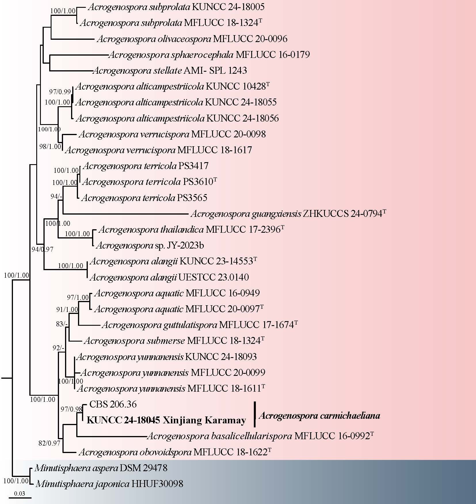
Minutisphaerales Raja, Oberlies, Shearer & A.N. Mill.
Acrogenosporaceae Jayasiri & K.D. Hyde
Acrogenospora M.B. Ellis
Acrogenospora was established by Ellis (1971). It is characterized by macronematous, mononematous, and brown conidiophores that frequently proliferate percurrently. The conidiogenous cells are monoblastic, either terminal or intercalary, and its conidia are globose, ellipsoid, or obovoid, ranging from olivaceous to brown (Hughes 1978; Goh et al. 1998c; Bao et al. 2020; Harrington et al. 2022; Li et al. 2024b). Initially, Acrogenospora comprised two species, A. carmichaeliana and A. sphaerocephala, both described solely by their asexual morphs. Goh et al. (1998c) expanded the genus by adding A. ovalia and A. subprolata, while Bao et al. (2020) further refined its taxonomy by accommodating nine species, including seven new taxa and two previously described ones, based on phylogenetic and morphological analyses. Currently, Acrogenospora encompasses 27 species, primarily from freshwater habitats (21 species), and terrestrial habitats (six species) (Bao et al. 2020; Li et al. 2024b; Cao et al. 2025; Xu et al. 2025).
1. Acrogenospora carmichaeliana (Berk.) Rossman & Crous, in Rossman, Crous & Hyde, IMA Fungus 6(2): 509 (2015) Fig. 2
Chinese name
Index Fungorum number: IF 814513; Facesoffungi number: FoF 08065
Saprobic on decaying wood submerged in freshwater stream. Sexual morph: Undetermined. Asexual morph: Colonies effuse on natural substrate, hairy, dark brown, with masses of conidia on conidiophores. Mycelium mostly immersed, composed of grayish-brown, septate, branched, smooth hyphae. Conidiophores 267–354 × 8–10 µm ( =308 × 8 μm, n = 10), mononematous, macronematous, solitary, erect, straight or slightly flexuous, cylindrical, unbranched, brown to dark brown, paler toward apex, septate, smooth-walled. Conidiogenous cells holoblastic, monoblastic, integrated, initially terminal, later becoming intercalary, cylindrical, smooth-walled, pale brown, proliferating percurrently. Conidia 31–38 × 25–31 µm ( = 35 × 28 μm, n = 25), acrogenous or acropleurogenous, solitary, spherical or subspherical, truncate at base, aseptate, hyaline when young, dark brown to black when mature, smooth-walled.
Material examined: CHINA, Xinjiang Autonomous Region, Karamay City, Lengshuigou, 43°58′11.99" N, 85°6′47.30" E, 1785 m a.s.l., on decaying wood submerged in a freshwater stream, 16 July 2021, R.J. Xu, MD-369 (HKAS 136217), living culture KUNCC 24-18045.
Notes: The taxonomic status of Acrogenospora carmichaeliana and its relationship with Farlowiella carmichaeliana have long been complex issues in mycological research. Historically, based on culture studies, Acrogenospora megalospora was considered as the asexual morph of Farlowiella carmichaeliana. Ellis (1971a) reported the asexual morph of F. carmichaeliana as Acrogenospora carmichaeliana, and Goh et al. (1998c) confirmed this relationship and synonymized A. carmichaeliana under Farlowiella carmichaeliana. The asexual-sexual connection between these two genera (Farlowiella and Acrogenospora) was also confirmed by Jayasiri et al. (2018) and Hyde et al. (2019) based on phylogenetic analysis. Therefore, Acrogenospora was given priority over Farlowiella. However, Jayasiri et al. (2018) found that Acrogenospora sphaerocephala clustered with Farlowiella carmichaeliana (the sexual morph of Acrogenospora carmichaeliana), supporting their connection. However, recent studies, Hyde et al. (2019) and Bao et al. (2020), showed that Acrogenospora sphaerocephala forms different clades from Acrogenospora carmichaeliana. Thus, whether A. sphaerocephala was the asexual morph of Farlowiella carmichaeliana and whether Acrogenospora megalospora was wrongly introduced as the asexual morph of Farlowiella carmichaeliana needs further study. In this study, our collection KUNCC 24-18045 is identified as Acrogenospora carmichaeliana based on morphological traits and molecular evidence (Fig. 1).
Pleosporales Luttrell ex M.E. Barr
Corynesporascaceae Sivan.
Corynespora Güssow, J
Corynesporasca, established by Sivanesan (1996) and typified by C. carotae, represents the sexual morph of a Corynespora species found on tropical plants. Corynespora typified by C. mazei and now synonymised with C. cassiicola, comprises about 200 species. The leaf spot fungus Corynespora cassiicola is particularly widespread, infecting numerous plant hosts predominantly in tropical regions (Smith et al. 2007). Due to the extensive usage and historical priority of the name Corynespora, especially in the context of plant pathogenic fungi, it is recommended to continue using this name. Corynespora species are mainly tropical and subtropical, found on a wide range of plant hosts, and known for causing leaf spot diseases. They exhibit notable morphological features such as distinct, determinate or percurrently extending conidiophores and monotretic, integrated, terminal conidiogenous cells producing solitary or sometimes catenate, distoseptate conidia. Liu et al. (2023a) and Zhang et al. (2025) provided a detailed description and explanation of the genus.
2. Corynespora yunnanensis Jing W. Liu & Jian Ma, in Liu, Hu, Luo, Castañeda-Ruíz, Xia, Xu, Cui, Shi, Zhang & Ma, Journal of Fungi 9 (no. 107): 11 (2023) Fig. 4
Chinese name: –
Index Fungorum number: IF 900078; Facesoffungi number: FoF 17390
Saprobic on decaying stems of wood submerged in a freshwater stream habitat. Sexual morph: Undetermined. Asexual morph: Colonies superficial, effuse, velvety, gregarious, pale brown, Mycelium immersed, subhyaline to pale brown, composed of branched, septate hyphae. Conidiophores 406–603 × 12–19 µm ( = 490 × 16 μm, n = 20), macronematous, mononematous, erect, straight or slightly flexuous, solitary or group, tapering towards the apex, septate, unbranched, cylindrical, dark brown or brown, becoming paler towards the apex, with several successive cylindrical extensions. Conidiogenous cells 21–38 × 10–16 µm ( = 29 × 12 μm, n = 20), integrated, terminal, monotretic, cylindrical, pale brown to brown, smooth. Conidia 122–154 × 20–28 µm ( = 134 × 23 μm, n = 25), acrogenous, solitary, obclavate, rostrate, rounded at the apex, straight or slightly curved, multi-distoseptate, olivaceous to brown, smooth-walled, truncate at the base.
Culture characteristics: Conidia germinating on PDA medium within 24 h, and germ tubes produced from both ends. Colonies grown on PDA reached 60 mm diameter at room temperature in two weeks, circular, with velvety, flat, brown mycelium on the surface, from below dark brown with entire margin.
Material examined: CHINA, Xizang Autonomous Region, Shannan Prefecture, Cuona City, Lebugou, 27°52′38.19" N, 91°47′54.64" E, 3560 m a.s.l., on decaying wood submerged in a freshwater stream, 3 August 2023, R.J. Xu, LJN-45 (HKAS 135995), living culture KUNCC 24-17961.
Notes: Corynespora yunnanensis was isolated from dead branches of an unidentified tree in Yunnan Province, China (Liu et al. 2023a). Morphologically, our collection (KUNCC 24-17961) shares identical morphological characters to the holotype of C. yunnanensis but has larger conidia (122–154 × 20–28 vs. 80–128 × 16–19 µm). The phylogenetic position of our strain is unclear (Fig. 3). However, comparisons of ITS sequences demonstrate 100% similarity between the ex-type strain HJAUP M2132 and KUNCC 24-17961. Therefore, based on the current evidence, we tentatively retain our strain as C. yunnanensis. Further collections and additional evidence are needed to clarify their relationship.
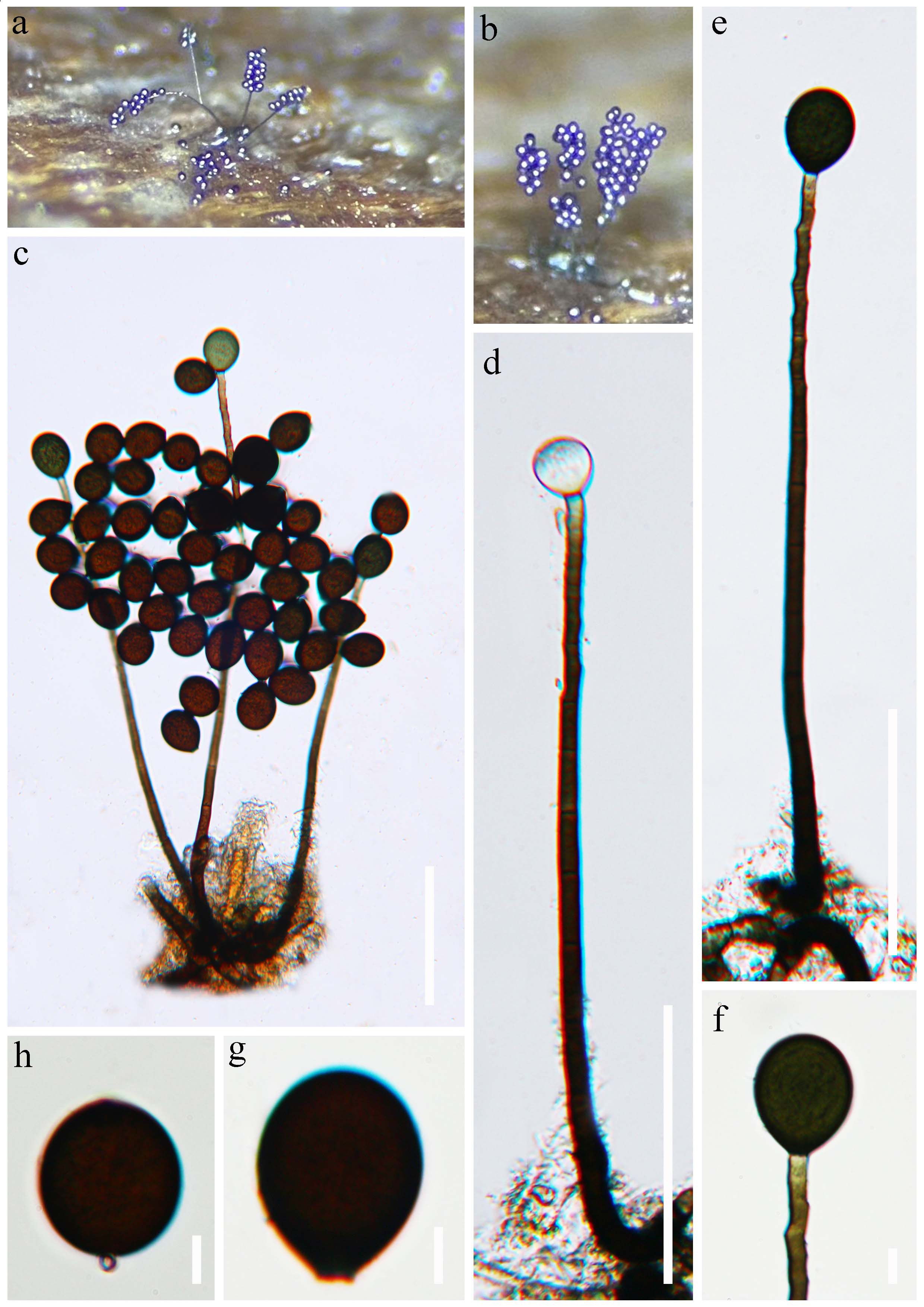
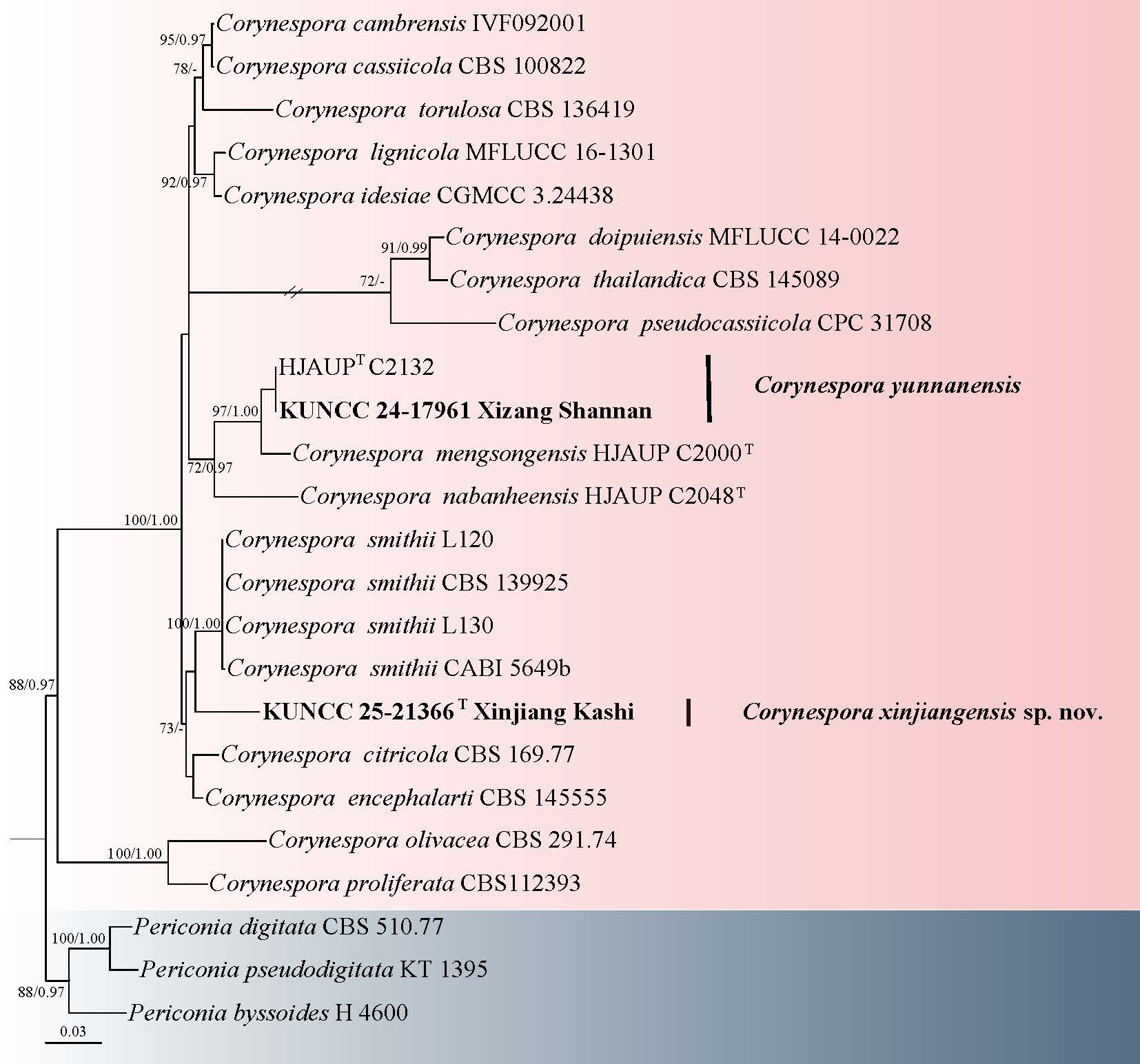
3. Corynespora xinjiangensis R.J. Xu, Q. Zhao & K.D. Hyde, sp. nov. Fig. 5
Chinese name: –
Index Fungorum number: IF 905026; Facesoffungi number: FoF 19296
Etymology: Refers to Xinjiang Autonomous Region in China, where the type specimen of this species was collected.
Holotype: HKAS 151641
Saprobic on decaying wood in a freshwater habitat. Sexual morph: Undetermined. Asexual morph: Colonies on natural substratum, superficial, effuse, filiform, pale brown. Mycelium mostly immersed, composed of branched, septate, pale brown to brown, smooth-walled hyphae. Conidiophores macronematous, mononematous, fasciculate, filamentous, cylindrical, septate, unbranched, pale brown to brown, sometimes with multiple consecutive proliferations. Conidiogenous cells monotretic, integrated, terminal, cylindrical, pale brown to brown, smooth-walled, often percurrently proliferating. Conidia 63–114 × 6–35 µm ( = 84 × 12 μm, n = 30), acrogenous, solitary, obclavato-cylindrical, straight to slightly flexuous, light olivaceous-brown, truncate at the base, obtuse to rounded at the apex, distoseptate, sometimes proliferates 1 to 3 times at the tip of the conidia.
Culture characteristics: Conidia germinating on PDA medium within 24 hours, with germ tubes arising from both ends of the conidium. Colonies grown on PDA reaching 60 mm diam at room temperature under natural light after two weeks, circular, flat, with velvety, with filiform edge, brown to dark brown from above and below.
Material examined: CHINA, Xinjiang Autonomous Region, Kashi Prefecture, Yecheng City, 37° 45' 8.87" N, 77° 25' 7.71" E, 1490 m a.s.l., on decaying wood submerged in a freshwater habitat, 18 July 2021, R.J. Xu, MD-172A (HKAS 151641, holotype), ex-type culture KUNCC 25-21366.
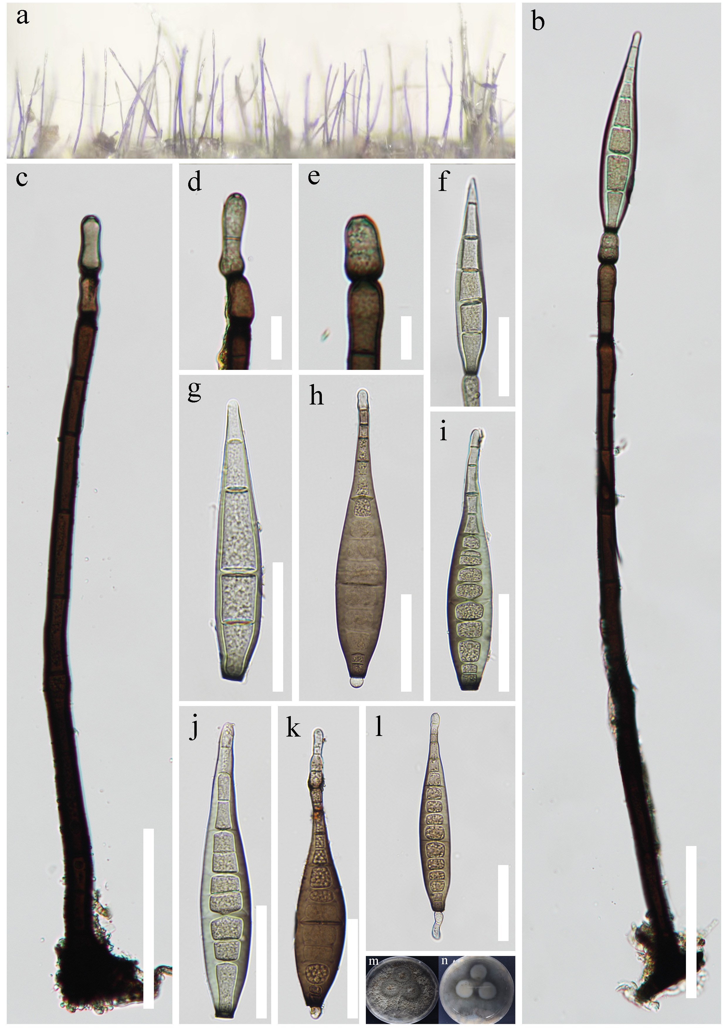
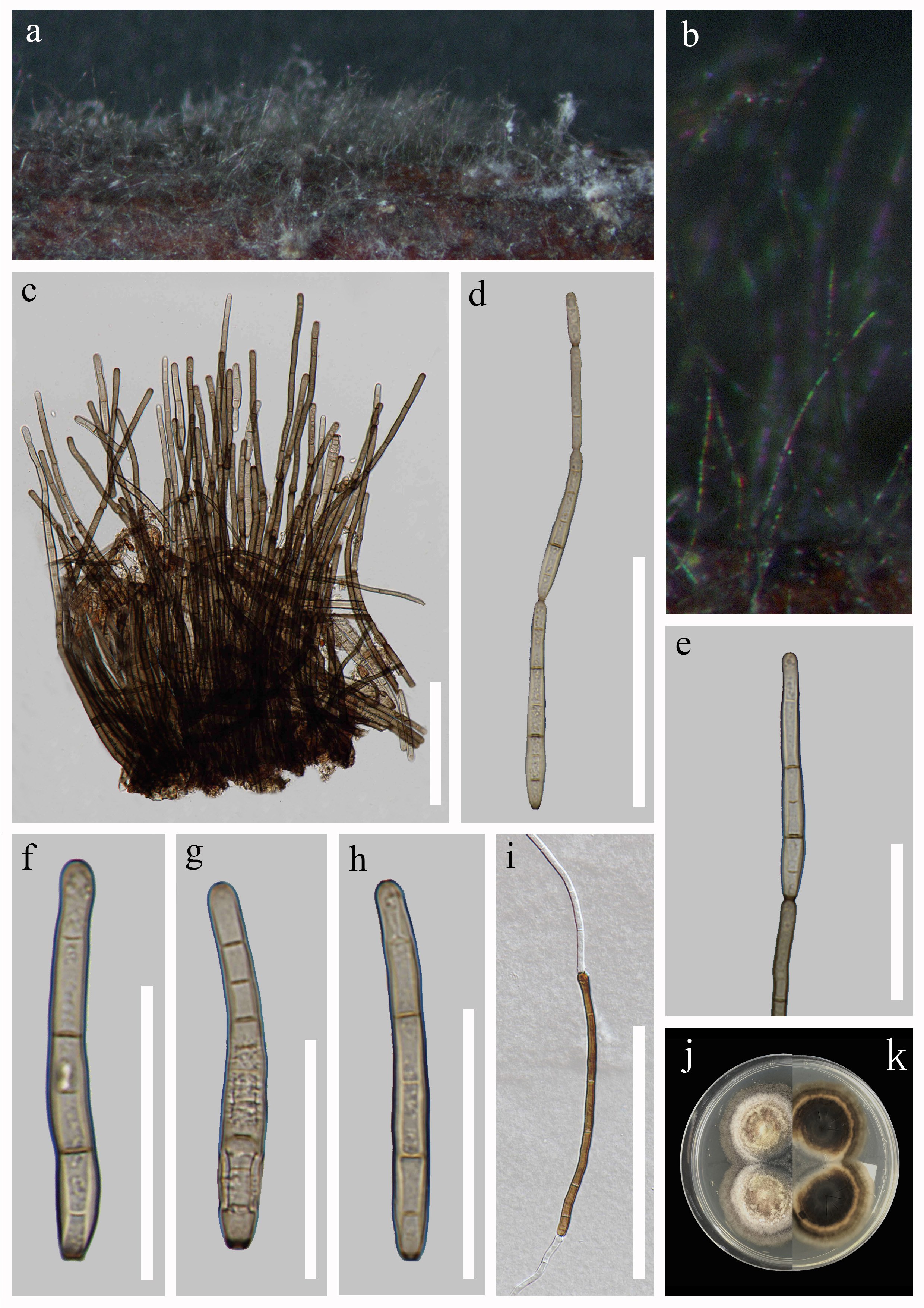
Notes: Corynespora xinjiangensis is morphologically typical of Corynespora, characterized by macronematous, mononematous conidiophores, monotretic conidiogenous cells, and obclavato-cylindrical, distoseptate conidia (Smith et al. 2007). The conidia of C. xinjiangensis are relatively large, measuring 63–114 × 6–35 µm, with an average size of 84 × 12 µm, with 1–3 proliferations at the apex. Corynespora xinjiangensis differs from C. mengsongensis and C. nabanheensis, by having short, multiple consecutive proliferations conidiophores and obclavato-cylindrical conidia (Liu et al. 2023a). Furthermore, a comparison of the ITS nucleotide bases in C. xinjiangensis (KUNCC 25-21366) and C. smithii (MFLU 19-0688) revealed that they differ in 83/869 bp (9.55%) of ITS, and the phylogenetic analyses (Fig. 3) place C. xinjiangensis in a distinct lineage, supporting its separation from the known species.
Dictyosporiaceae Boonmee & K.D. Hyde
Dictyocheirospora M.J. D'souza, S. Boonmee & K.D. Hyde
Dictyocheirospora was established by Boonmee et al. (2016) with D. rotunda as the type species. Dictyocheirospora is distinguished from its close relative genus Dictyosporium, by its unique conidial morphology. The conidia in Dictyocheirospora are cheiroid, non-complanate, or cylindrical, often tightly grouped at the apex, contrasting with the conidial arrangement in Dictyosporium (Boonmee et al. 2016; Wang et al. 2016a; Shen et al. 2022). The asexual morphs of Dictyocheirospora are characterized by dark sporodochial colonies producing aeroaquatic conidia, while its sexual morphs are noted for subglobose ascomata, cylindrical asci, and hyaline, fusiform, uniseptate ascospores, with or without a sheath (Boonmee et al. 2016; Tibpromma et al. 2018; Liu et al. 2023b; Tennakoon et al. 2023)
The phylogenetic placement of Dictyocheirospora within the Dictyosporiaceae family has been validated through analyses employing multiple-gene phylogenies in various studies (Liu et al. 2015a; Tanaka et al. 2015; Boonmee et al. 2016). Dictyocheirospora currently comprises 33 accepted species, identified through both morphological characteristics and phylogenetic analysis (Boonmee et al. 2016; Wang et al. 2016a; Hyde et al. 2017; Hyde et al. 2019; Yang et al. 2018a; Tibpromma et al. 2018; Jayasiri et al. 2019; Phookamsak et al. 2019; Dong et al. 2020; Phukhamsakda et al. 2020; Shen et al. 2022; Tennakoon et al. 2023; Wang et al. 2025c; Sun et al. 2025).
4. Dictyocheirospora luojiensis R.J. Xu, Q. Zhao & K.D. Hyde, sp. nov. Fig. 7
Chinese name: –
Index Fungorum number: IF 903471; Facesoffungi number: FoF 17391
Etymology: Referring to the location “Luoji River” where the holotype of this fungus was collected.
Holotype: HKAS 136148
Saprobic on decaying stems of wood submerged in a freshwater river. Sexual morph: Undetermined. Asexual morph: Colonies sporodochial, punctiform, scattered or in small groups, compactly, water-stained, dark brown to black. Mycelium immersed, subhyaline to pale brown, composed of branched, septate hyphae. Conidiophores macronematous, mononematous, septate, cylindrical, pale brown to subhyaline, smooth-walled, sometimes reduced to conidiogenous cells. Conidiogenous cells holoblastic, cylindrical to subglobose, hyaline to pale brown, smooth-walled. Conidia 56–71 × 22–40 µm ( = 64 × 28 μm, n = 25), acrogenous, solitary, cheiroid, brown or dark brown, closely appressed, composed of seven rows of cells, non-complanate, cylindrical, each row with 8–12 cells. Conidial secession was schizolytic.
Culture characteristics: Conidia germinating on PDA medium within 72 h and germ tubes arising from terminal end of conidium. Colonies grown on PDA reached 30 mm diam at room temperature in one month, circular, with velvety, raised, white mycelium on the surface, from below cream to light brown with entire margin.
Material examined: CHINA, Yunnan Province, Shangri-La City, Luoji River, 27°42′23.30" N, 99°59′31.51" E, 3539 m a.s.l., on decaying wood submerged in a freshwater river, 18 June 2022, R.J. Xu, LTS-25 (HKAS 136148, holotype), ex-type culture KUNCC 24-17999.
Notes: Phylogenetic analysis combining SSU, ITS, LSU and tef1-α sequence data showed that Dictyocheirospora luojiensis clustered within the Dictyocheirospora clade and formed an independent lineage (Fig. 6). Dictyocheirospora luojiensis is closely related to D. lithocarpi, D. multiappendiculata, and D. suae (Fig. 6). Dictyocheirospora luojiensis is morphologically similar to these species in having cheiroid, ellipsoid to cylindrical, not complanate, septate, closely appressed conidia, and with or without appendages (Jayasiri et al. 2019; Shen et al. 2022). However, D. luojiensis can be distinguished from D. lithocarpi by its larger conidia (56–71 × 22–40 µm vs. 35–40 × 12–18 μm) with appendages, and with more rows of cells (7 rows vs. 6 rows) (Jayasiri et al. 2019). Dictyocheirospora luojiensis differs from D. multiappendiculata by having larger conidia (56–71 × 22–40 µm vs. 55–62 × 19–22 µm) and rare appendages, whereas appendages are common in D. multiappendiculata (Shen et al. 2022). Dictyocheirospora luojiensis differs from D. suae by its smaller conidia (56–71 × 22–40 µm vs. 72–79 × 20–25 µm), and fewer cells in each row of cells (8–12 cells vs. 12–15 cells) (Shen et al. 2022). Conidial size is one of the important criteria for distinguishing dictyosporium-like taxa (Goh et al. 1999). Based on the unique morphological characters and phylogenetic distance, we therefore introduce D. luojiensis as a new species here.
Massarinaceae Munk
Helminthosporium Link
Helminthosporium was introduced by Link (1809) with H. velutinum as the type species. Species of Helminthosporium are widely distributed and mainly saprobic on a broad range of natural substrates such as decaying wood, plant stems, bark, dung, and occasionally insects, and some species have been reported as opportunistic human pathogens (Luttrell 1964; Alcorn 1988; Konta et al. 2023; Hyde et al. 2023, 2024). The genus is classified in Massarinaceae (Pleosporales), and is characterized by macronematous, cylindrical, septate, erect conidiophores with polytretic conidiogenous cells and acropleurogenous, clavate or obclavate, distoseptate conidia with a flat, ringed pore at the base (Luttrell 1964; Liu et al. 2022; Hyde et al. 2023). Although over 780 names have been listed under Helminthosporium (Index Fungorum 2026), many were reclassified based on morphological inconsistencies with the characteristics of the genus. In addition, recent phylogenetic analyses based on molecular data indicate that Helminthosporium is polyphyletic (Konta et al. 2021; Chen et al. 2022). In this study, we introduce a new species of Helminthosporium based on morphological characteristics and phylogenetic analysis.
5. Helminthosporium xinjiangense R.J. Xu, Q. Zhao & K.D. Hyde, sp. nov. Fig. 9
Chinese name
Index Fungorum number: IF 905028; Facesoffungi number: FoF 19297
Etymology: Refers to Xinjiang Autonomous Region in China, where the type specimen of this species was collected.
Holotype: HKAS 151642
Saprobic on decaying wood in a freshwater habitat. Sexual morph: Undetermined. Asexual morph: Colonies on natural substratum, superficial, effuse, scattered, hairy, dark brown. Mycelium mostly immersed, composed of branched, septate, brown to dark brown, smooth-walled hyphae. Conidiophores 214–433 × 9–12 μm ( = 329 × 11 μm, n = 20), macronematous, mononematous, solitary or in groups, erect, cylindrical, straight or flexuous, unbranched, dark brown to black, smooth-walled. Conidiogenous cells 47–112 × 5–9 μm ( = 75 × 8 μm, n = 20), polytretic, integrated, terminal and intercalary, cylindrical, brown, smooth-walled. Conidia 55–89 × 8–11 μm ( = 66 × 10 μm, n = 20), acropleurogenous, solitary, obclavate-rostrate, straight or curved, usually curved, tapering towards the rounded apex, brown, subhyaline at the apex, 6–9-distoseptate, smooth-walled, with a conspicuous black scar at the base.
Culture characters: Conidium germinating on PDA within 24 h and germ tubes produced from both ends or middle. Colonies on PDA reaching 10 mm diam., in a week at room temperature, effuse, hairy, mycelium radiating outwards, fimbriate edge, dense, pale. Mycelium superficial and partly immersed, light brown.
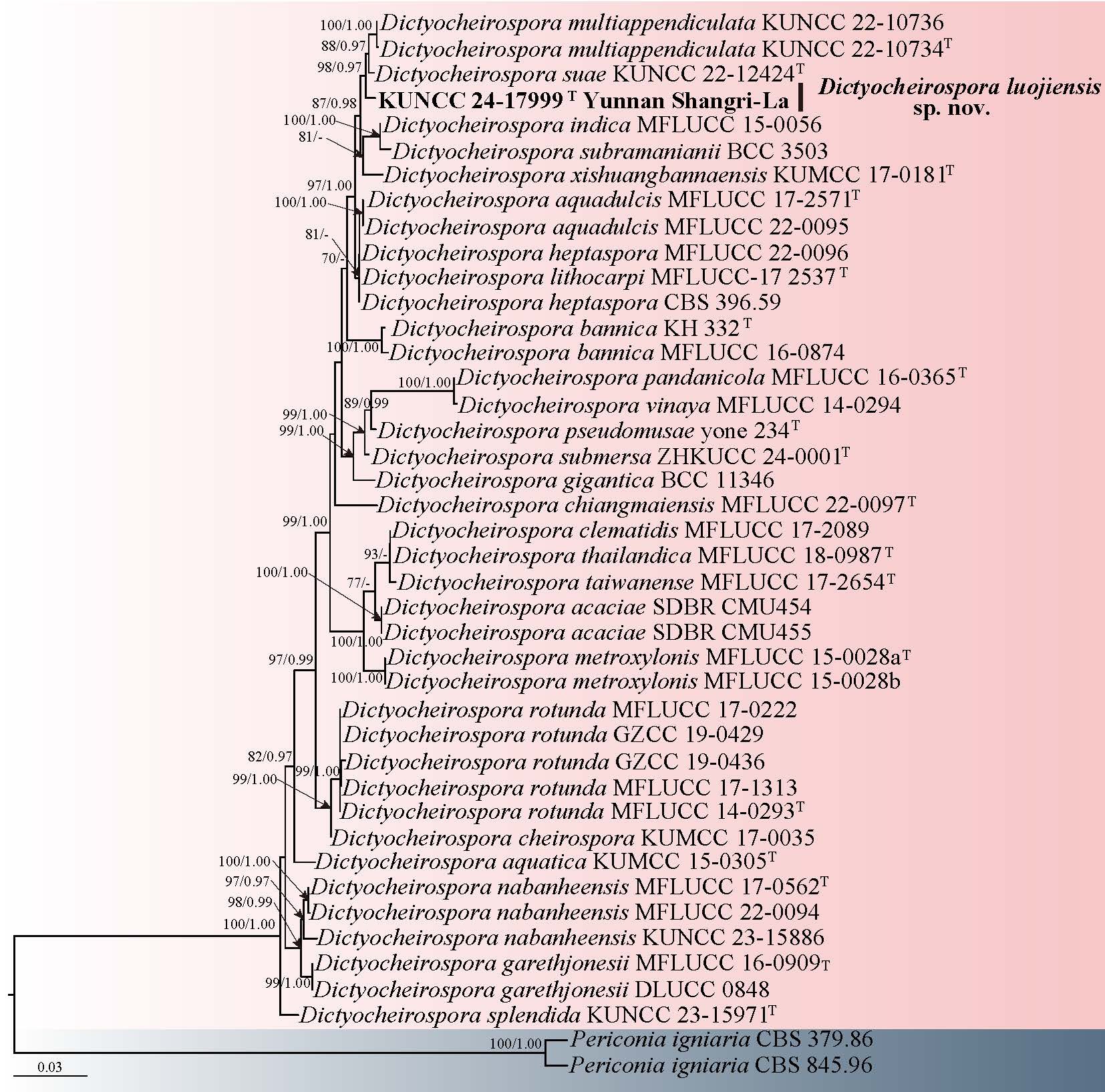
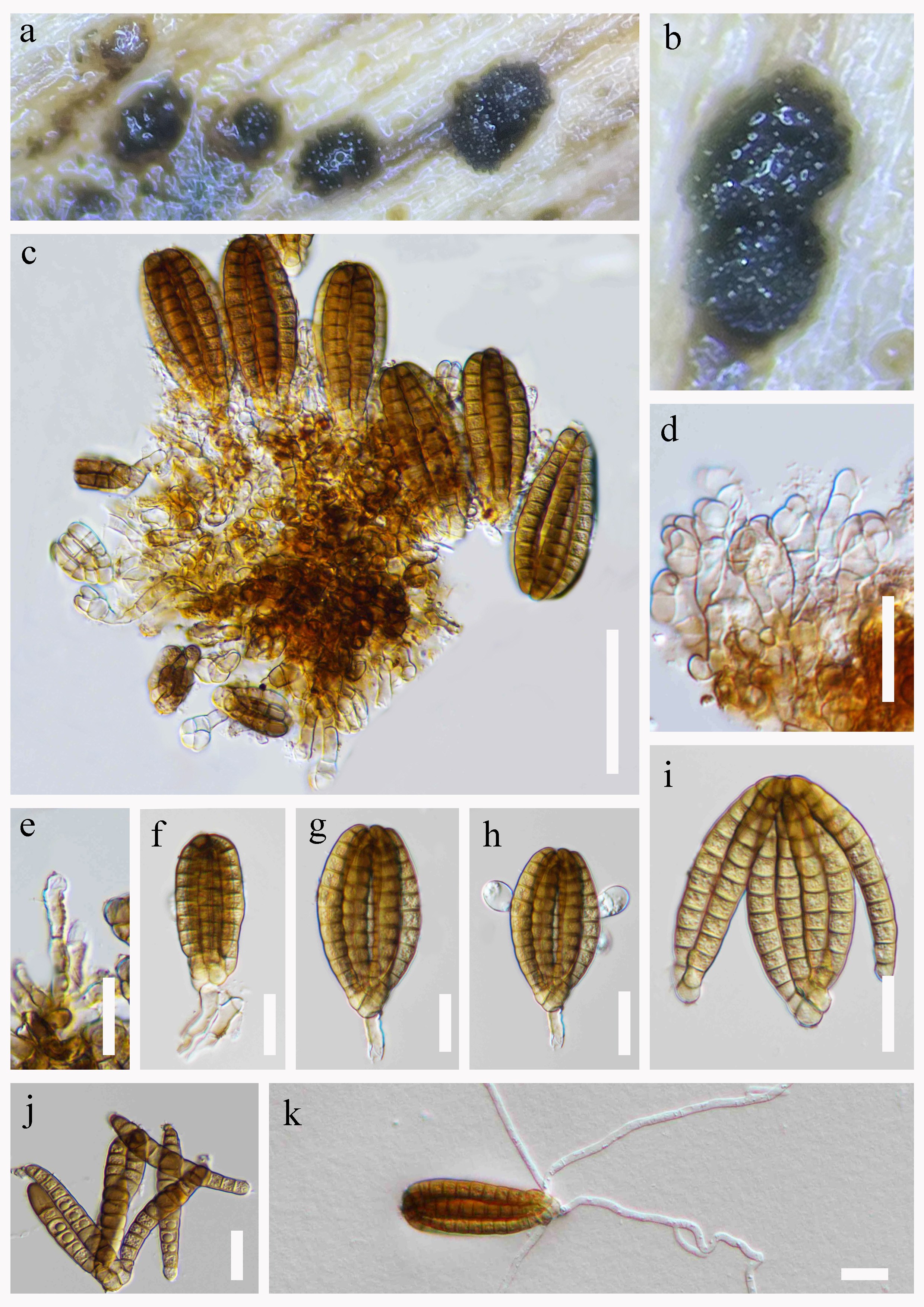
Material examined: CHINA, Xinjiang Autonomous Region, Kashi, Yecheng City, 37° 45' 8.87" N, 77° 25' 7.71" E, 1490 m a.s.l., on decaying wood submerged in a freshwater habitat, 18 July 2021, R.J. Xu, MD-176A (HKAS 151642, holotype), ex-type culture KUNCC 25-21367.
Notes: In phylogenetic analyses, our isolate formed a sister clade with Helminthosporium austriacum and H. yunnanense (Fig. 8). Sequence comparison of the ITS region
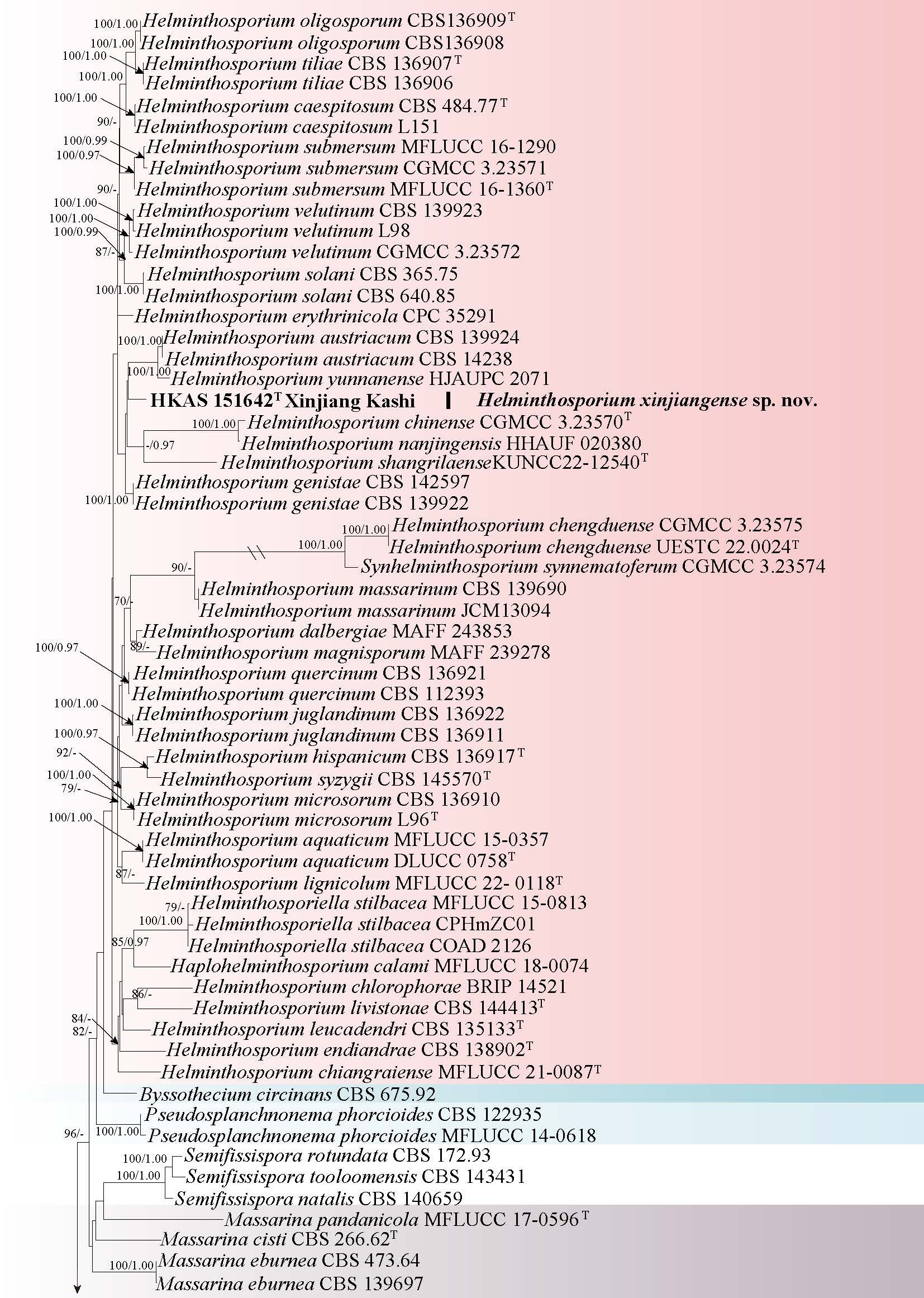
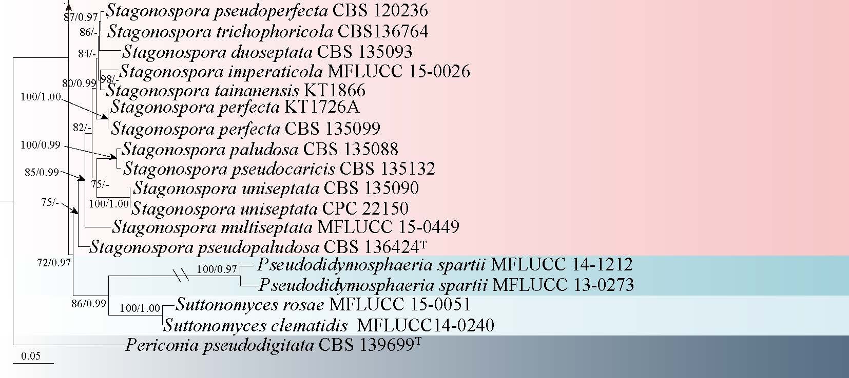
showed nucleotide differences of 13.64% (70/513 bp, excluding gaps) and 15.20% (78/513 bp, excluding gaps) between H. xinjiangense and H. austriacum, H. xinjiangense and H. yunnanense, respectively. Morphologically, H. xinjiangense differs from H. austriacum by having shorter conidiophores (214–433 × 9–12 μm vs. 275–700 × 11.5–19 μm), and more slender, usually curved conidia (55–89 × 8–11 μm vs. 35–48 × 13.7–16.5 μm) (Voglmayr et al. 2017), and differs from H. yunnanense in shorter conidiophores (214–433 × 9–12 μm vs. 560–680 × 12.5–15.5 μm) without one cylindrical, enteroblastic percurrent extension, and larger, obclavate-rostrate conidia (55–89 × 8–11 μm vs. 30.5–55.5 × 9–11 μm) with a higher number of distosepta (6–9 vs. 4–7) (Liu et al. 2022). Therefore, we introduce H. xinjiangense as a new species.
Hermatomycetaceae Locq. ex A. Hashim. & Kaz. Tanaka
Hermatomycetaceae was informally introduced by Locquin (1984) and later validated by Hashimoto et al. (2017) to accommodate the monotypic genus Hermatomyces. Members of Hermatomycetaceae are saprobic on various plant substrates. The sexual morph has not yet been observed. Asexual structures include sporodochial, pulvinate conidiomata that are dark brown to black. Conidiophores are pale brown and mononematous, bearing monoblastic, integrated, terminal conidiogenous cells that are cylindrical (Castañeda & Heredia 2000; Ren et al. 2021).
Hermatomyces Speg
Hermatomyces has a wide geographic range, occurring in both tropical and temperate regions, including Brazil, China, Cuba, Japan, Panama, the Philippines, Thailand, and Venezuela (Doilom et al. 2017; Hashimoto et al. 2017b; Koukol et al. 2018; Tibpromma et al. 2018). To date, host specificity within Hermatomyces remains unclear, as species have been identified on hosts from multiple plant families, including Acanthaceae, Arecaceae, Asteraceae, Fabaceae, and Pandanaceae (Doilom et al. 2017; Hashimoto et al. 2017; Koukol et al. 2018; Tibpromma et al. 2018). The sexual-asexual connection in Hermatomyces has not yet been established, since no sexual morph has been documented for any species in the genus (Hashimoto et al. 2017b; Tibpromma et al. 2018). Up to now, 34 species have been accepted within Hermatomyces (Hyde et al. 2024a; Shen et al. 2024; Koukol et al. 2025).
6. Hermatomyces motuoensis R.J. Xu & Q. Zhao, sp. nov. Fig. 11
Chinese name
Index Fungorum number: IF 905256; Facesoffungi number: FoF 19298
Etymology: The epithet refers to Motuo County, located in Xizang of China, where the type specimen was collected.
Holotype: HKAS 151643
Saprobic on decaying wood in a freshwater habitat. Sexual morph: Undetermined. Asexual morph: Colonies on natural substrate effuse, superficial, scattered, conspicuous, flattened, consisting of a sterile mycelial outer zone and a round, glistening, brown to black. Mycelium mostly immersed, composed of branched, septate, brown to dark brown, smooth-walled hyphae. Conidiophores micronematous, mononematous, unbranched, straight or flexuous, hyaline, septate, smooth. Conidiogenous cells 11–20 × 2–7 μm ( = 16 × 4 μm, n = 20), holoblastic, monoblastic, integrated, terminal, cylindrical, hyaline to subhyaline. Conidia 19–33 × 17–27 μm ( = 28 × 22 μm, n = 30), dimorphic, thick-walled, smooth. lenticular conidia, subglobose to ellipsoidal, muriform, smooth, central cells dark brown to black, peripheral cells narrow, flattened, subhyaline to pale brown, obovoid or oblong in side view, cylindrical conidia, 2–3-celled, symmetrical or asymmetrical, distinctly constricted at the septum, guttulate, straight or flexuous, hyaline, the upper cells usually subcylindrical and the apex rounded or slightly flattened, often slightly attenuated and dark brown or black, the lower cells are ellipsoidal.
Cultural characteristics: Conidia germinating on PDA and germ tubes produced from peripheral cells of conidium within 24 h. Colonies on PDA, reaching 30 mm after one month at room temperature, colony circular, edge smooth, surface hyphae grey-brown, fluffy, flocculent, reverse side grey-brown to brown, dense, cracked in the middle.
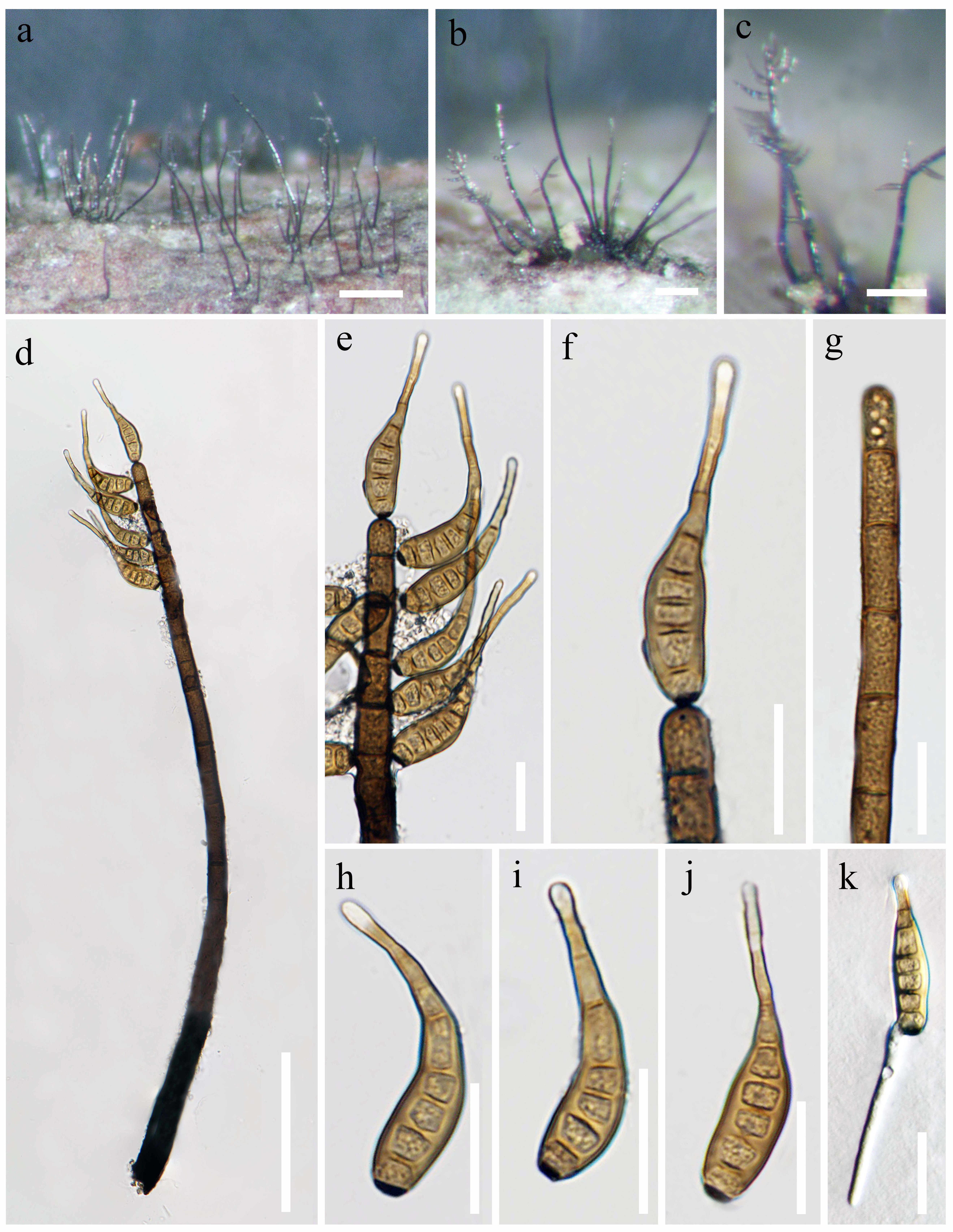
Material examined: CHINA, Xizang Autonomous Region, Linzhi City, Motuo County, Beibong Township, 29° 14′ 35.76″ N, 95° 10′ 6.94″ E, 1278 m a.s.l., on decaying wood submerged in a freshwater stream, 12 July 2022, R.J. Xu, MD-200A (HKAS 151643, holotype), ex-type culture KUNCC 25-21368.
Notes: Phylogenetic analysis revealed that Hermatomyces motuoensis clustered with H. hongheensis, H. constrictus, H. iriomotensis, H. krabiensis and H. griseomarginatus with 84% ML/0.68 BYPP support (Fig. 10). Morphologically, H. motuoensis displays dimorphic conidia, characterized by both lenticular and cylindrical forms. This morphological trait aligns with the conidial characteristics observed in H. constrictus, H. iriomotensis, and H. krabiensis, thereby indicating a potential phylogenetic relationship among these taxa (Spegazzini 1910; Tibpromma et al. 2016; Hashimoto et al. 2017; Koukol et al. 2018; Ren et al. 2021). However, H. motuoensis can be distinguished from H. constrictus in having larger conidiogenous cells (11–20 μm vs. 4–7 μm), and the cylindrical conidia with more cells (2–3-cells vs. 2-cells) (Koukol et al. 2018). Compared to H. iriomotensis, H. motuoensis has smaller lenticular conidia (28 × 22 μm avg. vs. 33.6 × 23.5 μm, avg.) and fewer septa (1–2-septate vs. 3–7-septate) in cylindrical conidia (Tibpromma et al. 2016). Furthermore, a megablast search of NCBI's GenBank nucleotide database, the best matching result of the ITS sequence is H. bifurcatus (voucher: PMA:116075, sequence ID: LS398263, similarities: 616/656 (94%), 10 gaps. Therefore, based on morphological and molecular sequence evidence, H. motuoensis is herein introduced as a novel species.
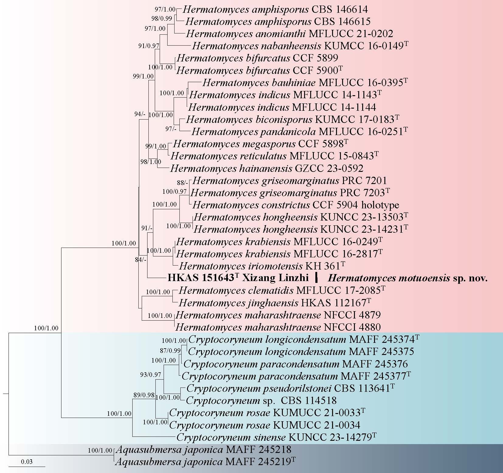
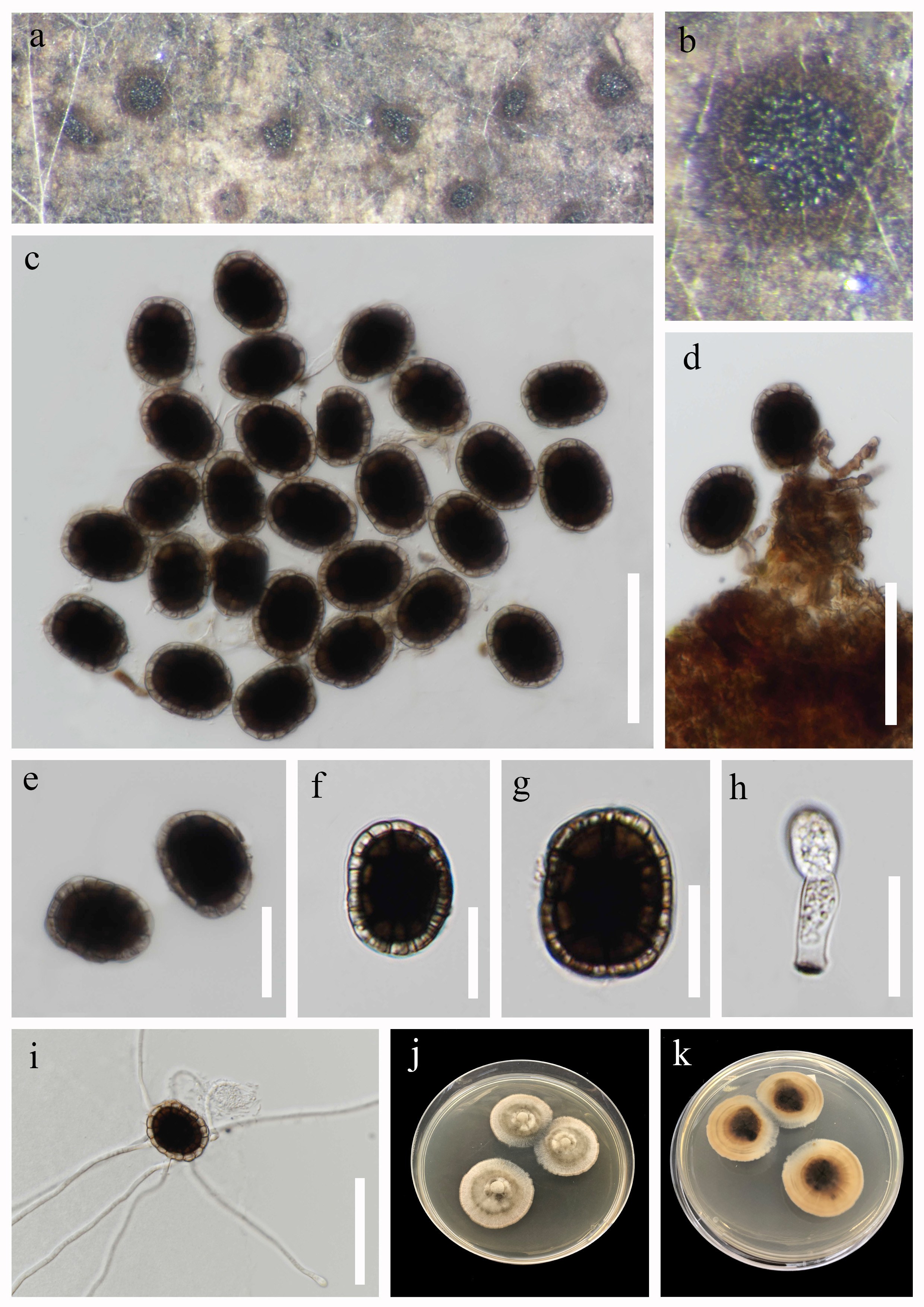
Periconiaceae Nann
Periconia Tode
Periconiaceae was delineated by Nannizzi (1934) and represented by the genus Periconia. The family was resurrected by Tanaka et al. (2015) and placed in Pleosporales based on morphological and phylogenetic evidence. Currently, the family comprises a single genus Periconia. Subsequent taxonomic reassessments by Yang et al. (2022a) led to the synonymization of Bambusistroma and Noosia under Periconia, based on morphological traits observed in sexual reproductive structures and multilocus phylogenetic studies. Additionally, Liao et al. (2024a) suggest that Flavomyces fulophazii should be synonymized with Periconia. However, further corroborative data is essential to affirm the taxonomic placement of F. fulophazii.
Periconia was introduced by Tode (1790) with P. lichenoides as the type species. Periconia was re-classified by Tanaka et al. (2015) based on the multi-locus phylogenetic analysis and placed in Periconiaceae. Species of Periconia are mostly reported as saprobes as their asexual morph, and also have some species as endophytes and pathogens, such as P. endophytica was isolated as an endophytic fungus from asymptomatic leaves of Wurfbainia villosa in China (Liao et al. 2024a). Periconia circinata was isolated as a pathogen causing blackening and rotting of wheat roots and stem bases (Goga 2000). To date, Periconia has 201 epithets listed in Species Fungorum.
7. Periconia xizangensis R.J. Xu, Q. Zhao & K.D. Hyde, sp. nov. Fig. 13
Chinese name
Index Fungorum number: IF 905030; Facesoffungi number: FoF 17397
Etymology: The epithet refers to Xizang, China, where the type specimen was collected.
Holotype: HKAS 136110
Saprobic on decaying wood submerged in a freshwater stream. Sexual morph: Undetermined. Asexual morph: Colonies effuse, hairy, velvety, scattered or in small groups, with glistening, pale brown to brown conidial masses at the apex. Mycelium is mostly superficial, partially immersed, consisting of branched, septate, smooth, subhyaline hyphae. Conidiophores 366–601 × 13–19 μm ( = 467 × 16 μm, n = 20), macronematous, mononematous, solitary, erect, unbranched or occasionally 2–3-branched, septate, straight or flexuous, cylindrical, tapering towards at the apex, with cylindrical to elliptical heads at the apex, often swollen at the base, dark brown at the base, light brown towards the apex. Conidiogenous cells polyblastic, pale brown to brown, terminal, subglobose or elliptical, smooth-walled. Conidia 6–8.5 × 5.6–8 μm ( = 7.3 × 7.3 μm, n = 40), solitary or aggregated in masses, globose, brown, aseptate, verruculose.
Culture characteristics: Conidia germinating on PDA medium within 24 h and germ tubes produced from both ends. Colonies grown on PDA reached 60 mm diam at room temperature in two weeks, filamentous, with velvety, flat, dark brown mycelium, aerial mycelium on the surface, from below dark brown with undulate margin.
Material examined: CHINA, Xizang Autonomous Region, Linzhi City, Motuo County, 29°19′43.69" N, 95°21′19.27" E, 798 m a.s.l., on decaying wood submerged in a freshwater stream, 12 July 2022, R.J. Xu, XK-14 (HKAS 136110, holotype), ex-type culture KUNCC 10433.
Notes: Phylogenetic analyses showed that our collection (KUNCC 10433) formed a distinct clade within Periconia, and formed a sister relationship with the ex-type strain of P. thailandica (MFLUCC 17-0065) (Fig. 12). Periconia xizangensis differs from P. thailandica in having conidiophores sometimes with 2–3-branched and cylindrical to elliptical heads (Liu et al. 2017).
Phaeosphaeriaceae M.E. Barr
Phaeosphaeriaceae, typified by Phaeosphaeria, currently comprises 84 genera and remains one of the largest families within Pleosporales (Wanasinghe & Maharachchikumbura 2023). Species in this family exhibit high diversity and are widely distributed in terrestrial environments worldwide, functioning as plant pathogens, saprobes, endophytes, and lichenicolous fungi (Phookamsak et al. 2014). Many species are significant plant pathogens that affect economically important crops, such as Phaeosphaeria and Paraconiothyrium which cause leaf spots on wheat and rice (Phookamsak et al. 2014). Although ongoing research integrating morphological and phylogenetic methods has led to considerable progress in identifying these taxa, the classification is still not fully resolved.
Setophoma Gruyter, Aveskamp & Verkley
Setophoma was originally published to accommodate Phoma terrestris and Pyrenochaeta sacchari (Liu et al. 2019a). Currently, 19 species are listed in Index Fungorum. They were thought to be well-resolved, since many of them are available of molecular data and colour illustrate (Phookamsak et al. 2014). Species in this genus are commonly found in asexual morph, living as plant pathogens (Crous et al. 2014, 2016). Morphologically, they are characterised by phialidic conidiogenous cells, and hyaline, ellipsoidal to subcylindrical, aseptate conidia (Liu et al. 2019a).
8. Setophoma manglietiae H.D. Yang, K.D. Hyde & Q. Zhao, sp. nov. Fig. 15
Chinese name: – 木莲壳霉 (mu lian ke mei)
Index Fungorum number: IF905257; Facesoffungi number: FoF 19299
Etymology: Referring to the host plant Manglietia fordiana (Magnoliaceae).
Holotype: HKAS 154724
Leaf spots amphigenous, angular, brown to grey. Sexual morph: Undetermined. Asexual morph: Colony on PDA grows slowly, reaching 66 mm after 65 days. Upper surface, flat, cottony, white at the beginning, forming pink patches near the centre at the later, undulate to entire edge, lower surface brown with pink plaque, white at the edge. Colony on SNA, upper white, flat, with abundant aerial mycelium, undulate to entire edge, lower surface white. Colony on WA, mycelium sparse, growth zone appears grey. The hyphae measure 1.8–3.3 μm in diameter, are septate, hyaline to lightly pigmented brown, and contain abundant guttules commonly aggregate to form inflated, globose, or bead-like structures.
Material examined: CHINA, Yunnan Province, Kunming City, on leaf of Manglietia fordiana, 14 March 2022, Hong-De Yang14 (HKAS 154724, holotype), ex-type culture KUNCC 23-14872.
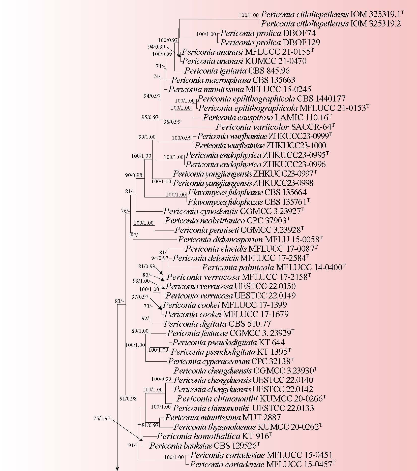
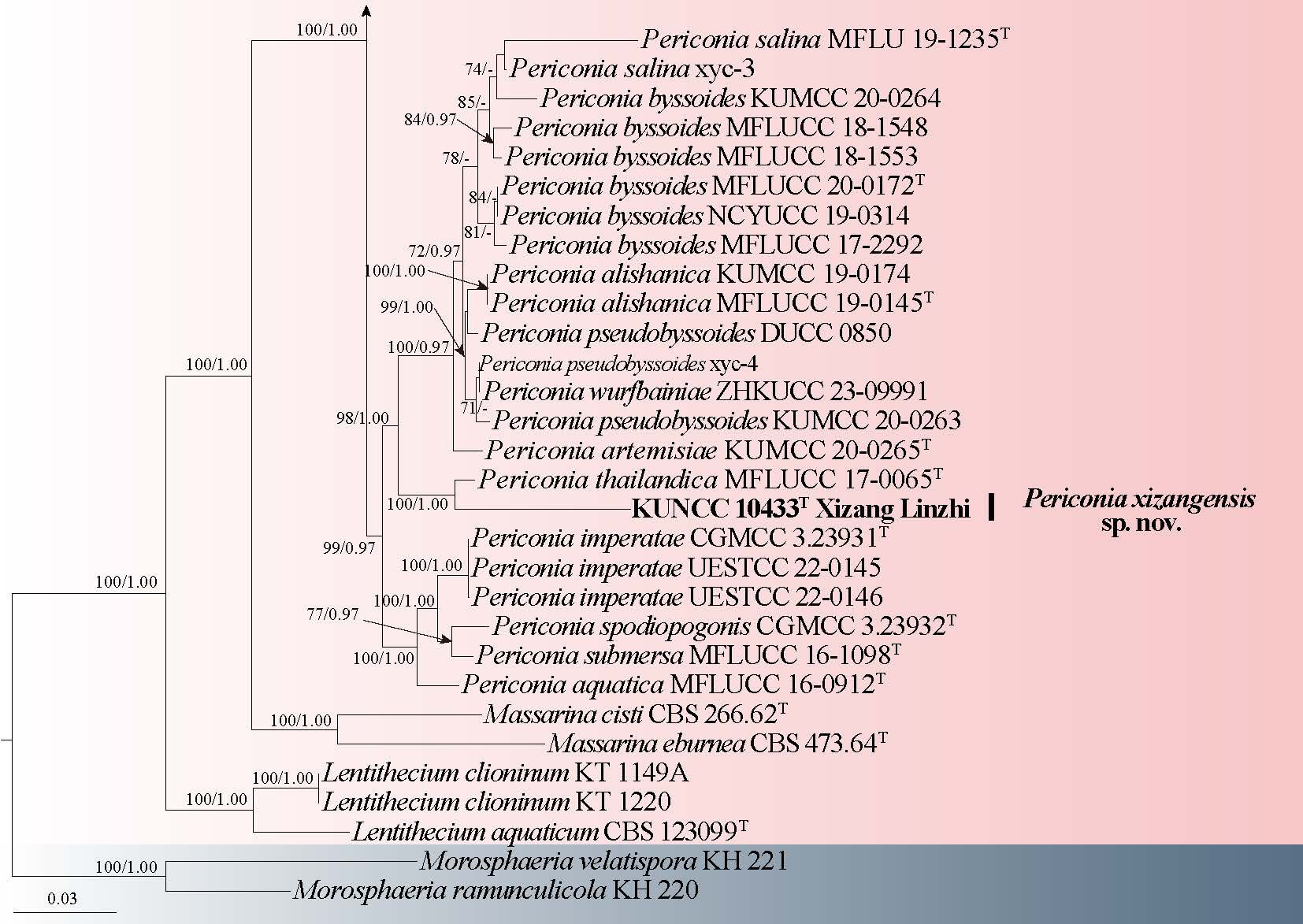
Notes: Setophoma manglietiae exhibits a phylogenetic affinity with S. chromolaenae, S. vernoniae, and S. syzygii (Fig. 14), all of which have been isolated from foliar tissues and associated with leaf pathologies. Our isolate presents shared phenotypic traits, such as the formation of colony patches and an undulate to entire margin; however, it is distinguished by its pigmentation, which varies from white to grey and pink (Quaedvlieg et al. 2013; Crous et al. 2014, 2021). Due to an inability to induce sporulation on various media, including PDA, SNA, and WA, micromorphological characteristics could not be assessed. Consequently, identification was primarily based on colony morphology and molecular data. It is noteworthy that S. chromolaenae, S. vernoniae and S. syzygii are characterized by the production of hyaline, aseptate, subcylindrical conidia (Quaedvlieg et al. 2013; Crous et al. 2014, 2021). Thus, further investigations are warranted to confirm the morphological attributes of our species.
Tetraplosphaeriaceae Kaz. Tanaka & K. Hiray.
Tetraploa Berk. & Broome
Tetraploa (synonym Tetraplosphaeria) was described by Berkeley & Broome in 1850 to accommodate the type species T. aristata. Species of the genus Tetraploa are predominantly isolated from bamboos, various herbaceous plants, and decaying wood across both terrestrial and aquatic ecosystems (Ando 1992; Dong et al. 2020; Hyde et al. 2020b; Phookamsak et al. 2022; Li et al. 2023; Liao et al. 2024b). This genus exhibits both sexual and asexual reproductive strategies. The asexual morph is distinguished by the absence of conidiophores, featuring monoblastic conidiogenous cells, and producing verrucose, short cylindrical to obpyriform conidia arranged in several columns, each adorned with setose appendages at the apex (Tanaka et al. 2009; Hyde et al. 2013). In contrast, the sexual morph is characterized by the presence of immersed to superficial, globose to subglobose ascomata, short-pedicellate asci, and septate, hyaline ascospores that are narrowly fusiform to broadly cylindrical, often enveloped by a mucilaginous sheath-like appendage (Tanaka et al. 2009; Hyde et al. 2013; He et al. 2025). There are about 30 species epithets linked to the genus Tetraploa (Hyde et al. 2024a).
9. Tetraploa hainanensis X. Tang, Jayaward., R. Jeewon & J.C. Kang, in Tang, Jeewon, Lu, Alrefaei, Jayawardena, Xu, Ma, Chen & Kang, MycoKeys 100: 189 (2023) Fig. 17
Chinese name
Index Fungorum number: IF 900952; Facesoffungi number: FoF 14667
Saprobic on the dead stem of Zea mays. Sexual morph: Undetermined. Asexual morph: Hyphomycetous. Colonies effuse, brown to dark brown, hairy, gregarious on the host substrate. Mycelium partly superficial, partly immersed, septate, branched and pale brown. Conidiophores absent. Conidiogenous cells integrated, monoblastic, determinate. Conidia 29–44 × 17–31 µm ( = 36 × 25 µm, n = 20), cylindrical with obtuse ends, dark brown, solitary, verrucose, smooth, composed of four columns of cells, sometimes five columns of cells, 2–6-septate in each column, with four apical appendages. Appendages 59–193 × 4–6 μm ( = 131 × 5 μm, n = 30), cylindrical, solitary, straight or slightly flexuous, smooth-walled, unbranched, pale brown to brown, guttulate, 4–8-septate in each column, wide at the base, tapering toward the apex with hyaline.
Material examined: CHINA, Yunnan Province, on a dead stem of Zea mays, 9 September 2022, Yanyan Yang, YYY333 (HKAS 144363), culture KUNCC 25-20221. Yunnan Province, Xishuangbanna, Mengyang Town, on a dead stem of Zea mays, 6 September 2022, Yanyan Yang, YYY333-1 (HKAS 144364), culture KUNCC 25-20222.
Notes: In the integrated phylogenetic analyses encompassing LSU, ITS, and SSU, our isolates exhibited a robust clustering with Tetraploa hainanensis (GZCC 23-0601), attaining statistical support values of 100% ML/1.00 BYPP (Fig. 16). Sequence comparison at the nucleotide level between our new isolate and T. hainanensis (GZCC 23-0601) highlighted divergences of 0.25% (807/809 bp) for LSU, 1.2% (492/498 bp) for ITS, and 0% (780/780 bp) for SSU. Furthermore, both taxa exhibit analogous morphological traits, characterized by cylindrical, verrucose conidia arranged in four columns of cells, accompanied by cylindrical and septate appendages. However, our isolate differs from the T. hainanensis (GZCC 23-0601) in having lower number of septate of appendages (4–8-septate vs. 5–16-septate) (Tang et al. 2023). Tetraploa hainanensis was reported from the unidentified decaying wood in Hainan Province, China. This study provides the first host record of T. hainanensis on a dead stem of Zea mays with evidence of morpho-molecular characterizations.
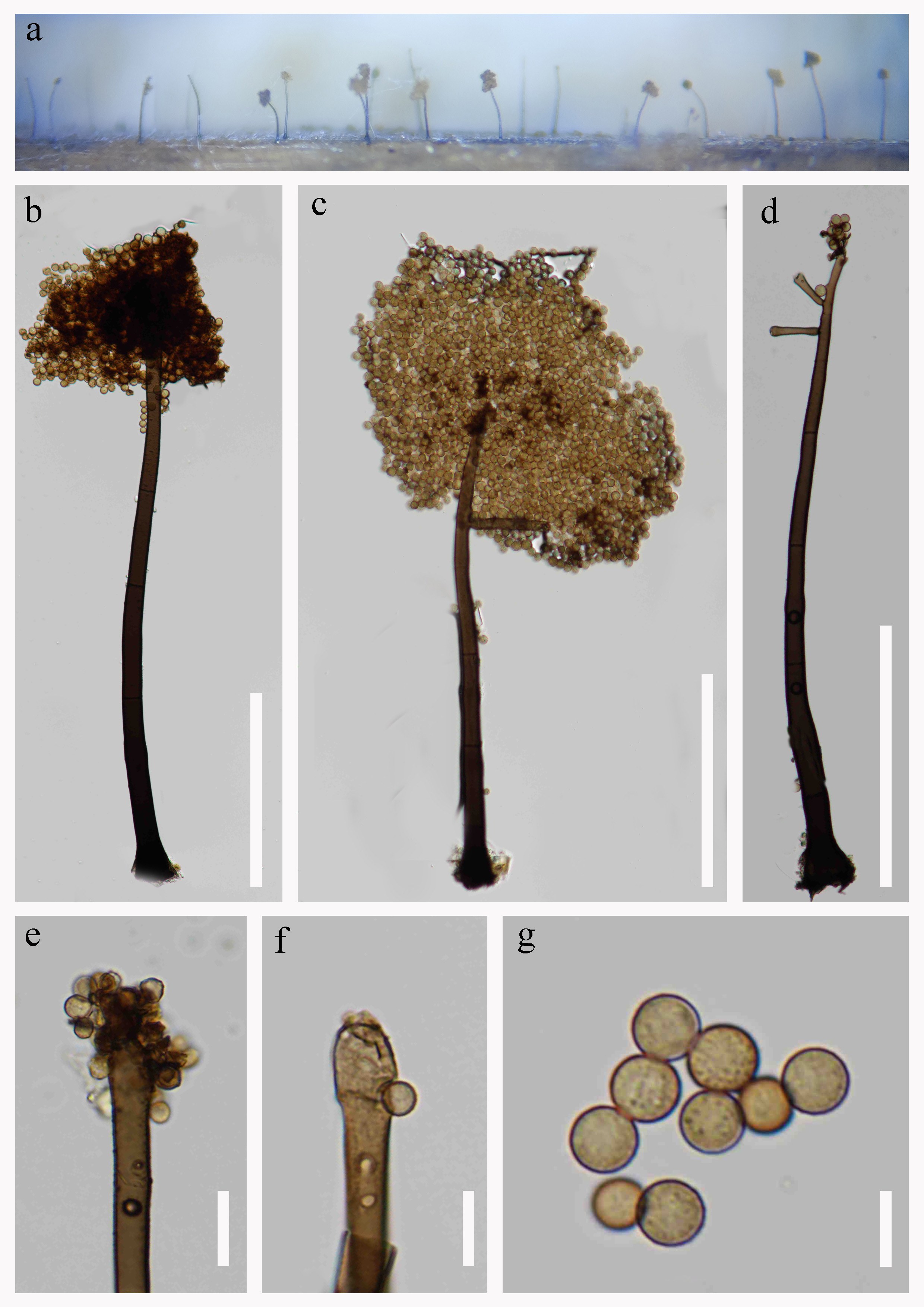
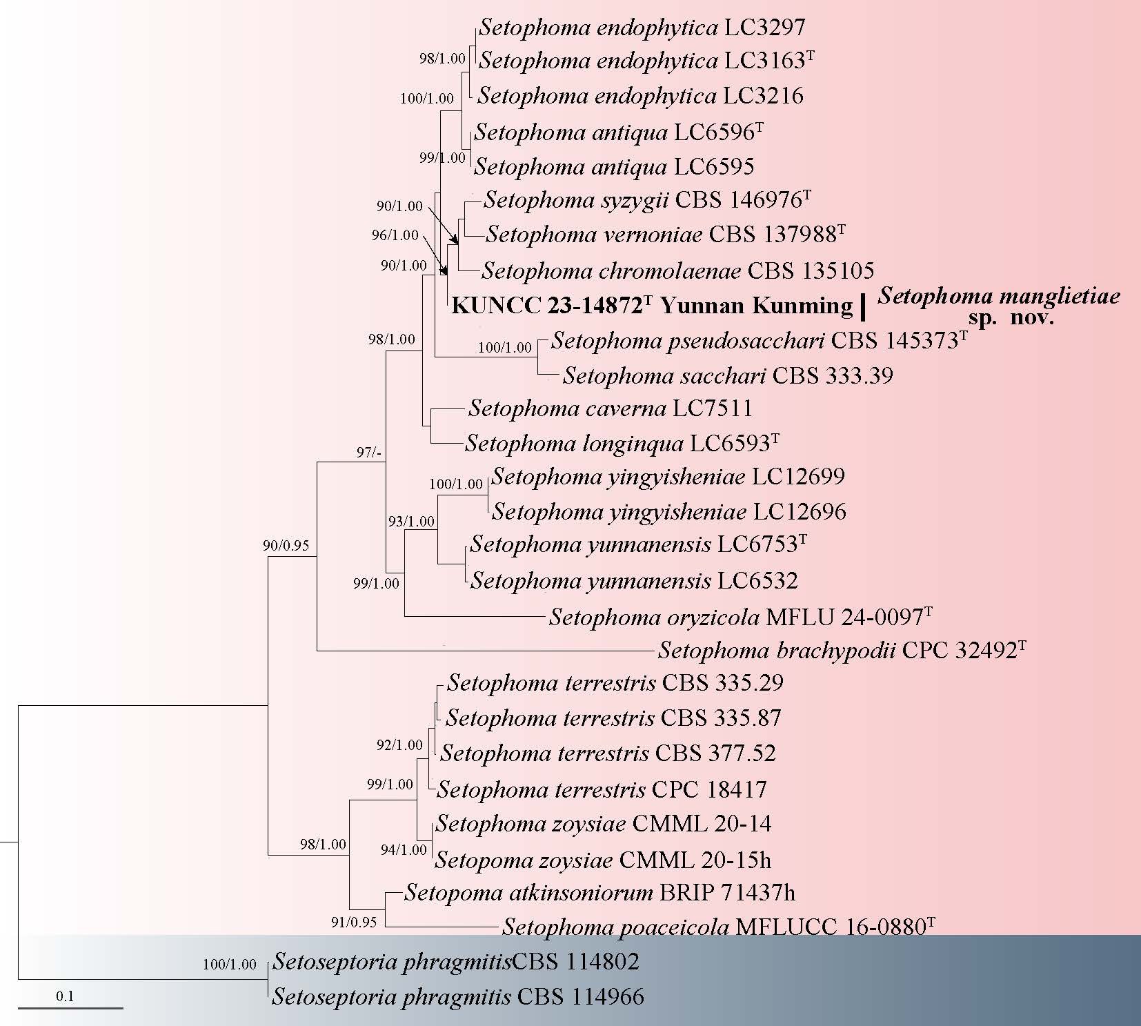
Torulaceae Corda
Torula Pers
Torula was introduced by Persoon in 1795, and is characterized by its type species T. herbarum. This genus is primarily recognized through its asexual reproductive structures, which include macronematous, mononematous conidiophores, holoblastic conidiogenous cells that exhibit a clavate to ellipsoidal morphology, and acrogenous, verrucose structures bearing septate conidia (Jayawardena et al. 2022; Hyde et al. 2023; He et al. 2024a, 2025). Most species within the Torula are found inhabiting freshwater and terrestrial environments (Crous et al. 2015a; Li et al. 2017a; He et al. 2024b). Torula is a widely distributed genus, having been documented across various regions including Asia (China, India, Thailand), Europe (France, Germany, Italy), North America (Crous et al. 2015a; Li et al. 2017a; Su et al. 2018; Boonmee et al. 2021; He et al. 2025; Zhang et al. 2025). It holds the distinction of being the largest genus within Torulaceae (He et al. 2024b), with 571 epithets in Index Fungorum (accessed on April 20, 2026). However, it is noteworthy that the majority of these species lack molecular data (Crous et al. 2015a), with only 34 species being corroborated by molecular data (He et al. 2025; Zhang et al. 2025). In this study, based on phylogenetic and morphological analysis and following the guidance of Maharachchikumbura et al. (2021), we introduced a new species, T. motuoensis, from Xizang, China.
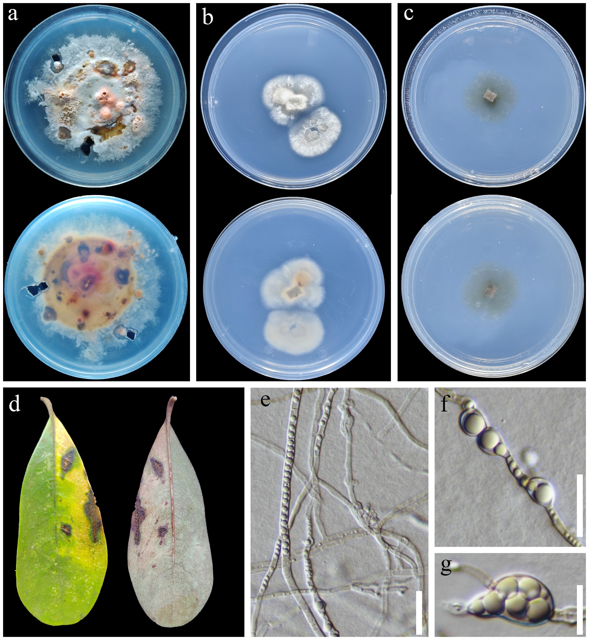
10. Torula motuoensis S.C. He, Q. Zhao & K.D. Hyde, sp. nov. Fig. 19
Chinese name
Index Fungorum number: IF 905258, Facesoffungi number: FoF 19300
Etymology: Referring to the location “Motuo County” where the holotype of this fungus was collected.
Holotype: HKAS 150299
Saprobic on dead stems of Ageratina adenophora. Sexual morph: Undetermined. Asexual morph: Colonies effuse, medium dense, hairy, black brown on the host. Mycelium immersed on the substrate, composed of septate, chained, branched, black brown hyphae. Conidiophores macronematous, mononematous, erect, flexuous, branched, verruculose, septate, cylindrical, brown to dark brown, 18.6–54.4 × 6–8.8 μm ( =32.5 × 7.2 μm, n = 20). Conidiogenous cells holoblastic, terminal, erect, ellipsoid, with melanized basal regions, bowl, pale brown to brown. Conidia phragmospores, catenate, acropleurogenous, monilioid, simple, dry, verruculose, guttulate, thick-walled, rough-walled, 0–multi-septate, pale brown to brown, 10–62 × 6–9 μm ( = 38 × 8 μm, n = 30).
Culture characteristics: germinating within 24 h on PDA at 25 °C, reaching 6.4–6.6 cm after 30 days incubation, above colony black, reverse dark brown, edge, entire, undulate, umbonate, surface rough, mycelia dense, no pigment.
Material examined: CHINA, Xizang Autonomous Region, Linzhi City, Motuo County, 29°35’N, 95°14′E, 1730 m a.s.l., on dead stems of Ageratina adenophora (Asteraceae), 12 July 2022, Shu-Cheng He, HSC821 (HKAS 150299, holotype), ex-type culture: KUNCC 23-17479. ibid. HSC805, (HKAS 150300), culture: KUNCC 23-17350.
Notes: The phylogenetic analysis indicated that our isolates of T. motuoensis (KUNCC 23-17479 and KUNCC 23-17350) clustered with T. calceiformis (HKAS 125551 and HKAS 125552) (Fig. 18). Torula calceiformis is known to exhibit saprobic associations with unidentified wood samples sourced from Guizhou, China (Hyde et al. 2023). Our isolates were collected from Ageratina adenophora in Linzhi City, Xizang, China. Morphologically, T. motuoensis resembles T. calceiformis in having phragmospores, and monilioid, verruculose, multi-septate conidia. Compared to T. calceiformis, T. motuoensis has longer conidiophores (32.5 × 7.2 vs. 5.5 × 6.5 μm) and conidia (38 × 8 vs. 27.6 × 9.2 μm). Therefore, based on phylogenetic and morphological analyses, we introduce a new species, T. motuoensis, from Linzhi City, Xizang, China.
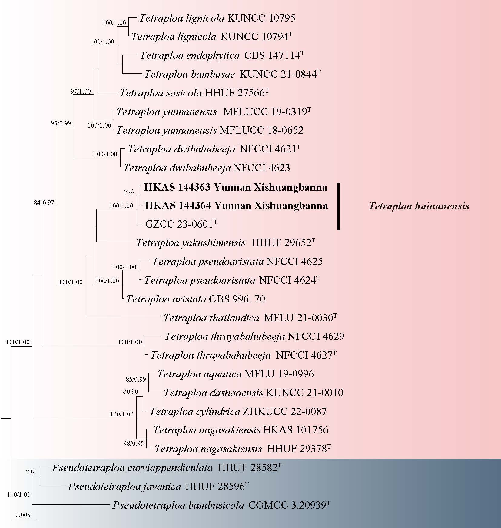
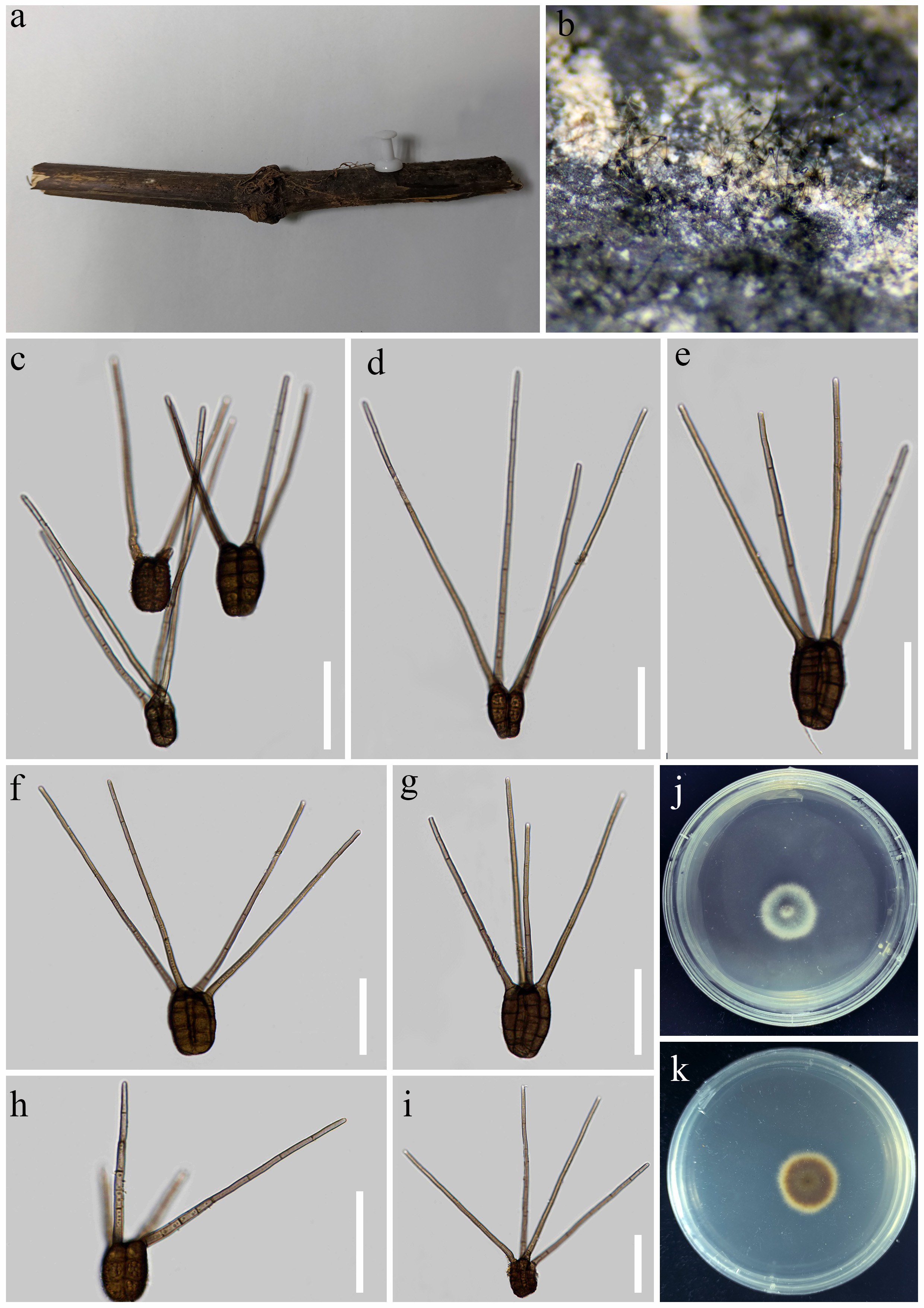
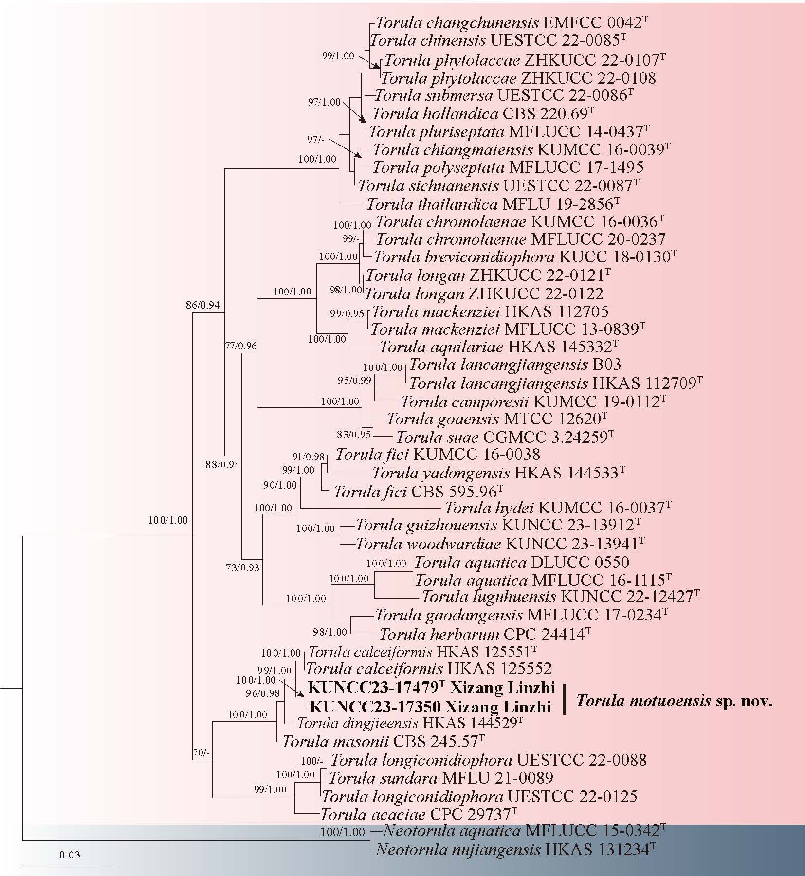
Tubeufiales Boonmee & K.D. Hyde
Tubeufiaceae M.E. Barr
Aquaphila Goh, K.D. Hyde & W.H. Ho
Aquaphila was established by Goh et al. (1998b) and typified by A. albicans based on morphological characteristics. Tsui et al. (2007) accepted Aquaphila as a member of Tubeufiaceae based on their phylogenetic analyses. Currently, Aquaphila comprises two species: A. albicans and A. edentata (Lu et al. 2018). Aquaphila albicans is characterized by semi-macronematous, mononematous, hyaline, delicate, septate, simple or branched, flexuous and geniculate conidiophores; integrated, terminal or intercalary, denticulate, monoblastic or polyblastic, proliferation sympodial conidiogenous cells; acrogenous, proliferation, hyaline, fusoid to falcate or sigmoid conidia (Goh et al. 1998b).
11. Aquaphila albicans Goh, K.D. Hyde & W.H. Ho, Mycol. Res. 102(5): 588 (1998) Fig. 21
Chinese name: – 白喜水孢 (bai xi shui bao)
Index Fungorum number: IF 443558; Facesoffungi number: FoF 02356
Saprobic on decaying wood submerged in freshwater habitats. Sexual morph: Undetermined. Asexual morph: Colonies superficial, hairy, velvety, white, conidia arising from aerial hyphae. Mycelium partly immersed in the woody substratum and partly superficial, consisting of septate, branched, hyaline, smooth, repent hyphae. Conidiophores 15–52 × 4–8 μm ( = 30 × 5 μm, n = 10), semi-macronematous, arising singly as lateral branches from the procumbent hyphae, cylindrical, simple or branched, septate, thin-walled, smooth, indistinctly septate, hyaline, flexuous or geniculate. Conidiogenous cells monoblastic or polyblastic, denticulate, sympodial proliferations, cylindrical. Conidia 58–78 × 8–10 μm ( = 65 × 9 μm, n = 30), holoblastic, solitary, borne acrogenously on conidiogenous denticles, becoming lateral after proliferation of the conidiophore, hyaline or very pale yellowish, predominantly fusoid to sickle-shaped, sometimes sigmoid, very rarely straight and obclavate, thin-walled, smooth, 9–12-euseptate, not constricted at the septa, heavily guttulate, conically rounded at both ends slightly broad at the base, basal cell obconical but not pedicellate.
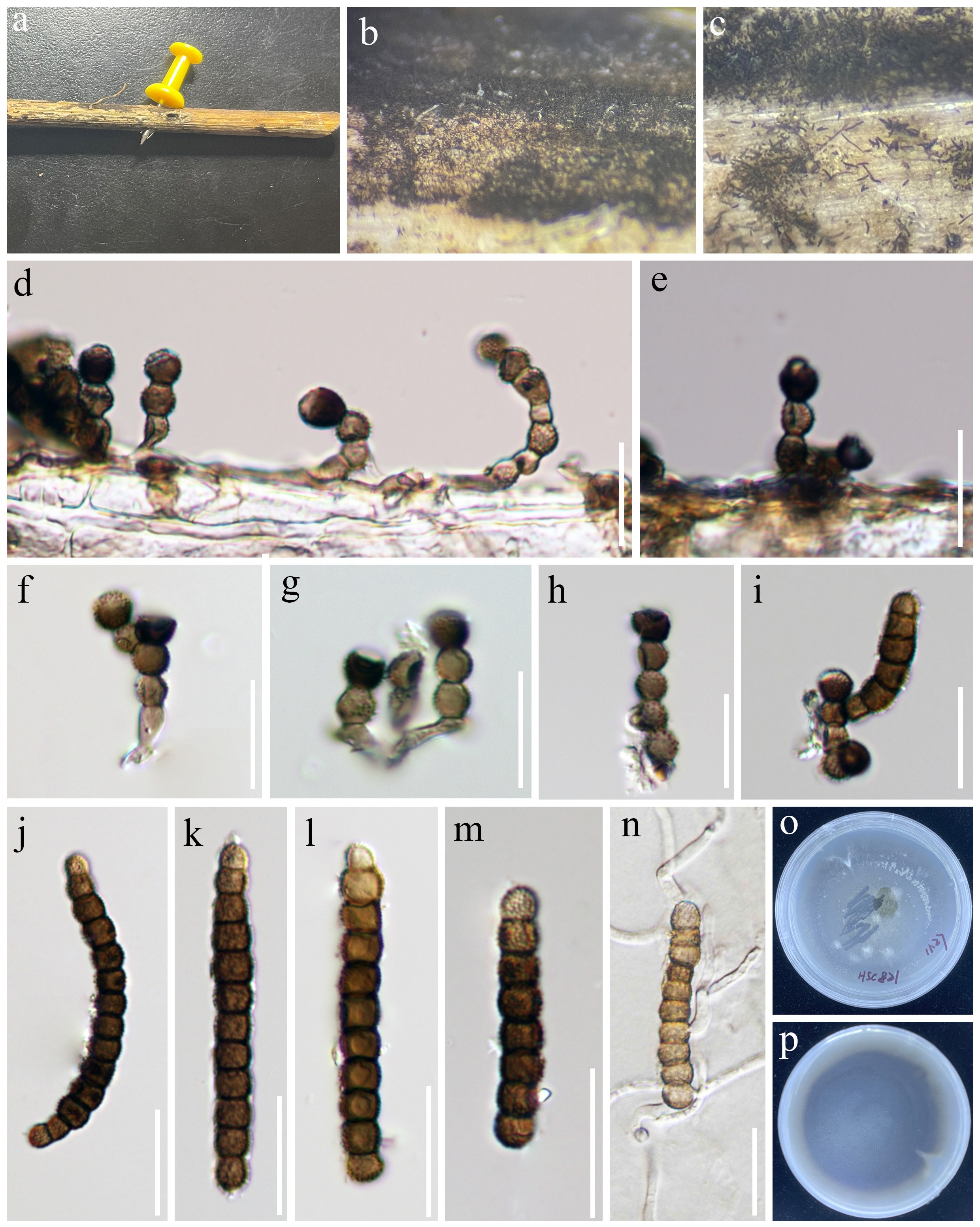
Culture characteristics: Conidia germinating on PDA medium within 48 h and germ tubes produced from both ends. Colonies grown on PDA reached 30 mm diam at room temperature in two weeks, circular, with velutinous appearance, flat, light brown aerial mycelium on the surface, from below dark brown with entire margin.
Material examined: CHINA, Xizang Autonomous Region, Shigatse City, Yadong County, Xiayadong Township, 27°23′38.03" N, 88°50′15.01" E, 4070 m a.s.l., on decaying wood submerged in a freshwater stream, 26 July 2023, R.J. Xu, LJN-56 (HKAS 135977), culture KUNCC 24-17963.
Notes: Goh et al. (1998b) introduced Aquaphila albicans from submerged wood in a tropical habitat. Our newly isolated strain (KUNCC 24-17963) clusters with A. albicans (MFLUCC 16-0010 and KUMCC 19-0078) with 100% ML/1.00 BYPP support (Fig. 20). Morphologically, our new isolate shares the same morphological characteristics with the holotype (HKU(M) 2856) of A. albicans, thus we identified our new isolate as A. albicans.
Eurotiomycetes O.E. Erikss & Winka
Chaetothyriales M.E. Barr
Herpotrichiellaceae Munk
Cladophialophora Borelli
Cladophialophora was established by Borelli (1980), with C. ajelaloi designated as the type species, now synonymized with C. carrionii (Badali et al. 2008). It is characterized by the production of branched or unbranched chains of conidia through blastic conidiogenesis, a process distinguished by hyaline conidial scars (Sutton et al. 2009). Members of this genus exhibit remarkable ecological adaptability, with approximately 85 accepted species inhabiting a range of environments, from soil and plants in terrestrial ecosystems to unusual substrates such as sports drinks and lichens (Obase et al. 2015; Tang et al. 2017; Wijayawardene et al. 2022; Chang et al. 2023; Thitla et al. 2023; Hyde et al. 2024a). Cladophialophora exhibits a widespread geographical distribution, being found across the continents of Africa, Asia, Europe, North America, Oceania, and South America (De Hoog et al. 1995; Thitla et al. 2023). Species such as C. nyingchiensis and C. tumulicola have been isolated from rocks, demonstrating the genus's ability to colonize various substrates, including aquatic and semi-aquatic environments (Isola et al. 2016; Kiyuna et al. 2017; Sun et al. 2020; Chang et al. 2023; Thitla et al. 2023).
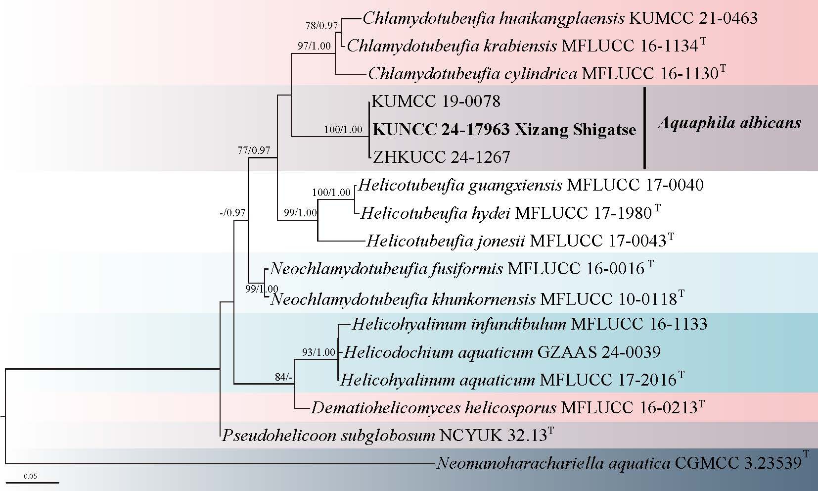
12. Cladophialophora biguttulata R.J. Xu & Q. Zhao, sp. nov. Fig. 23
Chinese name
Index Fungorum number: IF 903480; Facesoffungi number: FoF 17403
Etymology: the species epithet "biguttulata" refers to the presence of two large guttules within the conidia, which is a characteristic feature of this species.
Holotype: HKAS 136223
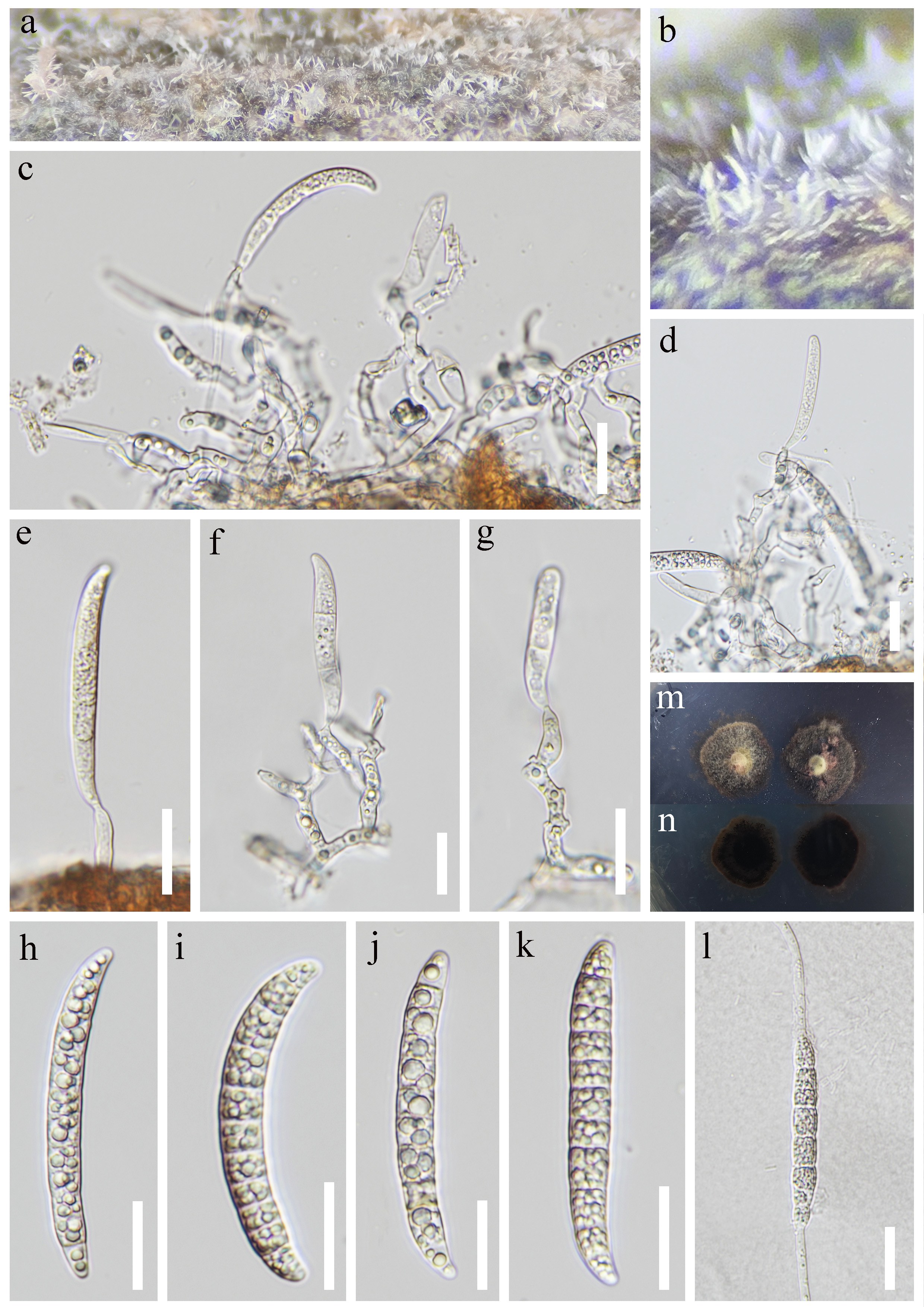
Saprobic on decaying wood submerged in freshwater habitats. Sexual morph: Undetermined. Asexual morph: Colonies superficial, effuse, velvety, gregarious, with masses white of conidia on the natural substrate. Mycelium immersed, consisting of hyaline, smooth-walled, branched, septate. Conidiophores semi-macronematous or macronematous, mononematous, cylindrical, straight or slightly flexuous, branched, dark brown or brown, capitate. Conidiogenous cells polyphialidic, with several sympodial proliferations, sometimes with conspicuous denticles, pale brown to subhyaline. Conidia 12–17 × 4–6 μm ( = 16 × 5 μm, n = 25), polyphialidic, reniform or cymbiform, curving, subhyaline, 1-septate, smooth, with two distinct guttules, short subhyaline pedicels.
Culture characteristics: Colonies grown on PDA reached 50 mm diam at room temperature in one month, circular, with dense, viscous, raised, dark brown mycelium on the surface, viscous, immersed mycelium on the surrounding, from below dark brown with entire margin.
Material examined: CHINA, Xinjiang Autonomous Region, Yili, Tacheng City, Dongdatang Scenic Spot, 43°52'42" N, 85°39'7.2" E, 1779 m a.s.l., on decaying wood submerged in a freshwater habitat, 17 July 2021, R.J. Xu, MD-345 (HKAS 136223, holotype), ex-type culture KUNCC 10438.
Notes: Recent morphological studies of Cladophialophora have primarily focused on the asexual forms produced on culture media (Obase et al. 2015; Sun et al. 2020; Boonmee et al. 2021; Cometto et al. 2023). Our collection was isolated from a natural substrate of submerged decaying wood. Morphologically, our collection differs from other species within Cladophialophora in having polyphialidic, with several sympodial proliferations conidiogenous cells and polyphialidic, reniform or cymbiform, with two distinct guttules, short subhyaline pedicels conidia (Sutton et al. 2009; Chang et al. 2023; Thitla et al. 2023). In addition, phylogenetic analyses also reveal that our collection C. biguttulata (KUNCC 10438) forms a separate, basal clade in Cladophialophora (Fig. 22), indicating it represents a new species.
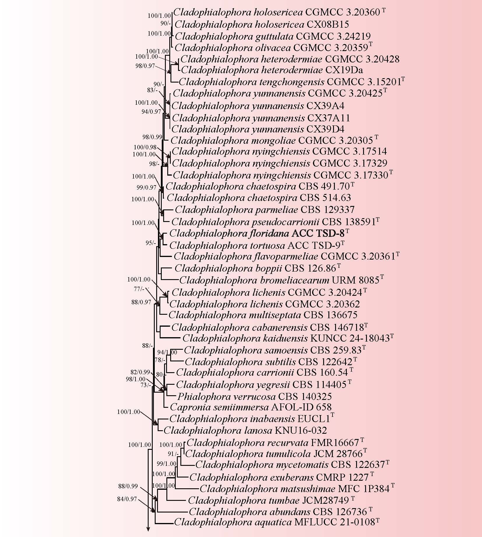
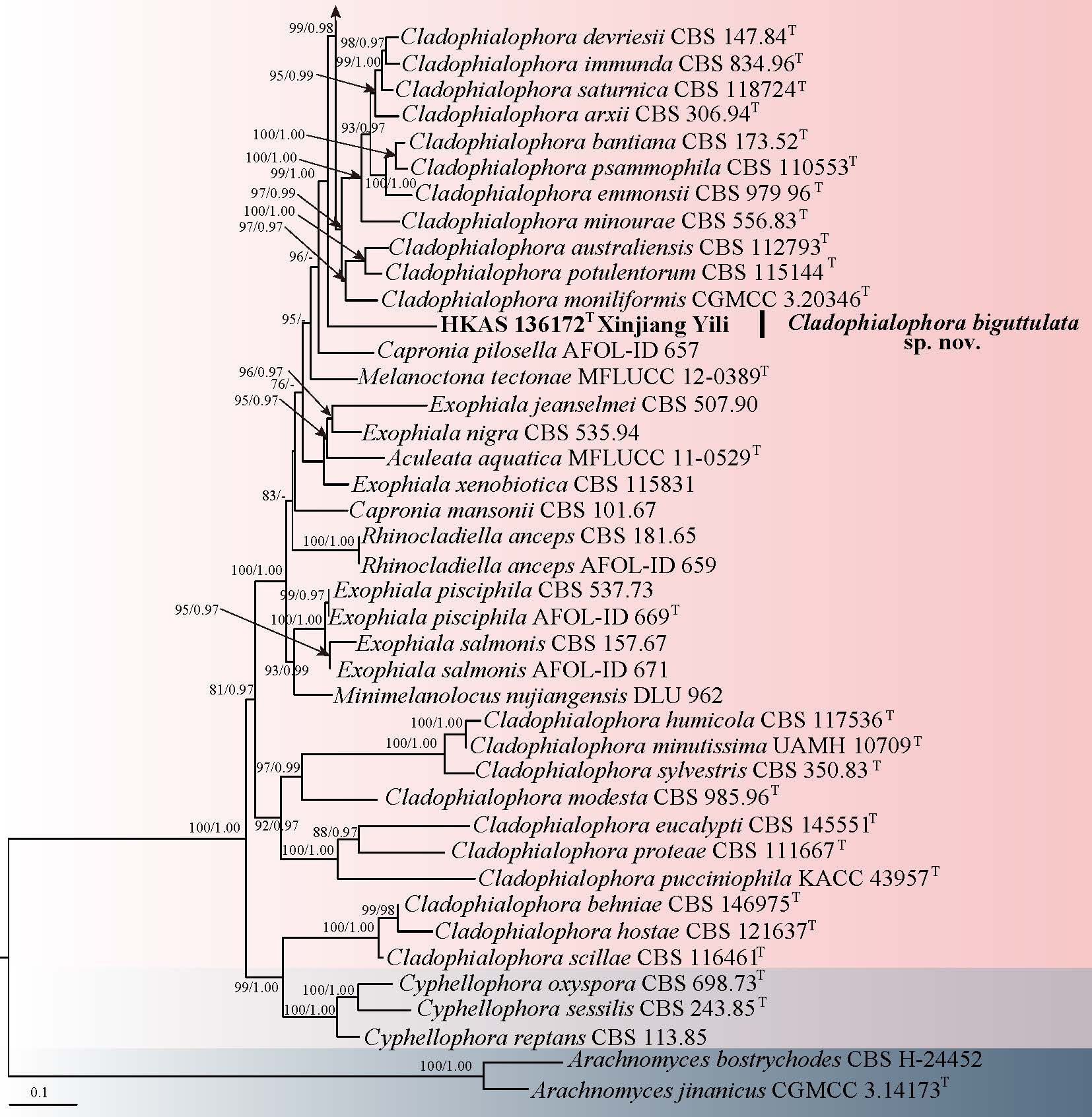
Thysanorea Arzanlou, W. Gams & Crous
Thysanorea was established by Arzanlou et al. (2007), with T. papuana, formerly known as Periconiella papuana, as the type species. Thysanorea is characterized by micro- or macronematous, erect conidiophores that are either simple or apically branched, occasionally proliferating percurrently at the apex. The conidiogenous cells are terminal or intercalary, polyblastic, and exhibit a color gradient, transitioning from brown at the base to a paler shade towards the apex. The conidia are generally pale brown and vary in shape, including oblong, obovoid, and cylindrical forms (Hernández-Restrepo et al. 2020; Yang et al. 2023). Hernández-Restrepo et al. (2020) later transferred species from Minimelanolocus to Thysanorea, citing their congeneric nature based on phylogenetic analyses. However, this reclassification has been debated, with Wan et al. (2021) challenging the move due to differences in the branching pattern of the conidiophores.
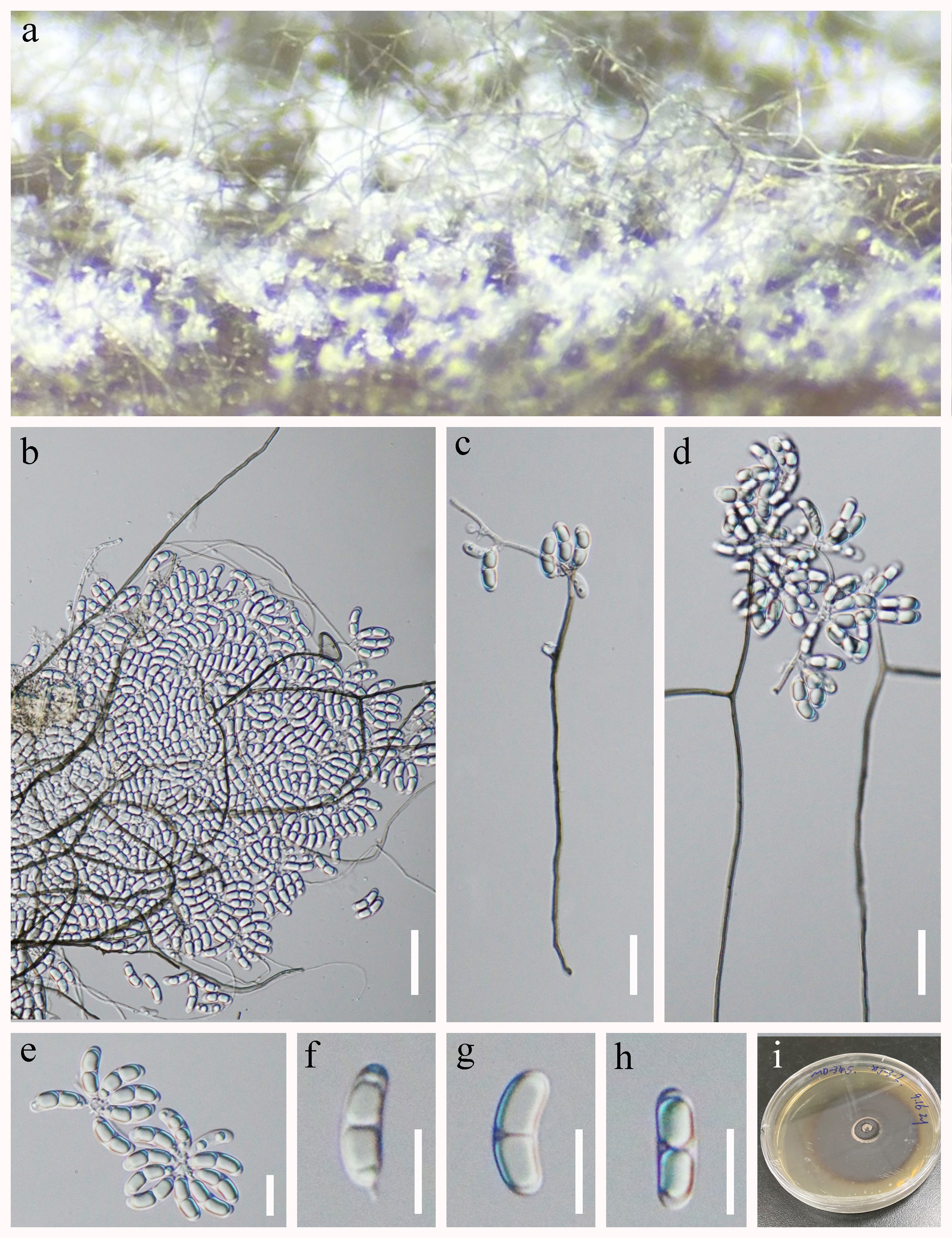
13. Thysanorea linzhiensis R.J. Xu, Q. Zhao & K.D. Hyde, sp. nov. Fig. 25
Chinese name
Index Fungorum number: IF 905033; Facesoffungi number: FoF 19301
Etymology: The epithet refers to Linzhi City, located in the Xizang Autonomous Region of China, where the type specimen was collected.
Holotype: HKAS 136140
Saprobic on decaying submerged wood in a freshwater stream. Sexual morph: Undetermined. Asexual morph: Colonies on natural substrates effuse, hairy, scattered or in small groups, brown, with glistening conidial masses at apex. Mycelium is partly superficial, partly immersed, composed of septate, smooth, pale brown hyphae. Conidiophores 201–320 × 3–9 μm ( = 263 × 6 μm, n = 20), macronematous, mononematous, erect, solitary, straight or flexuous, cylindrical, smooth-walled, septate, unbranched, dark brown, becoming pale brown or subhyaline towards the apex, thick-walled. Conidiogenous cells polyblastic, integrated, terminal, denticulate, sympodial proliferations, cylindrical, slightly constricted at septa, pale brown or subhyaline. Conidia 14–18 × 3–4 μm ( = 16 × 3 μm, n = 20), acrogenous or acropleurogenous, clavate, with a narrow truncate base, truncate at the base, aseptate when young, 1–4-septate when mature, smooth-walled, subhyaline.
Culture characteristics: Conidia germinating on PDA medium within 24 h and germ tubes produced from both ends. Colonies grown on PDA reached 30 mm diam at room temperature in two weeks, circular, with velvety, raised, grayish-green aerial mycelium on the surface, from below grayish-green with entire margin.
Material examined: CHINA, Xizang Autonomous Region, Linzhi City, Chayu County, 28°39′55.76" N, 97°28′15.71" E, 4118 m a.s.l., on decaying wood submerged in a freshwater stream, 14 July 2022, R.J. Xu, LTS-36 (HKAS 136140, holotype), ex-type culture KUNCC 24-18006.
Notes: Thysanorea linzhiensis resembles T. obscurus in possessing mononematous, macronematous, septate conidiophores, as well as polyblastic, integrated, sympodially proliferating conidiogenous cells, and clavate conidia with a narrow truncate base. However, T. linzhiensis can be distinguished from T. obscurus by its denticulate conidiogenous cells and longer conidiophores (201–320 × 3–9 μm vs. 123–219 × 5–6 μm) (Liu et al. 2015b). In the phylogenetic analysis using combined ITS, LSU, and SSU sequence data, T. linzhiensis formed a distinct clade within Thysanorea (Fig. 24), closely related to but clearly separate from T. obscurus, with a genetic divergence of 2.8% in ITS sequences (16/564 bp difference). The introduction of T. linzhiensis as a new species is supported by both morphological and phylogenetic evidence.
Leotiomycetes O.E. Erikss. & Winka
Helotiales Nannf. ex Korf & Lizoň
Mollisiaceae Rehm
Phialocephala W.B. Kendr.
Phialocephala was introduced by Kendrick (1961) to accommodate P. dimorphospora (type species) and P. bactrospora, the latter was recently transferred to Parasporendocladia (Wu & Diao 2022). Phialocephala is a polyphyletic genus throughout Mollisiaceae (Tanney & Seifert 2020; Visagie et al. 2024). Tanney & Seifert (2020) defined the Phialocephala sensu stricto which contains 14 species. Phialocephala is characterized by macronematous, mononematous, brown conidiophores, monophialidic, discrete conidiogenous cells arranged penicillately, and often catenate, subglobose to ellipsoidal, hyaline or pigmented, aseptate conidia (Ellis 1971b; Onofri et al. 1994; Tanney et al. 2016; Tanney & Seifert 2020; Senanayake et al. 2023). Phialocephala also has diplococcium-like synasexual morph, e.g., P. catenospora, and synnematous synasexual morph, e.g., P. oblonga (Tanney et al. 2016).
14. Phialocephala guizhouensis J. Ma & Y.Z. Lu, sp. nov. Fig. 27
Chinese name
Index Fungorum number: IF 573589; Facesoffungi number: FoF 19302
Etymology: the species epithet "guizhouensis" refers to the location where this species was collected
Holotype: GZAAS 23-0033
Saprobic on decaying wood in a terrestrial habitat. Sexual morph: Undetermined. Asexual morph: Colonies on natural substrate effuse, black, hairy. Mycelium mostly partly immersed, partly superficial, composed of brown, branched, septate, smooth hyphae. Conidiophores up to 220 μm long, macronematous, mononematous, erect, cylindrical, brown to reddish-brown, septate, thick-walled. Conidiogenous cells polytretic, integrated, terminal and intercalary, brown to reddish-brown. Conidia 6–13 × 3–5 μm ( = 8.5 × 4 μm, n = 30), catenate, formed in acropetal, branched chains, oblong with obtuse ends, brown to slight dark brown, 1–2-septate, darkened and slightly constricted at the septa, smooth.
Culture characteristics: Conidia germinate on water agar within 24 hrs. Germ tubes are produced from the apex or base. Colonies superficial, irregularly circular, flat, with entire edge, from above grey to whitish-grey in the center, brownish-gray at the edge, and from below, pale grey in the middle, brownish-grey at the edge.
Material examined: CHINA, Guizhou Province, Qiannan Buyi Autonomous Prefecture, Longli County, on decaying wood in a terrestrial habitat, 2 September 2020, Jian Ma, LLZB3 (GZAAS 23-0033, holotype), ex-type cultures GZCC 24-0159.
Notes: Our collection morphologically resembles the synasexual morph of Phialocephala catenospora, which has oblong and brown conidia (Tanney et al. 2016). However, the conidia of P. catenospora are larger than those of ours, (6–13 × 3–5 vs. 12–22.5 × 5–6 μm). Besides, P. catenospora (DAOMC 250108) is phylogenetically distant from our strain LLZB3 in the analyses (Fig. 26). In the phylogenetic tree, our strain LLZB3 formed an internal subclade with P. amethystea (DAOMC 251552) and P. compacta (CBS 507.94) with 99% ML/1.00 BYPP support (Fig. 26). ITS comparison shows there are 7.11% (53 out of 746 bp, including 7 gaps) and 4.97% (37 out of 745 bp, including 1 gap) differences with the strains DAOMC 251552 and CBS 507.94, respectively. Therefore, we introduce our collection as a new species.
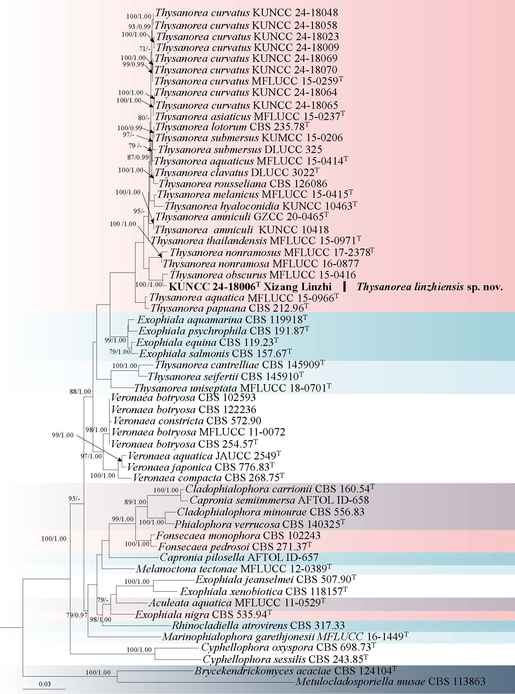
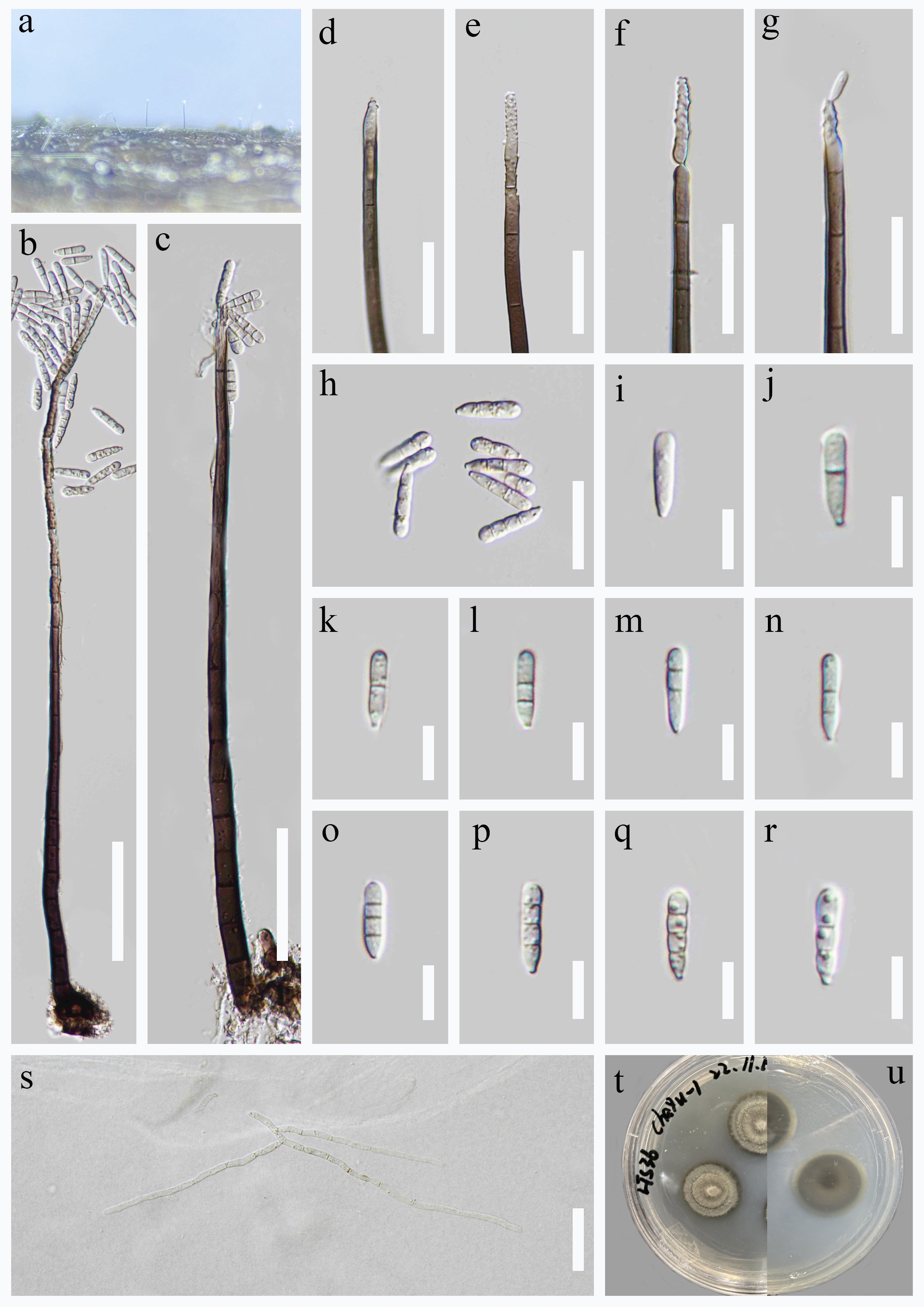
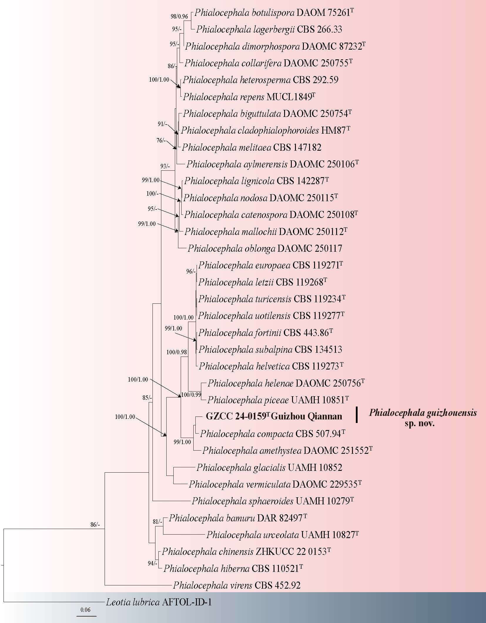
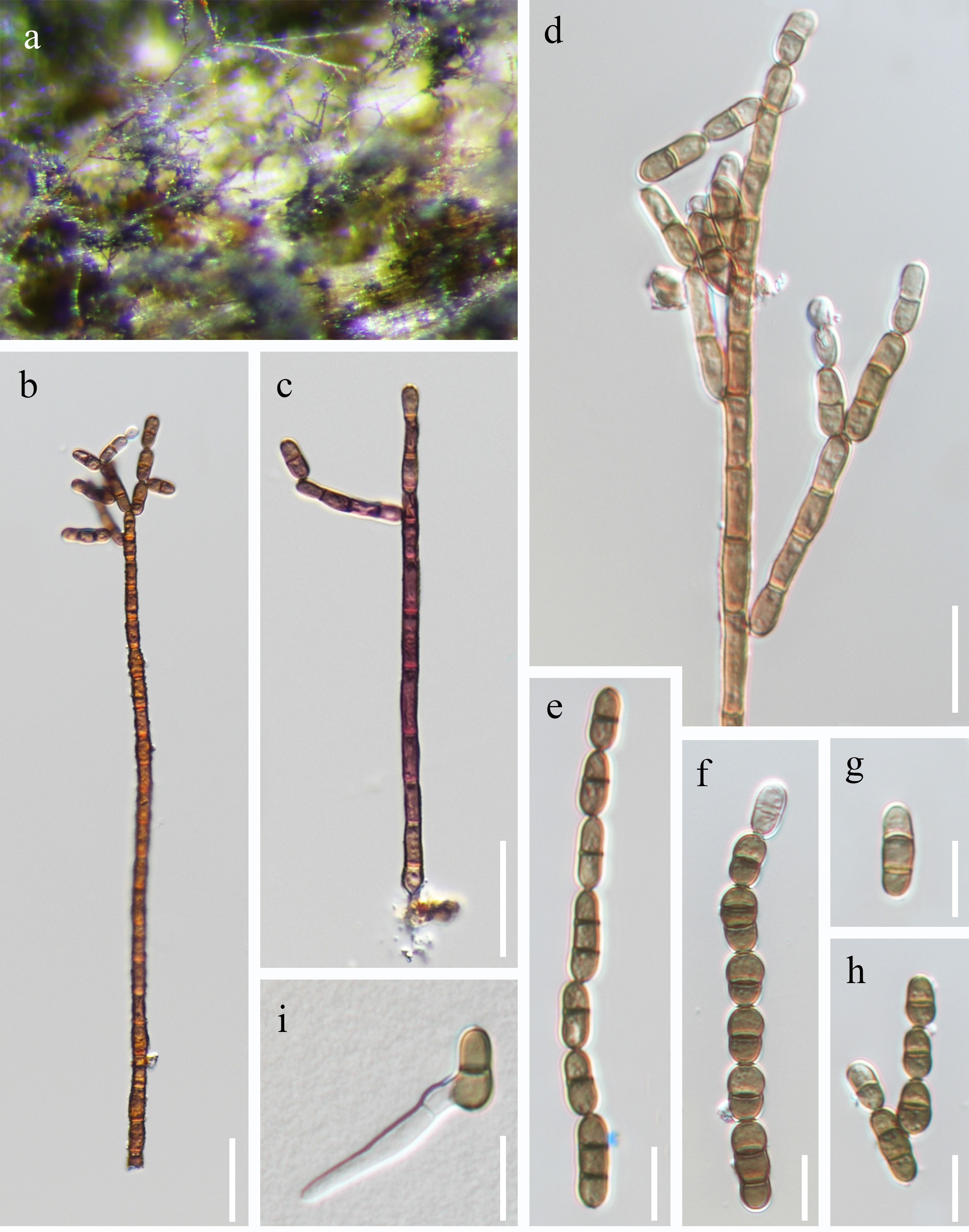
Pezizellaceae Velen
Chalara (Corda) Rabenh
Chalara was originally introduced by Corda (1837) as a subgenus of Torula to accommodate the hyphomycete Chalara fusidioides, characterized by a granular, effuse stroma, simple conidiophores, lageniform, cylindrical conidiogenous cells, and cylindrical, hyaline conidia. Rabenhorst (1844) elevated Chalara to the generic level. The genus Chalara comprises a group of hyphomycetes characterized by the presence or absence of stroma and setae; conidiophores are scattered or aggregated, erect, simple, subcylindrical to cylindrical, smooth or verrucose, septate or aseptate; conidiogenous cells ampulliform, lageniform, obclavate, ellipsoidal, or subcylindrical, with a venter and a collarette; conidia cylindrical to obclavate, hyaline, aseptate, and extruded in short chains (Nag Raj & Kendrick 1975; Wu & Diao 2023). Based on morphological and phylogenetic analyses, Wu & Diao (2023) revised the genus Chalara, typified by C. fusidioides, into a narrowly concept monophyletic genus to accommodate seven species that formed a distinct clade separate from other chalara-like fungi in their phylogenetic reconstruction. These species include Chalara africana, C. bambusicola, C. clidemiae, C. cylindrosperma, C. longiphora, C. platanicola, and C. qinlingensis (Wu & Diao 2023). In addition to these phylogenetically supported species, Chalara sensu stricto also includes nine other species for which molecular data are currently lacking (Wu & Diao 2023). Species of Chalara s. str. are saprobic and occur on various plant substrates and in soil (Nag Raj & Kendrick 1975; Kirk 1986; Christiansen 1993; McKenzie et al. 2002; Wu 2004; Pratibha et al. 2005; Wu & Diao 2023)
15. Chalara aquatica H.W. Shen, K.D. Hyde & Z.L. Luo, sp. nov. Fig. 28
Chinese name
Index Fungorum number: IF 905259; Facesoffungi number: FoF 19303
Etymology: The specific epithet “aquatica” refers to the aquatica habitat, which is where this species was collected.
Holotype: HKAS 151651
Saprobic on submerged decaying wood in a freshwater habitat. Sexual morph: Undetermined. Asexual morph: Colonies on natural substrate, superficial, effuse, scattered, hairy, dark brown, solitary or in small groups. Mycelium partly immersed and partly superficial, composed of branched, septate, pale brown hyphae. Stroma absent. Setae absent. Conidiophores (85–)115–171(–183) × 5.3–8.5 µm ( = 143 × 7.2 µm, n = 20), macronematous, mononematous, cylindrical, straight or slightly flexuous, unbranched, septate, basal cells slightly swollen, brown to dark brown, smooth-walled. Conidiogenous cells (30–)36–42(–44) × 7.4–9.3 µm ( = 39 × 8.3 µm, n = 25), integrated, terminal, lageniform, pale to medium brown, consisting of the wider venter and the narrower collarette, transition from venter to collarette abrupt. Venter 10–21 × 7–9 µm, ellipsoid to subcylindrical, medium brown. Collarette 18–26 × 4–6 µm, cylindrical, pale to medium brown. Conidia (3.5–)5.2–7.8(–8.6) × 3.5–5 µm ( = 6.5 × 4.2 µm, n = 30), endogenous, subglobose to barrel-shaped to rectangular, truncate or obtuse at both ends, hyaline, aseptate, guttulate, smooth-walled.
Culture characteristics: Conidia germinated on PDA within 12 h and germ tubes produced from around the conidium. Colonies on PDA reaching 2–3 cm diameter after one month at room temperature (around 22 °C) in the dark, dense, velvety, gray to brown, with smooth margins and bulge center from above, dense, gray to brown, with smooth margins from below.
Material examined: CHINA, Yunnan Province, Lijiang City, on the unidentified decaying wood, October 2021, L.L. Li, L944 (HKAS 151651, holotype), ex-type culture KUNCC 23-14304.
Notes: Phylogenetic analysis revealed that Chalara aquatica is closely related to C. cylindrosperma, C. longiphora, and C. platanicola, forming a robustly supported clade within the phylogenetic framework (Fig. 29). Morphologically, C. aquatica is similar to all three species, but can be distinguished from C. cylindrosperma by its larger venter (10–21 × 7–9 µm vs. 10–12.5 × 5–7 µm) and collarette (18–26 × 4–6 µm vs. 17.5–20 × 2.5–3.5 µm), as well as by its smaller conidia (5.2–7.8 × 3.5–5 µm vs. 10–17 × 1.5–2.5 µm) (Wu & Diao 2023). Chalara aquatica differs from C. longiphora in having a larger collarette (18–26 × 4–6 µm vs. 14–17 × 2.5–2.7 µm), wider conidia (3.5–5 µm vs. 2–2.5 µm), and subglobose to barrel-shaped or rectangular conidia, whereas the conidia of C. longiphora are cylindrical (Wu & Diao 2023). Compared to C. platanicola, C. aquatica has longer conidiogenous cells (36–42 µm vs. 26–27 µm), venter (10–21 × 7–9 µm vs. 10–12.5 × 5.7–6.5 µm), and collarette (18–26 × 4–6 µm vs. 17–18 × 2.8–3.3 µm), but produces shorter and wider conidia (5.2–7.8 × 3.5–5 µm vs. 9–10.5 × 2.2–2.5 µm) (Wu & Diao 2023). Based on both phylogenetic distinction and morphological differences, we introduce C. aquatica as a new species.
16. Chalara guttulata H.W. Shen, K.D. Hyde & Z.L. Luo, sp. nov. Fig. 30
Chinese name:
Index Fungorum number: IF 905260; Facesoffungi number: FoF 19304
Etymology: The specific epithet “guttulata” refers to the guttulate conidia of this species.
Holotype: DLU 3129
Saprobic on submerged decaying wood in a freshwater habitat. Sexual morph: Undetermined. Asexual morph: Colonies on natural substrate, superficial, effuse, scattered, hairy, dark brown, solitary or in small groups. Mycelium partly immersed and partly superficial, composed of branched, septate, pale brown hyphae. Stroma absent. Setae absent. Conidiophores 82–88 × 4.3–6 µm (x̄ = 85 × 5 µm, n = 15), macronematous, mononematous, cylindrical, straight or slightly flexuous, unbranched, 3–septate, basal cells slightly swollen, brown to dark brown, smooth-walled. Conidiogenous cells 50–60 × 5–6.4 µm ( = 56 × 5.5 µm, n = 15), integrated, terminal, long-lageniform, pale to medium brown, consisting of the wider venter and the narrower collarette, transition from venter to collarette abrupt. Venter 21–31 × 4.5–6 µm, subcylindrical to long-clavate, medium brown. Collarette 25.5–32 × 2.8–3.1 µm, cylindrical, pale to medium brown. Conidia 7.8–12 × 1.8–2.5 µm ( = 10 × 2.2 µm, n = 40), endogenous, cylindrical, truncate or obtuse at both ends, hyaline, aseptate, guttulate, smooth-walled.
Material examined: CHINA, Yunnan Province, Dali City, on undetermined decaying wood, May 2021, S. Luan, S3129 (DLU3129, holotype), ex-type culture DLUCC 3129.
Notes: Phylogenetic analysis placed Chalara guttulata within the Chalara clade, where it formed a distinct and with 100% ML/1.00 BYPP support (Fig. 29). Morphologically, C. guttulata resembles C. versicolor in having cylindrical, unbranched, septate conidiophores, integrated, terminal, lageniform conidiogenous cells, consisting of the wider venter and the narrower collarette, and cylindrical, hyaline, aseptate conidia, truncate or obtuse at both ends (Wu & Diao 2023). However, it differs by its longer conidiophores (82–88µm vs. 15–50 µm), longer conidiogenous cells (50–60 µm vs. 47–50 µm), and smaller conidia (7.8–12 × 1.8–2.5 µm vs. 6.5–15 × 2.5–3 µm) (Wu & Diao 2023). A BLASTn search of the ITS sequence against NCBI GenBank revealed the closest match to be Chalara sp. (Specimen voucher: HAY-F-011218, Sequence ID: PV742674.1), with 98% identity (485/496 bp, including 2 gaps). The closest match for the LSU sequence was Endoradiciella communis (Strain: P2333, Sequence ID: NG_149040.1), showing 99% identity (857/864 bp, including 1 gap). Based on both morphological characteristics and molecular phylogenetic evidence, we introduce C. guttulata as a novel species.
Sordariomycetes O.E. Erikss. & Winka
Sordariomycetes is one of the largest classes within Ascomycota, widely distributed worldwide and predominantly comprising terrestrial taxa, although several members also occur in aquatic habitats (Luo et al. 2019; Hyde et al. 2020c). In the present study, a phylogenetic framework of Sordariomycetes is reconstructed based on sequence data from three loci (LSU, rpb2 and tef1-α) using approximately 1,000 strains. Further details are provided in Supplementary (Fig. 2).
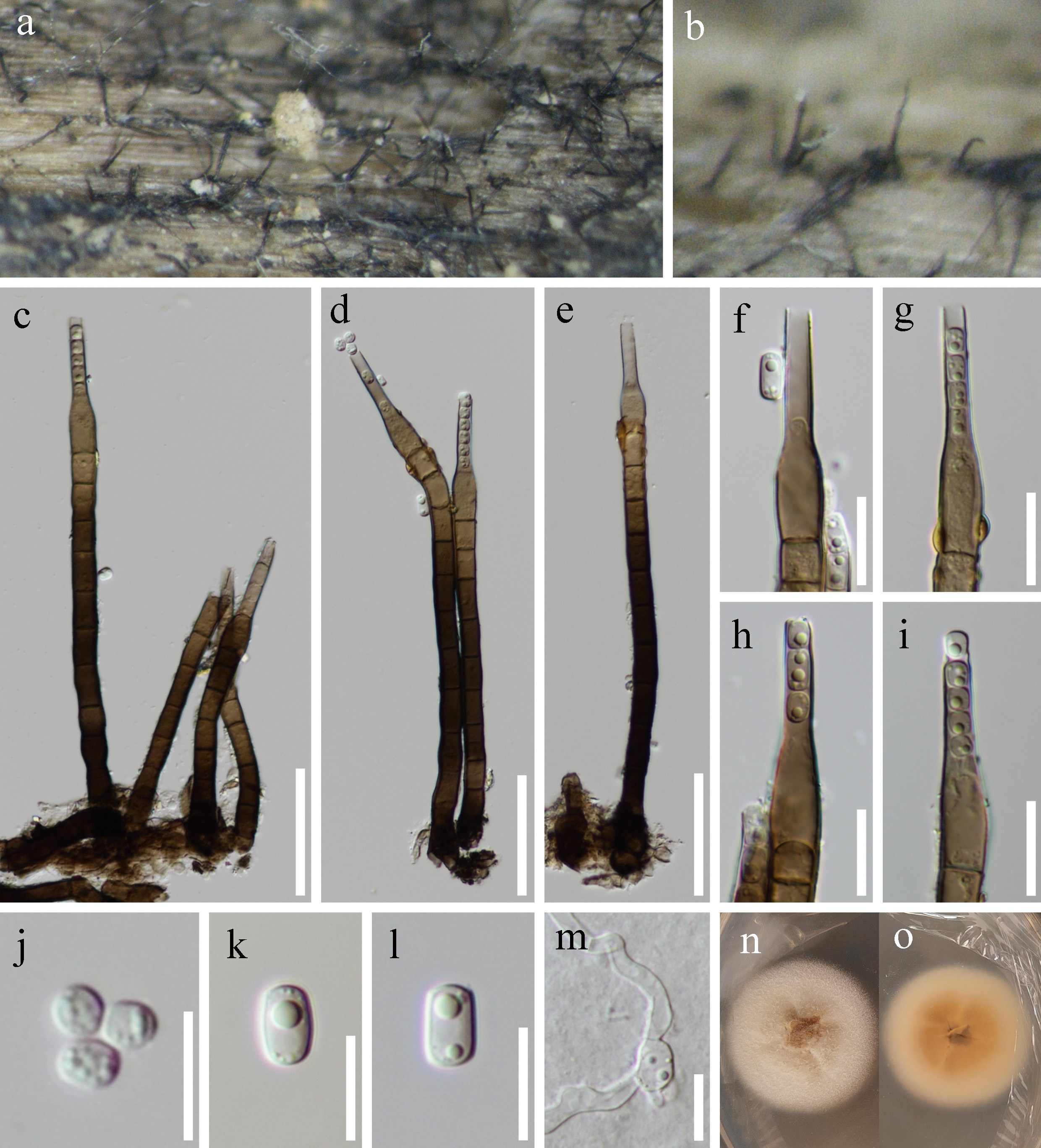
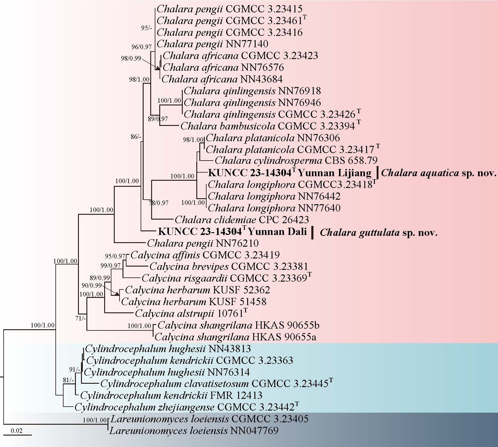
Chaetosphaeriales Huhndorf, A.N. Mill. & F.A. Fernández
Chaetosphaeriaceae Réblová, M.E. Barr & Samuels
Chaetosphaeriaceae was established by Réblová et al. (1999a) to accommodate Chaetosphaeria with the closely related genera including: Ascocodinaea, Melanochaela, and Sporoschisma. Chaetosphaeriaceae is a major family within Sordariomycetes, known for its complex pleomorphism and diverse asexual morphologies. Previous, the phylogenetic analyses have revealed that both Stanjehughesia and Linkosia are polyphyletic (Shenoy et al. 2006; Su et al. 2016a; Hyde et al. 2020c; Hsieh et al. 2021), and the type species S. hormiscioides lacks molecular data, leaving the phylogenetic position of the genus unresolved. However, Réblová et al. (2024) re-evaluated species previously classified in the genera Exserticlava, Phaeostalagmus, Phialocephala, and several Chalara and Stanjehughesia-like fungi, and reassigned them to Chaetosphaeriaceae based on living cultures and DNA sequence data obtained from numerous type species. Currently, 101 genera are accepted in Chaetosphaeriaceae and phylogenetic analysis based on combined ITS, LSU, and tef1-α sequence data (Hyde et al. 2024a; Réblová et al. 2024).
Chloridium Link
Chloridium is known for its cosmopolitan distribution, thriving on decaying plant tissues in terrestrial and freshwater habitats, notably on wood buried in moist soil or leaf litter. Chloridium was introduced with C. virescens as the type species, characterized by pigmented, unbranched, erect conidiophores with globose conidia forming green masses (Link 1809; Réblová et al. 2022). The taxonomy of Chloridium has been complex, with early classifications based on morphological features and later, molecular data. Recent molecular analyses have further refined the understanding of phylogeny of Chloridium, revealing its paraphyletic nature and close relationships with other genera such as Adautomilanezia and Sporoschisma (Réblová et al. 2022).
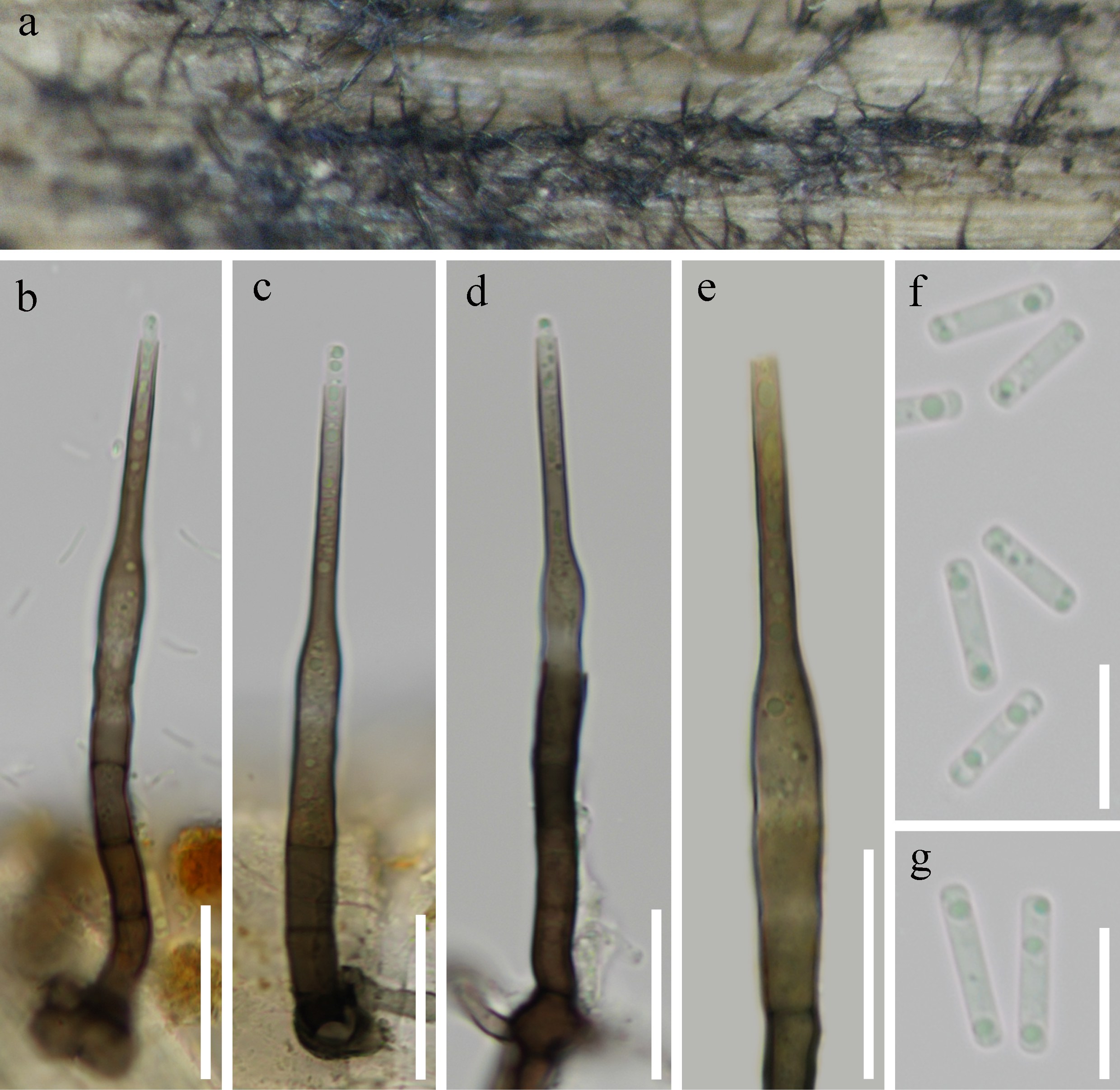
17. Chloridium fuscum (Corda) Réblová & Hern. -Restr., Stud. Mycol. 103: 176 (2022) Fig. 32
Chinese name
Index Fungorum number: IF 555386; Facesoffungi number: FoF 04950
Saprobic on decaying wood submerged in a freshwater habitat. Sexual morph: Undetermined. Asexual morph: Colonies effuse, dark brown, olivaceous brown to olivaceous grey, with long hairy mycelium. Mycelium partly immersed, partly superficial, consisting of branched, septate, brown hyphae. Conidiophores 158–241 × 3–6 µm ( = 195 × 5 μm, n = 15), macronematous, solitary or crowded, erect, straight, septate, unbranched, cylindrical, 4–6- septate, dark brown, pale brown to subhyaline towards the apex, slightly tapering towards the apex. Conidiogenous cells 11–31 × 3–5 µm ( = 21 × 4 μm, n = 20), monophialidic, discrete, phialide, sometimes extending percurrently, subulate to cylindrical, pale brown to subhyaline, hyaline towards the apex, in 5–10 cylindrical located in the two lower thirds of the conidiophore in the nodes at intervals. Conidia 4–5 × 3–4 µm ( = 5 × 3 μm, n = 20), acrogenous, ellipsoidal, subhyaline to pale brown, aseptate, smooth,
Culture characteristics: Conidia germinating on PDA medium within 24 h and germ tubes produced from both ends, Colonies grown on PDA reached 50 mm diam at room temperature in two weeks, circular, with velvety, flat, dark brown mycelium on the surface, from below dark brown to grayish-brown with entire margin.
Material examined: CHINA, Xizang Autonomous Region, Rikaze City, Yadong County, 27° 21' 11" N, 88° 58' 10" E, 3061 m a.s.l., on decaying wood submerged in a freshwater habitat, 01 July 2022, R.J. Xu, LTS-8 (HKAS 136133), culture KUNCC 24-17992. Linzhi City, Motuo County, Dexing Township, 29°21′14.97" N, 95°8′53.19" E, 2172 m a.s.l., on decaying wood submerged in a freshwater stream, 10 August 2023, R.J. Xu, LJN-42 (HKAS 135976), culture KUNCC 24-17960. Shannan City, Luozha County, 28°11′3.15" N, 90°59′57.88" E, 3491 m a.s.l., on decaying wood submerged in a freshwater habitat, 08 July 2022, R.J. Xu, XK-68 (HKAS 136102), culture KUNCC 24-18124. Linzhi City, Chayu County, Guyu Township, 29°19′31.89" N, 97°8′52.91" E, 3858 m a.s.l., on decaying wood submerged in a freshwater habitat, 12 August 2023, R.J. Xu, LJN-72 (HKAS 135984), culture KUNCC 24-17972.
Notes: Chloridium fuscum was initially described by Corda (1837) as Chaetopsis fusca. It was later synonymized with Gonytrichum and subsequently reclassified under Chloridium based on comprehensive phylogenetic analyses (Saccardo 1880; Hughes 1951; Réblová et al. 2022). Synonyms of this species include Mesobotrys simplex and Chl. aseptatum. Chl. fuscum is closely related to Chl. volubile, sharing similar conidiophore characters, but can be distinguished based on specific morphological differences (Réblová et al. 2022).
Chloridium fuscum is a widely distributed saprobic species that can be found in both terrestrial and freshwater environments, including soil, decaying wood and bark, thriving on a variety of plant remnants. This species exhibits a broad geographic presence, recorded in regions such as Australia, Canada, China, the Czech Republic, Germany, Italy, Korea, Puerto Rico, South Africa, Thailand, the United Kingdom, and the USA (Réblová et al. 2022). Morphologically, our collection fits well with the species concept of Chl. fuscum, and phylogenetic analyses showed that our collections of Chlo. fuscum clustered closely with Chl. fuscum (CBS 195.60) (Fig. 31). Consequently, we identified the four strains as Chl. fuscum and reported as a new collection from Xizang, China.
Reticulascus was established by Réblová et al. (2011a), based on a combined of ITS, LSU and SSU phylogeny and to accommodate two holomorph Chaetosphaeria species. Whereafter, Réblová et al. (2016a) given its widespread use, priority, and greater number of names, therefore recommend the use of Cylindrotrichum rather than Reticulascus. The genus is characterized by brown to black, hairy, effuse colonies in vivo. Setae are absent. Conidiophores are macronematous, mononematous, cylindrical, and straight. Conidiogenous cells are usually monophialidic, rarely polyphialidic with up to two lateral enteroblastic openings, with collarette hyaline to subhyaline. Conidia are cylindrical, slightly tapering, rounded at the apex, obtuse at the base, 1-septate, not constricted at the septum, hyaline, guttulate, and smooth-walled.
18. Cylindrotrichum clavatum W. Gams & Hol. -Jech., Stud. Mycol. 43: 54. 1976 Fig. 34
= Reticulascus clavatus Réblová & J. Fourn., in Réblová, Gams & Seifert, Stud. Mycol. 68: 181 (2011)
Chinese name
Index Fungorum number: IF 555386; Facesoffungi number: FoF 04950
Saprobic on decaying wood submerged in a freshwater river. Sexual morph: Reticulascus clavatus. Asexual morph: Colonies superficial, effuse, glistening, hairy or velvety, pale brown to brown. Mycelium immersed, consisting of hyaline, smooth-walled, branched, septate. Conidiophores 105–182 × 3–5 μm ( = 153 × 4 μm, n = 15), macronematous, mononematous, solitary or 2-group, erect, unbranched, 4–8-septate, straight or flexuous, cylindrical, dark brown at the base, light brown towards the apex. Conidiogenous cells 22–27 × 4–5 μm ( = 24 × 4 μm, n = 20), monophialidic, polyphialidic, collarette hyaline to subhyaline. Conidia 7–11 × 3–4 μm ( = 9 × 3 μm, n = 25), acrogenous, cylindrical or clavate, rounded at apex, slightly tapering, obtuse at base, 0–1-septate, hyaline, smooth-walled.
Culture characteristics: Conidia germinating on PDA medium within 24 h and germ tubes produced from both ends. Colonies grown on PDA reached 30 mm diam at room temperature in two weeks, irregular, with velvety, flat, aerial mycelium on the surface, from below dark brown with entire margin.
Material examined: CHINA, Xinjiang Autonomous Region, Aksu City, Aksu River, 41° 8' 13.8" N, 80° 8' 42.8" E, 1112 m a.s.l, on decaying wood submerged in a freshwater river, 02 August 2021, R.J. Xu, MD-306 (HKAS 136202), culture KUNCC 24-18038.
Notes: Réblová et al. (2011a) introduced the new holomorph genus Reticulascus for two species, based on multi-gene phylogenetic analyses. This included the new species R. clavatus, with Cylindrotrichum clavatum identified as its anamorph. However, Réblová et al. (2016a) later advised using the name Cylindrotrichum instead of Reticulascus. This recommendation was based on the discovery that the type species of Cylindrotrichum, Cy. oligospermum, is the asexual morph of Chaetosphaeria tulasneorum, which is the type species of Reticulascus (Réblová 1999; Réblová et al. 2011a). Consequently, Cylindrotrichum and Reticulascus are synonymous.
Cylindrotrichum clavatum, originally found on submerged wood in freshwater habitats in France, was later reported by Maharachchikumbura et al. (2018) in the Lancang River, Yunnan Province. Our phylogenetic analyses showed that the isolate (KUNCC 24-18038) clusters with Cy. clavatum (DLUCC 0575, DLUCC 0572, CBS 125296, CBS 125297, CBS 125239 and CBS 428.76) (Fig. 33). The morphology of our strain's conidiophores, conidiogenous cells, and conidia aligns with those of Cy. clavatum, leading to its identification as a new collection from Xinjiang, China.
Falholtia W.P. Wu & Y.Z. Diao
Falholtia is a monotypic genus established by Wu & Diao (2022) to accommodate a synnematous Stanjehughesia-like taxon previously described as Stanjehughesia kaohsiungensis (Hsieh et al. 2021). This genus is morphologically distinct within Chaetosphaeriaceae by producing cylindrical, septate conidiophores aggregated into conspicuous synnemata, and determinate, monoblastic conidiogenous cells. The conidia are holoblastic, obclavate-rostrate, multi-euseptate (up to 27 septa), and brown to dark olivaceous, with the base truncate and the apex paler and often slightly curved. These features, particularly the well-developed synnemata and the unique conidial morphology, clearly distinguish Falholtia from Stanjehughesia, in which conidiophores are usually solitary or in clusters, and conidia are less complex.
Phylogenetically, Falholtia forms a distinct, well-supported clade within Chaetosphaeriaceae (Hsieh et al. 2021; Wu & Diao 2022), separated from Stanjehughesia and other Sporidesmium-like genera such as Ellisembia, Morrisiella, and Paliphora (Réblová et al. 2021; Luo et al. 2019). Falholtia represents one of the few genera in Chaetosphaeriaceae with non-phialidic, holoblastic conidiogenous cells. It adds to the diversity of non-phialidic anamorphs in the family, which are otherwise represented mainly by Sporidesmium-like fungi. Other related genera with similar conidiogenesis include Paliphora and Chaetosphaeria, though their conidial and conidiophore structures are notably different (Sivanesan & Sutton 1985; Luo et al. 2019).
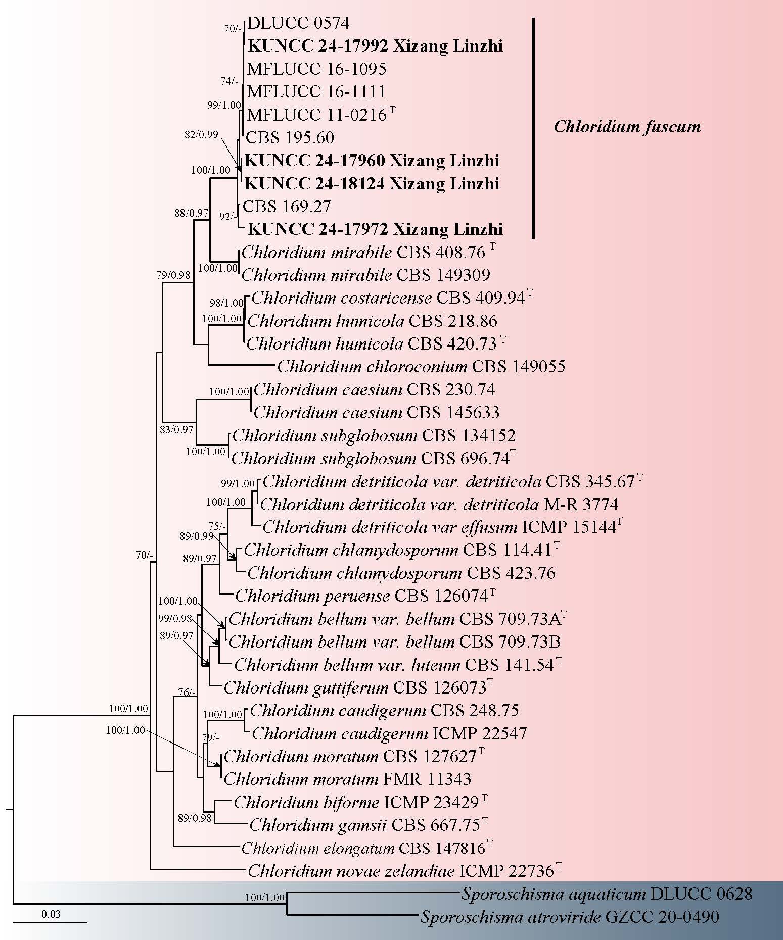
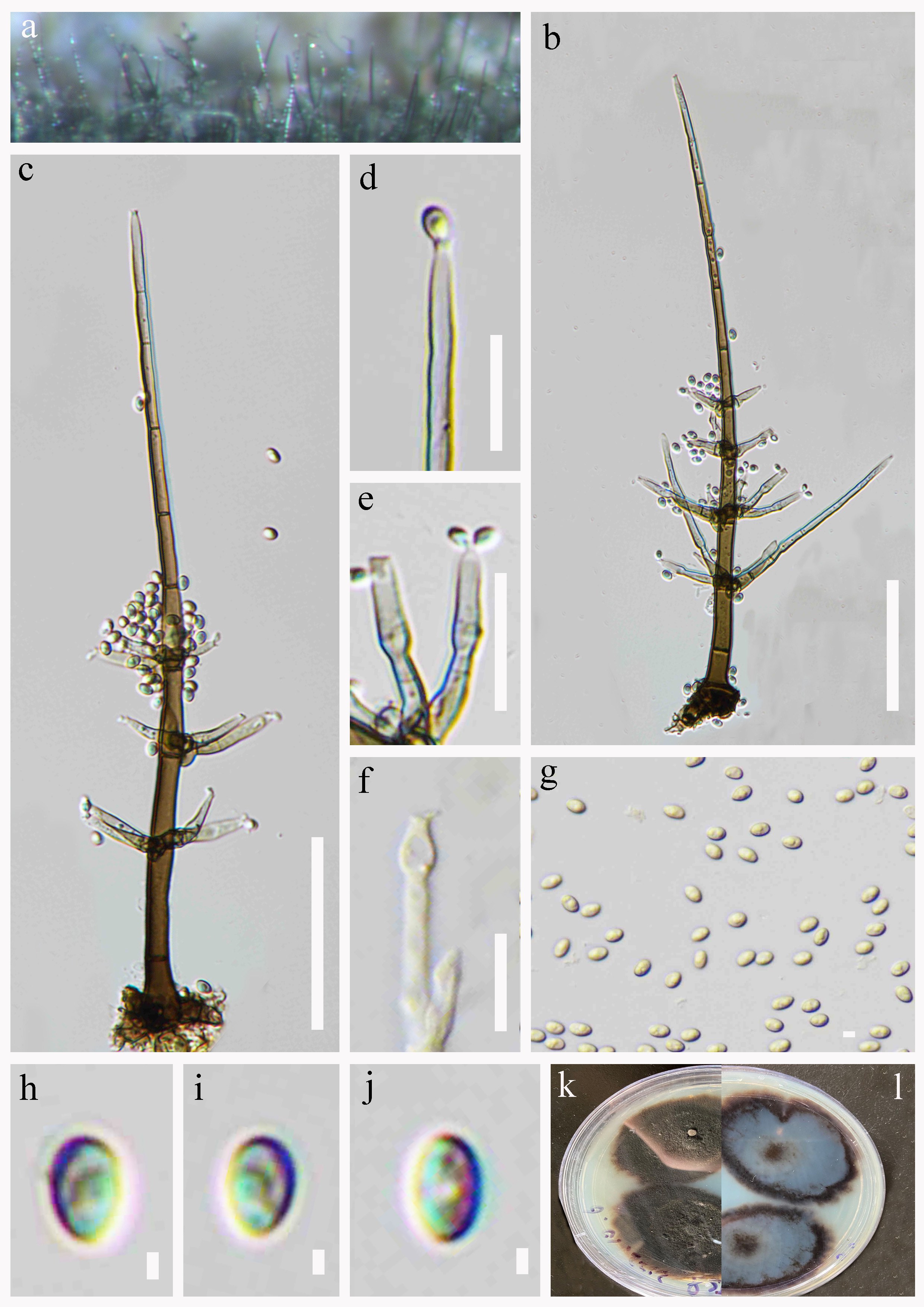
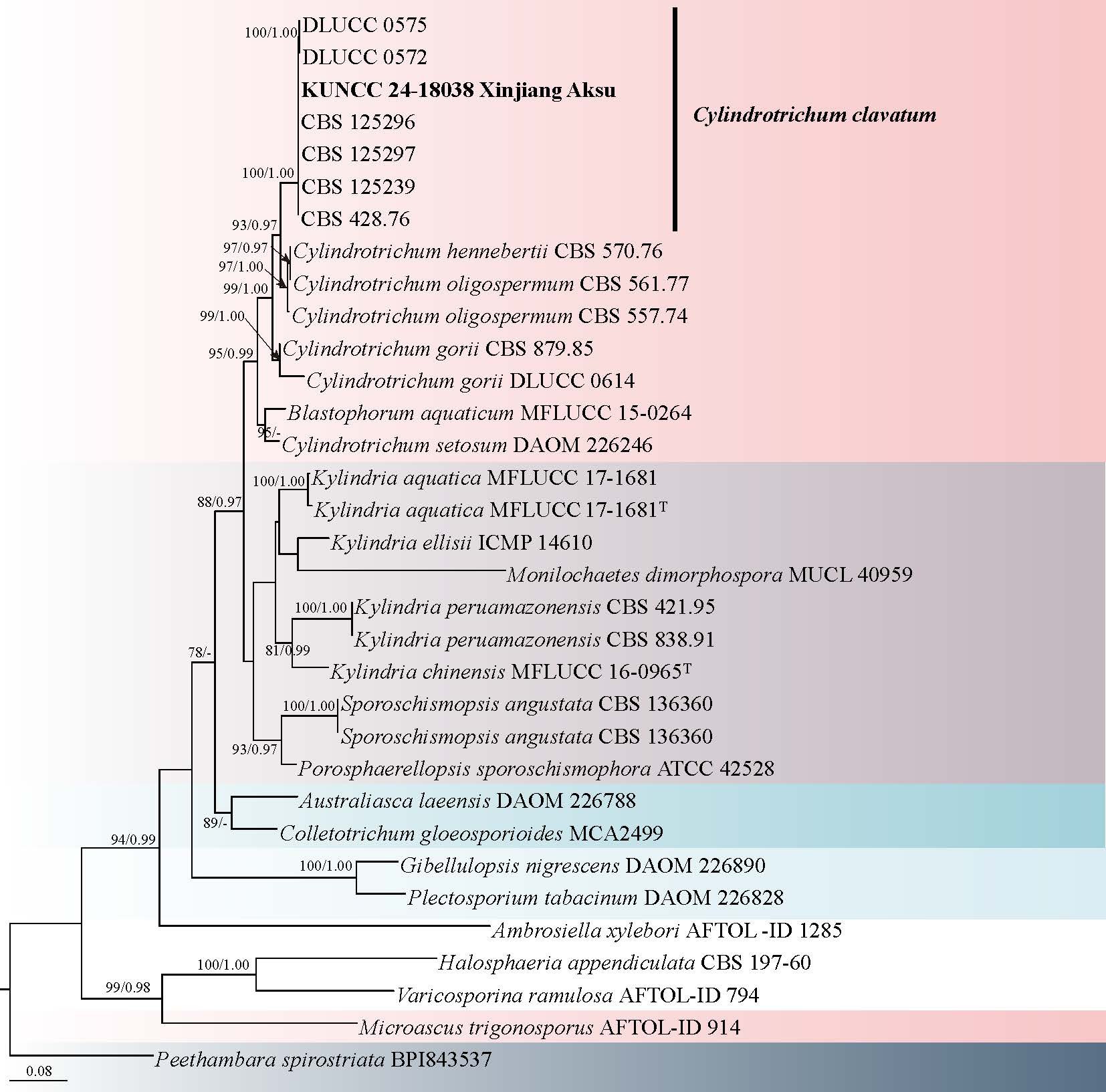
Chinese name
Index Fungorum number: IF 905035; Facesoffungi number: FoF 19305
Etymology: the term “linzhiensis” refers to the locality where the type specimen was found.
Holotype: HKAS 141644
Saprobic on decaying wood in a freshwater habitat. Sexual morph: Undetermined. Asexual morph: Colonies on natural substratum, superficial, effuse, scattered, hairy, dark brown. Mycelium partly immersed and partly superficial, composed of branched, septate, brown to dark brown, smooth-walled hyphae. Conidiomata 480–1128 × 34–50 μm ( = 640 × 40 μm, n = 20), synnematous, solitary or in group, erect, cylindrical, dark brown to black, tapering towards the apex, composed of closely compacted conidiophores and hyphae. Conidiophores 16–23 × 4–6 μm ( = 19 × 5 μm, n = 20), short, 1(2)-septate, cylindrical, unbranched, dark brown to black, smooth-walled. Conidiogenous cells monoblastic, integrated, terminal, determinate, cylindrical, brown to dark brown, smooth-walled. Conidia 73–152 × 11–15 μm ( = 98 × 13 μm, n = 20), acrogenous, solitary, straight to slightly flexuous, obclavate-rostrate, tapering towards the rounded apex, truncate at the base, brown to dark olivaceous brown, hyaline at the apex, base cells cuneiform, 8–15-euseptate, slightly constricted at the septa, sometimes with a sheath at the apex.
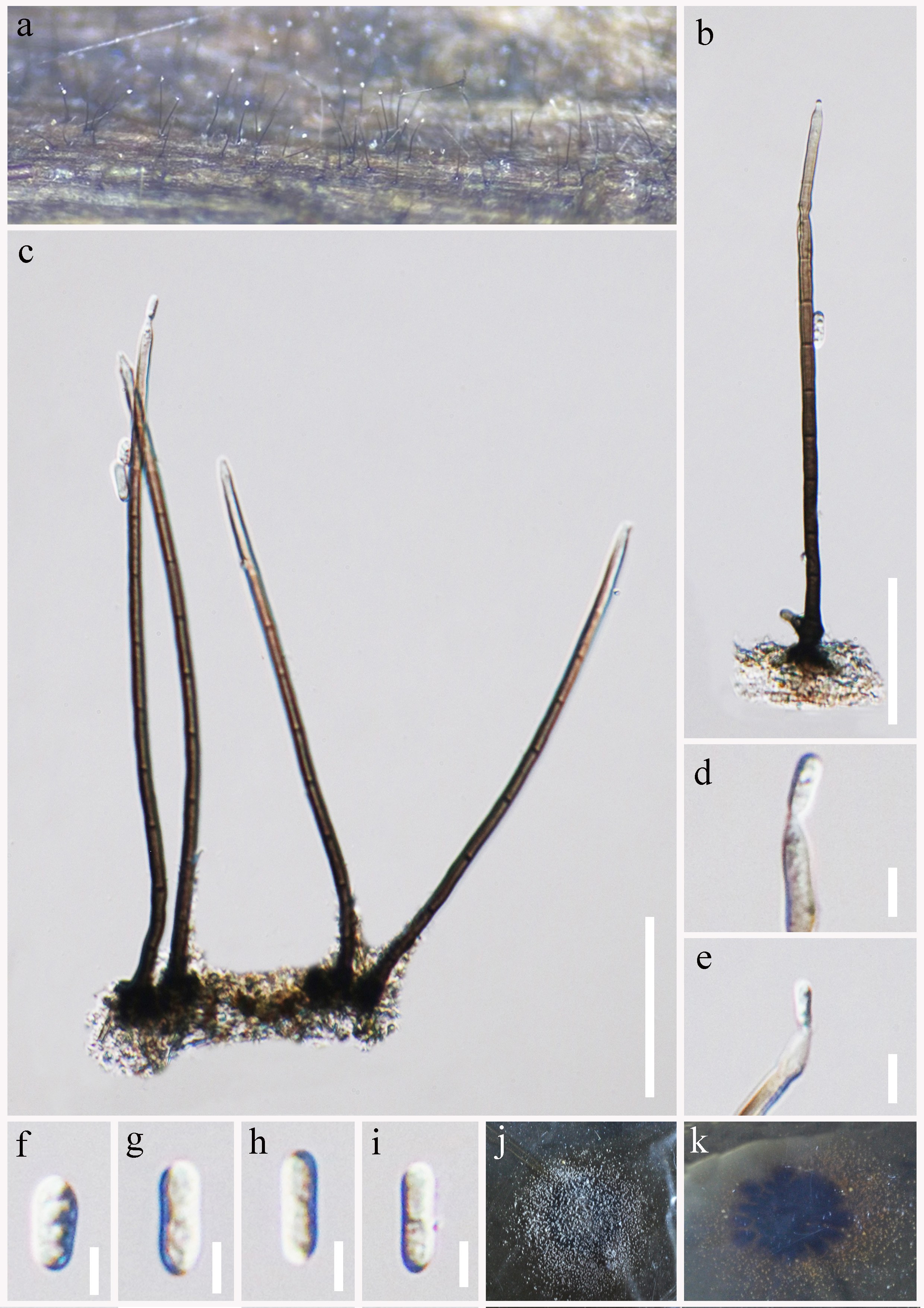
Culture characteristics: Conidia germinate on PDA within 48 h, with germ tubes emerging from a subtruncate base. Colonies on PDA reach a diameter of 3 cm within one month at room temperature. These colonies are circular with irregular margin, raised in the middle, cream-colored mycelium that covers the entire colony.
Material examined: CHINA, Xizang Autonomous Region, Linzhi City, Chayu County, 28° 29′ 39.27″ N, 96° 59′ 35.25″ E, 1537 m a.s.l., on decaying wood submerged in a freshwater stream, 14 July 2022, R.J. Xu, MD-152A (HKAS 141644, holotype), ex-type culture KUNCC 25-21369.
Notes: Falholtia linzhiensis shares the generic features of Falholtia by having compact synnemata composed of cylindrical, septate conidiophores, terminal, determinate, monoblastic conidiogenous cells, and holoblastic, obclavate-rostrate, euseptate conidia with a truncate base and paler apex (Hsieh et al. 2021; Wu & Diao 2022). However, F. linzhiensis is morphologically distinct from the type species F. kaohsiungensis in having shorter (16–23 μm vs. 13–34 μm), and conidia with fewer septa (8–15 vs. up to 27), often bearing an apical sheath, this feature not reported in the type. Additionally, F. linzhiensis forms conidiomata that are often scattered and hairy on the substrate surface, differing from the more compact and erect synnemata of F. kaohsiungensis.
Phylogenetic analysis based on ITS and LSU sequences places F. linzhiensis within Chaetosphaeriaceae, forming a well-supported and independent clade sister to F. kaohsiungensis (Fig. 35), thus supporting its recognition as a distinct taxon within the genus.
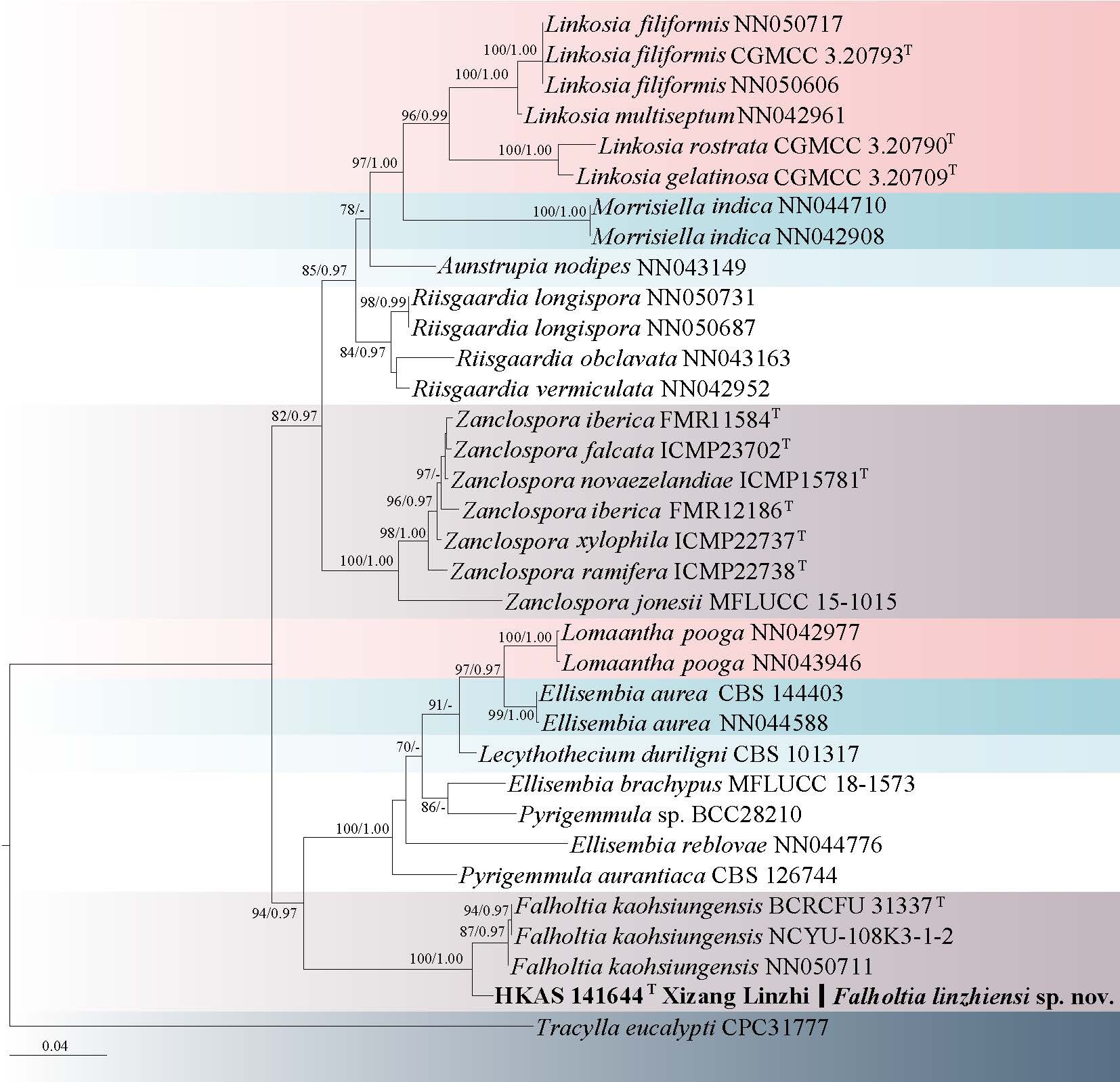
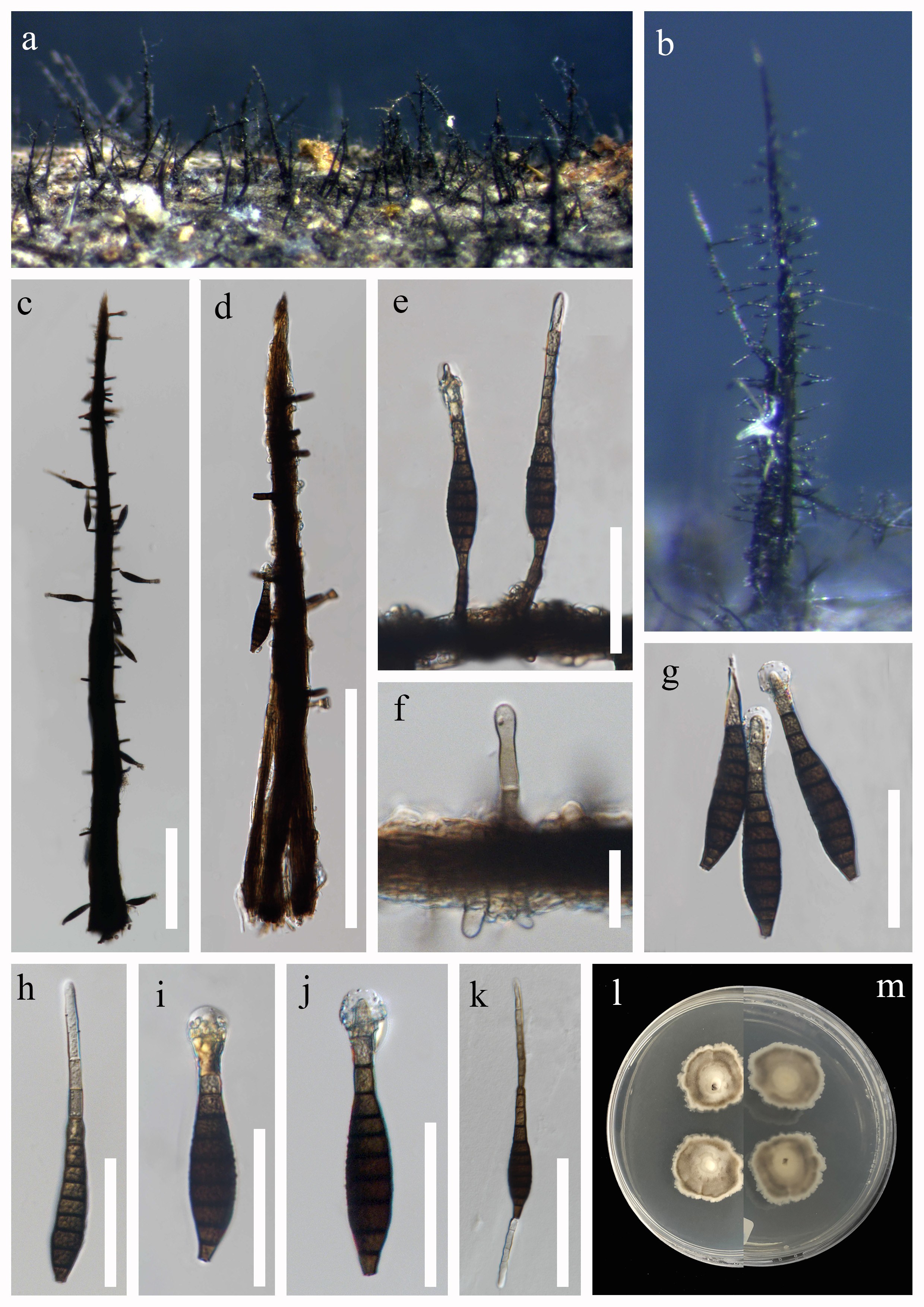
Phialosporostilbe Mercado & J. Mena
Phialosporostilbe was established by Sierra & Portales (1985) to accommodate P. turbinata. Members of Phialosporostilbe are characterized by the production of synnematous conidiophores bearing integrated, monophialidic or annellidic conidiogenous cells at the apices. The conidia are typically acrogenous, phialidic, catenate, and range from turbinate to cuneiform or cordiform in shape, colourless to pale brown, smooth-walled, and amerosporous, often with short subapical or apical appendages, such as setulae.
To date, seven species have been recognized worldwide: Phialosporostilbe catenata, P. dendroidea, P. gregariclava, P. scutiformis, P. setosa, P. turbinata and P. yadongensis, described from various habitats and geographic regions including Asia and the Americas (Sierra & Portales 1985; Bhat & Kendrick 1993; Shirouzu & Harada 2004; Sureshkumar et al. 2005; Wu & Zhang 2009; Yang et al. 2018b). Although similar in conidial shape and conidiogenesis to species of Nawawia, Phialosporostilbe is distinguishable by its synnematous rather than mononematous conidiophores, which has been the key feature supporting its separation at the generic level. Nonetheless, due to the similarities in conidial morphology and development, some authors previously considered Phialosporostilbe and Nawawia to be congeneric (Yang et al. 2018b), warranting further phylogenetic investigation to clarify their relationships.
20. Phialosporostilbe gaoligongensis R.J. Xu, Y. Li & Q. Zhao, sp. nov. Fig. 38
Chinese name: – 高黎贡瓶梗孢柱菌 (gao li gong ping geng zhu bao jun)
Index Fungorum number: IF905261; Facesoffungi number: FoF 19307
Etymology: The term “gaoligongensis” refers to the locality where the type specimen was found.
Holotype: HKAS 149332
Saprobic on decaying wood submerged in a freshwater stream. Sexual morph: Undetermined. Asexual morph: Colonies effuse, scattered or in small groups, dark brown, with masses of conidia on the apex of conidiophores. Mycelium immersed, consisting of pale brown, smooth-walled, branched, septate, smooth, subhyaline hyphae. Conidiophores 131–253 × 4–7 μm ( = 175 × 5 μm, n = 20), macronematous, mononematous, straight or slightly flexuous, solitary or 2–3-group, cylindrical, smooth-walled, 4–6-septate, unbranched, dark brown or brown, becoming paler towards the apex, rounded at the apex. Conidiogenous cells 26–44 × 4–7 μm ( = 35 × 6 μm, n = 20), monophialidic, terminal, integrated, pale brown, subhyaline towards the apex, cylindrical to cylindric-clavate, with an inconspicuous apical collarette. Conidia 8–12 × 6–10 μm ( = 10 × 8 μm, n = 30), acrogenous, solitary, round-tetrahedral, pyramidal, smooth, aseptate, thin-walled, guttulate, with 3-4 radiating from each corner appendages 1.6–3.3 μm ( = 2.4 μm, n = 30) long.
Culture characteristics: Conidia germinating on PDA medium within 48 h and germ tubes arising from terminal end of conidium. Colonies grown on PDA reached 30 mm diam at room temperature in three weeks, irregular, with velvety, dry, raised, grayish-white mycelium on the surface, from below cream to light brown, producing cream pigments, with entire margin.
Material examined: CHINA, Yunnan Province, Tengchong City, Gaoligong Mountain, 25° 12' 36.00" N, 98° 19' 48.00" E, 1852 m a.s.l., on decaying wood submerged in a freshwater stream, 12 November 2024, Y. Li, ly42 (HKAS 149332, holotype), ex-type culture KUNCC 25-19643.
Notes: Phialosporostilbe gaoligongensis possess its conidial characteristics that fit with Phialosporostilbe (Sierra & Portales 1985; Wu & Zhang 2009; Yang et al. 2018b), but differs from P. catenate, P. dendroidea, P. scutiformis, P. setosa, P. turbinata and P. yadongensis in having macronematous, mononematous, unbranched conidiophores. Phialosporostilbe gaoligongensis resembles P. gregariclava in having macronematous, mononematous, conidiophores, acrogenous, solitary, pyramidal conidia, with 3-4 radiating subapical appendages. However, P. gregariclava differs from P. gaoligongensis in having conidiophores of P. gregariclava sometimes with percurrently proliferate, and more septa (5–15 septa vs. 4–6 septa).
Phylogenetic analysis showed that Phialosporostilbe gaoligongensis (KUNCC 25-19643) isolates formed a distinct lineage within the genus, and sister clade with P. scutiformis (MFLU 18-1502 and MFLUCC 18-1288) (Fig. 37). but it differs from P. scutiformis in having unbranched conidiophores and shorter appendages (1.6–3.3 μm vs. 3.5–7.5 μm) (Yang et al. 2018b).
Sporoschisma Berk. & Broome
Sporoschisma was introduced by Berkeley (1847), with S. mirabile as the type species. Significant revisions of the genus were made by Hughes (1966) and Goh et al. (1997), who provided historical collection details and comprehensive illustrations. Sporoschisma is characterized by scattered, capitate setae and straight, smooth, unbranched brown conidiophores, typically consisting of a cylindrical stipe, a swollen venter, and a long cylindrical neck. The conidiogenous cells are phialidic, while the conidia are cylindrical, often truncate at both ends, and form in chains endogenously in basipetal succession (Seifert & Gams 2011; Wu & Diao 2022; Yang et al. 2023; Xu et al. 2024f).
21. Sporoschisma longicatenatum Jing Yang, Jian K. Liu & K.D. Hyde, in Yang, Liu, Hyde, Bhat, Gareth Jones & Liu, Phytotaxa 289(2): 152 (2016) Fig. 40
Chinese name
Index Fungorum number: IF 552182; Facesoffungi number: FoF 02243
Saprobic on decaying wood submerged in a freshwater habitat. Sexual morph: Undetermined. Asexual morph: Colonies effuse, black, hairy, chain-like conidia at the apex. Mycelium immersed, composed of pale to dark brown hyphae. Setae 105–156 × 5–7 µm ( = 127 × 6 μm, n = 10), scattered or in groups mixed with conidiophores, capitate, usually at the swollen apex, straight or flexuous, septate. Conidiophores 293–359 × 12–15 µm ( = 318 × 14 μm, n = 15), macronematous, mononematous, smooth, dark brown to black, paler at the torn apex, straight or slightly flexuous, solitary or in groups, setae, arising from dark brown to black bulbous base, composed of a cylindrical stipe and a swollen venter with a long cylindrical neck, erect. Conidiogenous cells monophialidic, percurrent, integrated, terminal, determinate, brown, lageniform, frayed at the apex. Conidia 66–87× 19–25 µm ( = 74 × 21 μm, n = 20), cylindrical, 5-septate, hyaline when young, olivaceous to brown at maturity, with hyaline to pale brown end cells, which are much shorter than the four inner cells, conspicuously darkened at the septa, rounded at both ends.
Culture characteristics: Conidia germinating on PDA medium within 72 h and germ tubes produced from both ends. Colonies grown on PDA reached 60 mm diam at room temperature in two weeks, irregular, with velvety, flat, dark brown mycelium on the surface, from below dark brown with undulate margin.
Material examined: CHINA, Xizang Autonomous Region, Linzhi City, Motuo County, 29°19'43" N, 95°21'19" E, 677 m a.s.l., on decaying wood submerged in a freshwater habitat, 13 July 2022, R.J. Xu, MD-109 (HKAS 136183), living culture KUNCC 24-18020.
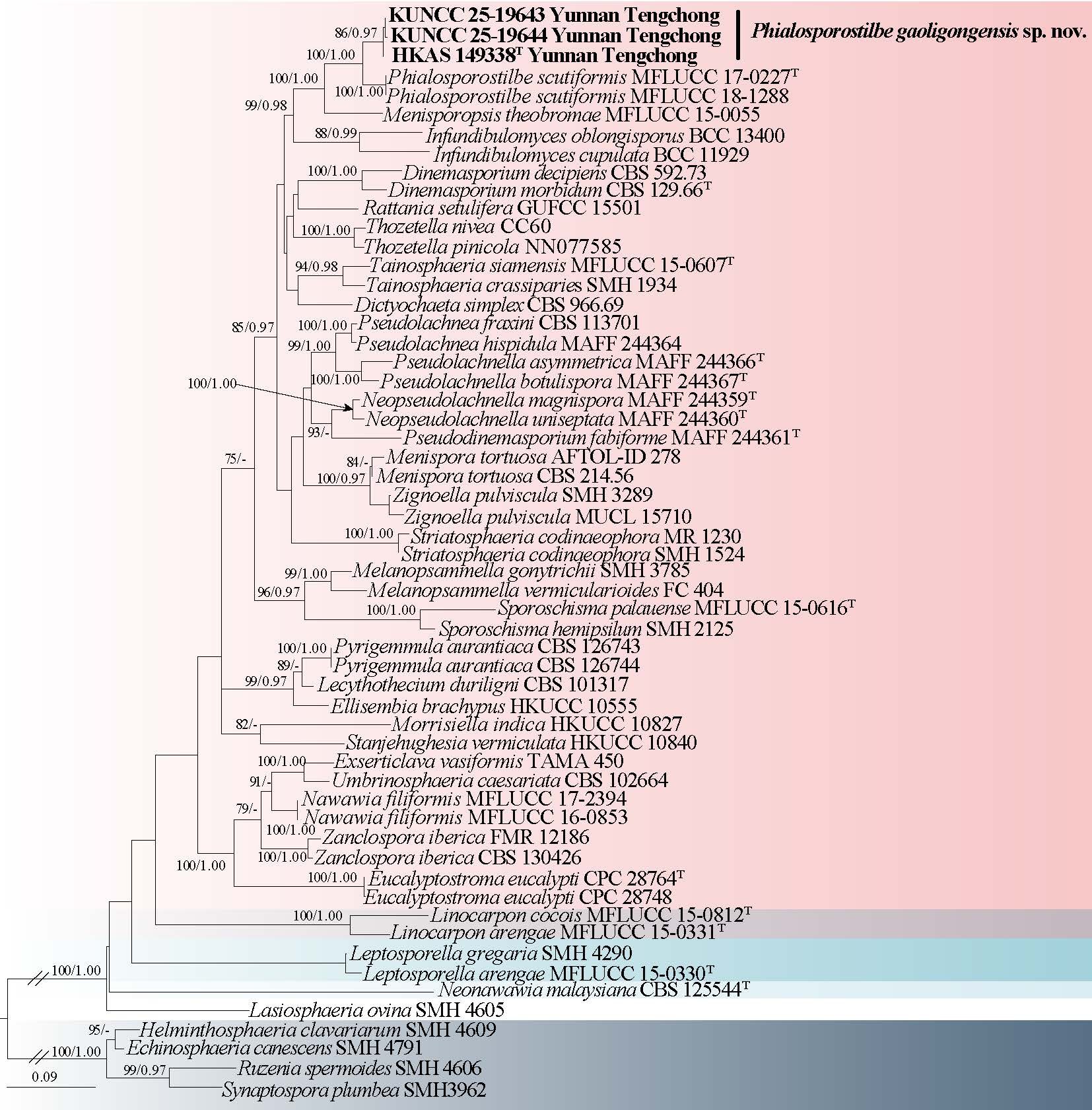
Notes: Sporoschisma longicatenatum was identified in a freshwater habitat in Thailand by Yang et al. (2016b), based on phylogenetic analyses and morphological comparisons. Subsequently, Yang et al. (2016a) and Bao et al. (2021) also reported the presence of S. longicatenatum in Thailand. Phylogenetic analyses showed that our isolated of S. longicatenatum (KUNCC 24-18020) clustered together with S. longicatenatum (MFLUCC16-0180 and GZCC15-0072) with strong statistical support (100% ML/1.00 BYPP) (Fig. 39). Therefore, we reported a new record for S. longicatenatum on the Q-X Plateau, China.
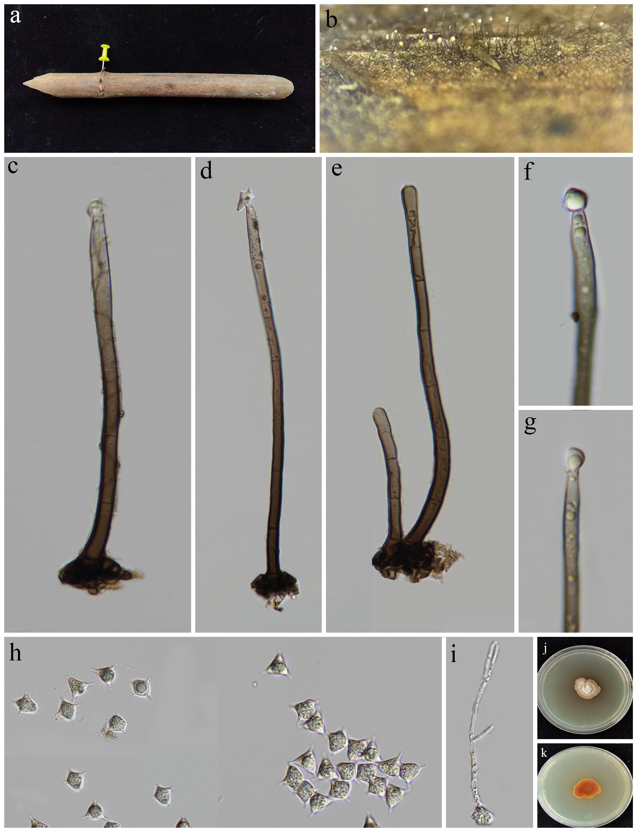
Stanjehughesia Subram
Stanjehughesia was established by Subramanian (1992) during his taxonomic revision of the heterogeneous Sporidesmium, in which he segregated several genera based on morphological features such as the presence or absence of conidiophores, the nature of conidial septation (euseptate vs. distoseptate), and percurrent proliferation. Species within Stanjehughesia are characterized by lacking well-developed conidiophores and producing obclavate to obclavate-rostrate, euseptate conidia, which may be smooth or verrucose, directly on simple, monoblastic conidiogenous cells (Subramanian 1992; Wu & Zhuang 2005; Seifert et al. 2011). To date, approximately 21 species have been reported in this genus (Hsieh et al. 2021). Cultural studies have linked the sexual morphs Umbrinosphaeria caesariata syn. Chaetosphaeria caesariata and Miyoshiella larvata linked to Stanjehughesia (Réblová 1999b). Morphologically similar genera such as Linkosia also possess reduced conidiophores and comparable conidial features. However, Linkosia can be distinguished by its distoseptate conidia (Hernández-Gutiérrez & Sutton 1997). Phylogenetic analyses have revealed that both Stanjehughesia and Linkosia as distinct linages in Chaetosphaeriaceae, Chaetosphaeriales (Hyde et al. 2024a; Réblová et al. 2024).
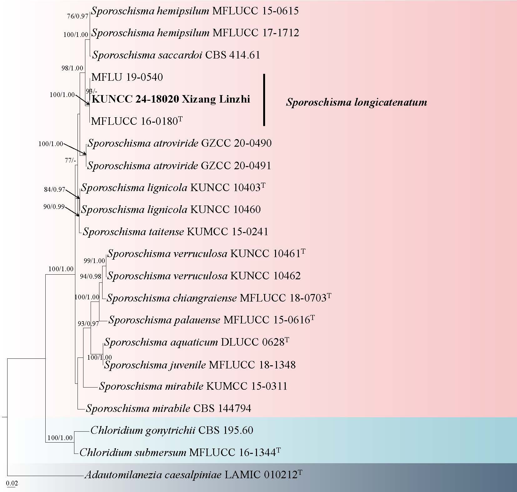
22. Stanjehughesia motuoensis R.J. Xu, Q. Zhao & K.D. Hyde, sp. nov. Fig. 42
Chinese name
Index Fungorum number: IF 905037; Facesoffungi number: FoF 19308
Etymology: the term “motuoensis” refers to the locality where the type specimen was found.
Holotype: HKAS 151645
Saprobic on decaying wood in a freshwater habitat. Sexual morph: Undetermined. Asexual morph: Colonies on wood effuse, scattered or in small groups, hairy, dark brown, glistening. Mycelium mostly immersed, composed of septate, branched, hyaline to brown, smooth-walled hyphae. Conidiophores reduced to conidiogenous cells. Conidiogenous cells 4–6 × 3–4 μm ( = 5 × 3 μm, n = 20), monoblastic, integrated, terminal, determinate, erect, solitary or fasciculate, straight, cylindrical or lageniform, aseptate, slight olivaceous green, brown to dark brown, smooth-walled. Conidia 30–110 × 9–12 μm ( = 90 × 11 μm, n = 20), acrogenous, solitary, lanceolate, rostrate, straight or slightly curved, 14–19-euseptate, brown to dark brown with olive green tint, pale brown to subhyaline at the apex, guttulate, tip swollen and gradually tapers towards the base part when young, the lower middle part swollen and gradually tapers towards both ends when mature, rounded at the apex and truncate at the base, smooth-walled.
Material examined: CHINA, Xizang Autonomous Region, Linzhi City, Motuo County, 29°19′43.6″ N, 95°21′19.2″ E, 798 m a.s.l., saprobic on submerged decaying wood in a freshwater habitat, 12 July 2022, R.J. Xu, MD-381A (HKAS 151645, holotype), ex-type culture KUNCC 10488.
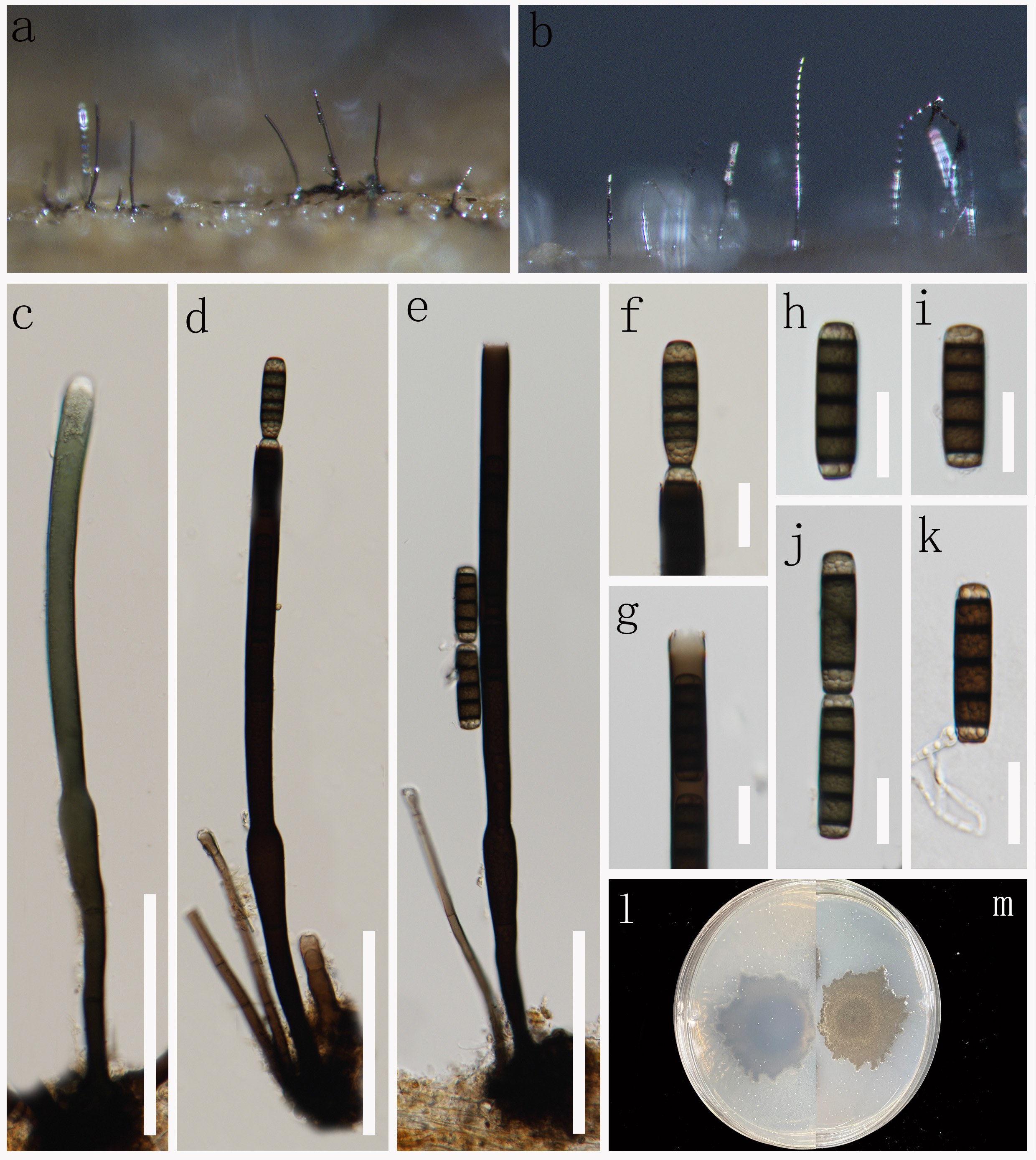
Notes: Stanjehughesia motuoensis was collected from decaying wood in a freshwater habitat in Q-X Plateau, China. In the multi-gene phylogenetic analysis based on combined LSU, SSU, ITS, and tef1-α sequence data, S. motuoensis forms a distinct, well-supported clade within Chaetosphaeriaceae (Fig. 41), clearly separated from other known species of the genus (Wu & Diao 2022). Although S. motuoensis shares the typical lanceolate, multi-septate conidia characteristic of Stanjehughesia, it is readily distinguished from S. hormiscioides by its longer conidia (30–110 μm vs. 30–60 μm), higher septation (14–19 vs. 7–14), and more pronounced tapering at both ends. Compared to S. kaohsiungensis, which has shorter (35–70 μm), less septate (7–13) conidia and a broader apex, S. motuoensis also exhibits a distinctive rostrate shape and a darker pigmentation with an olive tint. Stanjehughesia motuoensis is introduced herein as a new species based on its unique morphological characteristics and distinct phylogenetic placement within Stanjehughesia.
Glomerellales Chadef. ex Réblová, W. Gams & Seifert
Reticulascaceae Réblová & W. Gams
Kylindria DiCosmo, S.M. Berch & W.B. Kendr.
DiCosmo et al. (1983) established Kylindria to accommodate Cylindrotrichum triseptatumK. triseptata, type species), along with four other Cylindrotrichum species. The asexual morphs are characterized by solitary, unbranched, multi-septate, dark brown conidiophores, monophialidic, integrated, terminal, ampulliform conidiogenous cells with narrow apertures at the apex and with or without a conspicuous collarette, and unicellular or euseptate, hyaline conidia with or without an excentric lateral or basal hilum (DiCosmo et al. 1983; Zhang et al. 2010). Kylindria species are predominantly saprobic fungi that flourish on decomposing organic matter, including dead branches, decaying leaves, rotting rachides, and desiccated twigs, found in both aquatic and terrestrial ecosystems (Maharachchikumbura et al. 2018; Tian et al. 2024a). Currently, 18 species epithets are listed in Index Fungorum (accessed on 16 July 2025), however, sequence data are available for only 6 species.
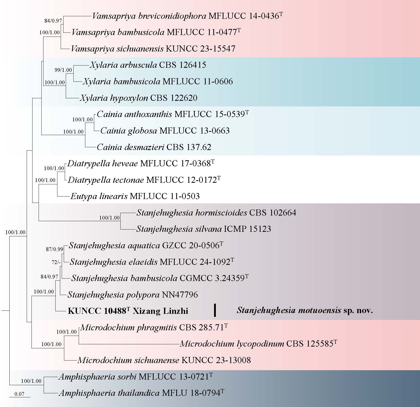
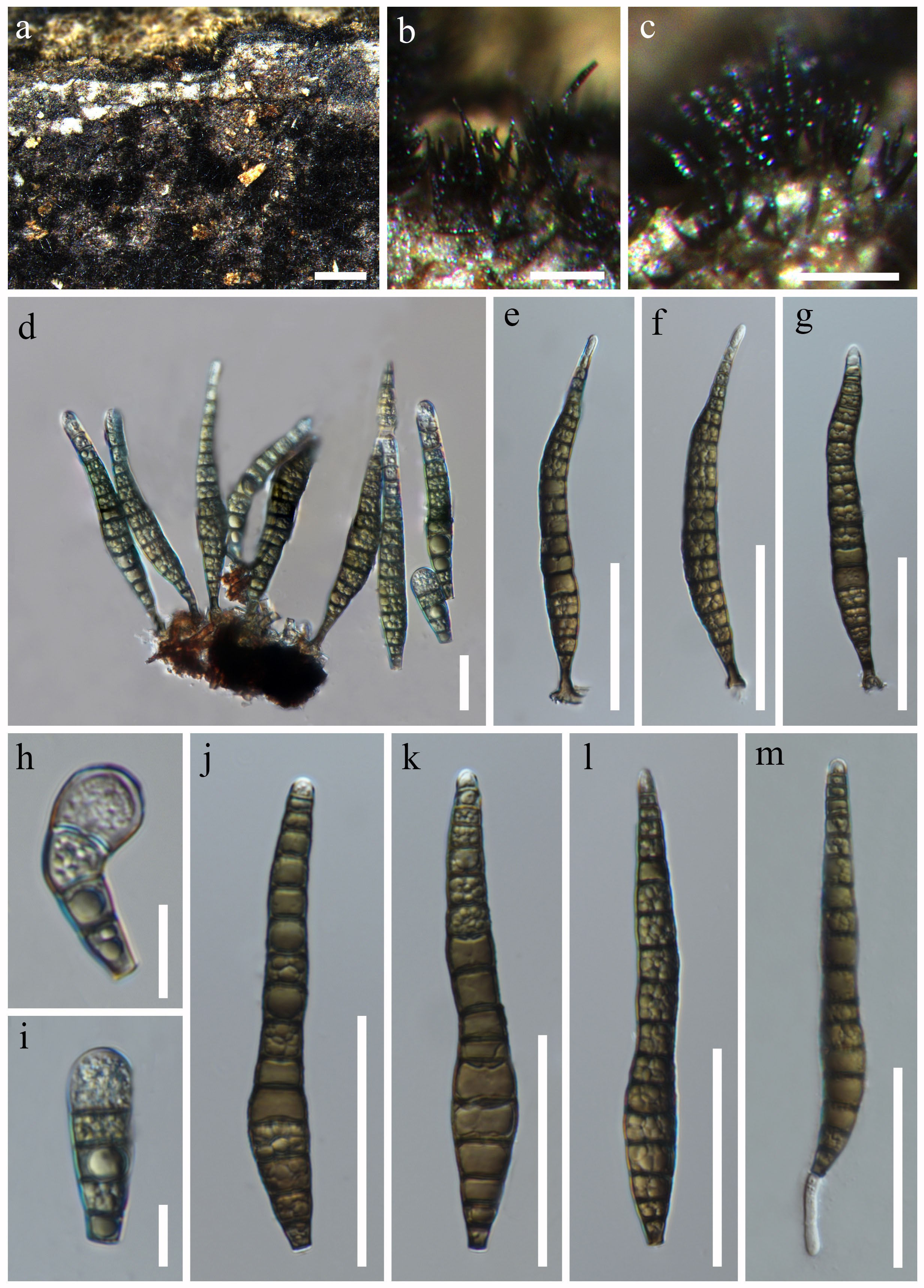
23. Kylindria cryptomeriae W.H. Tian, K.D. Hyde & Maharachch., Mycosphere 15 (1): 1861 (2024) Fig. 44
Chinese name
Mycobank number: MB 851891; Facesoffungi number: FoF 16281
Saprobic on decaying submerged wood in a freshwater habitat. Sexual morph: Undetermined. Asexual morph: Colonies on natural substrate, superficial, effuse, gregarious, brown to dark brown, hairy, with white glistening conidial masses at the apex. Mycelium partly immersed, composed of branched, septate, brown to dark brown, smooth hyphae. Conidiophores 204–305 × 9–16 µm ( = 268 × 12 µm, n = 20), mononematous, macronematous, straight to slightly curved, erect, unbranched, multi-septate, dark brown at base, paler toward the apex, thick-walled. Conidiogenous cells 41–53 × 9–15 µm ( = 46 × 13 μm, n=20), phialidic, terminal, integrated, ampuliform, broadest above the middle, tapering near the apex, with an inconspicuous apical collarette. Conidia 24–34 × 9–14 µm ( = 30 × 11 μm, n = 30), arranged in a slimy head, solitary, oblong with obtuse ends, hyaline, 0–3-septate, with a slimy mucilaginous coating around.
Culture characteristics: Conidia germinating on PDA medium within 24 hours, with germ tubes arising from polar cells of the conidium. Colonies grown on PDA reached a diameter of 12 mm at room temperature under natural light in one month, edges regular, flat, white to pale yellow on the surface, pale yellow on the reverse.
Material examined: CHINA, Guizhou Province, Liupanshui City, Shuicheng County, Yushe National Forest Park, N 26°27′17″ N, 104°48′26″ E, 2232 m a.s.l., on decaying submerged wood in a freshwater habitat, 27 February 2023, Xingjuan Xiao, YSS89 (HKAS 145867), culture GZCC 24-0088.
Note: Phylogenetically, our new collection is closely allied with Kylindria cryptomeriae (UESTCC 23.0231) with 96% ML/1.00 BYPP support (Fig. 43). Comparison of the ITS, LSU, and rpb2 sequences between the new collection and K. cryptomeriae (UESTCC 23.0231) showed 99% identity (481/483 bp, 1 gap), 100% identity (801/801 bp), and 99% identity (1019/1023 bp, 1 gap), respectively. Morphologically, the new collection displays nearly identical characteristics, including conidiophore dimensions (204–305 × 9–16 µm vs. 180–310 × 6–11 µm) and conidia (24–34 × 9–14 µm vs. 22–32 × 9–12 µm) with the holotype (HKAS 131279) of K. cryptomeriae (Tian et al. 2024a). Consequently, we classify our collection as K. cryptomeriae, marking the first report of this species from a freshwater habitat.
Conioscyphales Réblová & Seifert
Conioscyphaceae Réblová & Seifert
Conioscypha Höhn
Conioscypha was established by von Höhnel (1904), with C. lignicola as the type species. Species within this genus are widely distributed across terrestrial and freshwater habitats globally. The sexual morphs of Conioscypha are characterized by inconspicuous ascomata, which are either superficial or immersed, ostiolate, and subhyaline to pale orange. These ascomata contain hyaline, 8-spored, unitunicate asci with a refractive J-apical annulus, and fusiform, 3–7-septate ascospores that lack mucilaginous sheaths or appendages (Réblová & Seifert 2004; Zelski et al. 2014; Réblová et al. 2016b; Hyde et al. 2020b). The asexual morphs are defined by micronematous, mononematous, erect, hyaline, and smooth-walled conidiophores. The conidiogenous cells are enteroblastic, terminal, or intercalary, and exhibit cyathiform to doliiform shapes with multilayered, cup-like collarettes. The conidia are dark brown to black, ranging from globose to subglobose or elliptical, with smooth surfaces (Shearer & Motta 1973; Goh & Hyde 1998a; Chuaseeharonnachai et al. 2017; Luo et al. 2019). Currently, Conioscypha comprises 29 accepted species (Index Fungorum 2026; Liu et al. 2019b; Xu et al. 2024e, 2025; Li et al. 2024b; Sun et al. 2025).
24. Conioscypha aquatica (Z.L. Luo, K.D. Hyde & H.Y. Su) R.J. Xu, Q. Zhao & K.D. Hyde, comb. nov. Fig. 46
Chinese name
Index Fungorum number: IF 905262; Facesoffungi number: FoF 05450
Synonym: Conioscypha submersa Z.L. Luo, K.D. Hyde & Hong Y. Su, Fungal Diversity 99: 560 (2019)
Saprobic on decaying wood submerged in a freshwater habitats. Sexual morph: Undetermined. Asexual morph: Hyphomycetous. Colonies on natural substrate effuse, scattered, gregarious, punctiform, dark brown or black. Mycelium immersed, composed of hyaline, septate, branched, smooth hyphae. Conidiophores micronematous, mononematous, hyaline, often reduced to conidiogenous cells. Conidiogenous cells sessile or on short conidiophores, and arising directly from the hyphae. Conidia 17–23 × 14–22 μm ( = 20 × 18 μm, n = 40), acrogenous, globose, subglobose, or obovoid, aseptate, pale brown when young, dark brown to black when mature, smooth-walled, subtruncate at base.
Culture characteristics: Conidia germinating on PDA within 48 h and germ tubes produced from base cell. Colonies on PDA reaching 5–10 mm diam, at one month at room temperature, circular, with sparse dark brown mycelium in the middle, sparse pale brown mycelium on the outer ring with irregular margin. Sporulated conidia in cultures. Conidiophores micronematous, branched, sometimes reduced to a moniliform conidiogenous cell, hyaline to pale brown. Conidiogenous cells monoblastic, integrated, cylindrical, hyaline. Conidia 9–17 × 8–14 μm ( =12 × 11 μm, n = 40), acrogenous, subglobose or elliptical, olivaceous or pale brown.
Material examined: CHINA, Xizang Autonomous Region, Rikaze City, Nyalam County, 27° 59' 13.09" N, 85° 59' 0.93" E, 2236 m a.s.l., on decaying wood submerged in a freshwater stream, 06 July 2022, R.J. Xu, MD-680 (HKAS 136019), culture KUNCC 10475. Xizang Autonomous Region, Rikaze City, Dingjie County 27° 53' 8.72" N, 87° 27' 36.21" E, 3042 m a.s.l., on decaying wood submerged in a freshwater stream, 05 July 2022, R.J. Xu, MD-681 (HKAS 136027), culture KUNCC 10473.
Notes: Conioscypha aquatica and C. submersa were initially described by Luo et al. (2019), based on comprehensive phylogenetic analyses and morphological assessments. Our multigene phylogenetic analyses, coupled with meticulous morphological comparisons, indicate that our collected specimens (KUNCC 10475 and KUNCC 10473) group together with ex-type of C. aquatica (MFLUCC 18–1333) and ex-type of C. submersa (DLUCC 0904) (Fig. 45). Despite the observed differences in their ITS sequences, which amount to 7/528 bp (1.3%, excluding gaps), these intraspecific variabilities do not satisfy the standard thresholds commonly employed for species delineation (Jeewon & Hyde 2016; Chethana et al. 2021a). Furthermore, morphological analysis reveals a notable congruence among these specimens regarding conidiophore architecture and the morphology, dimensions, and shape of the spores, thereby reinforcing the hypothesis that they constitute a singular species.
Given these findings, we consolidate Conioscypha aquatica and C. submersa into a single species. In accordance with the International Code of Nomenclature for algae, fungi, and plants (ICN), we recommend the retention of the name C. aquatica, as it is supported by the earlier collection date of its type specimen (2015) in contrast to that of C. submersa (2016). Additionally, we have updated the molecular phylogenetic data for this species and provided a comprehensive description of its colonies cultivated on potato dextrose agar (PDA) medium.
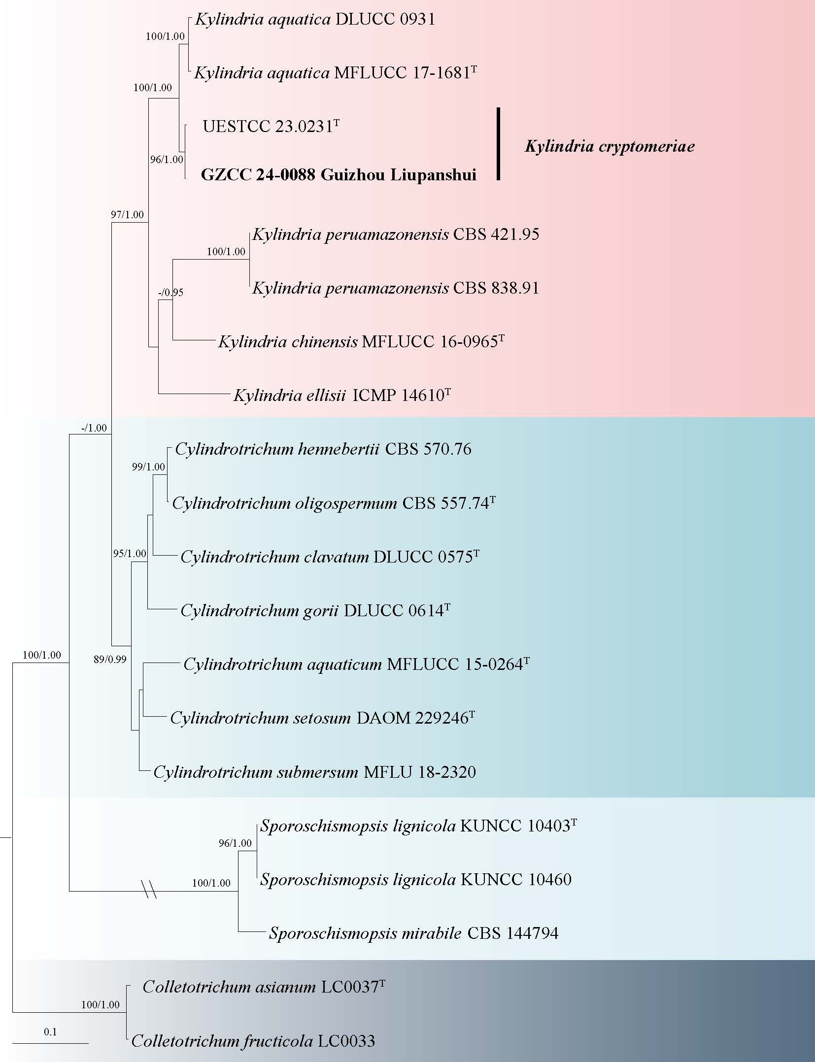
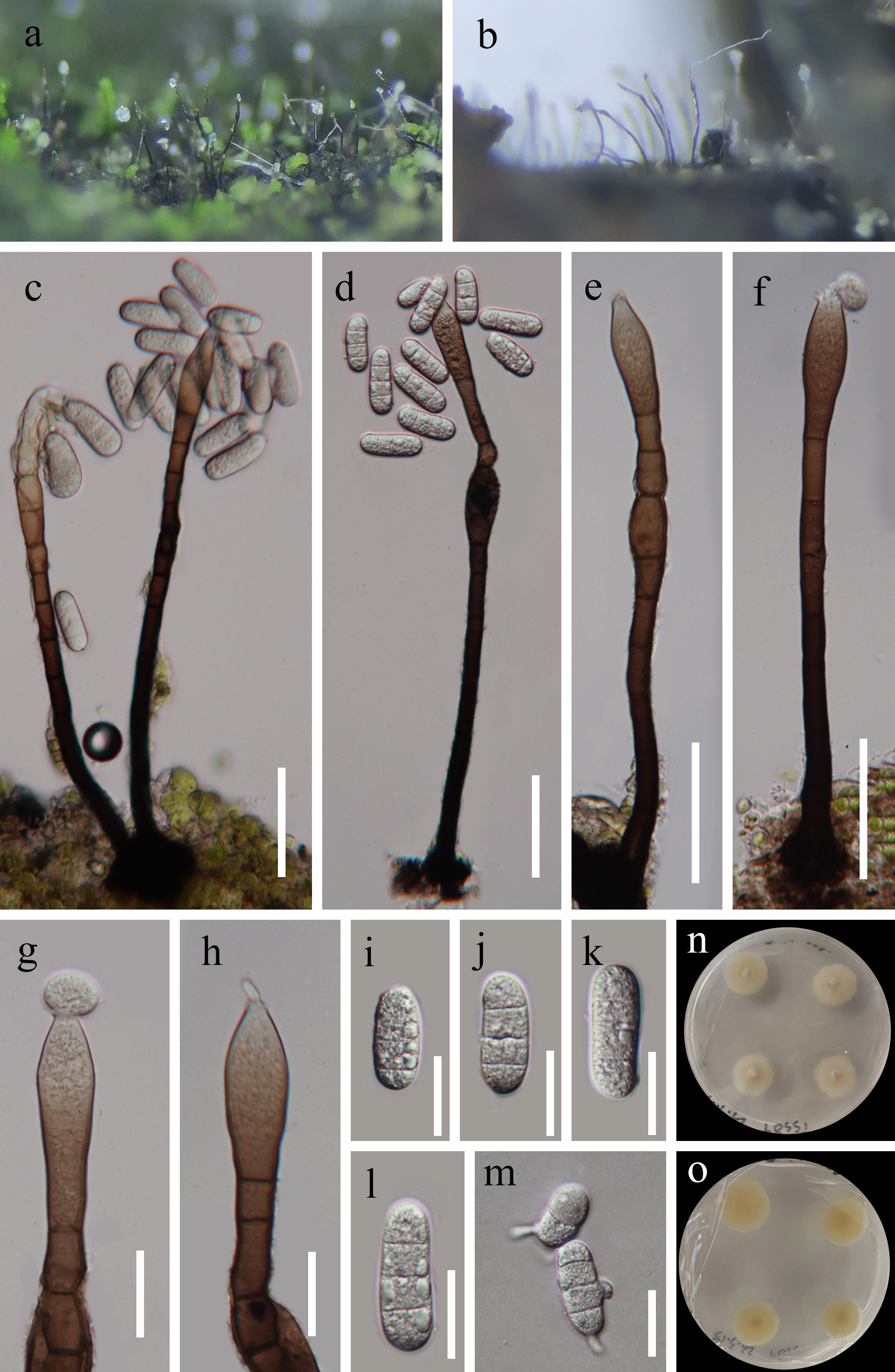
Coniochaetales Huhndorf, A.N. Mill. & F.A. Fernández
Cordanaceae Nann.
Cordana Preuss, Linnaea 24: 129 (1851)
Cordana was established by Preuss (1851) to include three species C. polyseptata, C. pauciseptata, and C. pedunculata, though no type species was initially designated (Hernández-Restrepo et al. 2014; Hyde et al. 2020c). Cordana is defined by macronematous, mononematous conidiophores with polyblastic, inflated, integrate or discrete conidiogenous cells that are terminal or intercalary, nodose, and swollen or umbellate at the apices. Its conidia are acropleurogenous, ovoid to ellipsoid, and typically bear a protuberant hilum (Castañeda Ruiz et al. 1999; Ai et al. 2019; Yang et al. 2022b).
Members of Cordana are globally distributed and commonly found on various substrates, including soil, plant debris, and other fungal organisms (De Hoog et al. 1983; Cai 2004; Soares et al. 2005). Currently, 21 species are recognized in the genus, with some species, such as C. abramovii, C. aquatica, C. reniformis, C. uniseptata and C. xizangensis also identified in freshwater environments (Cai 2004; Hernández-Restrepo et al. 2014; Zelski et al. 2014; Luo et al. 2019; Xu et al. 2024e, 2025).
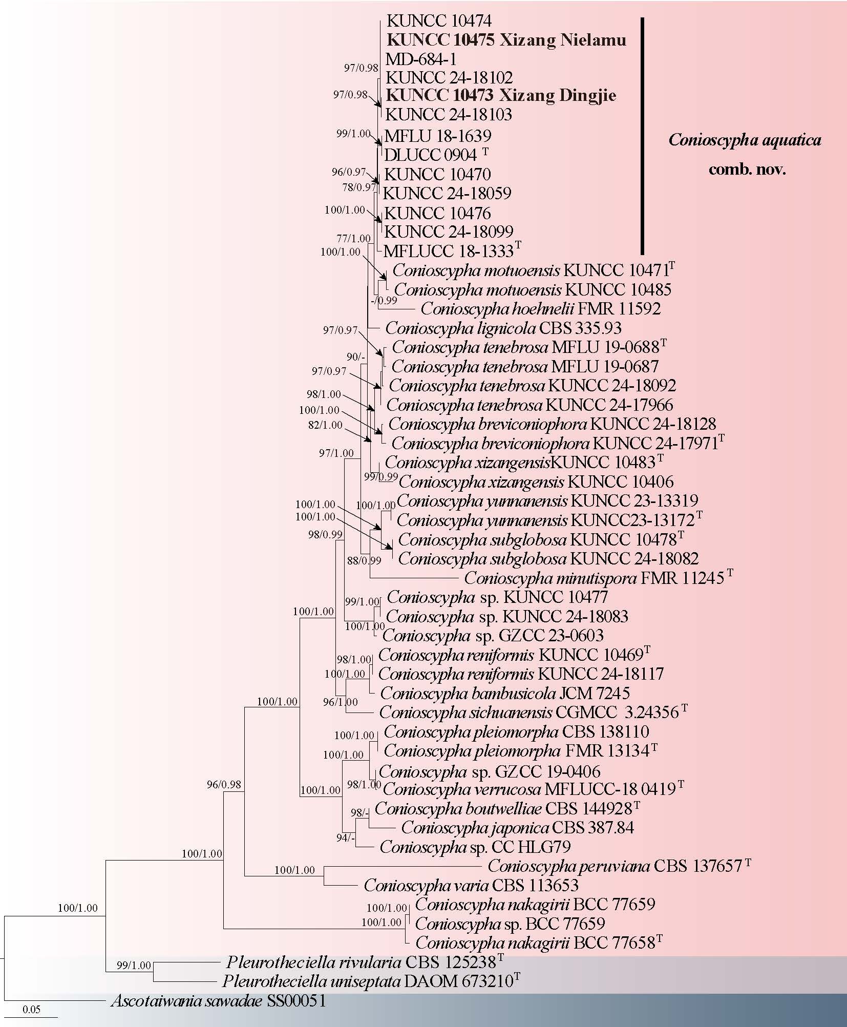
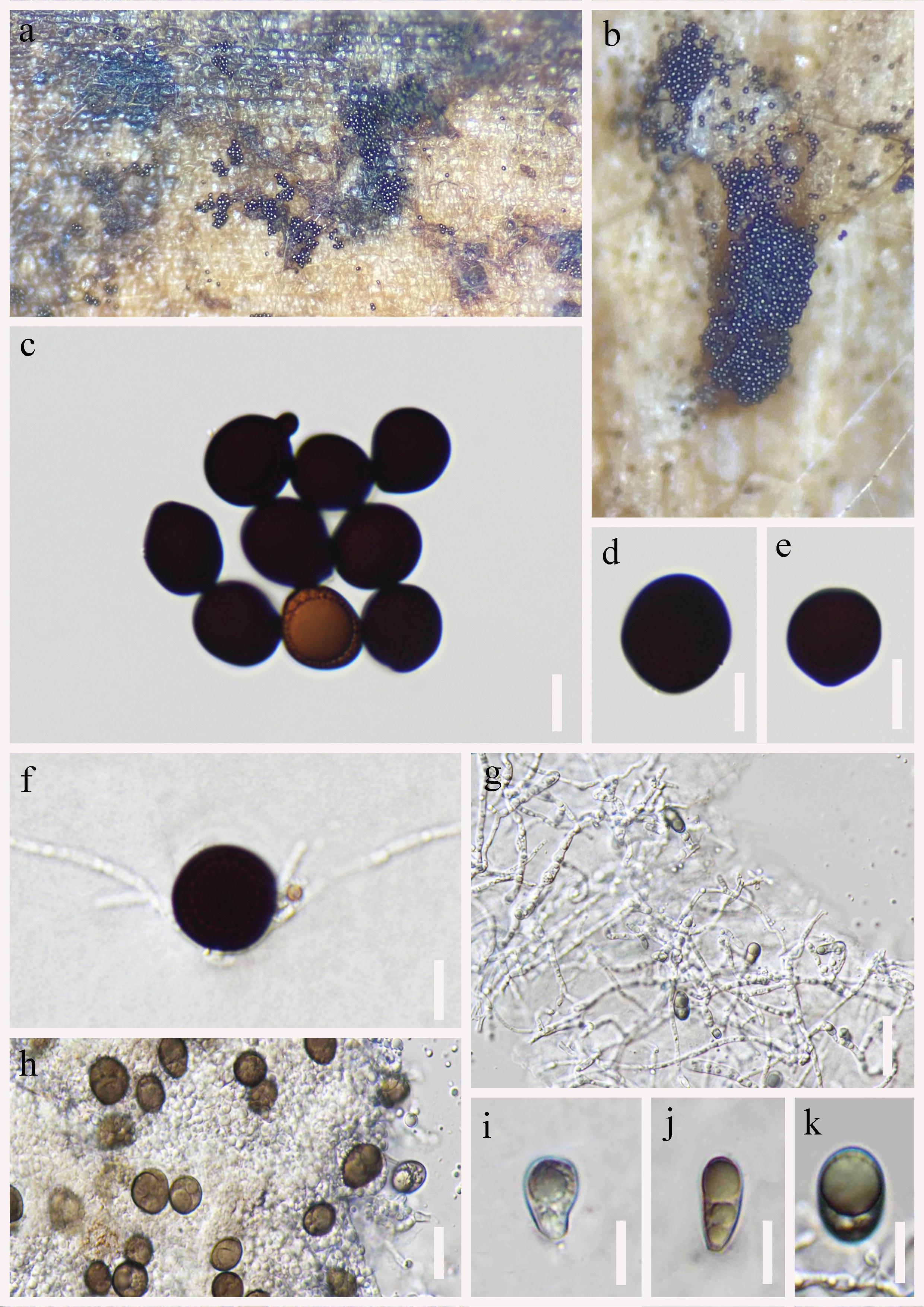
25. Cordana pohuashanensis S.C. He, Q. Zhao & K.D. Hyde, sp. nov. Fig. 48
Index Fungorum number: IF905263; Facesoffungi number: FoF 19309
Chinese name
Etymology: Name reflects the host, Sorbus pohuashanensis, from which the fungus was collected.
Holotype: HKAS 144626
Saprobic on decaying of Sorbus pohuashanensis (Rosaceae). Sexual morph: Not observed. Asexual morph: Hyphomycetous. Colonies on the host surface, effuse, medium sparse, dark brown. Mycelium immersed, septate, branched, smooth. Conidiophores macronematous, mononematous, erect, straight or slightly flexuous, branched, rough-walled, thick-walled, septate, cylindrical, 0-10–inflated, brown to hyaline, conidiophores have multiple conidiogenous loci, 51–260 × 4–7.7 μm ( = 160 × 6 μm, n = 20). Conidiogenous cell polyblastic, integrated, terminal. Conidia didymospores, solitary, acropleurogenous, simple, dry, oblong with obtuse ends, smooth, thin-walled, 1-septate, guttulate when immature, constricted at septate, darker at septum hyaline, hyaline to brown, 8.7–12 × 3.8–5.7 μm ( = 10.5 × 5 μm, n = 30).
Culture characteristics: Conidia germinating within 12 h on PDA media at 25 °C, reaching 2.2–2.4 cm after 15 days incubation, colony surface pale olivaceous-grey, reverse pale black, dense, circular, slightly raised, smooth, entire, hairy, pigmentation not produced.
Material examined: CHINA, Xizang, Linzhi City, Motuo County, 29°15’N, 95°15′E, 1663 m a.s.l., on the Sorbus pohuashanensis (Rosaceae), June 27, 2022, collected by Yun-Wei Zhao, ZYW221 (HKAS 144626, holotype), ex-type culture, KUNCC24-19008.
Notes: Cordana aquatica was introduced by Luo et al. (2019), based on phylogenetic and morphological analyses, and was collected from aquatic environments in Yunnan, China. Our specimen (HKAS 144626) was collected in Xizang, China, from a terrestrial environment of Sorbus pohuashanensis. Morphologically, our specimen (HKAS 144626) are similar to C. aquatica, with conidiophores that are macronematous, mononematous, and cylindrical, and conidia that are didymospores, acropleurogenous, and 1-septate (Luo et al. 2019). However, the conidiophores of our samples are shorter (160 × 6 μm vs. 209 × 6 μm), and the conidia are larger (10.5 × 5 μm vs. 9 × 4 μm). Through base pair comparison, the ITS and LSU sequence of C. aquatica differs from that of our specimen (HKAS 144626) in 17/482 (3.5 %) and 7/759 (1 %) respectively, and phylogenetic analysis showed that C. pohuashanensis sister group with C. reniformis (Fig. 47). Therefore, we introduce a new species, C. pohuashanensis, from Xizang, China, based on phylogenetic analysis, morphology, and habitat.
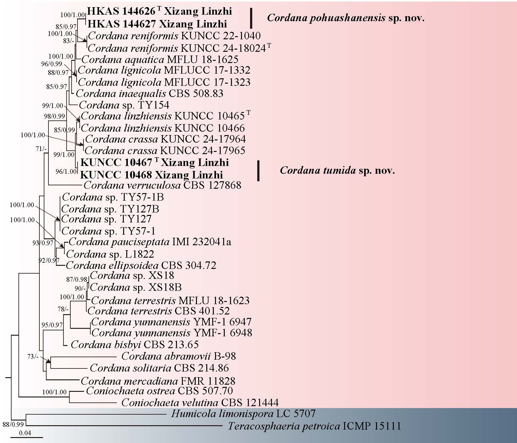
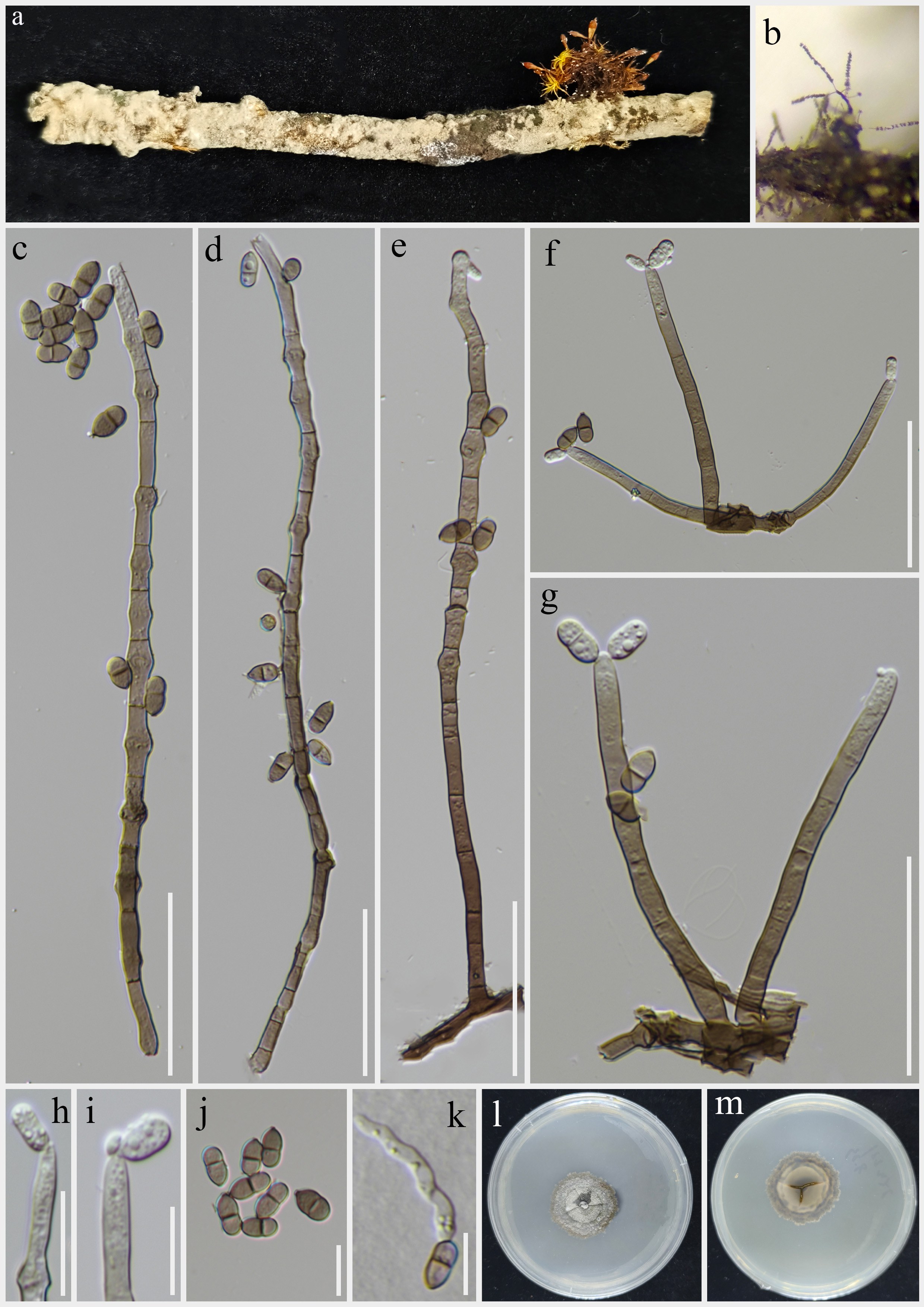
26. Cordana tumida R.J. Xu, Q. Zhao & K.D. Hyde, sp. nov. Fig. 49
Chinese name:
Index Fungorum number: IF 903488; Facesoffungi number: FoF 17412
Etymology: The epithet "tumida" refers to the swollen conidiophores.
Holotype: HKAS 136033
Saprobic on decaying wood submerged in a freshwater stream. Sexual morph: Undetermined. Asexual morph: Colonies superficial, effuse, scattered, hairy, dark brown. Mycelium immersed, consisting of pale brown, smooth-walled, branched, septate. Conidiophores 197–357 × 4–6 μm ( = 273 × 5 μm, n = 10), macronematous, mononematous, solitary, erect, straight or flexuous, unbranched, multi-septate, with swollen, fertile, intercalary nodes in the upper middle, cylindrical, slightly constricted at septa, dark brown at the base, lighter towards the apex. Conidiogenous cells 30–66 × 5–7 μm ( = 36 × 6 μm, n = 20), polyblastic, integrated, terminal, becoming intercalary, percurrent, sympodial, denticulate, usually swollen at the conidiogenous loci. Conidia 13–17 × 7–10 μm ( = 14 × 9 μm, n = 30), acropleurogenous, calabash-shaped, or cylindrical, smooth, 1-septate, pale brown, apparent constricted at the septa, elliptical at the apex, rounded at the base, guttulate.
Culture characteristics: Conidia germinating on PDA medium within 48 h and germ tubes arising from terminal end of conidium. Colonies grown on PDA reached 30 mm diam at room temperature in three weeks, circular, with velvety, flat, grayish-white mycelium on the surface, from below cream with entire margin.
Material examined: CHINA, Xizang Autonomous Region, Linzhi City, Motuo County, 29°19'36.31" N, 95°21'22.18" E, 1481 m a.s.l., on decaying wood submerged in a freshwater stream, 13 July 2022, R.J. Xu, MD-676 (HKAS 136033, holotype), ex-type culture KUNCC 10467. Linzhi City, Chayu County, 28°29'39.27" N, 96°59'35.25" E, 1537 m a.s.l., on decaying wood submerged in a freshwater stream, 14 July 2022, R.J. Xu, MD-676-2 (HKAS 136032, paratype), culture KUNCC 10468.
Notes: Among the known species of Cordana, only C. aquatica, C. lignicola, C. lushanensis and C. verruculose resemble C. tumida in having intercalary nodes (Hernández-Restrepo et al. 2014; Ai et al. 2019; Luo et al. 2019). However, C. aquatica differs from C. tumida by its smaller conidia (8–10 × 3–5 μm vs. 13–17 × 7–10 μm), and mostly oblong, sometimes ellipsoid conidia (Luo et al. 2019). Cordana lignicola differs from C. tumida by its narrower conidia (3.5–4.5 μm vs. 7–10 μm). Cordana lushanensis differs from C. tumida by its branched conidiophores and aseptate conidia (Ai et al. 2019). Cordana verruculose differs from C. tumida by its aseptate and verruculose conidia (Hernández-Restrepo et al. 2014).
In the phylogenetic analysis, Cordana tumida was grouped with C. linzhiensis and C. crassa forming a distinct clade separate from other species within Cordana (Fig. 47). However, C. tumida differs from C. linzhiensis in having smaller conidia (13–17 × 7–10 vs. 20–28 × 11–15 μm) and in the presence or absence of intercalary nodes in the conidiophores. Additionally, the conidia of C. linzhiensis are characterized by a hyaline mucoid sheath (Xu et al. 2024e). By contrast, C. crassa differs from C. tumida in that the conidiophores of the former absence intercalary nodes. In addition, a comparison of the ITS nucleotide bases in C. tumida (KUNCC 10467) and C. crassa (KUNCC 24-17964) revealed that they differ in 10/491 bp (2%) of ITS. Therefore, the new species C. tumida is introduced based on the recommendation of Jeewon & Hyde (2016) and the polyphasic approach recommended by (Chethana et al. 2021a).
Diaporthales Nannf
Lamproconiaceae Norph., T.C. Wen & K.D. Hyde
Lamproconiaceae was established to accommodate Lamproconium and Hercospora by Norphanphoun et al. (2016) based on both morphological and phylogenetic evidence. This family includes both pathogenic and saprobic fungi, typically found on dead herbaceous twigs (Norphanphoun et al. 2016; Crous et al. 2020). The sexual morph is characterized by prosenchymatous stromata surrounding the perithecia, externally demarcated by a greenish-black, dense pseudoparenchymatous layer, while the interior is whitish, composed of interwoven hyphae intermixed with host tissue, each stroma generally contains 3–5 perithecia. The asexual morph produces pycnidial conidiomata that are solitary, partially embedded in host tissues, and either uniloculate, multiloculate, or convoluted, conidiogenous cells are holoblastic, cylindrical to subcylindrical, forming single conidia at the apex, or annellidic, and may appear colorless to olivaceous with smooth walls, conidia are aseptate, fusiform to ellipsoid, thick-walled, containing granular contents (Norphanphoun et al. 2016). Currently this family contains 27 epithets in three genera, viz., Hercospora, Lamproconium, and Neolamproconium (Index Fungorum 2025; Hyde et al. 2024a).
27. Zangmuomyces R.J. Xu, K.D. Hyde & Q. Zhao, gen. nov.
Chinese name
Index Fungorum number: IF 905264; Facesoffungi number: FoF 19310
Etymology: The genus name Zangmuomyces is established to honor Professor Zang Mu for his outstanding contributions to the field of mycology in China.
Saprobic on decaying unidentified wood in terrestrial habitat. Sexual morph: Undetermined. Asexual morph: Conidiomata 378–481 µm high, 660–858 µm diam ( =428 × 740 μm, n = 15), pycnidial, solitary, scattered, semi-immersed or superficial, black, uni-loculate, ellipsoid or irregular when viewed from above, like a raised cylindrical barrel when viewed from the front, a large opening in the middle, glabrous. Peridium 69–120 μm wide, composed of brown to dark brown, irregular, thick-walled cells arranged in a textura angularis, the side walls are significantly thicker than the bottom, fused with the host tissue. Paraphyses interspersed within conidiophores. Conidiophores 15–78 × 2–4 μm ( = 47 × 3 μm, n = 20), arising from the outermost wall layer at the basal of pycnidium, filiform or cylindrical, hyaline, aseptate, unbranched, smooth-walled. Conidiogenous cells cylindrical to subcylindrical, filiform, hyaline, thin-walled, becoming swollen towards the apex. Conidia 26–35 × 10–13 μm ( = 31 × 12 μm, n = 25), hyaline, ellipsoidal to long ellipsoidal, rounded at both ends, straight or slightly curved, 3-septate, slightly constricted at septum, hyaline sheath around conidia, smooth-walled, guttulate.
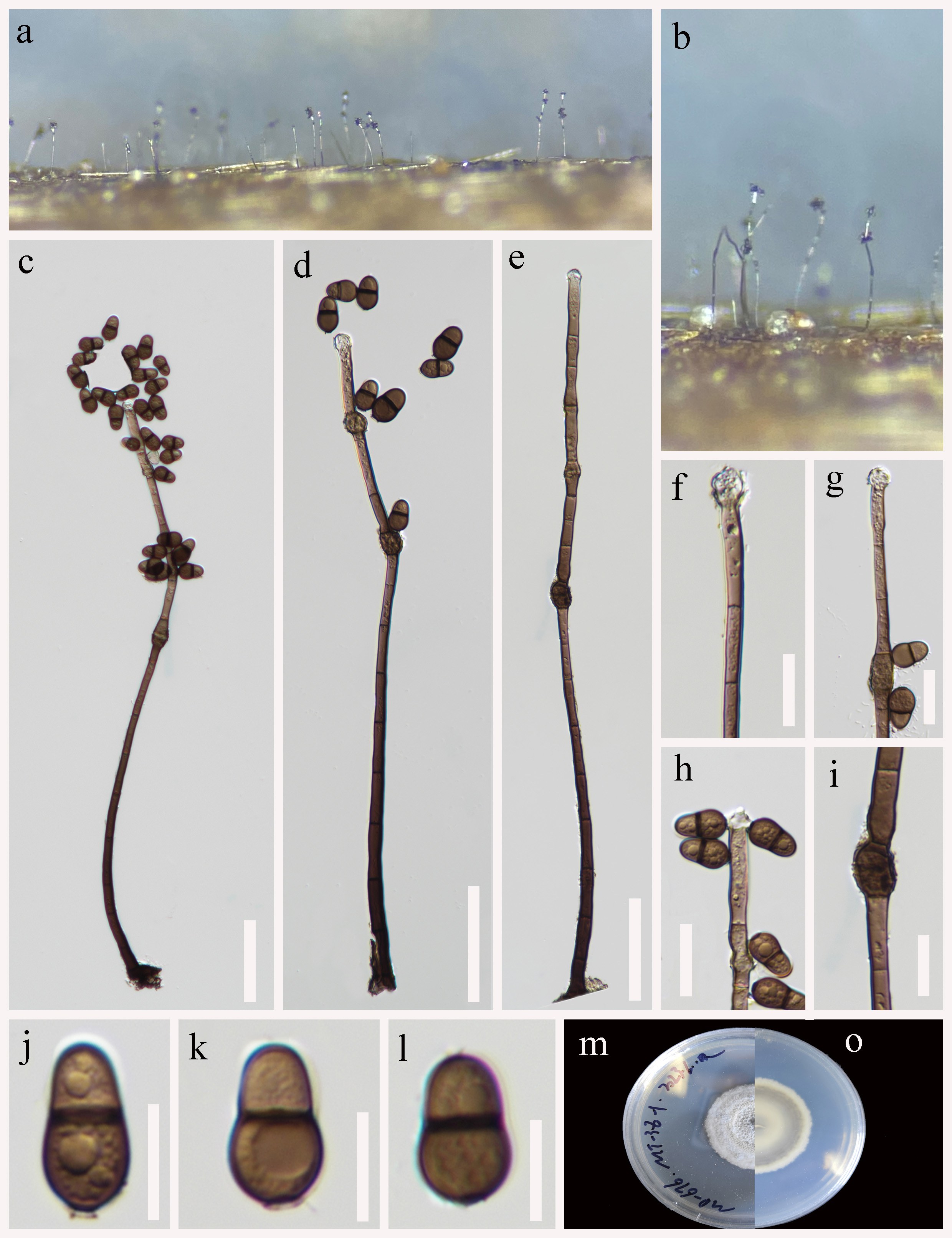
Type species: Zangmuomyces xizangensis R.J. Xu, K.D. Hyde & Q. Zhao (HKAS 151647)
Notes: In the phylogenetic analyses, strains of Zangmuomyces were clustered with Hercospora, Lamproconium, and Neolamproconium in the Lamproconiaceae. In morphology, Z. xizangensis shows unique features: conidiomata like a raised cylindrical barrel when viewed from the front, conidiogenous cells becoming swollen towards apex, and conidia ellipsoidal to long ellipsoidal, rounded at both ends, 3-septate. It can be distinguished from other three genera in Lamproconiaceae. Therefore, this study introduces Zangmuomyces as a new genus to accommodate Z. xizangensis.
28. Zangmuomyces xizangensis R.J. Xu, K.D. Hyde & Q. Zhao, sp. nov. Fig. 51
Chinese name
Index Fungorum number: IF 905043; Facesoffungi number: FoF 19311
Etymology: refers to Xizang Autonomous Region in China, where the type specimen was collected.
Holotype: HKAS 151647
Saprobic on decaying unidentified wood in a freshwater habitat. Sexual morph: Undetermined. Asexual morph: Conidiomata 378–481 µm high, 660–858 µm diam ( = 428 × 740 μm, n = 15), pycnidial, solitary, scattered, semi-immersed or superficial, black, uni-loculate, ellipsoid or irregular when viewed from above, like a raised cylindrical barrel when viewed from the front, a large opening in the middle, glabrous. Peridium 69–120 μm wide, composed of brown to dark brown, irregular, thick-walled cells arranged in a textura angularis, the side walls are significantly thicker than the bottom, fused with the host tissue. Paraphyses filamentous, aseptate, interspersed within conidiophores. Conidiophores 15–78 × 2–4 μm ( = 47 × 3 μm, n = 20), arising from the outermost wall layer at the basal of pycnidium, filiform or cylindrical, hyaline, aseptate, unbranched, smooth-walled. Conidiogenous cells cylindrical to subcylindrical, filiform, hyaline, guttulate, thin-walled, becoming swollen towards apex. Conidia 26–35 × 10–13 μm ( = 31 × 12 μm, n = 25), hyaline, ellipsoidal to long ellipsoidal, rounded at both ends, straight or slightly curved, 3-septate, slightly constricted at septum, hyaline sheath around conidia, smooth-walled, guttulate.
Material examined: CHINA, Xizang Autonomous Region, Linzhi City, Motuo County, 29°19′43.69″ N, 95°21′19.27″ E, 677 m a.s.l., on decaying wood submerged in a freshwater stream, 13 July 2022, R.J. Xu, MD-308A (HKAS 151647, holotype), ex-type culture KUNCC 25-21370.
Notes: In the phylogenetic tree, Zangmuomyces xizangensis clustered with strains of Hercospora, Lamproconium, and Neolamproconium with 99% ML/1.00 BYPP statistical support (Fig. 50). Morphologically, Zangmuomyces and other three genera of Lamproconiaceae show differences on conidiomata, conidiogenous cells, and conidia. The conidiomata of Z. xizangensis are like a raised cylindrical barrel when viewed from the front, conidiogenous cells become swollen towards the apex, and conidia are 3-septate, ellipsoidal to long ellipsoidal, often with a hyaline sheath, and rounded at both ends, while Hercospora has conidiomata with a superficial cap of sterile tissues, elongate conidiophores (conidiogenous cells not mentioned), and ovoid to ellipsoid, one-celled conidia (Norphanphoun et al. 2016). Lamproconium has dark blue conidiomata, with a raised centre, conidiogenous cells with flared periclinal thickenings in the collarette zone, and conidia are fusiform, ellipsoid, with narrowly rounded ends, aseptate, bluish to glistening dark blue (Norphanphoun et al. 2016). The stromata of Neolamproconium are characterized by aggregated rosettes of up to 15 pycnidia, phialidic conidiogenous cells, with prominent periclinal thickenings, and conidia are dimorphic, with macroconidia 1(–3)-septate, and microconidia aseptate (Crous et al. 2020).
Therefore, based on its obvious branching in the phylogenetic tree and unique morphological structure, this study introduces Zangmuomyces xizangensis as a new species of a new genus Zangmuomyces.
Hypocreales Lindau
Bionectriaceae Samuels & Rossman
Clonostachys
The asexual morph-typified genus Clonostachys, described by Corda (1839), is anchored by its type species, Clo. araucaria, distinguished by its penicillate conidiophores and imbricate conidia organized in columns (Schroers et al. 1999). Clonostachys is typified by its penicillate, sporodochial, or dimorphic conidiophores and phialidic conidiogenous cells, which generate hyaline conidia (Schroers 2001). The teleomorph, originally described as Bionectria (Spegazzini 1919), is characterized by ascomata typically situated on a pseudoparenchymatous stroma or directly arising from the substrate, ascomata exhibit a color range from white, pale yellow, to orange and dark brownish-orange, remaining unchanged in appearance when exposed to 3% KOH or lactic acid (Schroers 2001; Lechat & Fournier 2018; Perera et al. 2023). Currently, 136 records have been listed in the Index Fungorum (Index Fungorum Name Search, April 18, 2026).
29. Clonostachys habaensis Y.B. Wang, B.Z. Chen & Zhu L. Yang, sp. nov. Fig. 53
Chinese name
Index Fungorum number: IF 905265; Facesoffungi number: FoF 19312
Etymology: The epithet habaensis refers to Haba Snow Mountain (Haba Xueshan) in Shangri-La City, Yunnan Province, China, where the type specimen was collected.
Diagnosis: Clonostachys habaensis produces slightly smaller ellipsoidal conidia compared to its close relatives.
Holotype: HKAS 135029
Endophytic from the root of Rhododendron decorum. Sexual morph: Undetermined. Asexual morph: Conidiophores broadly penicillate, arising from the agar surface or aerial mycelium ter- to quinquiesverticillate, aggregated in pustules or sporodochia, phialides slightly divergent or adpressed, in whorls of 1–3, flask-shaped, widest in the lower third or almost cylindrical, slightly tapering in the upper part towards the tip, without a visible collarette, (11.3–) 11.4–18.9 (–19.2) × (1.9–) 2–3.2 (3.2) μm ( 15.5 × 2.7 μm, n = 20). Conidia aseptate, hyaline, smooth-walled, ellipsoid, some slightly curved or asymmetric with one more flattened side, with a laterally displaced hilum, 5.1–7.6 (–9) × (2.9–) 3–5.2 (–5.8) μm ( 6.3 × 4 μm, n = 30).
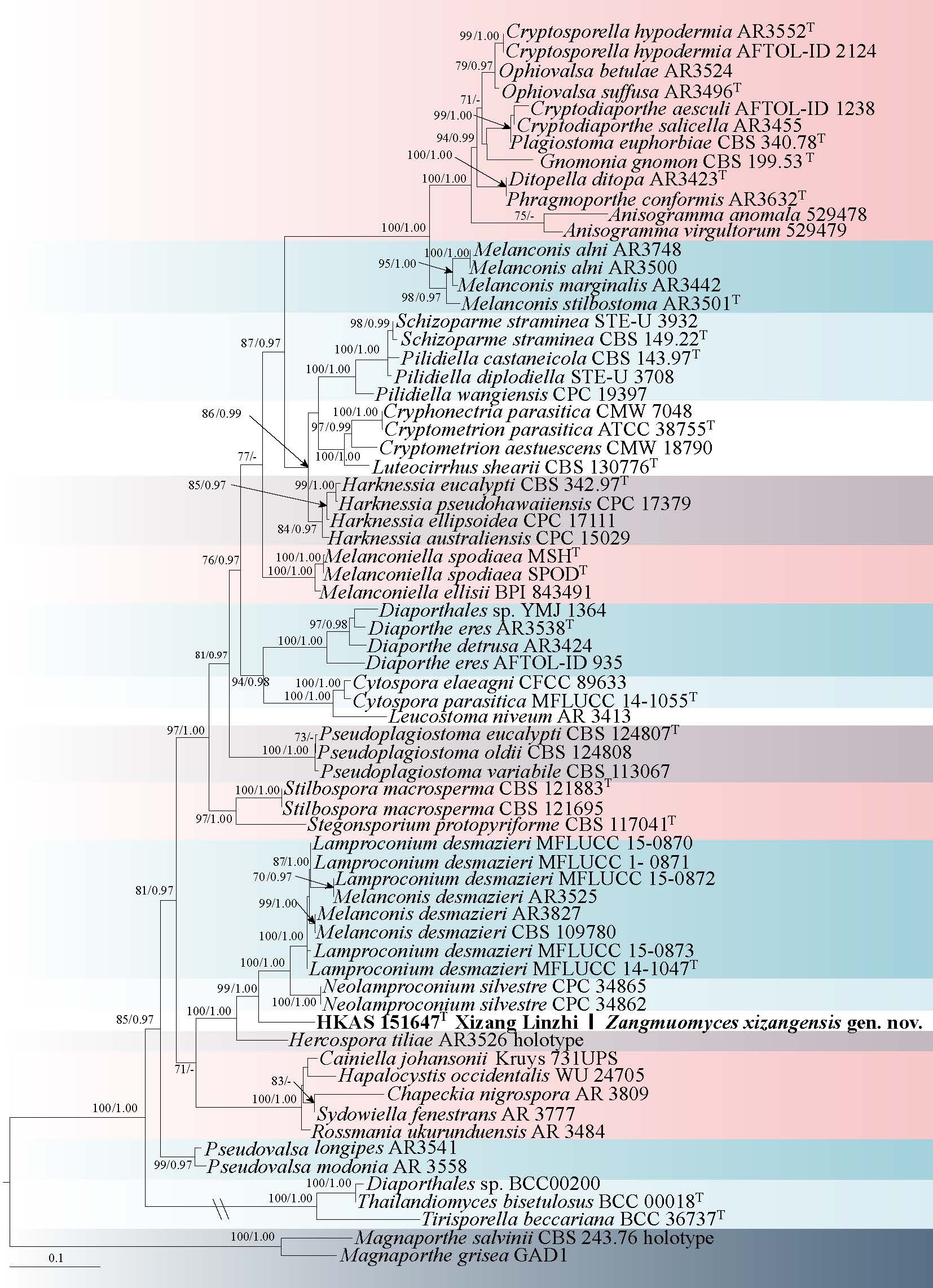
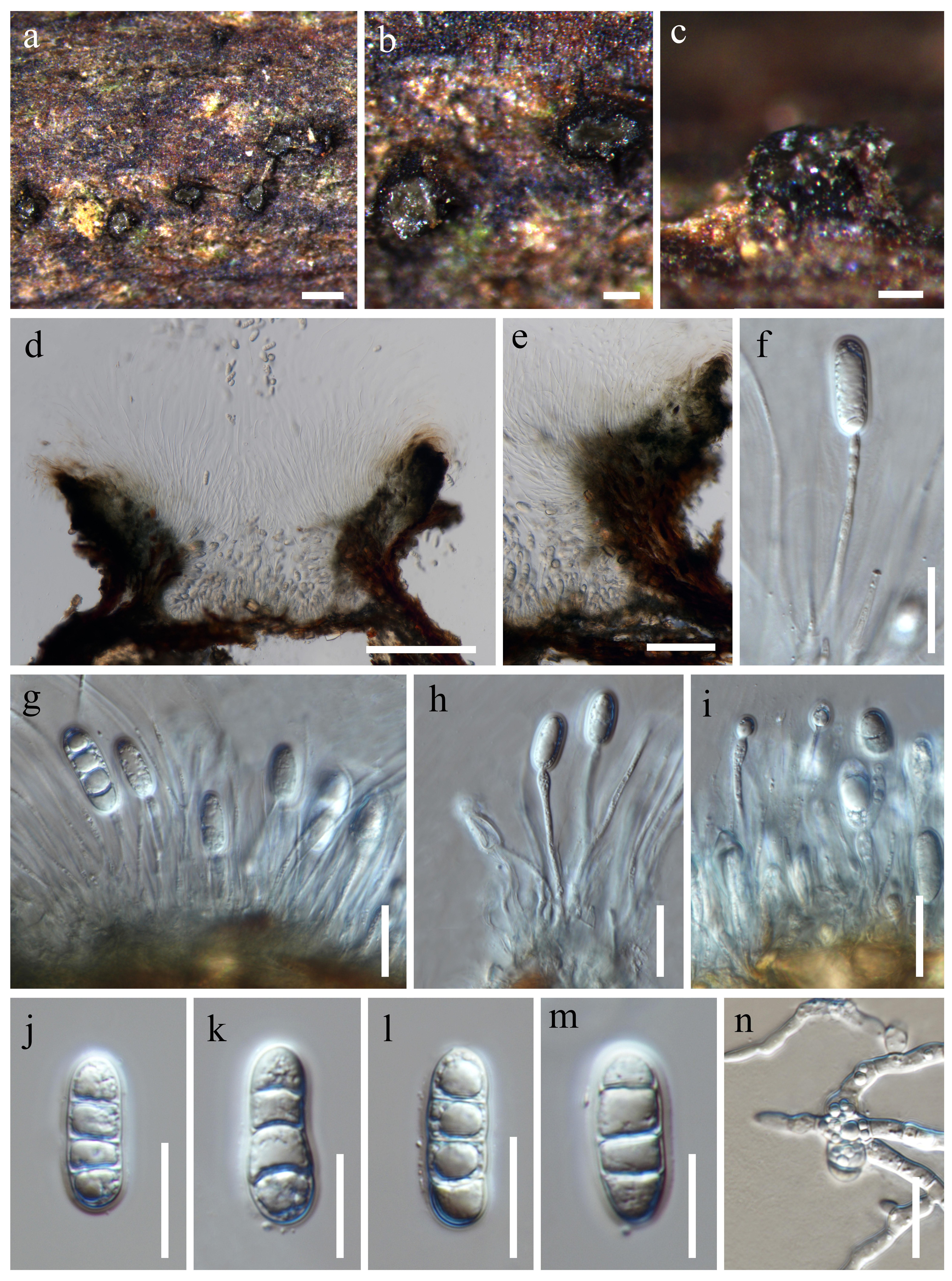
Culture characteristics: Colonies on PDA reaching 54–61 mm diameter after 8 days at 25 °C, flat, with entire margin, aerial mycelium abundant, cottony, pale yellow, reverse yellowish.
Material examined: CHINA, Yunnan Province, Diqing, Shangri-La City, Haba Snow Mountain, isolated as an endophyte from Rhododendron decorum root, 15 March 2023, collected by Q. Fan (HKAS 135029, holotype, dried culture on PDA, ex-type culture KUNCC 11234), other collection: Ibid., Q. Fan (culture KUNCC 11233).
Notes: Clonostachys habaensis is closely related to Clo. pseudostriata and Clo. palmae, as strongly supported by multi-gene phylogenetic inference. However, it shows clear sequence divergence from its sister species Clo. pseudostriata (CBS 120.87) (Fig. 52), with 99.4% identity in ITS (2 bp differences), 99.8% in LSU (1 bp), 98.7% in rpb2 (9 bp), 98.4% in tef1-α (12 bp), and 95.6% in tub (39 bp). Morphologically, Clo. habaensis produces ellipsoidal conidia that are slightly smaller than those of Clo. palmae (Schroers 2002; Zhao et al. 2023).
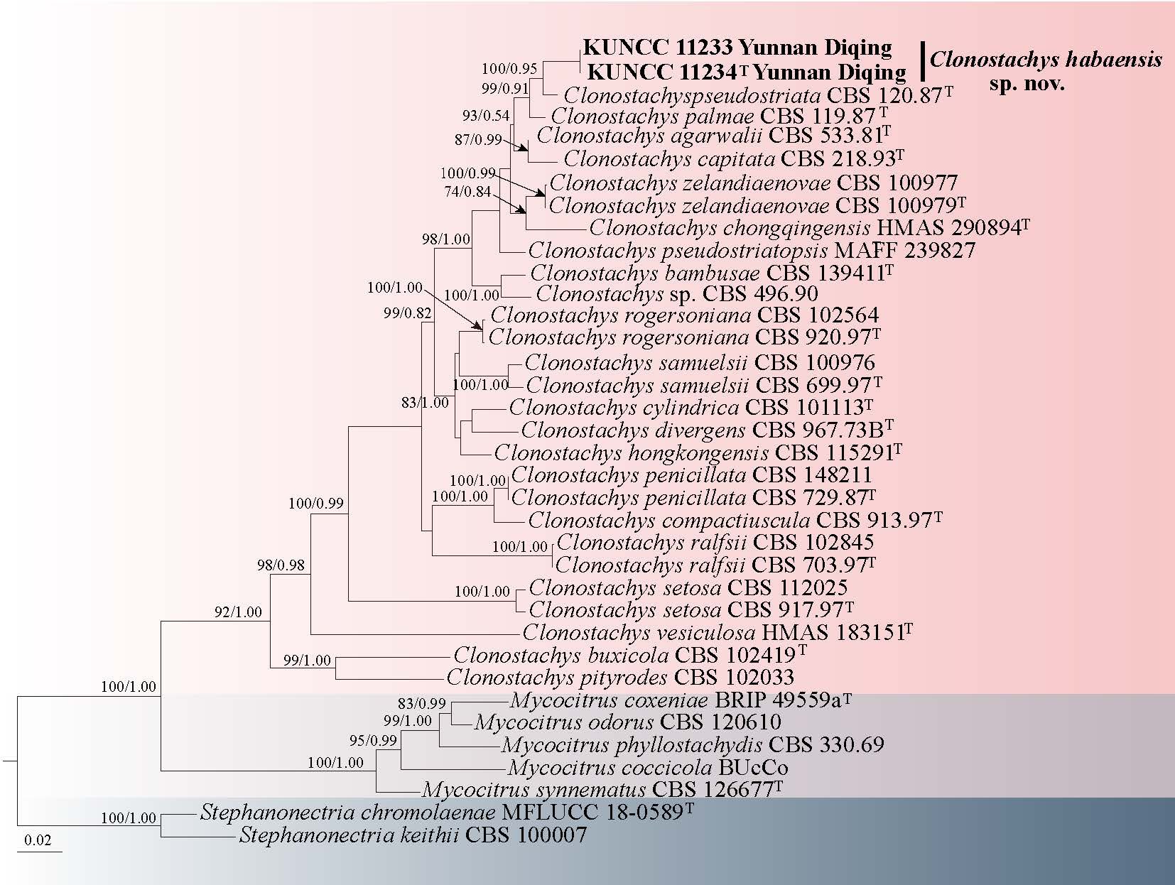
Clavicipitaceae Rogerson
Clavicipitaceae was initially treated as a subfamily of Hypocreaceae (Earle 1901). It was later placed within Hypocreales by Spatafora & Blackwell (1993). Sung et al. (2007) further divided it into three monophyletic families: Clavicipitaceae, Cordycipitaceae, and Ophiocordycipitaceae. Xiao et al. (2023) introduced a fourth family, Polycephalomycetaceae. Currently, Clavicipitaceae includes over 40 genera, such as Aciculosporium, Claviceps, Epichloë, and Metarhizium (Kepler et al. 2014). Many species are entomopathogenic, often parasitizing insects or forming symbiotic relationships with plants (Kepler et al. 2012; Khonsanit et al. 2021). The sexual morph features dark or brightly colored, fleshy to tough stromata or subiculum, with perithecial ascomata that can be superficial or immersed and are arranged in ordinal or oblique patterns, containing cylindrical asci with thickened apices and typically cylindrical, multiseptate ascospores that may disarticulate into part-spores (Sung et al. 2007; Kepler et al. 2014). The asexual morphs include genera like Aschersonia, Ephelis, Metarhiziopsis, Metarhizium, Neotyphodium, Nomuraea, Pochonia, and Rotiferophthora (Sung et al. 2007; Kepler et al. 2012; Mongkolsamrit et al. 2020).
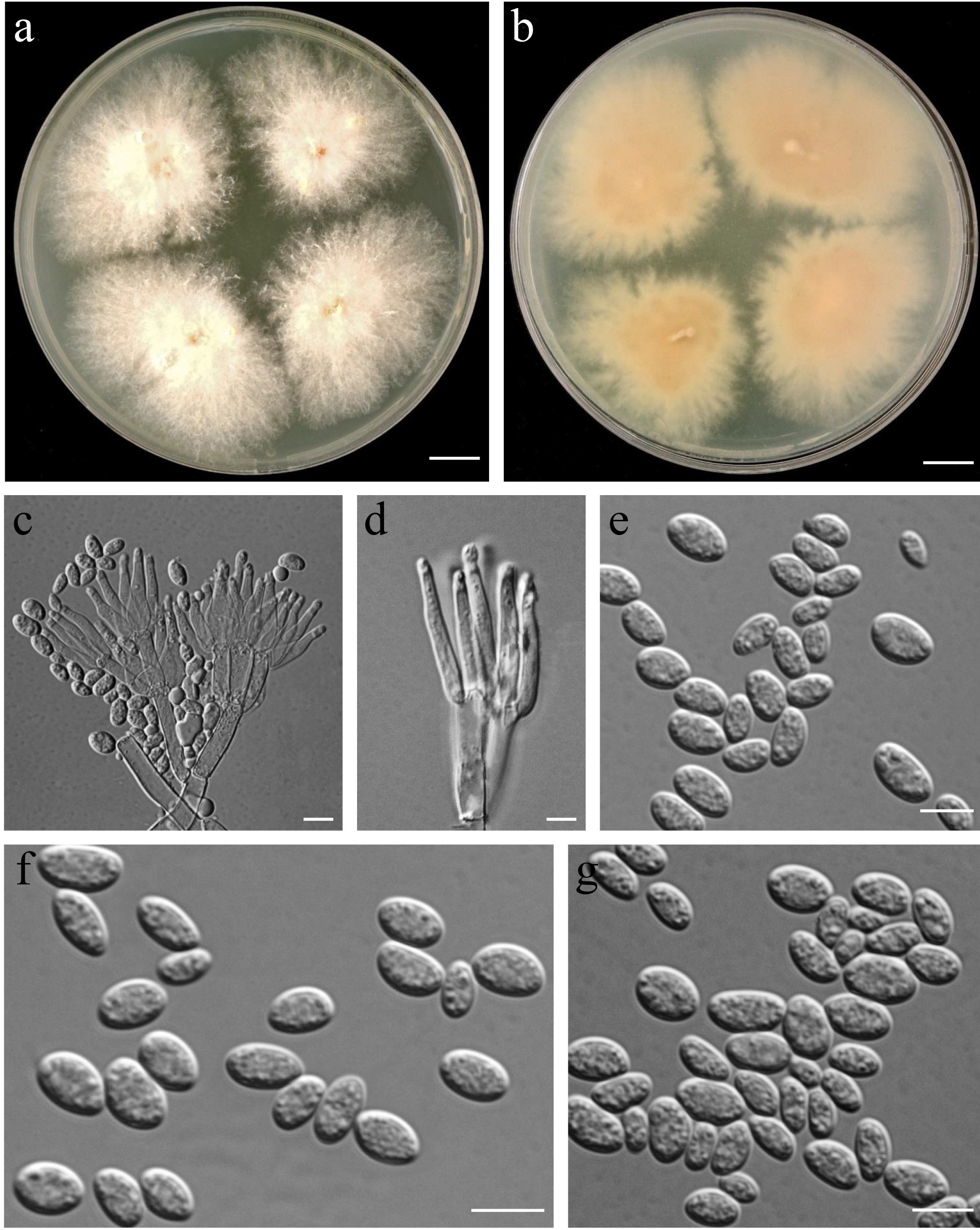
Metarhizium Sorokin
Metarhizium is of paramount significance in the study of entomopathogenic fungi, catalyzing two molecular phylogenetic revisions that have robustly validated and refined the morphological classification delineated by Rombach et al. (1987). The species classified within Metarhizium are acknowledged as entomopathogens, effectively targeting insect hosts and resulting in their mortality or incapacitation. Furthermore, these fungi have been documented to promote plant growth and enhance biomass accumulation (Elena et al, 2011; Khan et al, 2012; Mongkolsamrit et al. 2020).
30. Metarhizium luteum Y.B. Wang, C.Y. Wei, B.Z. Chen & Zhu L. Yang, sp. nov. Fig. 54
Chinese name:
Index Fungorum number: IF 905266; Facesoffungi number: FoF 19313
Etymology: The stromata are earthy yellow.
Diagnosis: Different from all other Metarhizium members by color of its stromata.
Holotype: HKAS 126260
Parasitic on larvae of Scarabaeidae. Sexual morph: Stromata from the head of the Scarabaeidae (Coleoptera) buried in soil, solitary, occasionally branched, fleshy, glabrous, 7–10 cm long. Stipes cylindrical, easily bent, white to orange, 2–4 mm wide. Fertile parts terminal, cylindrical, orange, tapering upwards, contains infertile tip, 70–100 × 2–4 mm. Perithecia obliquely immersed, dense, ovate, 600–800 × 100–400 µm. Asci linear, (100–) 150–300 (–350) × 3.5–5 (–5.5) µm ( 207.6 × 4.3 µm), apical caps prominent, hemispherical, (3–) 3.5–6 (–6.5) × 1.5–2 (–2.5) µm ( = 4.9 × 1.9 µm). Ascospores hyaline, filiform multiseptate, breaking into secondary ascospores, 15–25 (–30) × (0.5–) 1–1.5 µm ( 22.5 × 1.2 µm) Secondary ascospores cylindrical, rounded at both ends, (8.5-) 8.6 - 10.4 (-10.5) × 1 - 1.5 µm (x = 9.5 × 1.1 µm). Asexual morph: Conidiophores smooth-walled, cylindrical, apically branched. Phialides smooth-walled, cylindrical with semi-papillate apices, arranged in whorls of 3–5 at the apex of conidiophores, (6–) 6.5–10.5 (–11) × (1–) 1.5–3 (–3.5) µm ( 8.3 × 2.3 µm). Conidia hyaline, cylindrical, (6.5–)7–10(–10.5) × (2–)2.5–3.5(–4.0 μm ( = 8.3 × 2.9 μm).
Culture characteristics: Colonies on PDA attaining a diam of 23 mm in 14 d, white, flocculent, high mycelial density, intact edges, reverse white cream. Center of the colony is dark green due to the production of conidia. Hyphae smooth-walled, colorless, septate.
Material examined: CHINA, Yunnan Province, Baoshan City, parasite on larvae of Scarabaeidae buried in soil, 2 August 2022, C.Y. Wei, (HKAS 126260 holotype, ex-type culture KUNCC 10831), other collection: Ibid., C.Y. Wei (HKAS 126261, culture KUNCC 10832; HKAS 126262, culture KUNCC 10833).
Notes: As shown in the phylogenetic tree, Met. luteum forms a distinct terminal lineage (Fig. 57), strongly supported by multi-locus phylogenetic analyses. Metarhizium luteum shares similarity with Met. candelabrum and Met. cercopidarum in the production of cylindrical phialides with rounded ends (Mongkolsamrit et al. 2020). However, Met. luteum parasitize scarabaeidae larvae while Met. candelabrum and Met. cercopidarum infect audlt leafhopper. In Met. luteum the conidia on PDA are wider than in Met. candelabrum and Met. cercopidarum. Further, Met. luteum is distinguished from Met. candelabrum and Met. cercopidarum by growth rate on PDA, and Met. luteum is significantly faster than Met. candelabrum and Met. cercopidarum.
Moelleriella Bres
Moelleriella, an entomopathogenic genus within the Clavicipitaceae (Hypocreales, Ascomycota), is distinguished from related genera like Conoideocrella, Hypocrella and Samuelsia by unique features such as disarticulating ascospores and fusiform conidia produced by its anamorph (Bresadola 1897; Chaverri et al. 2008). Moelleriella species engage in a dual parasitic role, acting as necrotrophic parasites of scale insects and whiteflies, and biotrophic parasites of plants, often presenting as vibrant stromata on insect hosts (Chaverri et al. 2008; Mongkolsamrit et al. 2015). Molecular analyses, especially of LSU, tef1-α and rpb1 genes, have clarified their taxonomy, ecological roles, and evolutionary relationships, enhancing our understanding of fungal biodiversity and ecosystem dynamics (Khonsanit et al. 2021).
31. Moelleriella yusheensis Y. Yang, K.D. Hyde & Y.P. Xiao, sp. nov. Fig. 56
Chinese name
Index Fungorum number: IF 903156; Facesoffungi number: FoF 17061
Etymology: in reference to the place where the fungus was collected, China, Guizhou Province, Liupanshui City, Shuicheng County, Yushe forest Park.
Holotype: HKAS 132266
Sexual morph: ( = 296 × 110 µm, n = 30). Ostioles pale yellow to light orange. Asci hyaline, cylindrical, 130–180 × 5–11 μm ( = 162 × 7 µm, n = 50). Asci caps inconspicuous. Ascospores hyaline, disarticulating into part-spores. Secondary spores oblong to cylindrical with round or blunt ends, 7.2–9.7 × 2.3–3.5 μm ( = 8.6 × 2.9 µm, n = 50), smooth, hyaline, thin-walled, guttulate. Asexual morph: undetermined.
Material examined: CHINA, Guizhou Province, Liupanshui City, Shuicheng County, Yushe forest Park. Parasitism on scale insects (Coccidae), found on the upper surfaces of dicotyledonous leaves, 1 August 2022, Yu Yang SC186 (HKAS 132266, holotype).
Notes: Moelleriella yusheensis clusters with Moe. gracilispora and Moe. kanchanaburiensis in the phylogenetic tree, with 100% MLBP, 0.99 BYPP support (Fig. 55). The sequences of six loci of Moe. gracilispora and Moe. kanchanaburiensis share 97–98% identity with Moe. yusheensis in LSU, 94–95% in tef1-α, and 91–92% in rpb1. Moe. gracilispora infects whitefly nymphs (Hemiptera), while Moe. yusheensis and Moe. kanchanaburiensis both infect scale insects (Coccidae) (Yuan et al. 2020; Khonsanit et al. 2021). Morphologically, Moe. yusheensis can be easily distinguished from other Moelleriella species by its flat to umbonate, orange to dark orange teleomorphic stromata, numerous embedded perithecia, hyaline asci, and cylindrical thin-walled, and guttulate secondary spores. In this study, we introduce a novel species, Moe. yusheensis, from China.
Papiliomyces Luangsa-ard, Samson & Thanakitp. 2020
Papiliomyces was introduced by Mongkolsamrit et al. (2020) within the Clavicipiteae, to accommodate Pap. liangshanensis and Pap. shibinensis. The teleomorph of Papiliomyces encompass solitary to multiple (branched and robust to wiry) stromata, superficial to entirely immersed perithecia within the stroma, and ampulliform, ellipsoid to ovoid asci, with ascospores that are whole, exhibiting septation or fragmentation into cylindrical part-spores (Kepler et al. 2012a; Kepler et al. 2012b; Mongkolsamrit et al. 2020; Wang et al. 2021).
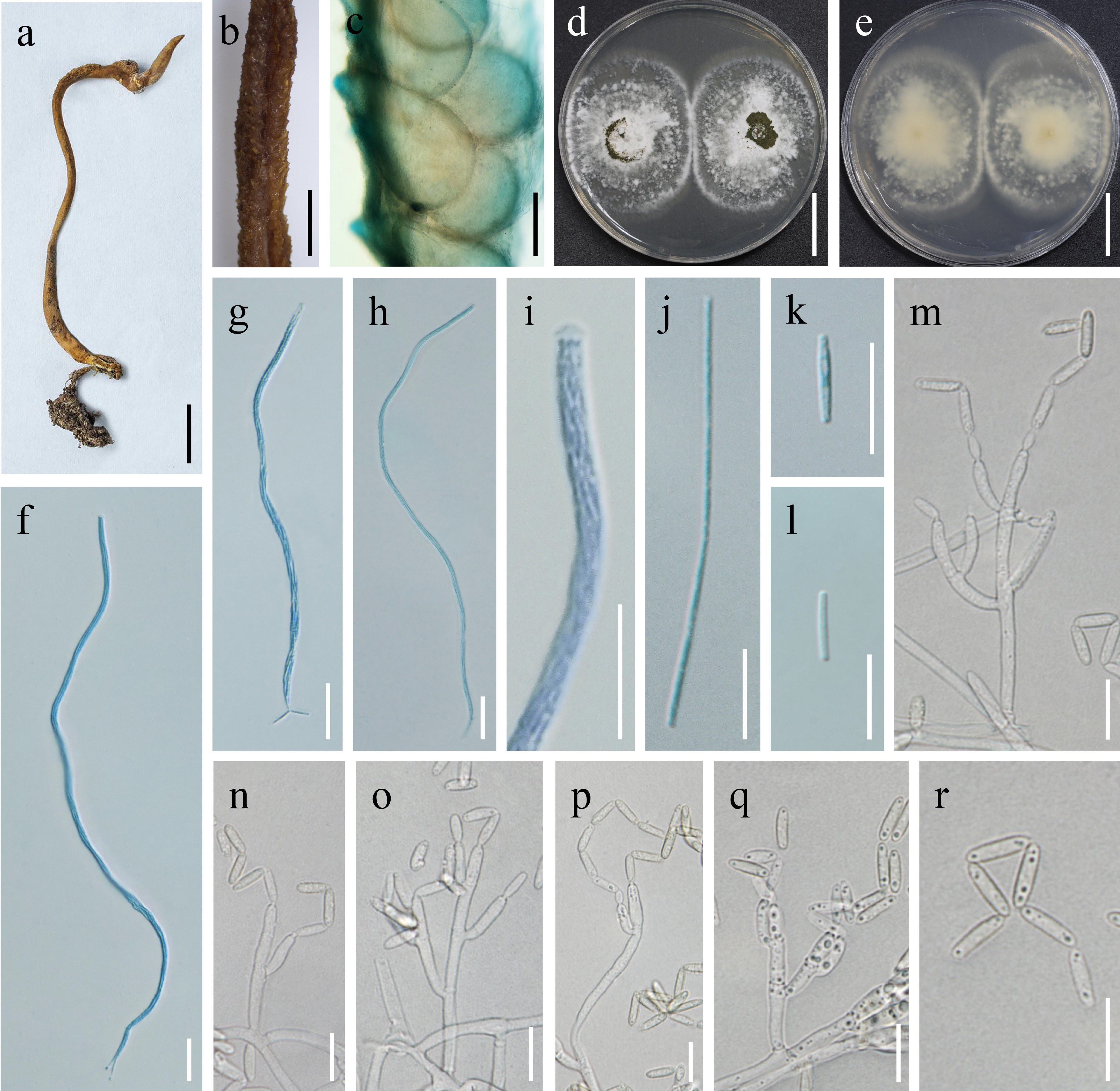
32. Papiliomyces aurantiacus Y.B. Wang, C.Y. Wei, B.Z. Chen & Zhu L. Yang, sp. nov.Fig. 58
Chinese name:
Index Fungorum number: IF 905267; Facesoffungi number: FoF 19314
Etymology: Fertile parts are orange
Diagnosis: Different from all other Papiliomyces members by color of its fertile parts.
Holotype: HKAS 126283
Parasitic on larvae of Scarabaeidae. Sexual morph: Stromata from the head of lepidopteran larvae buried in the soil, solitary, occasionally branched, 30–45 mm long. Stipe cylindrical, white to orange-yellow, 1.5–3 mm wide. Fertile parts cylindrical, orange, 5–10 × 2.5–3 mm. Perithecia embedded, dense, ovate or long oval, 445–503 × 176–239 µm ( 465.5 × 214.7 µm). Asci transparent, linear, 114–202 × 4.8–6.7 µm ( 152.7 × 5.5 µm), apical caps prominent, hemispherical, (4–) 4.5–5.5 (–6) × 2.5–3.5 (–4) µm ( 4.9 × 3 µm). Ascospores columnar, multiseptate, composed of numerous cells, 162–175 × 2.4–3.2 µm ( 168.8 × 2.8 µm), not breaking into secondary ascospores. Asexual morph: Conidiophores smooth-walled, cylindrical, top branch. Phialides smooth-walled, colorless, flask-shaped, base thinning upward, arising singly from the hyphae or in whorls of 3-4 at the top of the conidiophores, (11–) 11.5–15 (–15.5) × (0.5–) 1–2 (–2.5) µm ( 13.4 × 1.6 µm). Conidia smooth-walled, unicellular, globose to subglobose, surface tooth teeth, often arranged into chains, and 3.5–4.5 (–5) × 3.5–4 (–4.5) µm ( 4.2 × 4.1 µm).
Culture characteristics: Colonies on PDA attaining a diam of 30 mm in 14 d, dense, pulvinate, white, intact edges. Hyphae smooth-walled, colorless.
Material examined: CHINA, Yunnan Province, Nujiang, parasite on larvae of Hepialidae buried in soil. 12 August 2023, C.Y. Wei (HKAS 126283 holotype, ex-type culture KUNCC 10852), other collection: Ibid., C.Y. Wei (HKAS 126282).
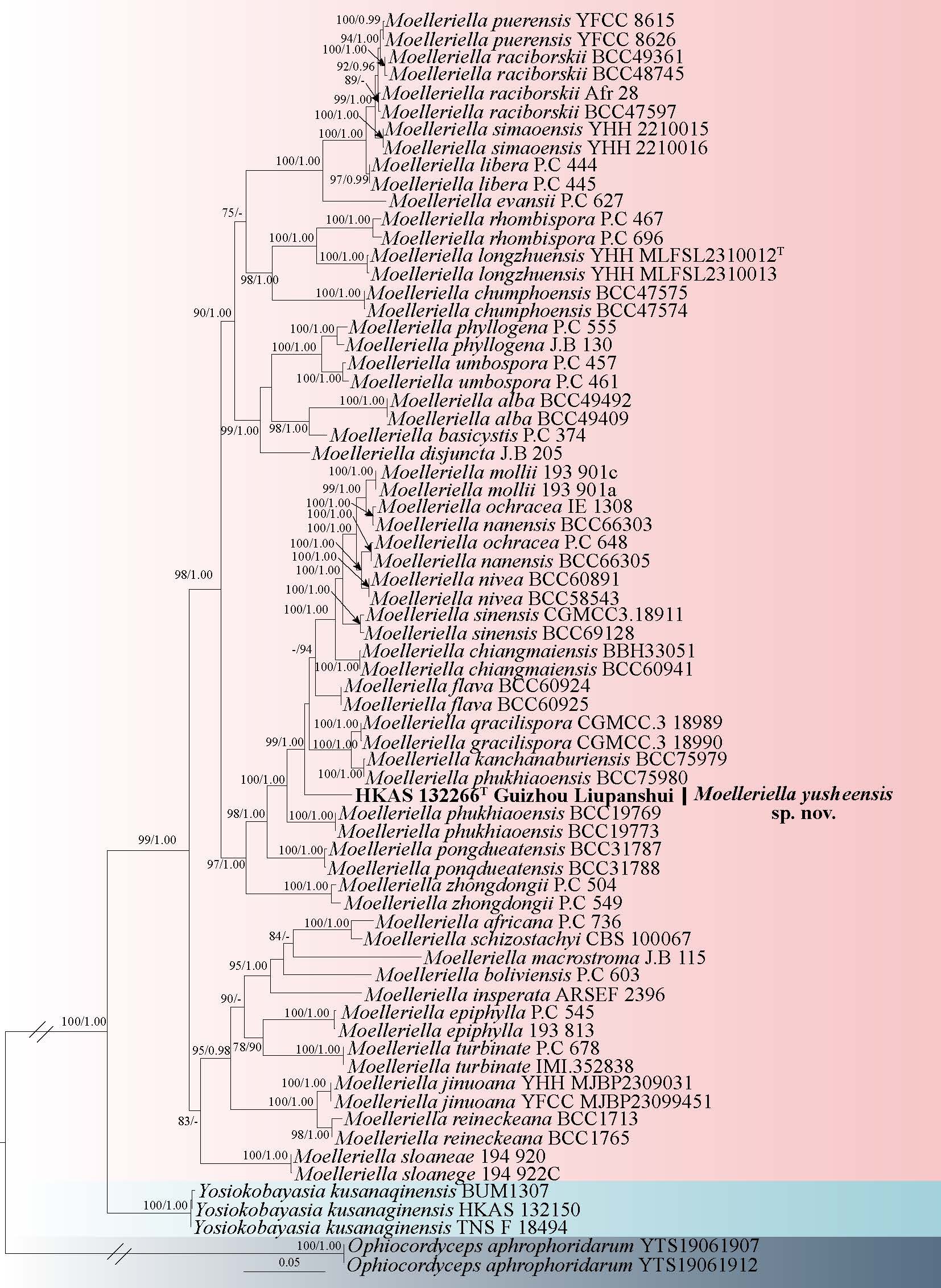
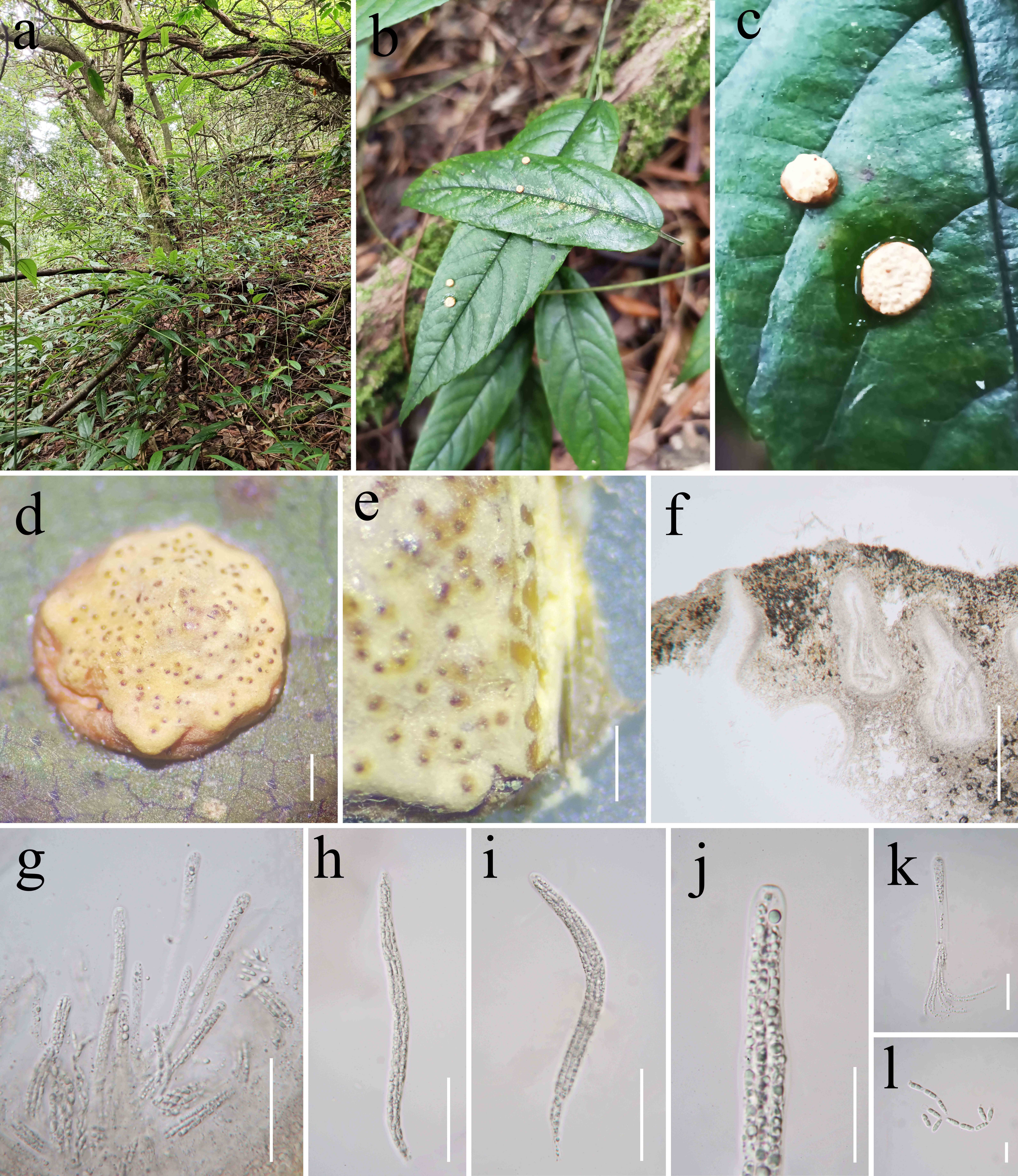
Notes: As shown in the phylogenetic tree, Pap. aurantiacus represents a distinct terminal lineage (Fig. 57), strongly supported by multi-locus phylogenetic analyses. Papiliomyces aurantiacus exhibits similarities to Pap. shibinensis, as both species share the same host, namely the larvae of Lepidoptera. However, they differ in the size and
color of fertile parts and ascopores. Papiliomyces aurantiacus has orange fertile parts, whereas Pap. shibinensis is faint yellow (Mongkolsamrit et al. 2020). In Pap.aurantiacus (162–175 × 2.4–3.2 µm), the fertile parts are wider than in Pap. shibinensis (120–170 × 1.4–2.1 μm).
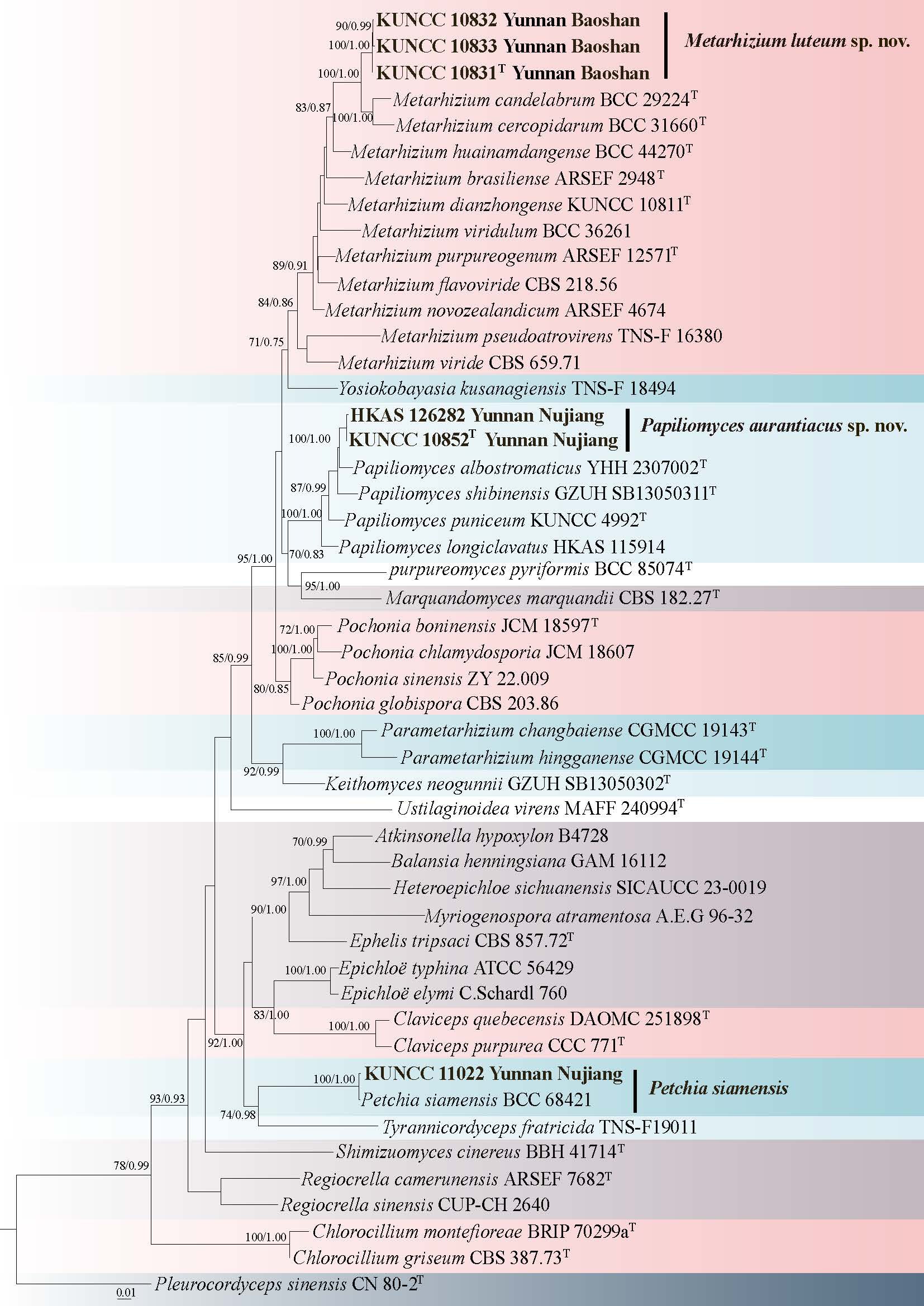
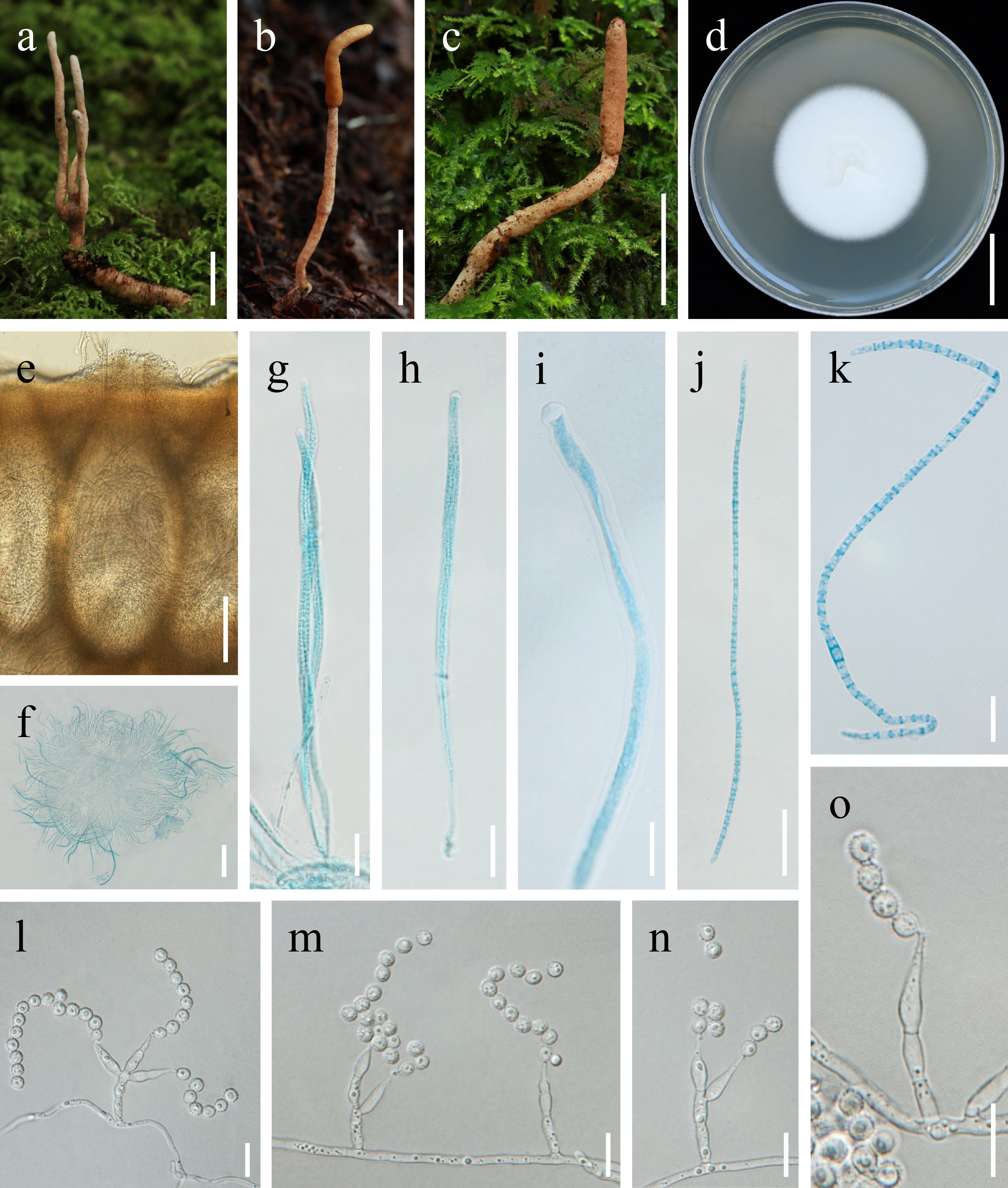
Petchia Thanakitp., Mongkols. & Luangsa-ard 2020
Petchia was proposed by Mongkolsamrit et al. (2020) within the Clavicipiteae. Petchia is distinguished from related genera by stromata arising from the egg cases of praying mantis (Mantidae), multiple, mostly erect up to 2 cm high, fertile region is located at the apical end of the stroma, exhibiting a pale cream coloration, with a globose morphology measuring 1.5–1.8 cm in diameter, perithecia are narrowly ovoid, exhibiting a brown hue, and are immersed with dense mycelial encasement around each perithecium (Mongkolsamrit et al. 2020). Currently, only two species in the genus, all are saprotrophic, on the egg cases of praying mantis (Mongkolsamrit et al. 2020).
33. Petchia siamensis Thanakitp., Mongkols. & Luangsa-ard, in Thanakitpipattana, Tasanathai, Mongkolsamrit, Khonsanit, Lamlertthon & Luangsa-ard, Persoonia 44: 154 (2020) Fig. 59
Chinese name:
Index Fungorum number: IF 830168; Facesoffungi number: FoF 14494
Diagnosis: The specimen examined in this study shares the same phylogenetic position as Petchia siamensis.
Parasitic on the ootheca of praying mantis (Mantidae). Sexual morph: Stromata grow from the ootheca of praying mantis (Mantidae), several produced on a single host, each stroma unbranched, easily bent, length 5-7 mm. Stipe cylindrical, curved, smooth, glabrous, light yellow, with a diameter of 0.8-1 mm. The fertile part is globose apical, light yellow, measuring 2 × 1.5 mm. Perithecia are immersed, oval, brown, (550–)578–624(–650) × (400–)413–434(–450) µm, ( 596 × 434 µm). Asci are cylindrical, slender, (170–)179–250 × 3–4.8(–5) µm ( 203 × 4 µm). The apical cap is prominent, hemispherical, thickened, measuring 3–3.5 × 1.5–2 µm, ( 3.1 × 1.8 µm). Ascospores are transparent, thread-like, multi-septate, (200–)215–275(–300) × 1–1.5 µm, ( 246 × 1.3 µm), and break into secondary ascospores when mature. Secondary ascospores are short-cylindrical, (5.5–)5.7–9.8(–10) × (0.5–)0.7–1.2(–1.5) µm, ( 8.1 × 0.9 µm). Asexual morph: Hyphae smooth-walled, colorless, branched. Conidiophores smooth-walled, cylindrical, branched at the apex. Phialides smooth-walled, base cylindrical and gradually tapering upwards, bottle-shaped, whorled at the top of the conidiophores, 12.5–18.8(–20) × (1–)1.3–3.3(–3.5) µm, ( 14.9 × 1.9 µm). Conidia unicellular, colorless, ellipsoidal, 3–5 × (1.5–)1.7–2.8(–3) µm, ( 4.4 × 2.3 µm).
Culture characteristics: Colonies on PDA reach 25 mm in diameter within 14 days, tightly attached, flat, cushion-like, fuzzy, gray-purple, clear edges.
Material examined: CHINA, Yunnan Province, Nujiang, Gongshan County, on the ootheca of praying mantis (Mantidae), August 3, 2024, J.L. Jia (HKAS 135119, culture KUNCC 11022).
Notes: Thanakitpipattana et al. (2020) discovered a fungus parasitizing the oothecae of praying mantises in Khao Yai National Park, Thailand. Morphologically, this fungus showed similarities to Cor. mantidicola, but exhibited significant differences in the size of its perithecia and the shape of its ascospores. Based on a combination of multigene phylogenetic analysis and morphological data, researchers confirmed that it belongs to the family Clavicipitaceae and is distinct from other known genera. Consequently, Thanakitpipattana et al. (2020) established the new genus Petchia, with Pet. siamensis as the type species. In this study, phylogenetic analyses (Fig. 57), clearly demonstrated that the newly collected specimen belongs to the same species as Pet. siamensis were observed emerging from various parts of the host’s body, and their microscopic characteristics were highly consistent with those previously reported. This specimen represents a new distribution record for the genus Petchia, specifically for the species Pet. siamensis.
Cordycipitaceae Kreisel ex G.H. Sung, J.M. Sung, Hywel-Jones & Spatafora
Akanthomyces Lebert 1858
Akanthomyces, described by Lebert in 1858, is typified by Aka. aculeatus, a species originally identified on a moth in Europe (Mains, 1950). Specific insect targeted by Akanthomyces include Hemiptera (Aka. lecanii), Coleoptera (Aka. neocoleopterorum), Lepidoptera (Aka. pistillariiformis), and Orthoptera (Aka. fragilis) (Hodge et al, 2003; Mongkolsamrit et al, 2018; Chen et al, 2020). Overall, the host range of Akanthomyces remains consistent for both its teleomorph and anamorph forms. Additionally, the genus encompasses Cordyceps tuberculata, which parasitizes adult moths and is taxonomically linked to the anamorphic stage Aka. pistillariiformis. Furthermore, Akanthomyces holds taxonomic precedence over Lecanicillium, a member of the family Cordycipitaceae characterized by its verticillium-like morphology.
34. Akanthomyces phariformis Khons., Thanakitp. & Luangsa-ard, in Khonsanit, Thanakitpipattana, Mongkolsamrit, Kobmoo, Phosrithong, Samson, Crous & Luangsa-ard, Fungal Syst. Evol. 14: 285 (2024) Fig. 60
Chinese name
Index Fungorum number: IF 842455; Facesoffungi number: FoF 19315
Diagnosis: The molecular data showed a high level of identity with Akanthomyces phariformis.
Parasitic on the adult of moth clinging to the stone. Sexual morph: Stromata numerous, arising from the head, thorax and abdomen of the insect host, 3.6 mm long, 0.6 mm wide. Stipes cylindrical to clavate, white. Fertile parts terminal, slightly curved, yellow, 2 mm long, 1.2 mm wide. Perithecia superficial, ovoid, 424–678 × 217–401 μm. Asci cylindrical, 8-spored, 176–433 × 4–9 μm. Ascal cap hemispherical, 3.6–4.9 × 2.2–3.5 μm. Ascospores hyaline, entire, cylindrical, 39–187 × 0.6–1.8 μm, with 10–16 septa. Secondary ascospores cylindrical, 5–10 μm long, smooth, hyaline. Asexual morph: Hyphae smooth-walled, hyaline, measuring 1.4–4.5 μm in width. Phialides 11.2–21.2 μm long, arising singly from vegetative hyphae; base cylindrical, middle portion swollen, tapering gradually or abruptly towards the apex; base 1.9–4.8 μm wide, neck 0.2–1.5 μm wide. Conidia 4.3–6.5 × 2.4–3.8 μm, smooth-walled, hyaline, globose to ellipsoidal.
Culture characteristics: Colonies on PDA attaining 15–30 mm in diameter after 14 days at 25 ℃, with dense, centrally elevated mycelia, margins radiating and creeping. Colony center pale yellow, margins white, reverse white to pale yellow.
Material examined: CHINA, Yunnan Province, Nujiang, Fugong County, on the adult of moth clinging to the stone, 5 September 2023, M. Tang, (HKAS 135021, culture KUNCC 11410).
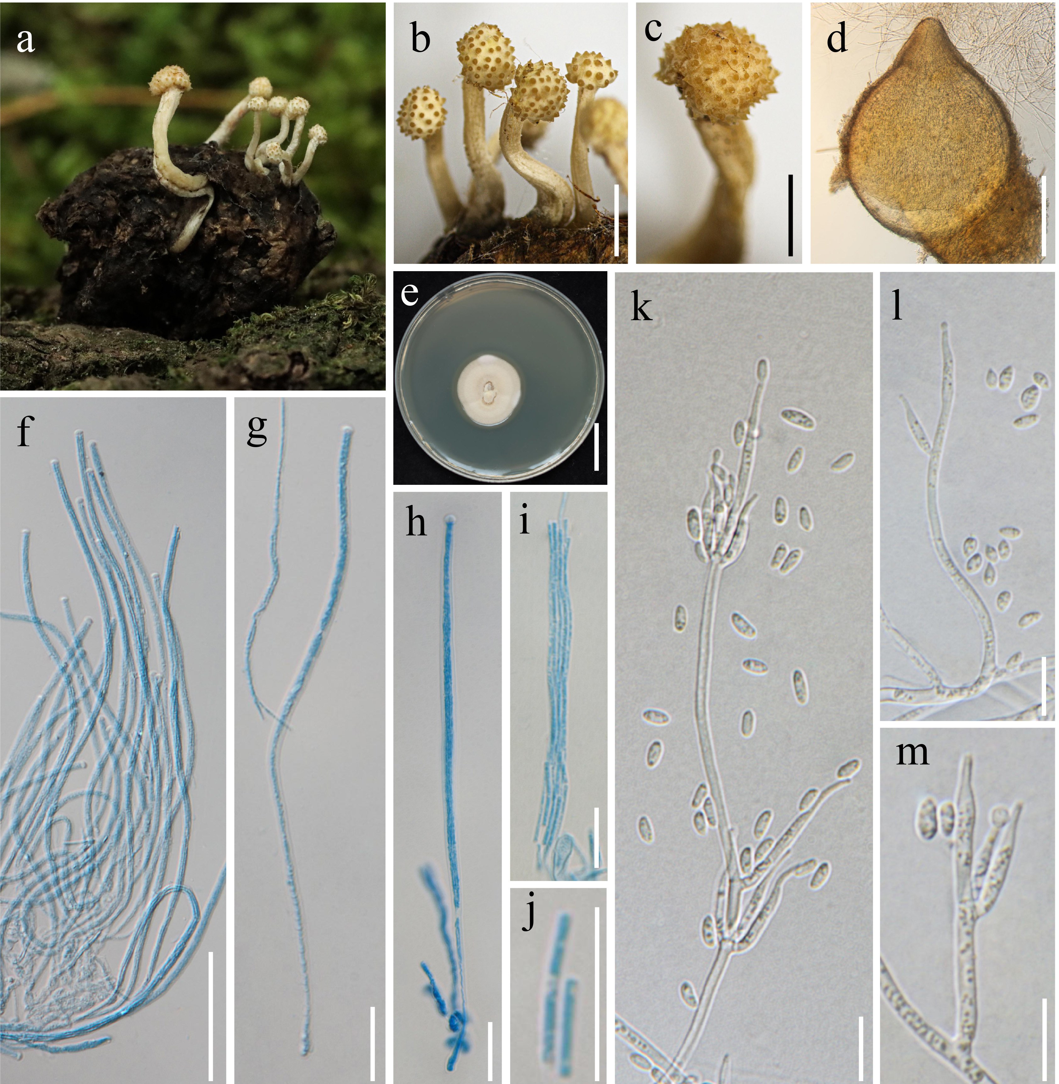
Notes: Akanthomyces phariformis is characterized by numerous stromata, with a terminal, slightly curved, yellow fertile part, superficial ovoid perithecia, and cylindrical asci bearing filiform ascospores. Phylogenetic analyses placed the specimen within Akanthomyces and showed that it is closely related to Aka. taiwanicus. Morphologically, Aka. phariformis can be distinguished from Aka. taiwanicus by its phialides. In Aka. phariformis, phialides arise singly from vegetative hyphae, with a cylindrical base, a swollen middle portion, and a tapering apex that narrows gradually or abruptly, whereas these features are absent in Aka. taiwanicus. In addition, Aka. phariformis has a wider phialide base (1.9–4.8 μm) compared to that of Aka. taiwanicus (2.5–3 μm) (Chuang et al. 2024). Phylogenetic analysis of the present specimen showed that its clusters with Aka. phariformis, and morphological observations are consistent with the original description. Therefore, Aka. phariformis is reported here as a new record from China (Fig. 63).
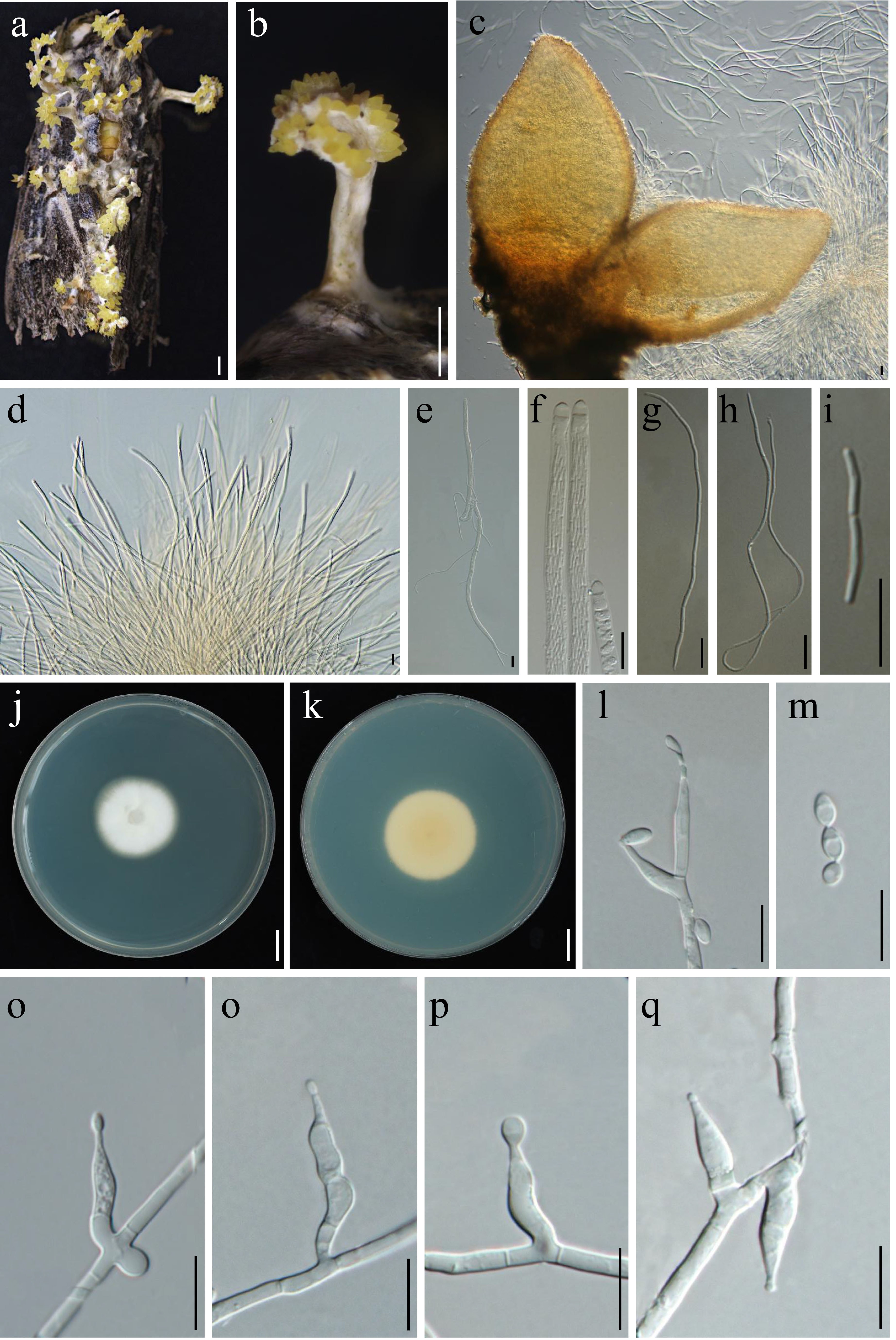
Beauveria Vuill. 1912
Beauveria was proposed by Vuillemin in 1912, with B. bassiana (Bals.-Criv.) Vuill. as the type species. Since its inception, numerous investigations have been conducted to elucidate and delineate the distinct taxa within Beauveria (Petch 1926; MacLeod 1954; De Hoog 1972; Rehner & Buckley 2005). The identification of species within Beauveria utilizing solely ITS as a DNA barcode has reached a resolution threshold. Recently, there has been a discernible shift towards employing an integrative multi-locus delimitation framework for closely related species. To date, 81 records in the Index Fungorum (Index Fungorum July 18, 2025), and 17 species have been accepted in Beauveria (Imoulan et al. 2017).
35. Beauveria lanceolata Y.B. Wang, B.Z. Chen & Zhu L. Yang, sp. nov. Fig. 62
Chinese name:
Index Fungorum number: IF 905268; Facesoffungi number: FoF 19316
Etymology: The specific epithet “lanceolata” refers to the narrowly elongated and tapering stroma, resembling a spearhead.
Diagnosis: Beauveria lanceolata is distinguished by narrowly elongated, spearhead-shaped stroma arising from coleopteran larva, pale yellow to orange, with superficial, densely arranged ovoid to ellipsoidal perithecia.
Holotype: HKAS 135020
Parasitic on the larva of Coleoptera buried in soil and adult of Dermaptera clinging to the fallen leaves. Sexual morph: Stroma 2–5 cm long, solitary, unbranched, arising from larvae of Coleoptera, pale yellow to orange. Fertile part 1–4 cm long, orange, cylindrical or flattened, 2–10 mm wide. Perithecia 200–540 × 160–320 µm, superficial, densely aggregated, and ovoid to ellipsoidal. Asci 120–250 × 2–3 µm, cylindrical, slender. Ascospore caps 2–3.5 × 1.5–2.5 µm, hemispherical. Asexual morph: Hyphae 0.5–4 µm wide, hyaline, septate, smooth-walled. Conidiophores 8–12.5 × 1–2 µm, smooth, hyaline, cylindrical, branched at the apex. Phialides 4–18 × 1–2 µm, smooth-walled, cylindrical or flask-shaped, produced in clusters at the tips of conidiophores or directly from hyphae, with zigzag-shaped apices. Conidia 3–6 × 2–4 µm, single-celled, hyaline, smooth, ovoid to ellipsoidal, produced at the tips of phialides.
Culture characteristics: Mycelia emerge from the entire host body, forming white to pale yellow powdery conidia. Colonies on PDA reach 30–40 mm in diameter after 14 days at 25 °C, surface white to pale yellow, darkening outward, with smooth margins and a dense or slightly floccose texture, reverse side darker, centrally pale yellow and fading toward the edge.
Material examined: CHINA, Yunnan Province, Kunming City, 12 September 2023, on the adult of Dermaptera clinging to the fallen leaves, W.B Zeng (HKAS 135020 holotype, ex-type culture KUNCC 11286). CHINA, Yunnan Province, Kunming City, on the larva of Coleoptera buried in soil, 12 September 2023, Y.B. Wang (culture KUNCC 8665).
Notes: In the multigene phylogenetic analyses based on tef1-α, rpb1, rpb2, and Bloc sequences, B. lanceolata clusters closely with B. pseudobassiana within the same clade but is clearly separated with strong support (Fig. 61). Morphologically, B. lanceolata is distinct from B. pseudobassiana and B. varroae in producing ovoid to ellipsoidal conidia, whereas the latter species bear globose to broadly ellipsoidal conidia (Rehner et al. 2011).
Cordyceps Fr.
Cordyceps, a fascinating group of fungi, has garnered significant attention within scientific and medical communities due to its unique parasitic lifestyle and potential therapeutic properties. The diversity within Cordyceps is vast, encompassing over 300 identified species, with ongoing research continually broadening our understanding of this intriguing fungal group (Wang et al. 2020a). To date, 607 records have been listed in the Index Fungorum (Index Fungorum July 18, 2025).
36. Cordyceps biclavata Y.B. Wang, B.Z. Chen, M. Tang & Zhu L. Yang, sp. nov. Fig. 64
Chinese name
Index Fungorum number: IF905269; Facesoffungi number: FoF 19317
Etymology: The epithet “biclavata” refers to the two clavate stromata emerging from the head of lepidopteran pupa.
Diagnosis: Cordyceps biclavata differs from related species by producing two clavate stromata from the head of lepidopteran pupa, and the verticillium-like asexual morph.
Holotype: HKAS 135022
Parasitic on the pupae of Lepidoptera. Sexual morph: Stromata arising from the head of lepidopteran pupa buried in soil, two in number, unbranched, 30–35 mm long and 3–8 mm wide, orange to orange-yellow, cylindrical or flattened at the base and cylindrical toward the apex. Fertile parts cylindrical to clavate, 15–20 mm long and 2–6 mm wide, orange-yellow. Perithecia semi-immersed, ovoid, 280–499 × 134–379 μm. Asci eight-spored, hyaline, cylindrical. Ascal cap hemispherical, 2.8–4 × 1.3–3.3 μm. Asexual morph: Hyphae 1.5–3.3 μm wide, smooth, hyaline. Phialides 15.7–47.2 μm long, typically solitary or arising laterally from vegetative hyphae or terminally from conidiogenous cells; base cylindrical, tapering gradually toward the apex; basal width 1.3–3 μm, neck 0.3–1.2 μm wide. Conidia 3.4–5.2 × 1.8–3.3 μm, hyaline, ellipsoidal. Two types of conidia are observed, all are one-celled, occurring singly or more commonly aggregated into subglobose to ellipsoidal heads at the apex of phialides.
Culture characteristics: Colonies on PDA at 25 °C for 14 days grow rapidly, reaching 30–35 mm in diameter, with a floccose texture, white to pale yellow.
Material examined: CHINA, Yunnan Province, Baoshan City, the host is a pupa of Lepidoptera found underground. 5 September 2023, M. Tang, (HKAS 135022 holotype, ex-type culture KUNCC 11366), other collection: Ibid., M. Tang (culture KUNCC 11367).
Notes: Cordyceps biclavata is phylogenetically closely related to Cor. militaris, Cor. shuifuensis, and Cor. changbaiensis (Fig. 63), but forms a distinct and well-supported clade within this lineage, indicating that it represents a separate species. Morphologically, the stromata of Cor. biclavata are cylindrical or flattened at the base and cylindrical toward the apex, differing from the solitary, cylindrical to slightly clavate stromata observed in Cor. militaris and Cor. shuifuensis (Wang et al. 2020a). Among these related species, Cor. biclavata is most similar to Cor. militaris, sharing characteristics such as cylindrical to slightly clavate stromata with yellowish to reddish-orange coloration, superficial perithecia, and a verticillium-like asexual morph. However, it can be distinguished by its overall smaller size and the absence of an isaria-like asexual morph, which is present in Cor. militaris (Wang et al. 2020a). In contrast, Cor. changbaiensis differs significantly from Cor. biclavata in having longer stromata covered with white mycelia and in parasitizing coleopteran larvae rather than lepidopteran hosts (Hu et al. 2021). These phylogenetic and morphological distinctions support the recognition of Cor. biclavata as a novel species within the genus Cordyceps.
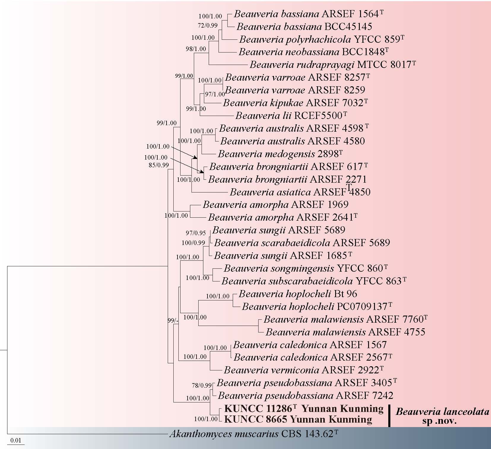
Samsoniella Mongkols., Thanakitp., Spatafora & Luangsa-ard
Samsoniella, as described by Mongkolsamrit et al. (2018), was established through molecular phylogeny of isaria-like morphotypes within the Cordycipitaceae. This genus is characterized by the presence of oval to fusiform conidia, along with striking bright red-orange teleomorphic stromata and anamorphic synnemata (Mongkolsamrit et al. 2018). Additionally, the species Penicillium alboaurantium G. Sm. was reclassified under the genus Samsoniella, and two novel species, Sam. aurantia and Sam. inthanonensis, have been formally described (Wang et al. 2020a). Currently, 45 species, viz. Sam. alboaurantia, Sam. alpina, Sam. anhuiensis, Sam. antleroides, Sam. aranea, Sam. asiatica, Sam. aurantia, Sam. cardinalis, Sam. coccinellidicola, Sam. coleopterorum, Sam. cristata, Sam. doupengshanensis, Sam. duyunensis, Sam. erucae, Sam. farinospora, Sam. formicae, Sam. fusiformispora, Sam. guizhouensis, Sam. haniana, Sam. hepiali, Sam. hymenopterorum, Sam. inthanonensis, Sam. kunmingensis, Sam. lanmaoa, Sam. lasiocampidarum, Sam. lepidopterorum, Sam. lurida, Sam. neopupicola, Sam. pseudogunnii, Sam. pseudotortricidae, Sam. pupicola, Sam. ramosa, Sam. sanmingensis, Sam. sapaensis, Sam. scoliopterygis, Sam. sinensis, Sam. subasiatica, Sam. tiankengensis, Sam. torquatistipitata, Sam. tortricidae, Sam. vallis, Sam. winandae, Sam. yuanzuiensis and Sam. yunnanensis, species have been accepted in Samsoniella (Index Fungorum July 18, 2025). This study introduces two new Samsoniella species that was reported during the survey of microfungi in Southwestern China. The new species is established based on the evidence from morphological characterization and multigene phylogenetic analyses.
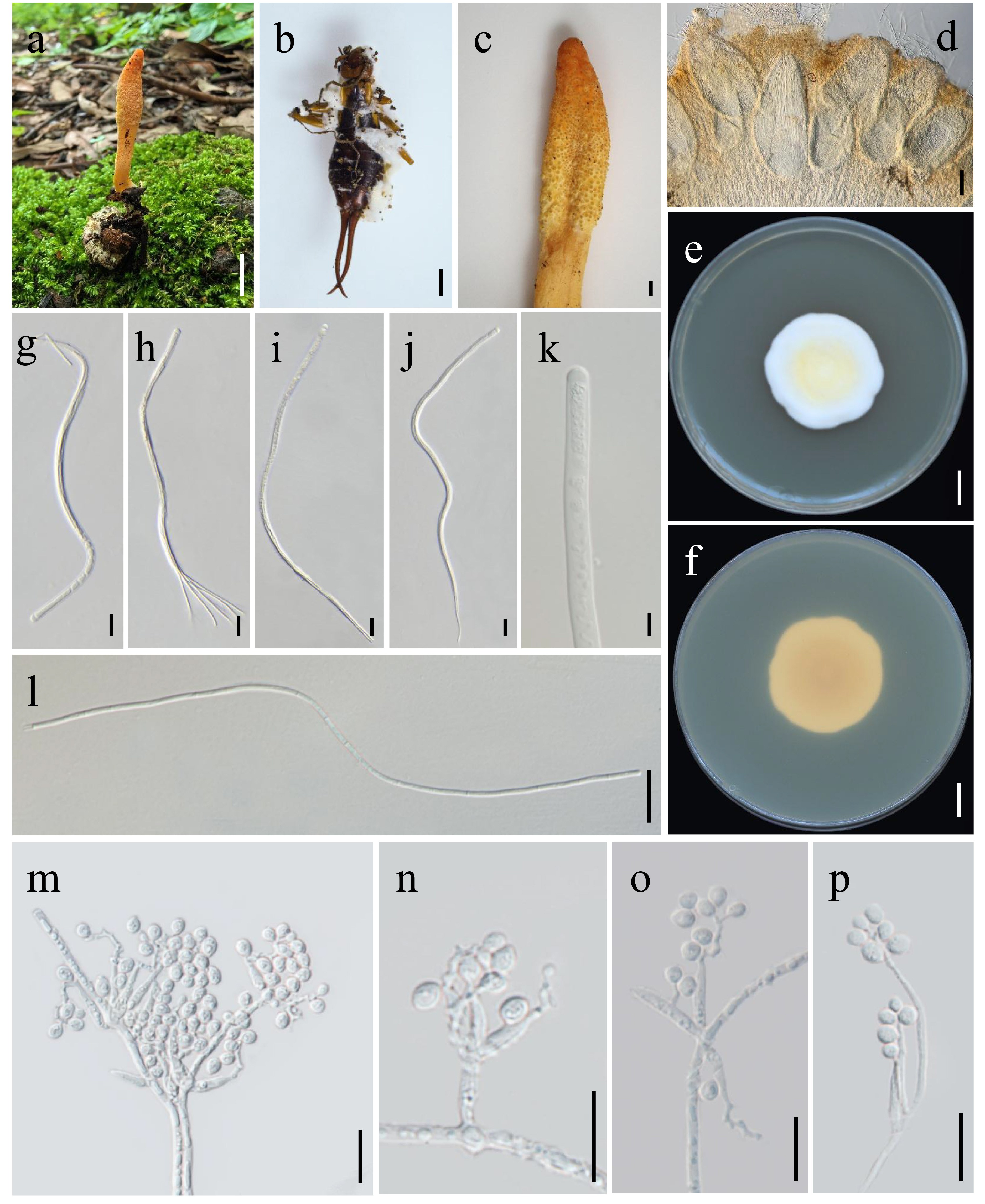
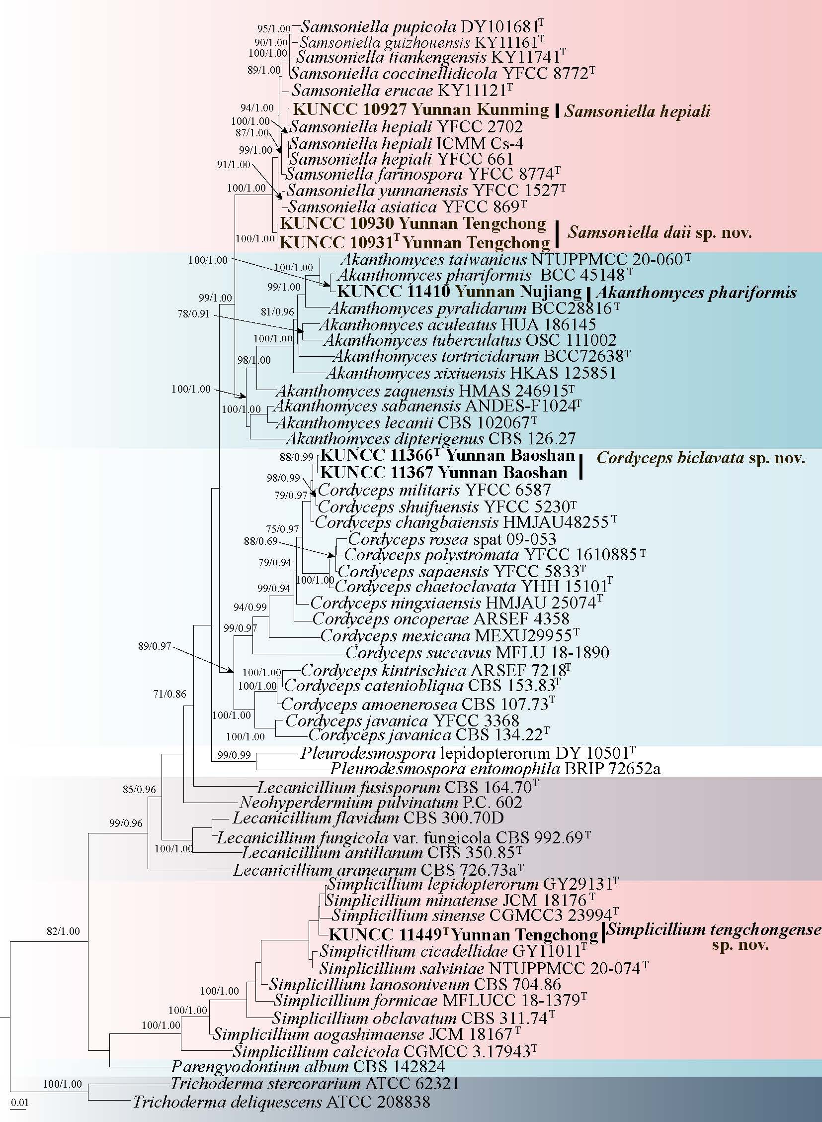
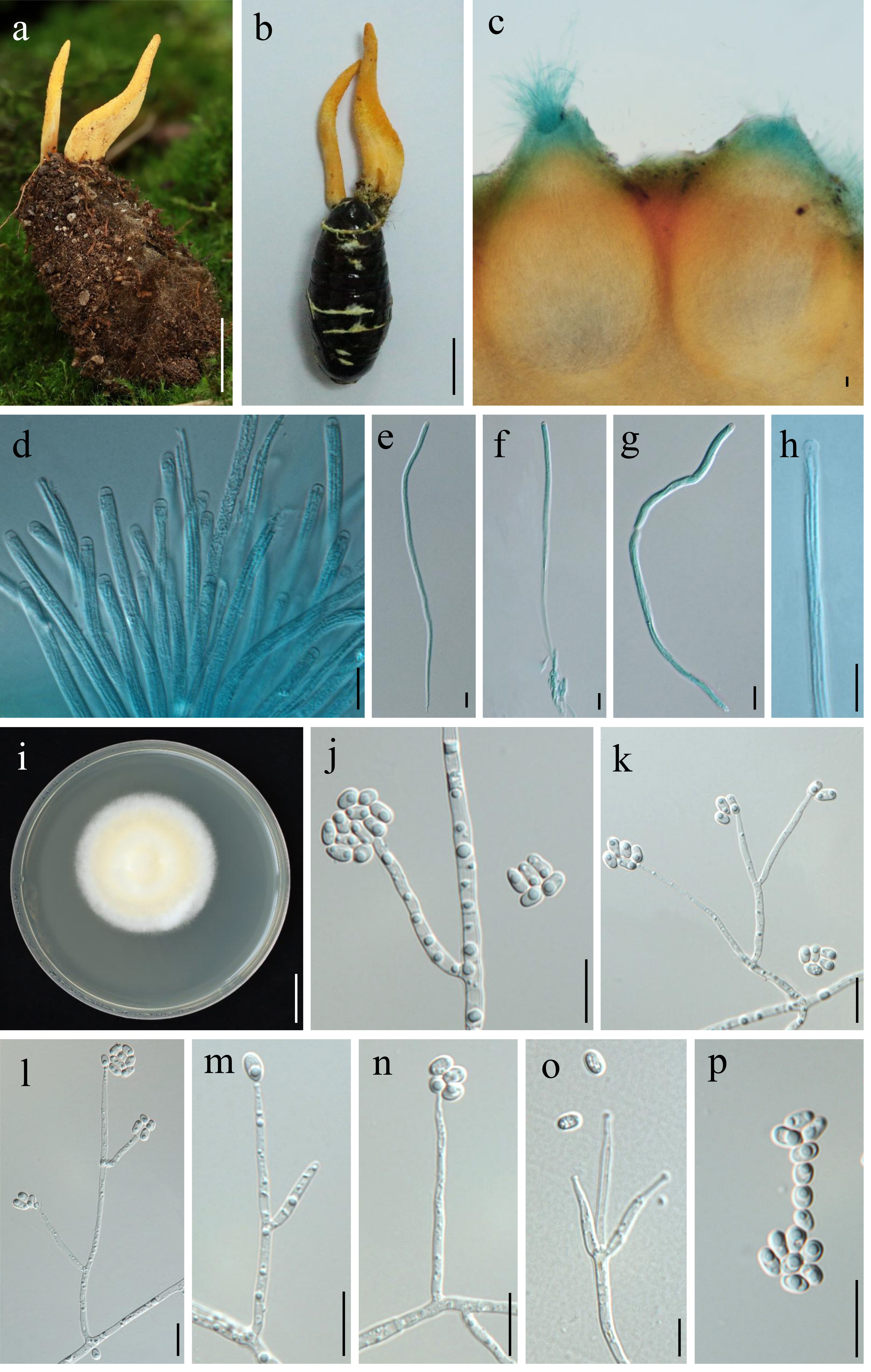
37. Samsoniella hepiali (Q.T. Chen & R.Q. Dai ex R.Q. Dai, X.M. Li, A.J. Shao, Shu F. Lin, J.L. Lan, Wei H. Chen & C.Y. Shen) H. Yu, R.Q. Dai, Y.B. Wang, Y. Wang & Zhu L. Yang. Fig. 65
Chinese name:
Index Fungorum number: IF 833114; Facesoffungi number: FoF 13965
Diagnosis: The molecular data were consistent with Samsoniella hepiali
Parasitic on the pupa of Lepidoptera. Sexual morph: Undetermined. Asexual morph: The strain was isolated from the stroma of Cordyceps militaris. Hyphae smooth-walled, hyaline, 1.4–2.5 μm wide. Conidiophores smooth-walled, cylindrical, 5.1–20.8 × 1.7–3.5 µm. Phialides on conidiophores verticillate, in whorls of two to six, basal portion cylindrical to narrowly lageniform, tapering gradually or abruptly toward the apex; 6.9–19.6 μm long, 1–1.8 μm wide at the base, 0.5–0.8 μm wide at the apex. Conidia smooth, hyaline, one-celled, fusiform or oval, 2.3–3.4 × 1.5–2.2 μm, often in chains.
Culture characteristics: Colonies on PDA fast-growing, 35–40 mm diameter in 14 days at 25 ℃, white to yellowish, cottony with high mycelial density, Reverse white to yellowish.
Material examined: CHINA, Yunnan Province, Kunming City, Kunming Institute of Botany, isolated from Cor. militaris on the pupa of Lepidoptera buried in soil, 10 September 2023, M. Tang, (HKAS 126362, dried culture on PDA, living culture KUNCC 10927).
Notes: Samsoniella hepiali has isaria-like asexual morph and is characterized by branched or unbranched synnemata arising from the whole body of lepidopteran insects, cylindrical or clavate stipes with powdery conidia clusters at the apex (Wang et al. 2020a). However, a notable difference is that Sam. hepiali was isolated from a stroma of Cor. militaris in this study. Similarities include conidiophores cylindrical, phialides on conidiophores verticillate, basal portion cylindrical to narrowly lageniform, conidia fusiform or oval, often in chains. Phylogenetic analysis showed that the specimen clustered with Sam. hepiali (Fig. 63), and morphological studies confirmed that the isolated strain was Sam. hepiali. Therefore, Cor. militaris is reported as the new host of Sam. hepiali.
38. Samsoniella daiae Y.B. Wang, B.Z. Chen, M. Tang & Zhu L. Yang, sp. nov. Fig. 66
Chinese name:
Index Fungorum number: IF 905270; Facesoffungi number: FoF 17504
Etymology: Named after Prof. Ruqin Dai for her contributions to cordycipitoid fungi.
Diagnosis: The species is characterized by reddish-orange and occasionally branched stromata, narrowly ovoid perithecia.
Holotype: HKAS 126364
On the larvae of Lepidoptera. Sexual morph: Stromata arising from the head of insects buried in soil, solitary, occasionally branched, 25 mm long, 2 mm wide. Stipes orange, cylindrical. Fertile parts reddish-orange, cylindrical to clavate, 5 mm long, 2.5 mm wide. Perithecia semi-immersed, narrowly ovoid, 301–481 × 99–240 µm. Asci cylindrical, eight-spored, 84–230 × 2.5–4.5 µm. Ascal caps hemispherica, 1.8–3.9 × 1.8–2.9 µm. Ascospores hyaline, filiform, 45–110 × 0.7–0.9 µm. Asexual morph: Hyphae smooth-walled, hyaline, 1.1–2.7 μm wide. Conidiophores smooth-walled, cylindrical, solitary, 6.5–14.9 × 1.2–2.6 μm. Phialides on conidiophores verticillate, in whorls of three to six, basal portion cylindrical, tapering gradually or abruptly toward the apex; 6.2–22.6 μm long, 1.3–2.4 μm wide at the base, 0.2–1.4 μm wide at the apex. Conidia hyaline, smooth, one-celled, fusiform or oblong, 2–3.9 × 1–2.6 µm, often in chains.
Culture characteristics: Colonies on PDA fast-growing, 40–45 mm diameter in 14 days at 25 ℃, cottony with high mycelium density, white. Reverse white to yellowish.
Material examined: CHINA, Yunnan Province, Tengchong City, Wuhe Township, on the larva of Lepidoptera buried in soil, 5 September 2023, M. Tang (HKAS 126364, holotype, ex-type culture KUNCC 10931), other collection: Ibid., M. Tang (HKAS 126363, culture KUNCC 10930).
Notes: Samsoniella daiae is characterized by solitary stromata, occasionally branched, clavate, fertile parts reddish-orange, cylindrical to clavate, perithecia semi-immersed, narrowly ovoid, and cylindrical asci with filiform ascospores. Samsoniella daiae was phylogenetically closely related to Sam. farinospora (Fig. 63). However, the difference is that stromata of Sam. daiae arise from the head of insects, whereas Sam. farinospora on the hepialid hosts occurs asexual synnemata bearing powdery conidia at the apex, along with isaria-like conidiogenous structures (Wang et al. 2020a). Morphological observations reveal a significant difference in conidia sizes between Sam. daiae (2–3.9 × 1–2.6 µm) and Sam. farinospora (1.6–2.8 × 0.6–1.2 μm).
Simplicillium W. Gams & Zare
Simplicillium was initially established with Sim. lanosoniveum as the type species, aiming to define a putatively monophyletic group comprising Sim. lanosoniveum, Sim. lamellicola, Sim. obclavatum, and Sim. wallacei within the family Clavicipitaceae (Wang et al. 2020a). Simplicillium exhibits a broad host range and is distributed globally. Several species within this genus are associated with plant pathogenic rust fungi and are recognized for their considerable potential in biological control (Gomes et al. 2018). To date, 37 species and 2 varietal taxa, viz. Sim. album, Sim. aogashimaense, Sim. araneae, Sim. calcicola, Sim. chinense, Sim. cicadellidae, Sim. coccinellidae, Sim. coffeanum, Sim. coleopterorum, Sim. cylindrosporum, Sim. filiforme, Sim. formicae, Sim. formicidarum, Sim. guizhouense, Sim. humicola, Sim. hymenopterorum, Sim. lamellicola, Sim. lanosoniveum, Sim. lanosoniveum var. lanosoniveum, Sim. lanosoniveum var. tianjinense, Sim. larvatum, Sim. lepidopterorum, Sim. minatense, Sim. neolepidopterorum, Sim. niveum, Sim. obclavatum, Sim. pechmerlense, Sim. pseudocercosporicola, Sim. puwenense, Sim. salviniae, Sim. scarabaeoidea, Sim. sinense, Sim. spumae, Sim. subtropicum, Sim. sympodiophorum, Sim. wallacei and Sim. yunnanense were restricted to Simplicillium, based on the phylogenetic analyses of ITS sequence data and strong morphological evidence (Index Fungorum, July 18, 2025).
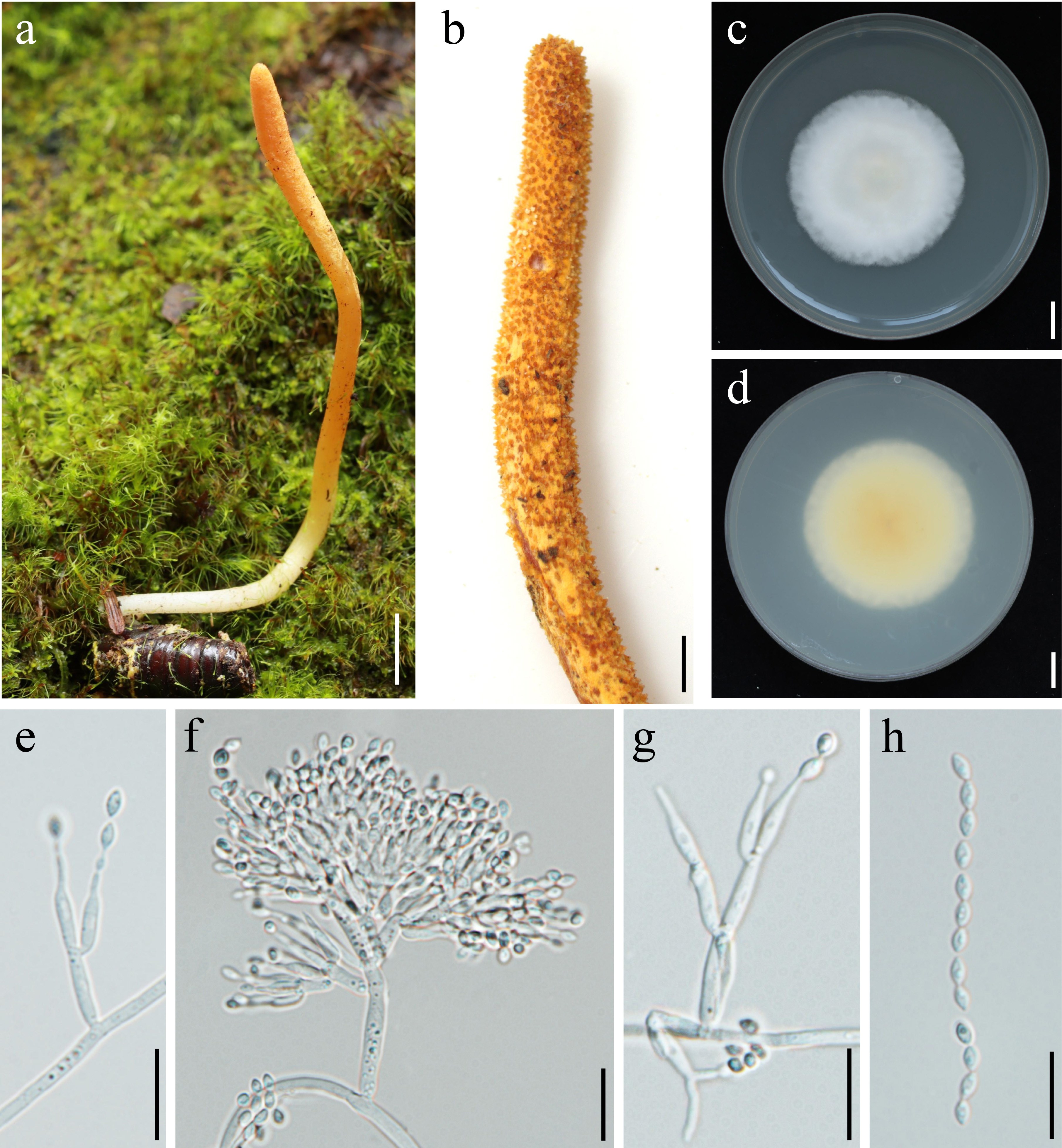
39. Simplicillium tengchongense Y.B. Wang, B.Z. Chen, M. Tang & Zhu L. Yang, sp. nov. Fig. 67
Chinese name
Index Fungorum number: IF 905271; Facesoffungi number: FoF 19318
Etymology: The epithet tengchongense refers to Tengchong City, Yunnan Province, China, where the holotype specimen was collected.
Diagnosis: Simplicillium tengchongense is distinguished by its solitary phialides on hyphae or paired on conidiophores, and ellipsoidal to subglobose conidia in chains.
Holotype: HKAS 135023
Endophytic from Nigelia martialis which parasite on the larva of Lepidoptera. Sexual morph: Undetermined. Asexual morph: Hyphae are 1.5–3.1 μm wide, smooth-walled, and hyaline. Phialides 9.4–32.4 μm long, typically solitary on vegetative hyphae or in pairs at the apex of conidiophores; base cylindrical, tapering gradually toward the apex; basal width 1.3–3.3 μm, neck 0.5–1.4 μm wide. Conidia 1.9–5.6 × 2.2–4.5 μm, smooth, hyaline, ellipsoidal to subglobose, commonly arranged in chains.
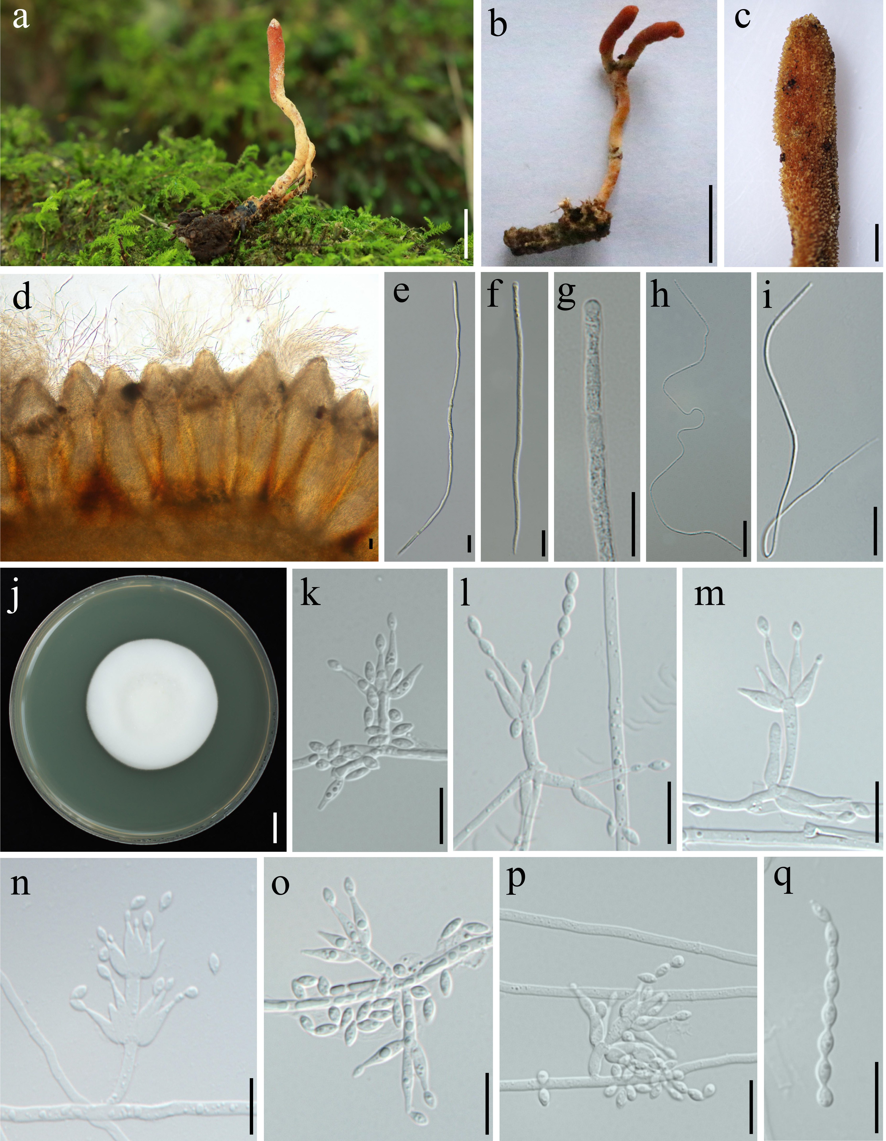
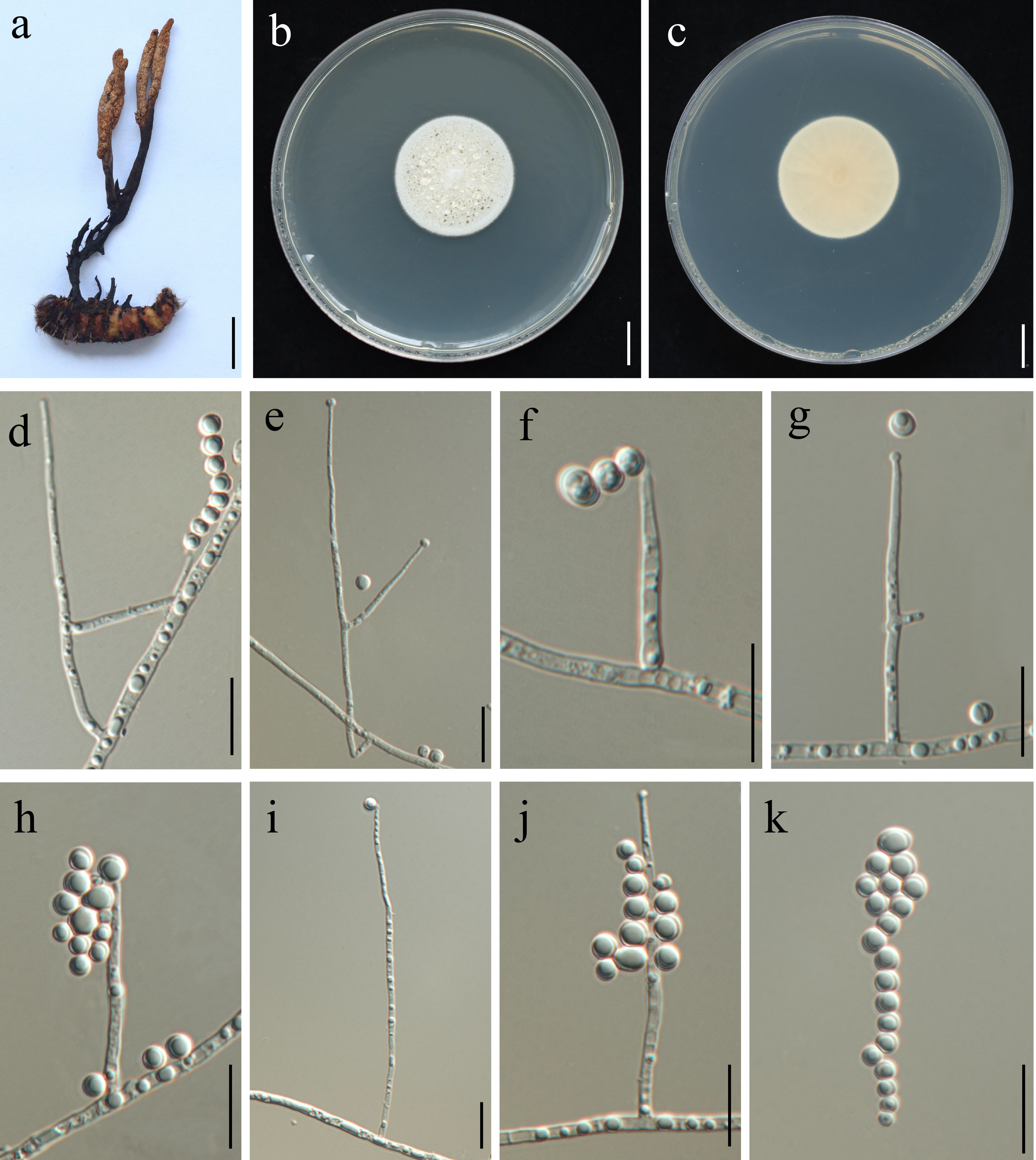
Culture characteristics: Colonies on PDA at 25 °C for 14 days reach 30–35 mm in diameter, with dense mycelia and abundant production of hyaline exudate droplets. Colonies are white to pale yellow on the surface, and white to cream on the reverse, with conspicuous longitudinal furrows.
Material examined: CHINA, Yunnan Province, Tengchong City, Houqiao Township, isolated from the sclerotium of Nig. martialis parasitic on the larva of Lepidoptera buried in soil, 5 September 2023, M. Tang, (HKAS 135023 holotype, ex-type culture KUNCC 11449).
Notes: Simplicillium tengchongense was described as a member of the genus Simplicillium due to its solitary phialides and conidia adhering in globose slimy heads. In our five-gene phylogenetic analyses (Fig. 63), Sim. tengchongense forms a distinct clade within Simplicillium and is closely related to Sim. lepidopterorum and Sim. sinense (Chen et al. 2019; Yan et al. 2023). However, Sim. tengchongense can be distinguished from Sim. lepidopterorum by its production of ellipsoidal to subglobose conidia, whereas Sim. lepidopterorum produces ellipsoidal to fusiform conidia (Chen et al. 2019). It also differs from Sim. sinense in its ecological origin, having been isolated from the sclerotium of Nig. martialis rather than from human skin (Yan et al. 2023).
Hypocreaceae De Not
Trichoderma Pers
Trichoderma is a genus of widely distributed filamentous fungi that exhibit considerable biotechnological promise, primarily due to their remarkable ability to colonize a variety of substrates across diverse environmental contexts (Jaklitsch 2011). This genus is also known for its proficiency in secreting an array of hydrolytic enzymes, including cellulases, chitinases, glucanases, and proteases, alongside a variety of secondary metabolites such as pyrones, peptaibols, and terpenes (Jaklitsch & Voglmayr 2015). Certain species like Tri. reesei serve as vital sources of enzymes for various biotechnological applications. Currently, 563 described within Trichoderma (Index Fungorum July 18, 2025).
40. Trichoderma habaense Y.B. Wang, B.Z. Chen & Zhu L. Yang, sp. nov. Fig. 69
Chinese name
Index Fungorum number: IF 905272; Facesoffungi number: FoF 19319
Etymology: The epithet “habaense” refers to Haba Snow Mountain (Haba Xueshan) in Shangri-La City, Yunnan Province, China, where the type specimen was collected.
Diagnosis: Trichoderma habaense produces smaller, globose to ellipsoid conidia without oblong forms.
Holotype: HKAS 135027
Endophytic from the root of Rho. decorum. Sexual morph: Undetermined. Asexual morph: Conidiophores typically straight, verticillium-like or regularly branched in a treelike pattern, mostly slender, rarely forming long, distinct main axes. They emerged radially from the reticulum and occasionally extended as short, fertile outgrowths bearing 1–3 terminal phialides. Branches (2–)2.5–4.5(–5) μm wide, sometimes widening to 8 μm; lateral branches arranged at right angles or slightly inclined upward, loose and short, typically single-celled and terminated by a solitary phialide; higher-order branches often formed 2–3 rebranching levels. Phialides solitary or in divergent whorls of 2–4, lageniform, with or without distal wall thickening, measuring (6.9–)7.6–14.8(–19) × (2.6–)3.1–4.1(4.4) μm ( = 10.7 × 3.6 μm, n = 29). Terminal phialides in each whorl often distinctly longer; most phialides straight, rarely curved. Conidia (3.5–)3.8–5.2(–5.5) × (3.2–)3.4–4.8(–5.5) μm ( =4.4 × 4.1 μm, n = 34), green, globose to ellipsoid, smooth-walled, containing a few small guttules. Chlamydospores not observed.
Culture characteristics: Colonies on PDA reaching 29–30 mm in radius at 25 °C and 6–9 mm at 30 °C after 72 h, covering the entire plate after 7 days at 25 °C. Colonies circular, dense, with abundant aerial hyphae forming a thick white cottony mat that ascended in concentric zones and reached the lid of the Petri dish. Autolytic exudates inconspicuous, hyphal coilings absent, no diffusing pigments or distinct odour observed.
Material examined: CHINA, Yunnan Province, Diqing, Shangri-La City, Haba Snow Mountain, isolated as an endophyte from Rho. decorum root, 15 March 2023, F. Xiong (HKAS 135027 holotype, ex-type culture, KUNCC 11229), other collection: Ibid., Q. Fan (culture KUNCC 11230).
Notes: As shown in the phylogenetic tree, Tri. habaense forms a distinct terminal lineage (Fig. 68), strongly supported by multi-locus phylogenetic analyses (Cai & Druzhinina 2021). BLAST comparisons against the NCBI database revealed 98.8% identity to Tri. cremeoides S112 in tef1-α (14 bp differences) and 99.3% identity in rpb2 (5 bp differences). Morphologically, the conidia of Tri. cremeoides are larger than those of Tri. habaense. In addition, the conidia of Tri. habaense are predominantly globose to ellipsoid, with oblong forms rarely observed (Jaklitsch & Voglmayr 2015).
41. Trichoderma xinpingense Y.B. Wang, B.Z. Chen & Zhu L. Yang, sp. nov. Fig. 70
Chinese name:
Index Fungorum number: IF 905273; Facesoffungi number: FoF 19320
Etymology: The epithet xinpingense refers to Xinping Yi & Dai Autonomous County, Gasa Town in Yuxi City, Yunnan Province, China, where the type specimen was collected.
Diagnosis: Differs from T. phellinicola by shorter, wider phialides and smaller conidia.
Holotype: HKAS 135028
Endophytic from Ophiocordyceps sp. Sexual morph: Undetermined. Asexual morph: Conidiophores simple or rebranching 1 (–2) times, i.e. 1 main axis of variable length, tapering from 2 to 3 μm at the base to 1–2 μm wide terminally, with 1–2 celled, often asymmetric terminal branches, replaced by phialides in apical regions. Phialides solitary or divergent in whorls of 1–2, often distinctly inclined upwards, arising from cells 2–4 μm thick. Phialides (10.1–)10.5–20.8(–21.2) × (1.9–)2–3.3(3.4) μm ( =15.3 × 2.5 μm, n = 30), subulate or cylindrical, widest at or slightly above the base, straight or curved. Conidia formed in minute wet heads, rarely exceeding 50 μm diam, distributed across the whole plate, denser around the margin. Conidia (3.2–)3.9–7(–7.4) × (2–)2–3(–3.3) μm ( =4.9 × 2.5 μm, n = 30), oblong, cylindrical, less commonly sub-ellipsoidal, hyaline, smooth, with few minute guttules; scar indistinct.
Culture characteristics: Colonies on PDA 53–57 mm in radius after 72 h at 25℃, covering the plate after 4-5 d. Colonies circular, aerial hyphae abundant. No pigment and distinct odor produced on this medium.
Material examined: CHINA, Yunnan Province, Yuxi City, Xinping, Gasa Town, isolated as an endophyte from Ophiocordyceps sp., 19 July 2024, F.W. Lou (HKAS 135028, holotype, ex-type culture KUNCC 11231), other collection: Ibid., Q. Fan (culture KUNCC 11232).
Notes: As shown in the phylogenetic tree, Tri. xinpingense forms a distinct terminal lineage (Fig. 68), strongly supported by multi-locus phylogenetic analyses. BLAST comparisons against the NCBI database revealed 88% identity to Tri. phellinicola in tef1-α (88 bp differences) and 95.6% identity in rpb2 (33 bp differences). Morphologically, the conidia of Tri. xinpingense, measuring (3.2–)3.9–7(–7.4) × (2–)2–3(–3.3) μm, are smaller than those of Tri. phellinicola, which measure (5–)6–11(–15) × (2–)2.2–2.7(–3) μm. Furthermore, the phialides of Tri. xinpingense (10.1–)10.5–20.8(–21.2) × (1.9–)2–3.3(3.4) μm are notably shorter and slightly wider than those of Tri. phellinicola (11–)19–33(–41) × (1.8–)2–3(–3.2) μm (Jaklitsch 2011).
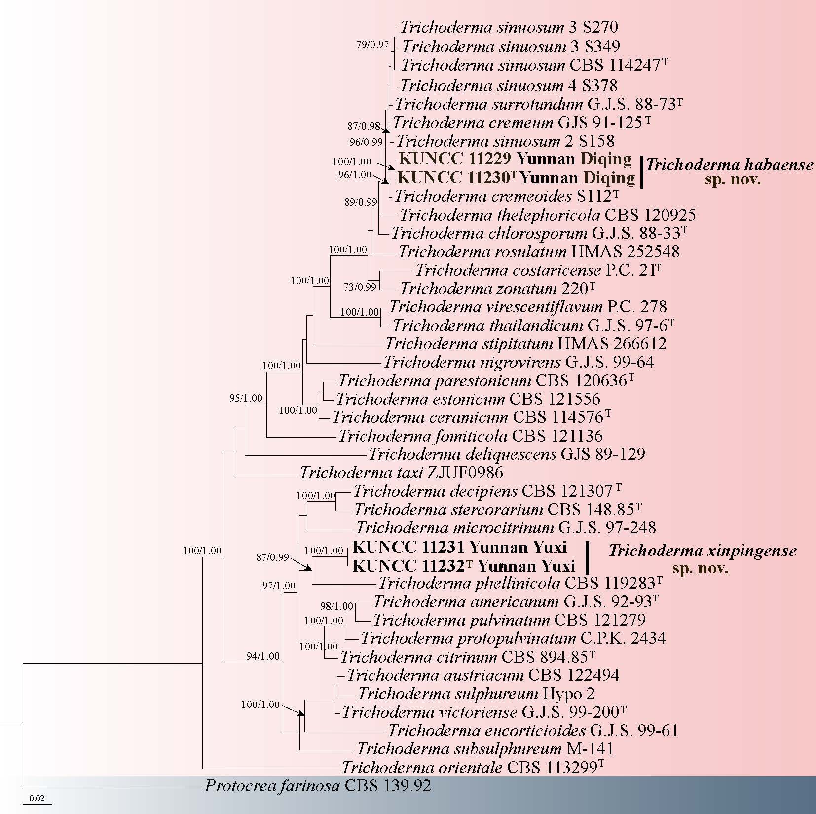
Hypocreales Lindau
Nectriaceae Tul. & C. Tul.
Fusarium Pers
Fusarium is recognized as one of the most widespread filamentous fungal pathogens globally. Multi-locus sequence analysis encompassing tef1-α, tub2, calmodulin, and rpb2 genes has elucidated the existence of multiple cryptic species within each morphospecies of clinically significant Fusarium taxa (John F. Leslie & Summerell 2006; Balajee et al. 2009). Fusarium, currently encompasses more than 712 names (Crous et al. 2021; Fusarium Database), and 1863 lists within the indexfungorum (Index Fungorum July 18, 2025).
42. Fusarium highlandense Y.B. Wang, B.Z. Chen, Q. Fan & Zhu L. Yang, sp. nov. Fig. 72
Chinese name:
Index Fungorum number: IF 905274; Facesoffungi number: FoF 19321
Etymology: "highlandense" refers to samples collected from high-altitude plateau regions.
Diagnosis: Fusarium highlandense is distinguished by producing cream to orange, translucent sporodochia and 2–7-septate falcate macroconidia with blunt to slightly curved apical cells and foot-shaped basal cells.
Holotype: HKAS 135030
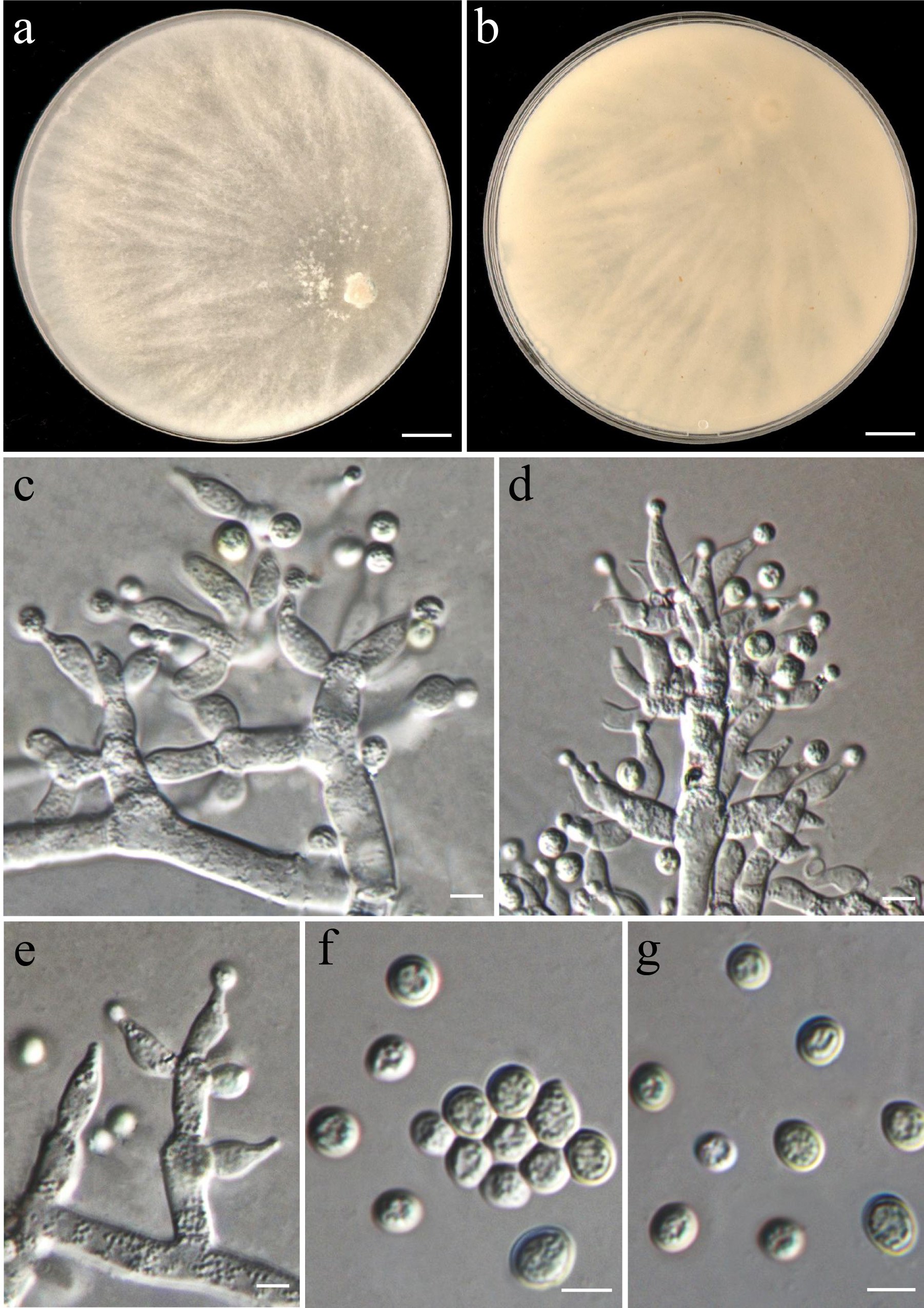
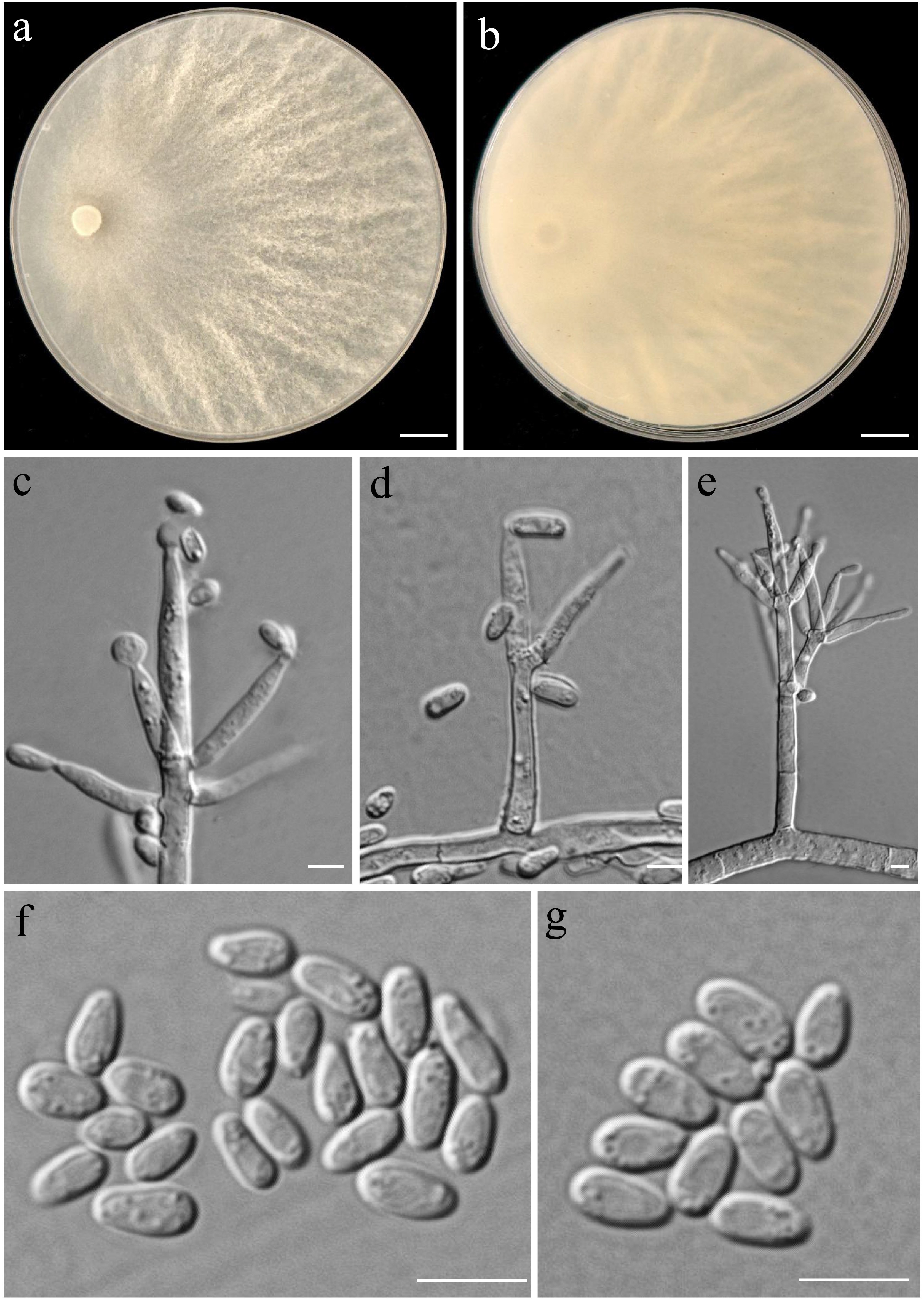
Parasitic from the symptomatic leaves of Erianthus rufipilus. Sexual morph: Undetermined. Asexual morph: Sporodochial conidiophores densely, irregularly branched, 12–21.5 × 3–4.5 μm, bearing apical whorls of 2–3 phialides, sporodochial phialides monophialidic, subulate to subcylindrical, 14.5–22.5 × 3–3.5 μm, ( = 17 × 3.5 μm), smooth and thin-walled, apical collarettes absent and periclinal thickening inconspicuous. Sporodochial microconidia absent. Sporodochial macroconidia falcate, straight to slightly dorsiventrally curved, broadest at the middle portion, tapering towards both ends, apical cell blunt to slightly curved, basal cell stunted to well-developed, foot-shaped, 2–6(–7)-septate, predominantly 5-septate, hyaline, smooth- and thick-walled; 2-septate conidia: 21–24 × 3.5–4.5 μm, ( = 23 × 4 μm); 3-septate conidia: 29–38.5 × 3–4.5 μm ( = 34 × 3.5 μm); 4-septate conidia: 40–47.5 ×3.5–4 μm, ( = 44 × 3 μm); 5-septate conidia: 45–60(–64.5) ×3.5–5 μm ( = 52.5 × 4 μm); 6-septate conidia: 58–68 × 4–4.5 μm ( = 62 × 4 μm); 7-septate conidia: 69.5–78.5 × 3.5–4.5 μm, ( = 73.5 × 4 μm). Chlamydospores not observed.
Culture characteristics: Colonies on PDA exhibiting 6.5–7 cm diam in 7 d at 25℃, aerial mycelia abundant, flat, colony margin regular, surface white to magenta, reverse cream to magenta. On SNA reaching 3.5–4 cm diam in 7 d, aerial mycelia scant, flat, colony margin regular, surface white to cream, reverse white to cream. On OA reaching 5.5–6 cm diam in 7 d, felty, with abundant aerial mycelia, colony margin lightly regular, surface white, reverse pale brown. Aerial conidiophores and conidia were not observed. Sporodochia cream to orange, translucent, formed richly on the carnation leaves and on the agar, often covered with aerial mycelia.
Material examined: CHINA, Yunnan Province, Shangri-La City, from the symptomatic leaves of Erianthus rufipilus, August 2023, Q. Fan (HKAS 135030 holotype, dried culture on PDA; ex-type culture KUNCC 11103), other collection: Ibid., Q. Fan, (culture KUNCC 3449).
Notes: Phylogenetic analysis based on the concatenated dataset of CaM, rpb1, rpb2, tef1-α, ITS and tub2 loci resolved the Fus. highlandense isolates as a monophyletic clade within the Fusarium, with strong statistical support (Fig. 71). Fusarium highlandense is closely related to Fus. avenaceum, but differs by 63 bp from Fus. avenaceum in the 3-locus (rpb1-rpb2- tef1-α) dataset (ITS, CaM and tub2 sequences are not available for Fus. avenaceum). Morphologically, Fus. highlandense can be distinguished from related species through the septation (3–5-septate in F. avenaceum vs. 2–7-septate in Fus. highlandense) (John F. Leslie & Summerell 2006).
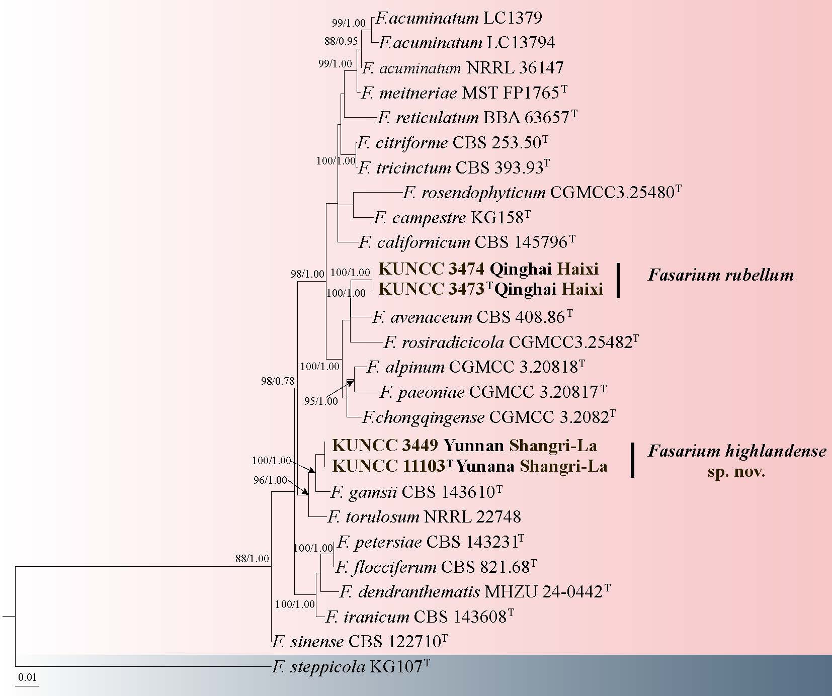
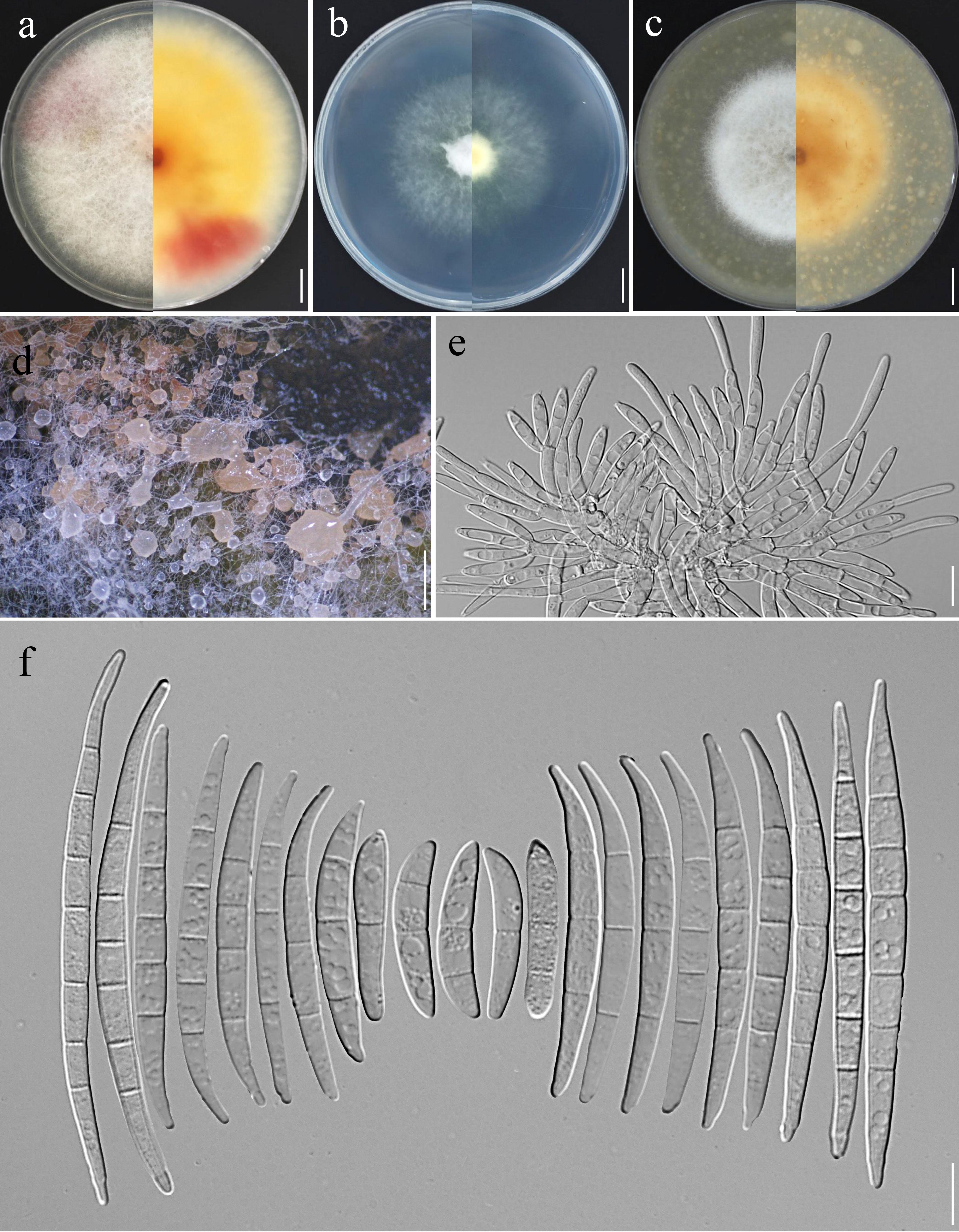
43. Fusarium rubellum P. Zhao & L. Cai. Fig. 73
Chinese name
Index Fungorum number: IF 572668; Facesoffungi number: FoF 19322
Endophytic from Ophicordyceps sinensis. Sexual morph: Undetermined. Asexual morph: Conidiophores on aerial mycelia 12–28 μm tall, simple or rarely irregularly branched, bearing terminal single phialides or whorls of 1–2 phialides, sometimes reduced to solitary conidiogenous cells borne laterally on hyphae; aerial conidiogenous cells monophialidic, subulate to subcylindrical or lageniform, smooth- and thin-walled, 11–24 × 3–4.5 μm (= 18 × 4 μm), apical collarettes absent and periclinal thickening inconspicuous. Aerial macroconidia falcate to navicular, hyaline, smooth- and thin-walled, almost straight to slightly dorsiventrally curved ,tapering towards both ends, apical cell blunt or papillate, basal cell stunted to well-developed, foot-shaped, 1–6-septate, predominantly 5-septate, 1-septate conidia: 22–30 × 4–5 μm ( = 26 × 4.5 μm); 2-septate conidia: 29.5–41 × 4–6 μm ( 34 × 5 μm); 3-septate conidia: 31.5–43.5 × 4.5–6 μm ( = 37.5 × 5 μm); 4-septate conidia: (35–) 37–52 (–55) × 4–6 μm, ( = 43.5 × 5 μm); 5-septate conidia: 38.5–56.5 × 5–6.5 μm, ( = 46.5 × 5.5 μm); 6-septate conidia: 43.5–52.5 × 5–6.5 μm, ( 48 × 6 μm); overall: 22–56.5 × 4–6.5 μm ( = 39.5 × 5 μm). Sporodochia and Sporodochial conidia not observed. Chlamydospores obovoidal, subglobose to globose, hyaline, smooth-walled to slightly roughened, thick-walled, 6–8.5 μm, terminal or intercalary.
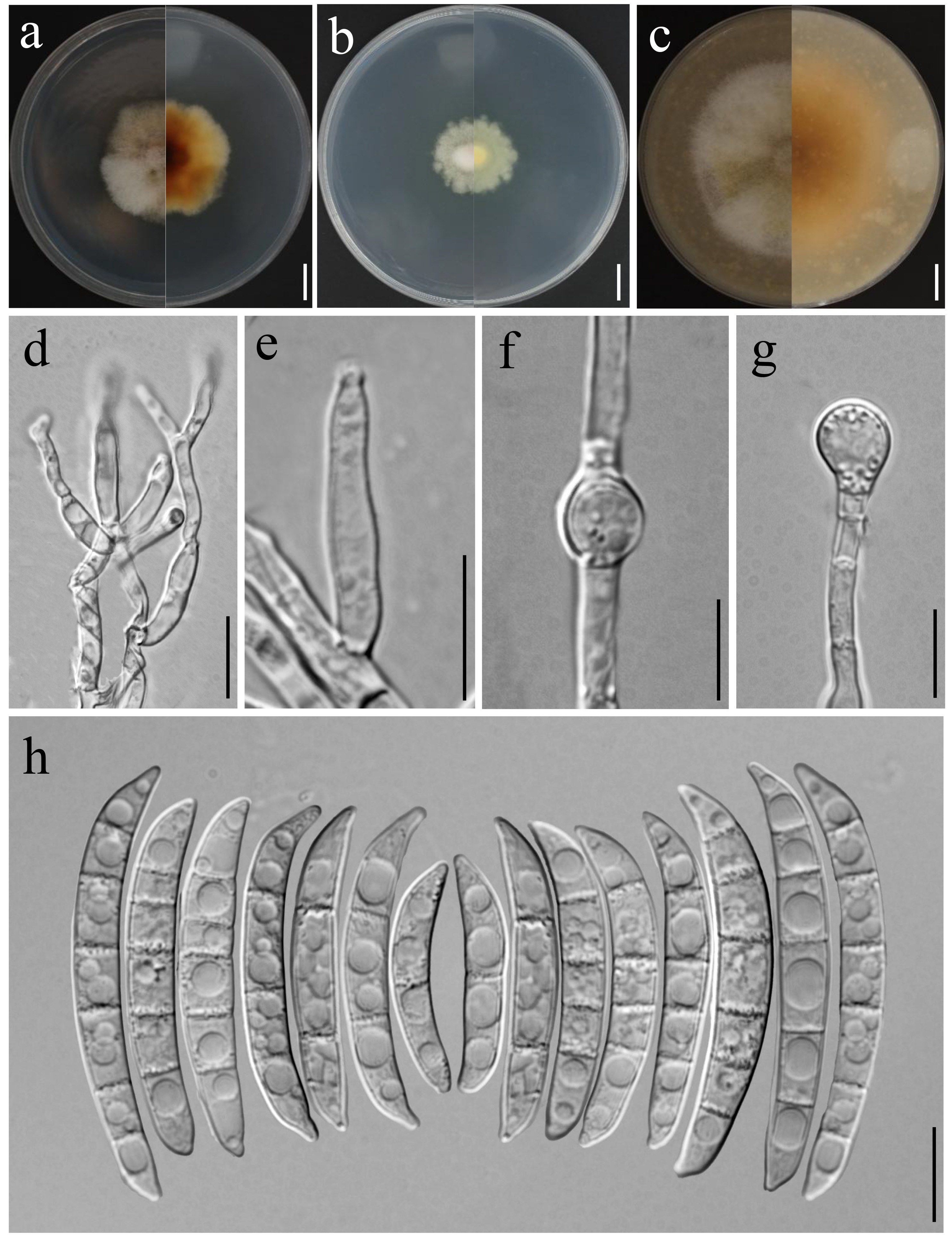
Culture characteristics: Colonies on PDA exhibiting 4–4.5 cm diam in seven days at 25℃, cottony, with abundant aerial mycelium, colony margin undulate, surface white to pale brown, reverse pale brown to brown. On SNA reaching 2.5–3 cm diam in seven days, flat, colony margin undulate, surface white to cream, reverse cream. On OA reaching 5.5–6 cm diam in seven days, cottony, with abundant aerial mycelium, colony margin regular, surface cream to pale brown, reverse cream to pale brown.
Material examined: CHINA, Qinghai Province, Haixi, isolated from O. sinensis, May 2022, Q. Fan (HKAS 126169 holotype, ex-type culture KUNCC 3473), other collection: Ibid., Q. Fan (culture KUNCC 3474).
Note: Phylogenetic analysis based on the concatenated dataset of CaM, rpb1, rpb2, tef1-α, ITS, and tub2 loci revealed that the isolates form a monophyletic clade with Fus. rubellum, with strong statistical support (Fig. 71). Fusarium rubellum is phylogenetically closely related to Fus. gamsii but differs by 14 bp in the combined tef1 and rpb2 dataset. It should be noted that sequences of rpb1, tub2, and CaM are unavailable for Fus. gamsii, which limits further comparative analyses. Morphologically, Fus. rubellum can be distinguished from related species by the size of macroconidia (22–56.5 × 4–6.5 μm vs. 17–46 × 4–6 μm in Fus. gamsii) and septation (1–6-septate vs. 1–4-septate in Fus. gamsii) (Torbati et al. 2019). Based on both phylogenetic and morphological evidence, the isolates are identified as Fus. rubellum. In this study, this species was isolated from O. sinensis, representing a new host record for Fus. rubellum (Torbati et al. 2019; Zhao et al. 2025).
Ilyonectria P. Chaverri & C. Salgado
Ilyonectria was introduced with type species I. radicicola, having asexual morphs and belonging to Neonectria radicicola-group (Booth 1959, 1996; Chaverri et al. 2011; Lombard et al. 2014). Sexual morphs are mostly found on herbaceous material, rarely on woody parts while asexual morphs are in soil, with diseased roots (Chaverri et al. 2011). Ilyonectria sexual morph are characterized with perithecia loosely attaching to substratum, red, globose to subglobose, and having smooth, ellipsoidal, 1- septate ascospores (Chaverri et al. 2011; Lombard et al. 2014). Asexual morphs are produced cylindrocarpon-like conidia, characterized by ellipsoidal to ovoid, 0–1-septate, chlamydospores present, microconidia and straight, 1–3-septate macroconidia (Chaverri et al. 2011; Lombard et al. 2014). Recent studies have evaluated the paraphyletic nature of Ilyonectria (Cabral et al. 2012a; Cabral et al. 2012b; Lombard et al. 2014).
44. Ilyonectria liriodendri (Halleen, Rego & Crous) P. Chaverri & Salgado, in Chaverri, Salgado, Hirooka, Rossman & Samuels, Stud. Mycol. 68: 71 (2011) Fig. 74
Chinese name:
Index Fungorum number: IF 518561; Facesoffungi number: FoF 14355
Saprobic on decaying wood submerged in freshwater habitats. Sexual morph: See Halleen1 et al. (2006). Asexual morph: Mycelium immersed, consisting of hyaline, smooth-walled, branched, septate, smooth, subhyaline hyphae. Conidiophores 68–149 × 3–5 μm ( = 95 × 4 μm, n = 15), macronematous, semi-macronematous, irregularly branched, solitary or group, phialides cylindrical, smooth, 2–3-septate, subhyaline. Conidiogenous cells monophialidic, integrated, cylindrical, subhyaline, truncate at the apex. Conidia 22–43 × 5–9 μm ( = 31 × 7 μm, n = 25), acrogenous, solitary, fusiform or obclavate, 1–2-septate, smooth, subhyaline, rounded at the apex.
Culture characteristics: Conidia germinating on PDA medium within 24 h and germ tubes arising from terminal end of conidium. Colonies grown on PDA reached 50 mm diam at room temperature in one month, circular, with velvety, glistening, dry, flat, cream to light brown mycelium on the surface, from below dark brown with entire margin.
Material examined: CHINA, Xizang Autonomous Region, Linzhi City, Motuo County, 29°19'36.31" N, 95°21'22.17" E, 1481 m a.s.l., on decaying wood submerged in a freshwater stream, 13 July 2022, R.J. Xu, XK-2 (HKAS 136098), culture (KUNCC 24-18108).
Notes: Ilyonectria liriodendri was described as Neonectria liriodendri and was isolated from Vitis vinifera in France, Portugal and South Africa, as well as from Liriodendron tulipifera in the USA. It is the ex-type strain of Cylindrocarpon liriodendri by (Halleen et al. 2006). Later, Chaverri et al. (2011) synonymized N. liriodendri as I. liriodendri due to molecular phylogenetic analysis revealing five distinct genera, with Ilyonectria including some species from the N. radicicola group. Morphologically, our collection perfectly matches the ex-type strain of I. liriodendri. Therefore, we identify our collection as I. liriodendri and report it as a new record for China.
Mariannaea G. Arnaud ex Samson
Mariannaea was established by Samson (1974) and includes species M. camptospora and M. elegans var. punicea, with M. elegans designated as the type species. Mariannaea is characterized by branched, septate conidiophores and flask-shaped phialides that produce 1–2-celled, hyaline conidia, typically arranged in overlapping chains or slimy clusters (Samson 1974, Samson and Bigg 1988, Hu et al. 2017). The taxonomy of Mariannaea was later reassessed by Hu et al. (2017) using both morphological and molecular data, and an epitype was designated for M. elegans. More recently, Bao et al. (2023) introduced two new species, M. yunnanense and M. suae, and reported two additional Mariannaea species from freshwater habitats based on phylogenetic and morphological analyses. To date, 23 species have been recognized within the genus (Hyde et al. 2024a).
45. Mariannaea elegans (Corda) Samson, Stud. Mycol. 6: 75 (1974) Fig. 76
Chinese name
Index Fungorum number: IF 300242; Facesoffungi number: FoF 17424
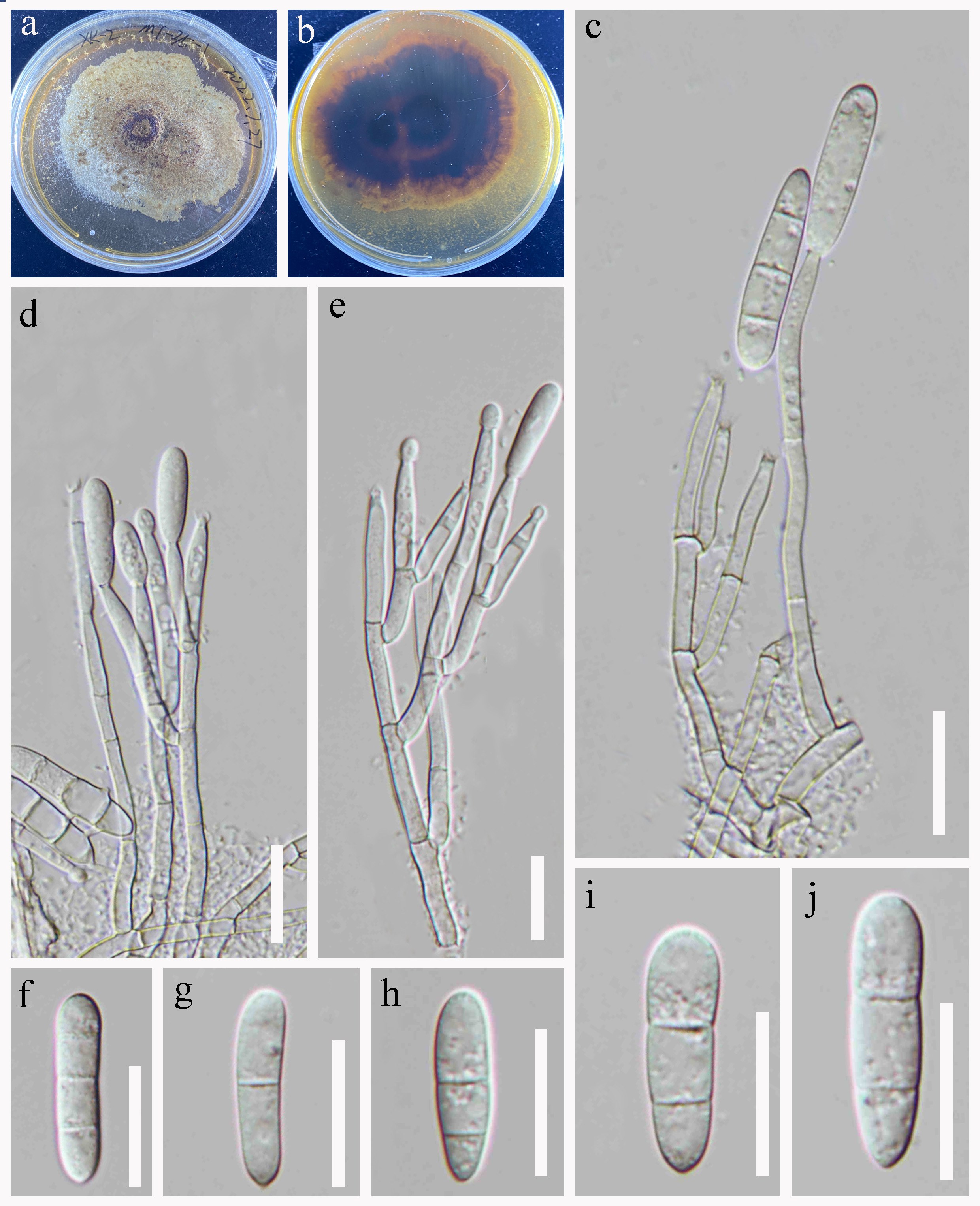
Saprobic on submerged decaying wood. Sexual morph: Undetermined. Asexual morph: Colonies on the substratum superficial, effuse, white, hairy. Mycelium immersed, subhyaline to pale brown, composed of branched, septate hyphae. Conidiophores 202–636 × 6–10 µm ( = 481 × 8 μm, n = 15), macronematous, mononematous, cylindrical, erect, straight or slightly flexuous, septate, smooth-walled, hyaline when young, becoming orange brown at base and gradually paler towards apex, hyaline at the apex. Conidiogenous cells phialides, flask-like, hyaline, slightly swollen at base, tapering at apex, smooth-walled. Conidia 5–6 × 2–3 µm ( = 5 × 3 μm, n = 10), ellipsoidal to fusiform, acuminate at apex, pointed at base, aseptate, hyaline.
Culture characteristics: Conidia germinating on PDA medium within 24 h and germ tubes produced from both ends. Colonies grown on PDA reached 30 mm diam at room temperature in two weeks, circular, with velvety, flat, light brown mycelium in the middle and sparse, dark brown at the edge, producing reddish-brown pigment, from below dark brown with entire margin.
Material examined: CHINA, Yunnan Province, Shangri-La City, Gonda Village, 27°58′24.94" N, 99°46′39.32" E, 3222 m a.s.l., on decaying wood submerged in a freshwater stream, 19 June 2022, R.J. Xu, LTS-10 (HKAS 136147), culture (KUNCC 24-17993).
Notes: Mariannaea elegans was originally described by (Samson 1974), based on samples collected from the bark of rotten trunks of fir trees (Pinaceae) in Brezina. Based on strain (CBS 217.73A). Hu et al. (2017) confirmed that the resulting colony generally matches the description of M. elegans, thereby designating it as the epitype. Mariannaea elegans is saprobic and lignicolous, occurring on submerged decaying woody debris in terrestrial environments. It has been reported from the Brezina, Czechoslovakia, Europe and, Netherlands (Hu et al. 2017).
In the phylogenetic analyses (Fig. 75), our collection (KUNCC 24-17993) clustered with Mariannaea elegans (DUCC 400). Morphologically, these new isolates closely resemble M. elegans Hu et al. (2017), therefore, we have identified our new isolates as M. elegans. This represents the first report of M. elegans from a freshwater habitat on the Yunnan Province, China.
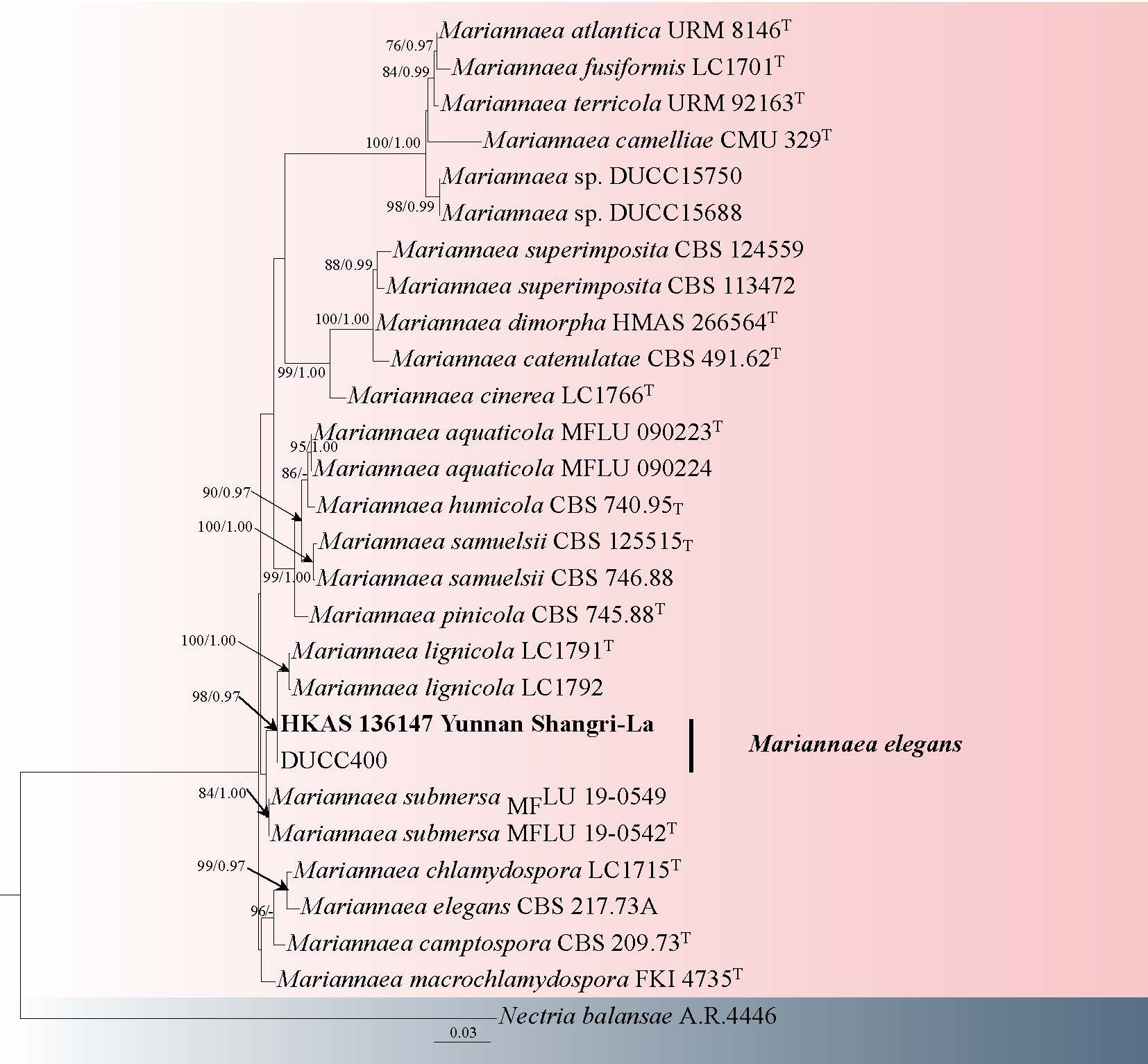
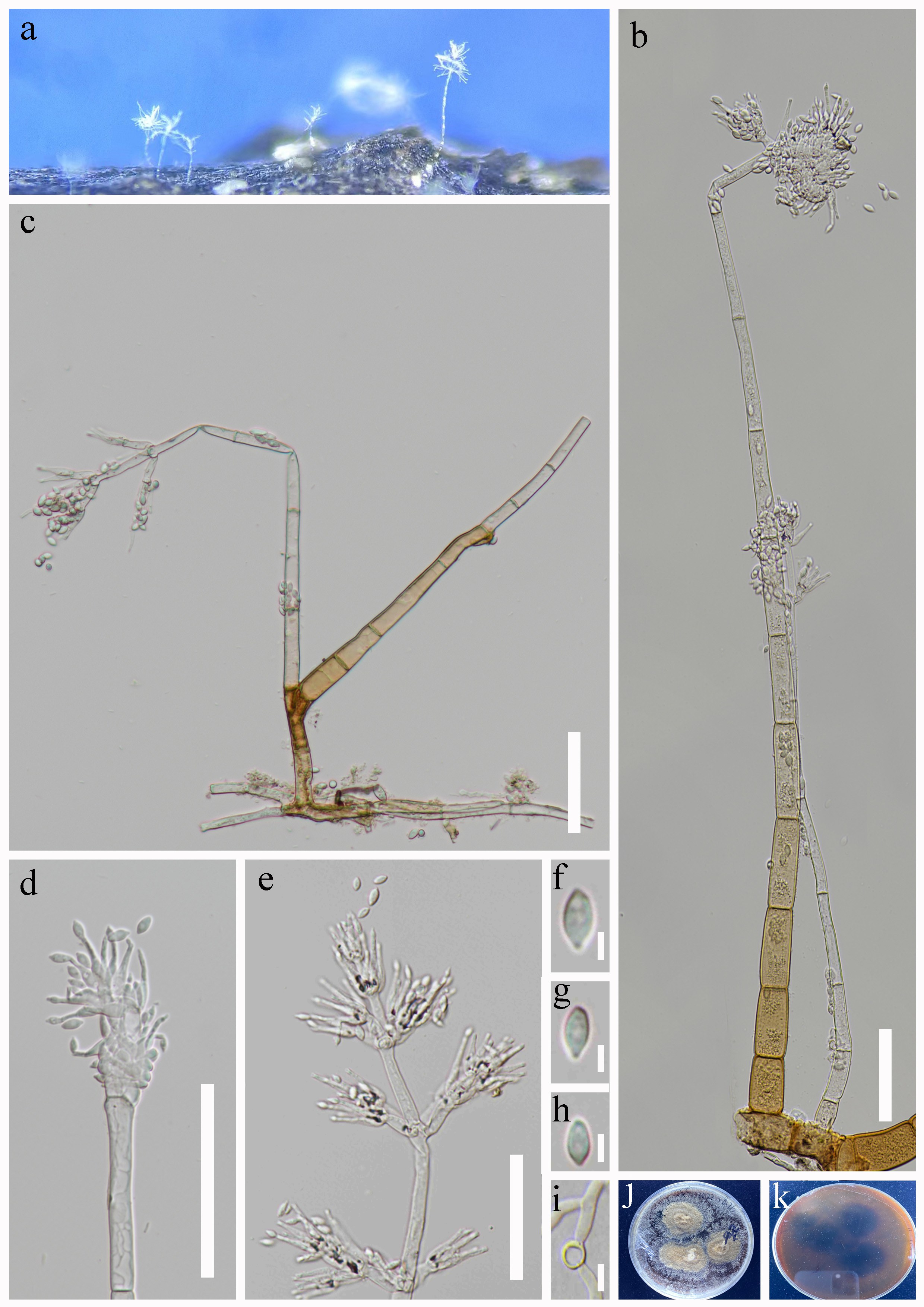
Ophiocordycipitaceae G.H. Sung, J.M. Sung, Hywel-Jones & Spatafora
Ophiocordycipitaceae separated from Clavicipitaceae to include nine genera based on morphological and phylogenetic analyses (Sung et al. 2007; Quandt et al. 2014; Simmons et al. 2015). Asexual morphs display varied synnemata, conidiophores, and conidia, while sexual morphs feature dark or bright, tough stromata or subicula with cylindrical, unitunicate asci and multi-septate, often disarticulating ascospores (Mains 1950; Samson & Evans 1975; Sung et al. 2007; Simmons et al. 2015; Maharachchikumbura et al. 2016, 2021). Currently, this family contains over 500 distinct species. However, more than 30 species were introduced within two years, indicating that the taxonomic diversity of this family has not been sufficiently investigated (Tang et al. 2023; Mongkolsamrit et al. 2024; Yang et al. 2024)
Ophiocordyceps Petch, Trans. Br
Ophiocordyceps, established by Petch to classify Cordyceps species with non-disarticulating ascospores (Petch 1931). Later, it was designated as a subgenus based on spore morphology (Kobayasi 1941) and subsequently included in Cordyceps sensu lato (Mains 1958). The genus, known for infecting insects, sexual morphs is characterized by darkly pigmented, tough, fibrous stromata that may be stipitate or sessile, with perithecia that range from superficial to fully immersed and ascospores that either remain intact or disarticulate into secondary spores (Sung et al. 2007; Simmons et al. 2015). Asexual morphs associated with Ophiocordyceps include Hirsutella, Syngliocladium, Stilbella, Paraisaria, Hymenostilbe and Sorosporella (Evans & Samson 1982; Araújo & Hughes 2016; Xiao et al. 2019; Yang et al. 2021).
46. Ophiocordyceps fusispora Y.B. Wang, H.F. Liao & Zhu L. Yang, sp. nov. Fig. 78
Chinese name
Index Fungorum number: IF 905275; Facesoffungi number: FoF 19323
Etymology: The species is named for its fusiform secondary ascospores.
Diagnosis: Different from other Ophiocordyceps species by its fusiform secondary ascospores.
Holotype: HKAS 126122
Parasitic on ants buried in soil. Sexual morph: Stromata arising from the heads of hymenopteran ants, 25–58 × 1.2–1.9 mm ( = 41 × 1.5 mm, n = 10), solitary, pale yellow to yellow. Fertile parts 1.6–2.5 × 1.8–2.8 mm ( = 2.1 × 2.3 mm, n = 10), nearly spherical, terminal, spinous surface due to protruding ostioles, distinctly demarcated from the stipe, colored yellow to orange-yellow. Perithecia 566.8–825.6 × 85.3–118.6 µm ( = 692 × 102 μm, n = 20), longitudinal, immersed, lageniform, and parallel to the stipe. Asci 189.6–394.7 × 5.8-9.6 µm ( = 292.2 × 7.8 μm, n = 20), hyaline, cylindrical, 8-spores. Ascospores disintegrated into secondary ascospores, 8.6–11.4 × 1.8–3.2 µm ( = 10.0 × 2.5 μm, n = 20), hyaline, fusiform. Asexual morph: Undetermined.
Culture characteristics: Undetermined
Material examined: CHINA, Yunnan, Nujiang Prefecture, Lanping County, Fuhe Village, on ants buried in soil, 8 August 2022, H.F. Liao (HKAS 126122, holotype), ibid. (HKAS 126123).
Notes: Phylogenetic analysis based on five genes indicates that Ophiocordyceps fusispora is most closely related to O. formicarum (Fig. 77). These two species share similar morphological characteristics and are both parasitic on hymenopteran ants, with their perithecia being immersed (Yahagi et al. 2004). The differences are reflected in the arrangement of the perithecia, O. fusispora has longitudinal perithecia, whereas O. formicarum has obliquely immersed perithecia. Furthermore, the perithecia (566.8–825.6 × 85.3–118.6 µm), asci (189.6–394.7 × 5.8–9.6 µm), and secondary ascospores (8.6–11.4 × 1.8–3.2 µm) of O. fusispora are comparatively larger than those of O. formicarum, which measures perithecia (300–500 × 100–150 µm), asci (100–160 × 3–5 µm), and secondary ascospores (6–9 × 1–2 µm) (Yahagi et al. 2004).
47. Ophiocordyceps hutiaoxiaensis Y.B. Wang, H.F. Liao & Zhu L. Yang, sp. nov. Fig. 79
Chinese name:
Index Fungorum number: IF905276; Facesoffungi number: FoF 19324
Etymology: The species is named after the Hutiaoxia Town in Shangri-La City, where the type specimen was collected.
Diagnosis: Different from related Ophiocordyceps species by its smaller perithecia, more narrower and slender asci
Holotype: HKAS 126128
Parasitic on adult stink bugs buried in fallen leaves. Asexual morph: Undetermined. Sexual morph: Stromata arising from the thoracic sides of the adults of Pentatomidae, 80-138 × 0.8-1.6 mm ( = 105 × 1.2 mm, n = 5), woody, solitary, straight, black or dark brown, a red-orange stipe below the fertile part. Fertile parts 12-16 × 2.1-2.8 mm ( = 14 × 2.5 mm, n = 5), cylindrical, unbranched, longer than the stroma's end, yellow to orange. Perithecia obliquely immersed, narrowly obpyriform or lageniform, 360-480 × 160-220 µm ( = 420 × 195 μm, n = 20). Asci 420-780 × 1.8-3.8 µm ( = 600 × 2.8 μm, n = 20), hyaline, filiform. Apical caps hemispherical 3.5-5.1 × 4.9-6.6 µm ( = 4.3 × 5.8 μm, n = 10). Ascospores broke into secondary ascospores, hyaline, cylindrical, 10.2-12.8 × 0.7-1.2 µm ( = 11.4 × 1.1 μm, n = 20).
Culture characteristics: Undetermined.
Material examined: CHINA, Yunnan, Shangri-La City, Hutiaoxia Town, on adult stink bugs buried in fallen leaves, 16 July 2021, Y.B. Wang (holotype, HKAS 126128); ibid. (HKAS 126060).
Notes: Phylogenetic analysis based on five genes indicates that Ophiocordyceps hutiaoxiaensis is closely related to O. tessaratomidarum and O. asiana (Fig. 77), yet it forms a distinct new evolutionary clade (Khao-ngam et al. 2021). These species exhibit morphological similarities, parasitize on insects of stink bugs (Hemiptera) produce orange-colored stromata, possess obliquely immersed perithecia, and their ascospores undergo fragmentation into secondary ascospores (Khao-ngam et al. 2021). However, the perithecia of O. hutiaoxiaensis (360–480 × 160–220 µm) are smaller compared to those of O. asiana (), and its asci are slender than O. asiana (420–780 × 1.8–3.8 µm). The asci of O. tessaratomidarum (240–790 × 5–6 µm) are wider than those of O. hutiaoxiaensis (420–780 × 1.8–3.8 µm) (Khao-ngam et al. 2021).
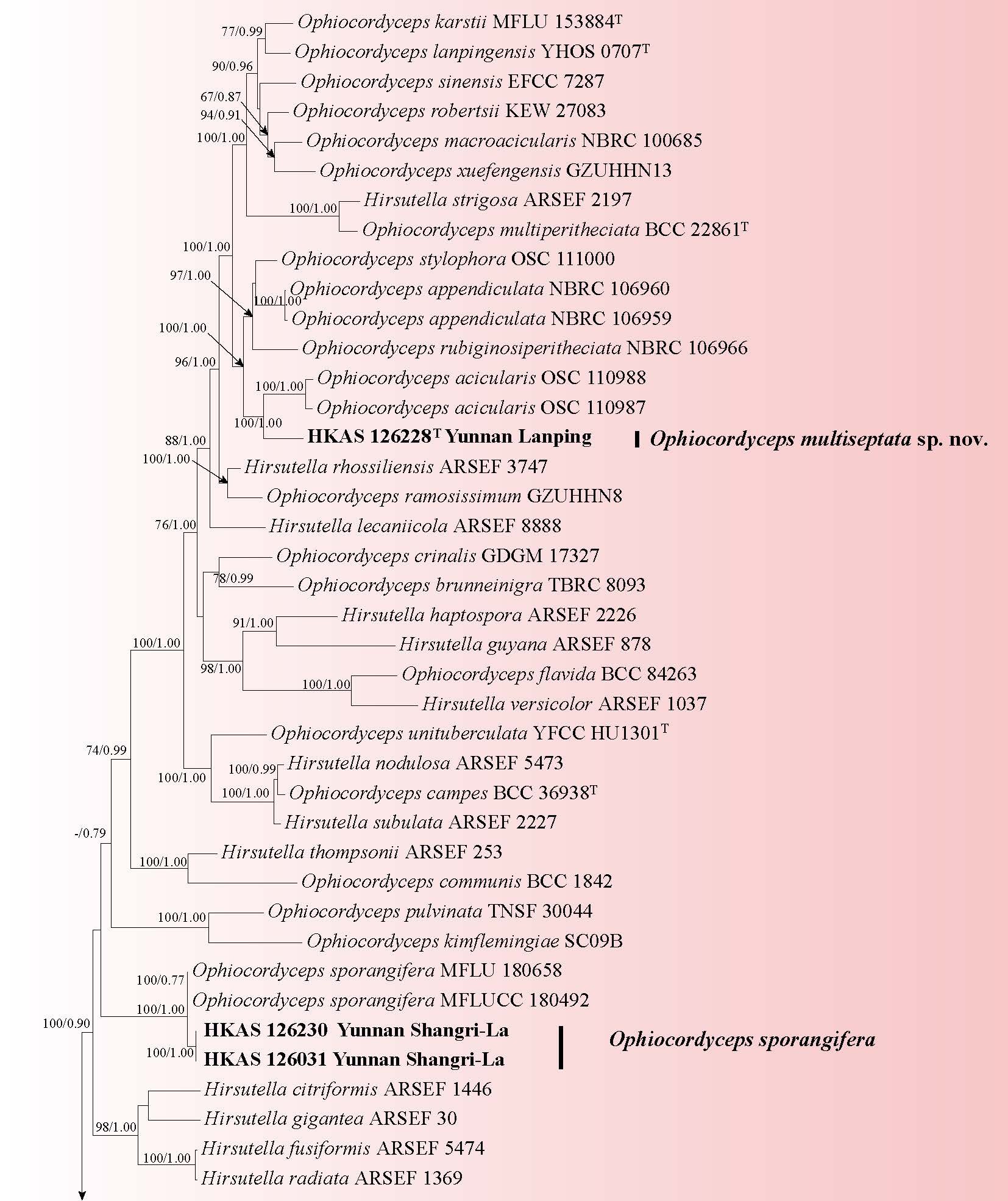
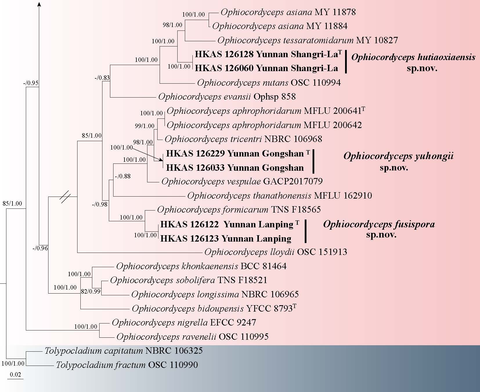
48. Ophiocordyceps multiseptata Y.B. Wang, H.F. Liao & Zhu L. Yang, sp. nov. Fig. 80
Chinese name
Index Fungorum number: IF905277; Facesoffungi number: FoF 19325
Etymology: The species is named after the ascospore with multiple septa.
Diagnosis: Different from related Ophiocordyceps species by its slender stromata and ascospores with multiple septa.
Holotype: HKAS 126228
Parasitic on larvae of Buprestidae buried in soil. Sexual morph: Stromata arising bilaterally from the head of larva of Buprestidae, 63 mm in length and 0.7 mm in width, woody and rigid, solitary, with the lower half black and the upper half brown. Fertile heads 12 mm in length and 0.6 mm in width. Perithecia 490–560 × 380–440 µm ( = 525 × 412 μm, n = 20), are vertical, initially yellowish-brown and mature to dark brown. Asci 210–280 × 12–16 µm ( = 253 × 15 μm, n = 20), hyaline, cylindrical, 8-spores, with an incomplete apical cap. Ascospores 168–220 × 3.1–3.7 µm ( = 188 × 3.2 μm, n = 20), hyaline, multiseptate, non-disarticulating, septate cells 4.2–7.6 µm ( = 5.1 μm, n = 20). Asexual morph: Undetermined.
Culture characteristics: Undetermined.
Material examined: CHINA, Yunnan, Nujiang Prefecture, Lanping County, Jinding Town, on a larva of Buprestidae buried in soil, 9 August 2022, H.F. Liao (holotype, HKAS 126228).
Notes: Phylogenetic analysis based on five-gene data reveals Ophiocordyceps multiseptata is closely related to O. acicularis (Fig. 77), as both species exhibit identical morphological characteristics, and are parasitic on larvae of Buprestidae (Coleoptera) (Petch 1933). Ophiocordyceps acicularis was introduced by Petch in 1933 and was later redescribed in the "Field Guide to the Natural World of Cordyceps" by the Japanese Cordyceps Society (Petch 1933, Japanese Cordyceps Society 2018). The difference between O. multiseptata and O. acicularis is that the stromata of O. multiseptata (63 mm long, 0.7 mm wide) are slenderer than those of O. acicularis (3–10 cm long, 1–1.5 mm wide). Additionally, the perithecia of O. multiseptata are immersed and predominantly clustered at the upper portion of the stroma, exhibiting a clear demarcation from the stipe, and are significantly wider (490–560 × 380–440 µm) (Japanese Cordyceps Society 2018).
49. Ophiocordyceps polyphialidica Y.P. Xiao, K.D. Hyde & Y. Yang, sp. nov. Fig. 81
Chinese name: – 多瓶梗线虫草 (duo ping geng xian chong cao)
Index Fungorum number: IF 903157; Facesoffungi number: FoF 17062
Etymology: in reference to polyphialidic of conidiogenous cells.
Holotype: HKAS 144591
Parasitism on Larvae of Lepidoptera, buried in the leaf litter. Sexual morph: Undetermined. Asexual morph: Hirsutella-like. Synnemata 1.5–3.5 cm long, 0.3–0.8 mm wide, white to pale gray, solitary or multiple, on the insect body, without fertile head. Phialide solitary along synnemata, monophialidic or polyphialidic, 8.3–14.5 × 1.3–4.5 µm ( = 11.4 × 2.9 µm, n = 50), mostly with pyriform inflated basal portion 4.5–8.8 × 2.3–4.5 µm ( = 6.6 × 3.4 µm, n = 50), tapering gradually or abruptly into a thin neck 3.5–6.8 µm long. Conidia 4.4–6.2 × 1.4–2.3 µm ( = 5.3 × 1.8 µm, n = 60), fusiform, 1-celled, hyaline, smooth–walled.
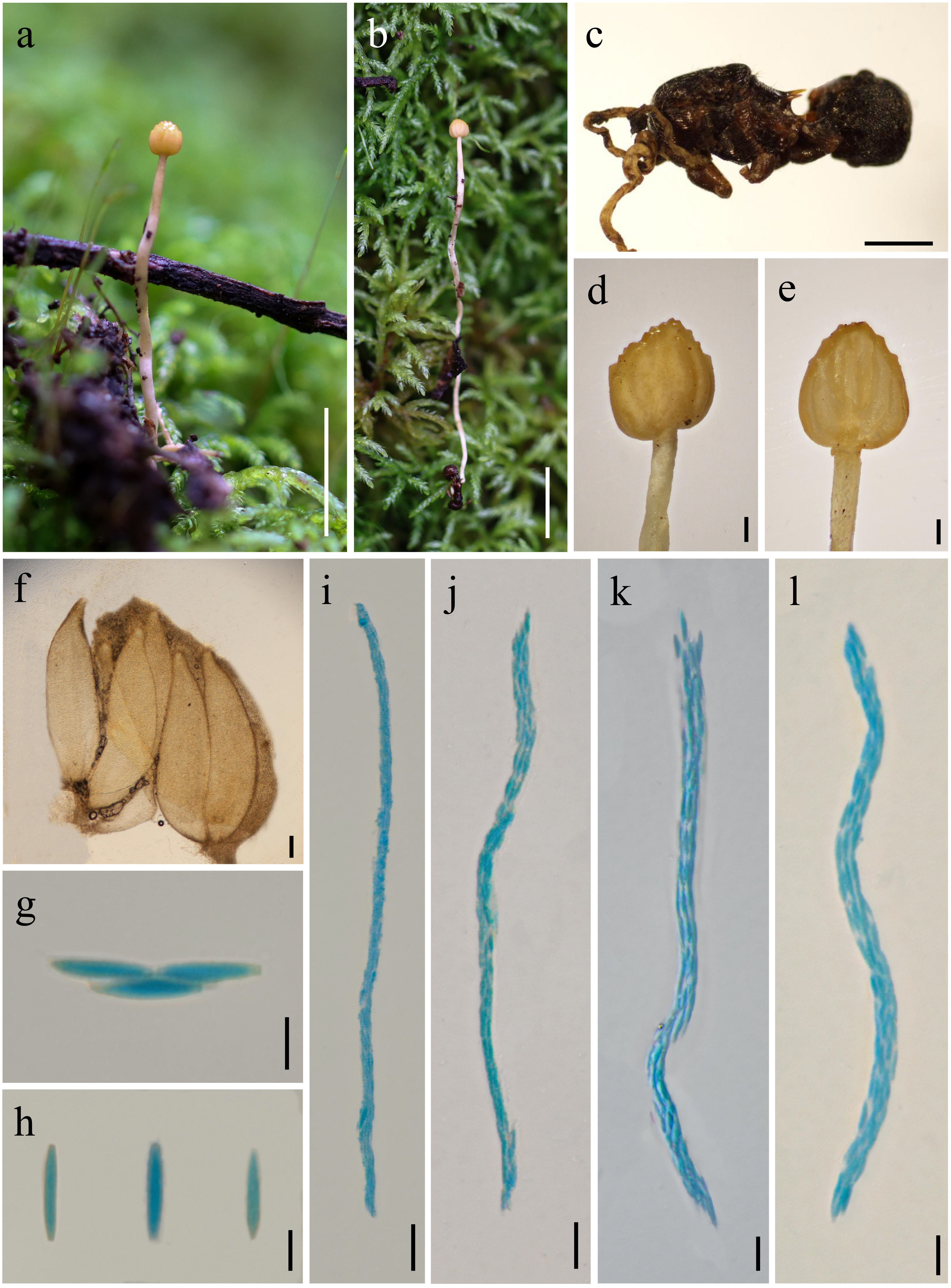
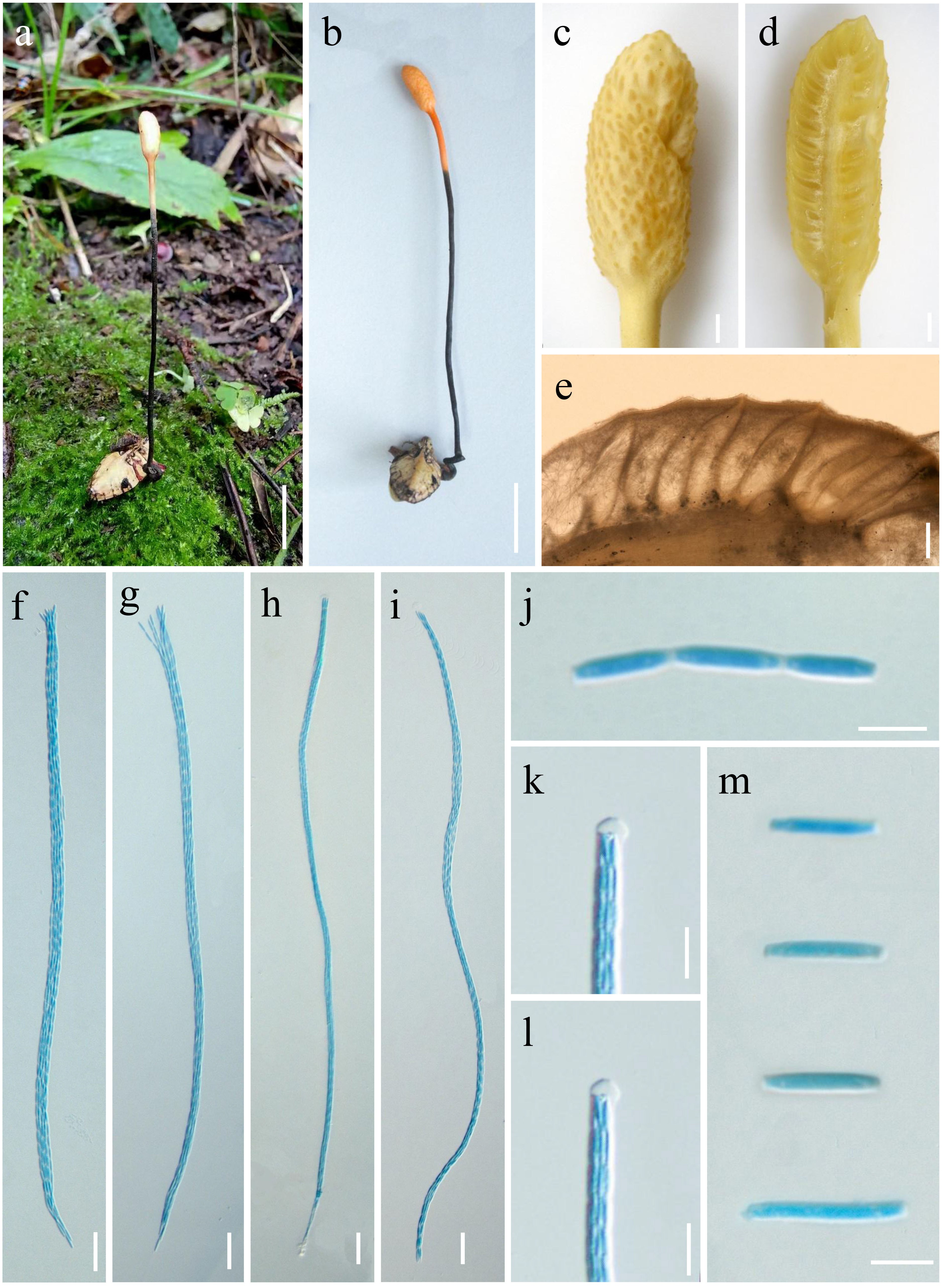
Material examined: CHINA, Xizang Autonomous Region, Linzhi, Bomi County, parasitism on Larvae of Lepidoptera, 31 August 2024, Xingjuan Xiao 242605 (HKAS 144591, holotype), 242607 (HKAS 144589, paratype).
Notes: Ophiocordyceps polyphialidica was phylogenetically retrieved as a sister species of O. unituberculata with strong statistical support (100% ML/1.00 BYPP) (Fig. 82). Base pair differences of ITS, tef1-α, rpb1 and rpb2 genes loci of O. unituberculata are 3.09% (15/485), 6.12% (42/686), 3.05% (20/655) and 3.16% (29/916) respectively. Morphologically, O. unituberculata produces monophialidic, longer phialide (31.9–128.3 × 1.2–5 μm vs. 8.3–14.5 × 1.3–4.5 μm), bigger conidia (6.3–10.6 × 1.9–3.7 vs. 4.4–6.2 × 1.4–2.3 μm), compared to O. polyphialidica (Wang et al. 2018). Both morphological observation and phylogenetic analyses of combined ITS, SSU, tef1-α, rpb1 and rpb2 sequence data support that this fungus is a distinctive species in Ophiocordyceps.
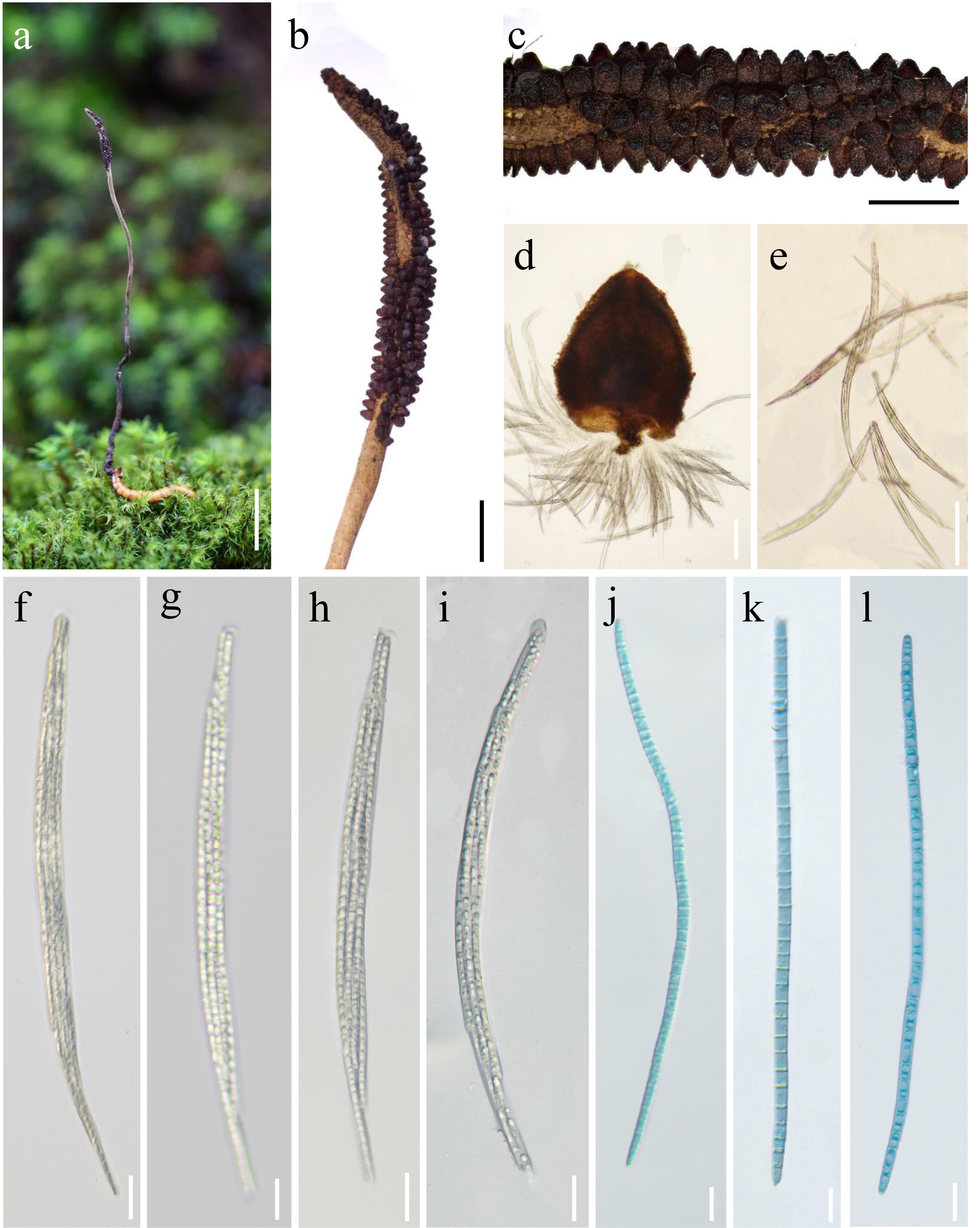
50. Ophiocordyceps sporangifera Y.P. Xiao, T.C. Wen & K.D. Hyde, in Xiao, Hongsanan, Hyde, Brooks, Xie, Long & Wen, MycoKeys 47: 63 (2019) Fig. 83
Chinese name
Index Fungorum number: IF 555324; Facesoffungi number: FoF 04865
Sexual morph: Unknown. Asexual morph: Primary synnemata arise from the head region of bugs (Cydnidae, Hemiptera), paired to multiple, simple, cylindrical, Flexuous, not smooth, brown to deep brown, with a white fertile head at the apex, 80–120 × 0.9–1.2 mm ( = 100 × 1.1 mm, n = 10). Fertile heads cylindrical, 0.5–1.8 × 0.3–0.9 mm ( = 1.1 × 0.7 mm, n = 10), white to brown, with sporangia on their surfaces. No other morphological features were observed.
Culture characteristics. Colony growth on PDA medium is slow; after 5 weeks of incubation at a constant temperature of 25°C, the diameter reaches 2 cm, with a raised white center and felty margins. The surface features circular, dense mycelia. After 7 weeks, the colony color deepens gradually from the periphery towards the center, shifting from white to brown, with circular rings. By the 9th week, vesicular structures are produced. The mycelium is septate and smooth. Phialides 52–96 × 2–6.3 µm ( = 74 × 4.2 µm, n = 20), enlarged base, ampulla, gradually tapering towards an attenuated apex, displaying slight verrucose protrusions. Most are solitary, with a few dichotomous branching. Conidia 6.1–9.4 × 2.3–4.8 μm ( = 7.8 × 3.6 µm, n = 20) 1 cell, hyaline, ovoid, bound in mucilaginous spheres. Mucilaginous spheres 7.6–11.2 × 4.2–7.3 µm ( = 9.5 × 6.1 µm, n = 20), hyaline to brown, 1–3 conidia, lemon-shaped or semispherical.
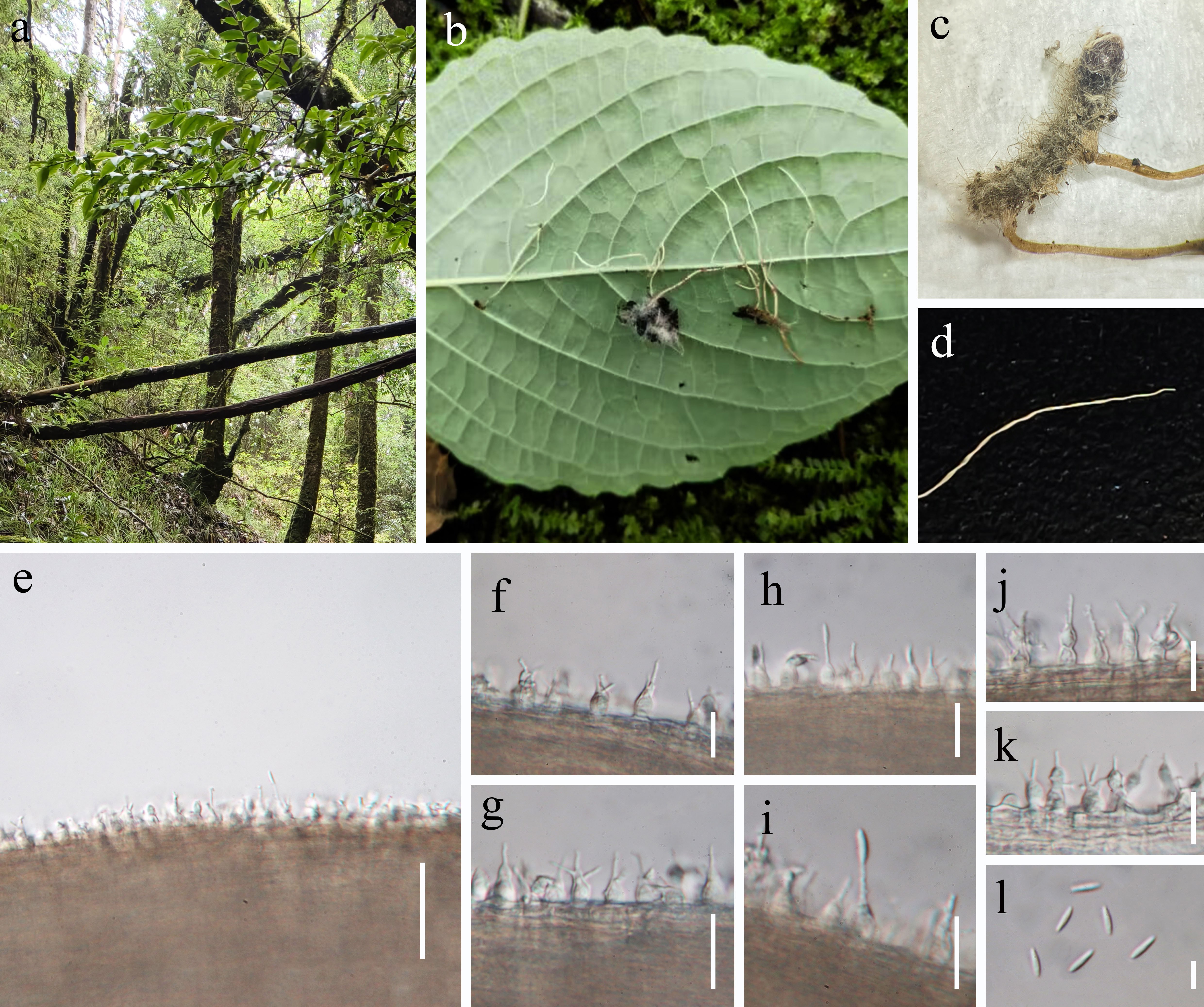
Material examined: CHINA, Yunnan, Shangri-La City, Hutiaoxia Town; collected from the surface layer of leaf litter in a broad-leaved forest, 8 August 2023, Y.B. Wang & H.F. Liao, (HKAS 126230, KUNCC 10936); ibid. (HKAS 126031).
Notes: Ophiocordyceps sporangifera was discovered as a new species by Xiao et al. (2019) in Thailand. Multigene phylogenetic analysis demonstrates that this species, along with O. sporangifera Y.P. Xiao, T.C. Wen & K.D. Hyde (Fig. 77), is closely related within the same clade on the Hirsutella subclade (Xiao et al. 2019). They exhibit a very close phylogenetic relationship. Morphologically, both species are remarkably similar in the size and shape of their primary synnemata and fertile heads. In culture characteristics, the growth characteristics on PDA medium are nearly identical, with phialides and mucilaginous spheres that are similar in size, shape, and color. Therefore, they should be considered the same species. However, they differ in their host species; the hosts for the specimens collected in this study are adults of Cydnidae, Hemiptera, whereas the hosts for O. sporangifera are larvae of Elateridae (Coleoptera). Furthermore, this species was discovered in China for the first time, indicating an expansion in both its host range and geographical distribution (Xiao et al. 2019).
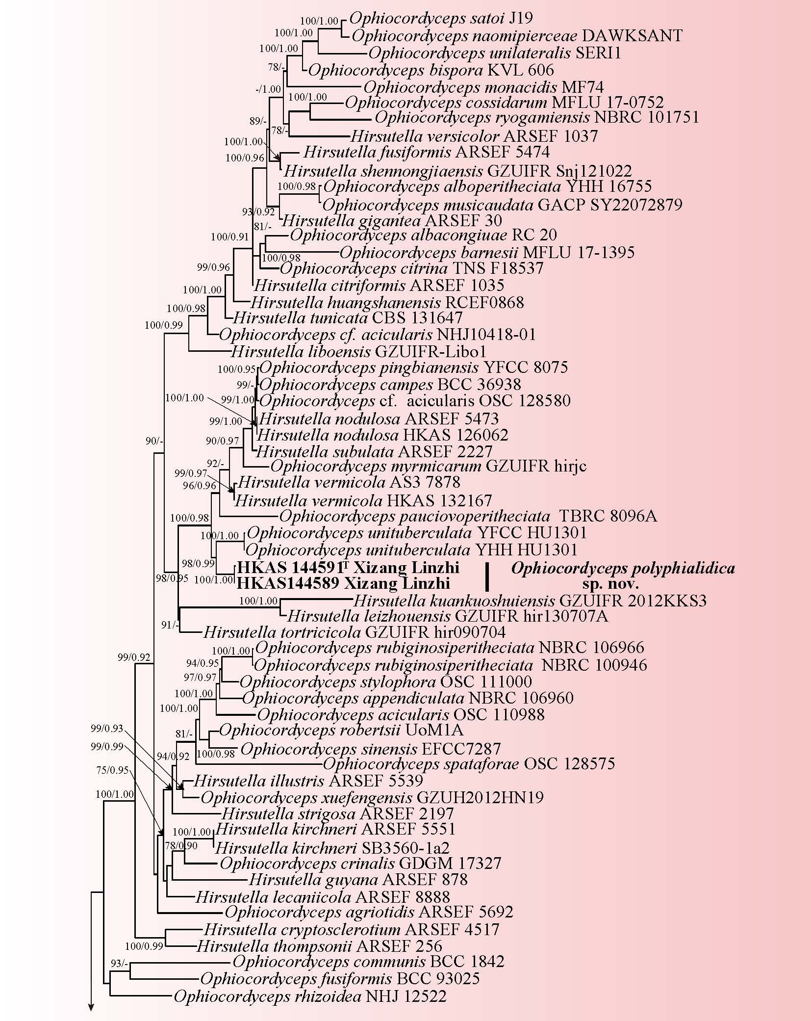
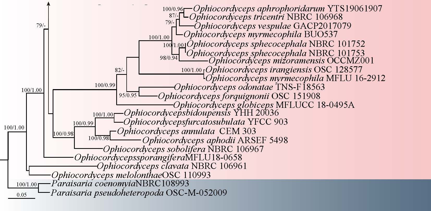
51. Ophiocordyceps yuhongii Y.B. Wang & H.F. Liao, sp. nov. Fig. 84
Chinese name:
Index Fungorum number: IF 905278; Facesoffungi number: FoF 19326
Etymology: The species is named after Professor Yu Hong, acknowledging his contributions to the research on cordycipitoid fungi.
Diagnosis: Different from related Ophiocordyceps species by its shorter stromata and larger perithecia, occuring from the head of insect hosts.
Holotype: HKAS 126229
Parasitic on adult of Aphrophoridae cling to the surface of leaf litter. Sexual morph: Stromata 25–50 × 0.4–0.6 mm ( = 32 × 0.5 mm, n = 5), arising from the heads of adult froghopper insects (Hemiptera), solitary, simple, yellow to orange-yellow. Fertile heads 3.4–4.2 × 1.8–2.2 mm ( = 3.8 × 2.0 mm, n = 5), ovoid, surface spinous due to the protruding ostioles, orange-yellow. Perithecia 765–910 × 210–290 µm ( = 837 × 250 µm, n = 20), immersed, inclined aggregated, ostioles opening on the upper surface of the head, flask-shaped to elongated obpyriform. Asci 450–698 × 6.1–7.7 µm ( = 574 × 6.9 µm, n = 20), hyaline, filiform, thickened apices, with the apical cap disappearing upon maturation. Apical caps 6.1–6.8 × 5.2–5.5 µm ( = 6.5 × 5.3 μm, n = 20), hyaline, conspicuous, hemispherical. Ascospores 280–610 × 0.9–1.8 µm ( = 498 × 1.4 µm, n = 20), filiform, multiseptate, which upon maturation, breaking into secondary ascospores. Secondary ascospores 11.1–13.1 × 1.9–2.4 µm, ( = 12.5 × 2.1 µm, n = 20) hyaline, lanceolate. Asexual morph: Undetermined.
Culture characteristics: Undetermined
Material examined: CHINA, Yunnan Province, Nujiang, Gongshan County, Dulongjiang Township, on adult of Aphrophoridae cling to the surface of leaf litter, 24 July 2023, H.F. Liao (HKAS 126229, holotype); ibid. (HKAS 126033).
Notes: Phylogenetic analysis based on the five-gene data indicates that O. yuhongii is closely related to O. aphrophoridarum and O. tricentri (Fig. 77), sharing similar morphological characteristics (Shrestha & Sung 2005; Yang et al. 2021). The hosts for all three species are the adult froghopper insects of Hemiptera. Ophiocordyceps tricentri was initially described in Japan as Cor. tricentri Yasuda (1922) and was subsequently redescribed by Shrestha and Sung (Shrestha & Sung 2005). Ophiocordyceps yuhongii differs from O. aphrophoridarum and O. tricentri by its thinner and shorter stromata (25–50 × 0.4–0.6 mm), as well as larger perithecia (765–910 × 210–290 µm), asci (450–698 × 6.1–7.7 µm), ascospores (280–610 × 0.9–1.8 µm), and secondary ascospores (11.1–13.1 × 1.9–2.4 µm). Additionally, while O. aphrophoridarum emerges from the thorax of its insect host, O. yuhongii occurs from the head of its insect host. This distinction highlights notable differences in morphological characteristics as well as host interactions across the genus (Yang 2021; Shrestha & Sung 2005). Therefore, both morphological and molecular phylogenetic analyses provide strong evidence to support O. yuhongii as a new species within the genus Ophiocordyceps.
Polycephalomycetaceae Y.P. Xiao, Y.B. Wang, T.C. Wen, H. Yu & K.D. Hyde
Polycephalomycetaceae was separated from Ophiocordycipitaceae by Xiao, based on morphological and phylogenetic analyses, to accommodate three genera: Polycephalomyces, Perennicordyceps, and Pleurocordyceps, comprising 30 species (Xiao et al. 2023, 2024; Liu et al. 2024c). Unlike Ophiocordycipitaceae, Polycephalomycetaceae parasitizes insects or fungi and has a thick peridium (Xiao et al. 2023). Species within this family have a cosmopolitan distribution, with most species found in tropical and subtropical regions (Xiao et al. 2023). The insect orders most commonly associated with this family include Coleoptera, Formicidae, Hemiptera, Hymenoptera, Lepidoptera, Orthoptera, Pentatomidae, and Psocoptera (Kobayasi 1939; Kobayasi & Shimizu 1982; Ban et al. 2009; Xiao et al. 2018).
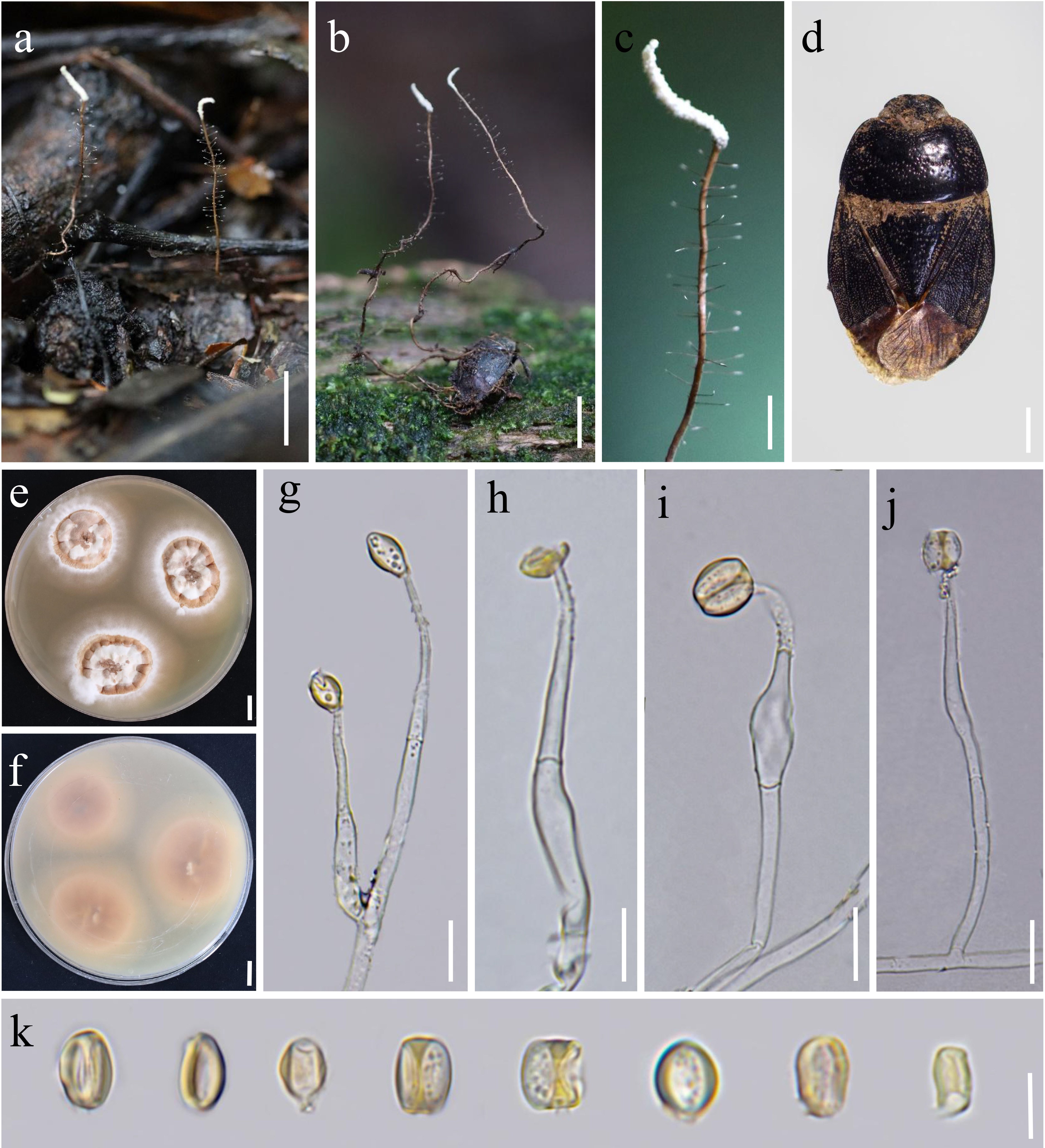
52. Perennicordyceps woodihabitata Y.B. Wang, B.Z. Chen, X.M. Wang & Zhu L. Yang, sp. nov.
Chinese name
Index Fungorum number: IF 905279; Facesoffungi number: FoF 19327
Etymology: Collected from rotten wood.
Diagnosis: Perennicordyceps woodihabitata differs from related species by having smaller perithecia, larger ascal caps and secondary ascospores, and larger ellipsoidal to subellipsoidal conidia.
Holotype: HKAS 135026
Parasitic on coleopteran larva inhabiting decayed wood. Sexual morph: Stromata arising from the dorsal surface or both ends of coleopteran larvae, leathery, clavate. Fertile parts located at the apical region of the stroma, solitary, orange, 0.2–0.5 mm long, with irregular protuberances, pale yellow to orange. White ostioles of perithecia present on the protruding areas. Perithecia superficial, ovoid, 180.54–241.45 μm long, 69.16–113.13 μm wide at the base, 75.88–169.59 μm at the middle, and 26.85–33.56 μm at the neck. Asci narrowly clavate, 35.68–47.12 × 8.5–16 μm. Apical caps hemispherical, 33–4.89 × 0.48–2.66 μm. Secondary ascospores hyaline, short-clavate, 140–310 × 1.7–2.41 μm. Asexual morph: Hyphae branched, hyaline, smooth-walled, 2.3–4.8 μm wide. Conidiophores cylindrical, 7.88–17.71 μm long, 1.43–23 μm wide at the base, 1.8–2.25 μm at the middle, and 1.79–2.86 μm at the neck. Phialides solitary or in whorls of 2–4 on hyphae, mostly lanceolate, occasionally narrowly flask-shaped, 18.26–21.5 μm long, 1.47–2.19 μm wide at the base, 1.81–2.41 μm at the middle, and 1.1–1.23 μm at the neck. Conidia hyaline, smooth-walled, aseptate, ellipsoidal to subellipsoidal, 2.44–3.88 × 0.72–1.69 μm.
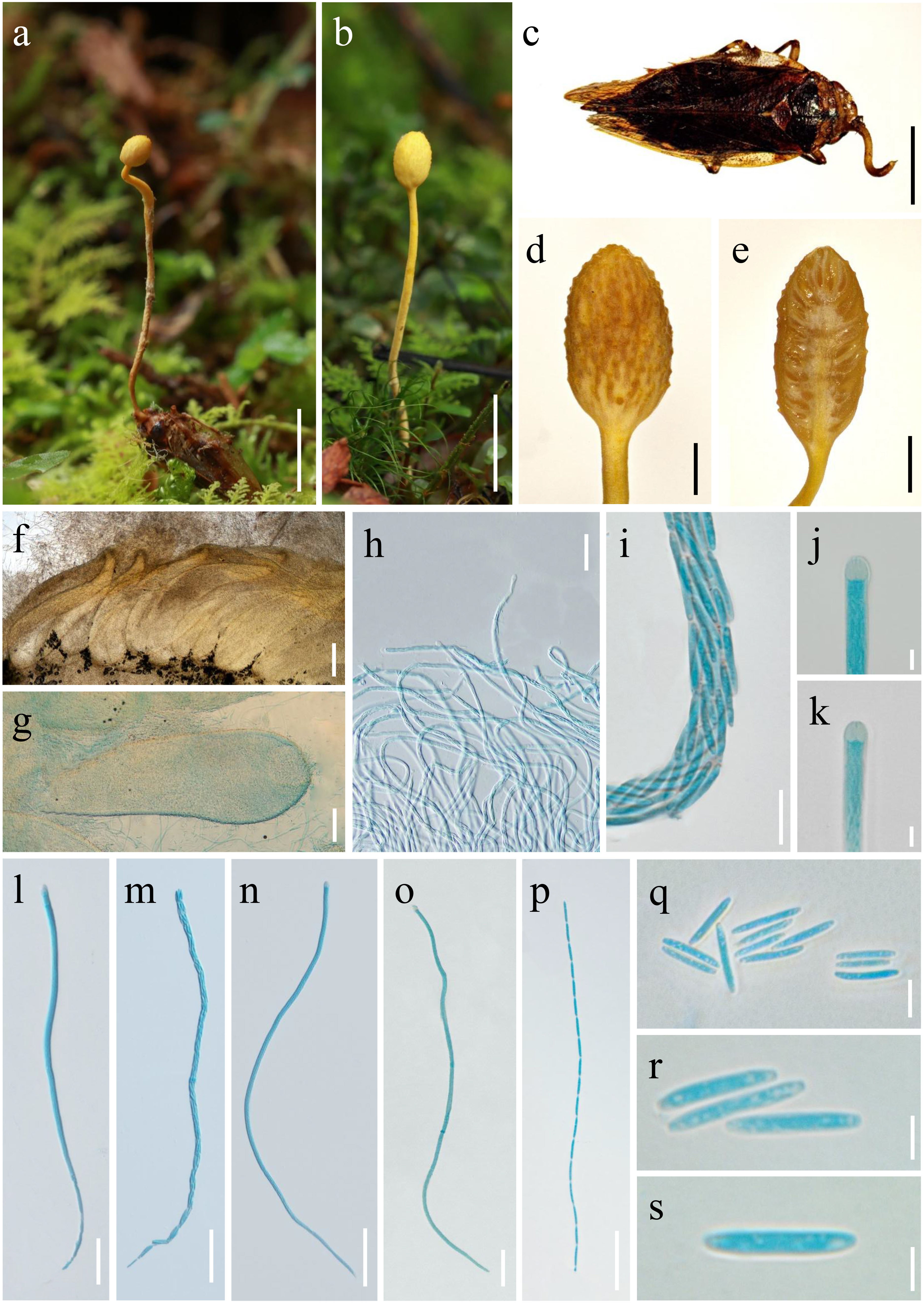
Culture characteristics: Colonies on PDA grew slowly, reaching only 2.8 cm in diameter after 21 days at 25 °C. Reverse surface yellow-brown, surface floccose, spreading from the center to the margin, changing from pale yellow to white. No synnemata observed. After 40 days, dark yellowish-brown aggregated structures formed on the colony surface.
Material examined: CHINA, Yunnan Province, Tengchong City, on larva of Coleoptera inhabiting decayed wood, 26 May 2024, X.M. Wang (HKAS 135026, holotype, ex-type culture, KUNCC 11459), ibid., X.M. Wang (culture KUNCC 11458).
Notes: Perennicordyceps woodihabitata is phylogenetically closely related to P. paracuboidea (Matočec et al. 2014), as strongly supported by multi-gene phylogenetic inference (Fig. 85). However, it shows clear sequence divergence from P. paracuboidea (CBS 120.87), with 96.1% identity in ITS (26 bp differences), 99.9% in LSU (21 bp differences), 97.8% in rpb2 (21 bp differences), 98.7% in rpb1 (8 bp differences), and 98% in tef1-α (20 bp differences). Morphologically, P. woodihabitata differs from P. paracuboidea by having significantly smaller perithecia, but larger ascal caps and ascospores. Its secondary ascospores exhibit a more pronounced length-to-width ratio, and its conidia are larger. In the asexual morph, the phialides are borne in whorls of 2–4 in P. woodihabitata, and the conidia are slightly smaller than those of the other two related species.
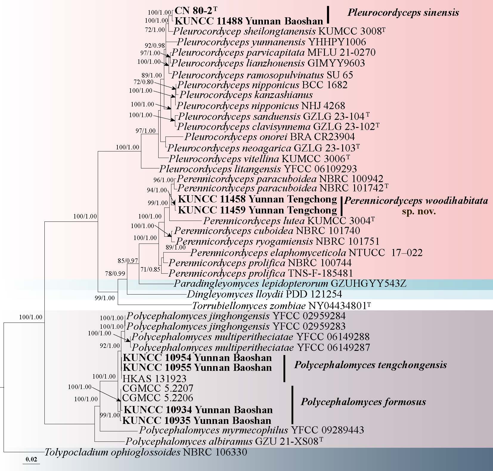
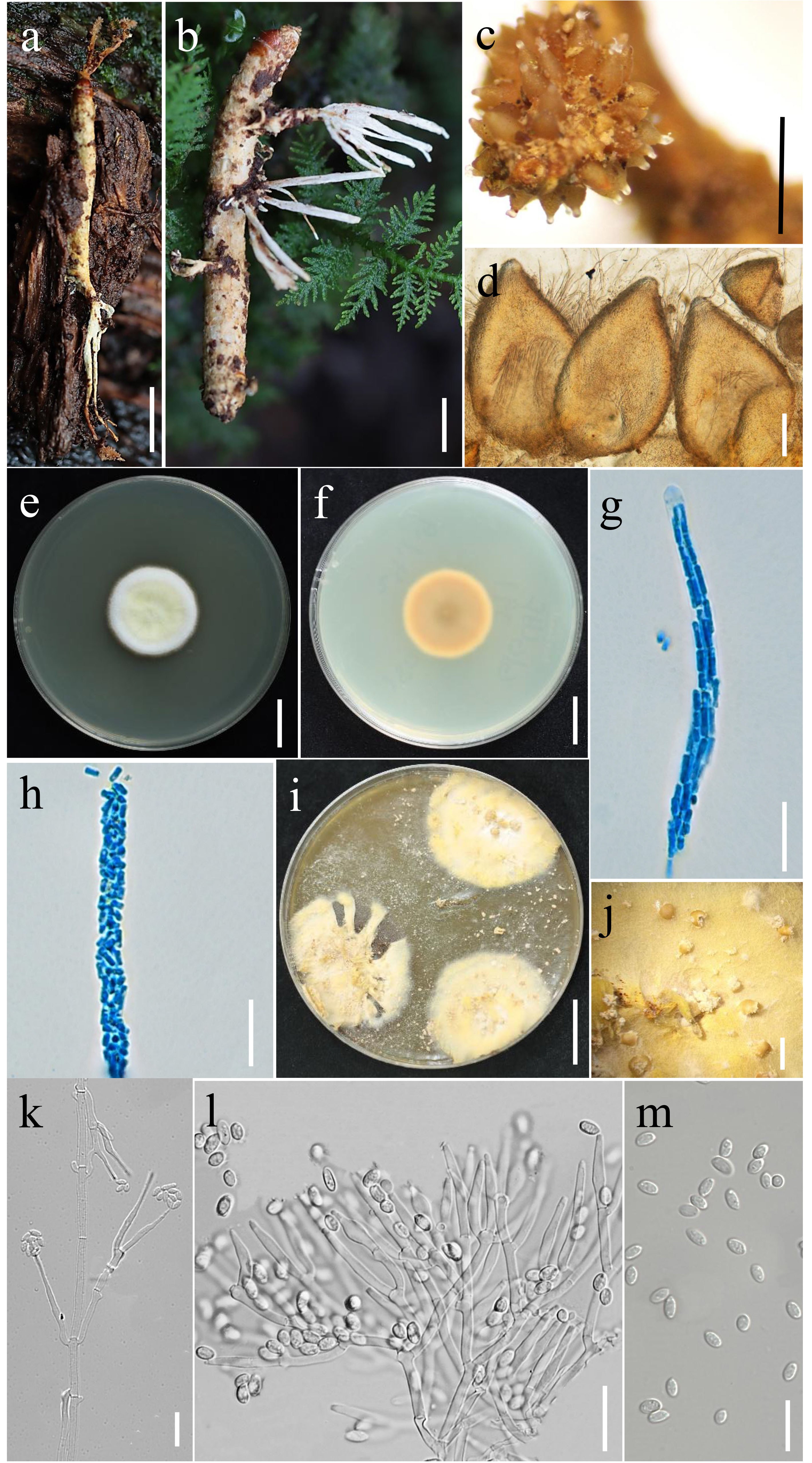
Pleurocordyceps Y.J. Yao, Y.H. Wang, S. Ban, W.J. Wang, Yi Li, Ke Wang & P.M. Kirk
Pleurocordyceps, established by Wang et al. (2021) with the type species Ple. sinensis, is characterized by its lateral fertile pulvinate stromata near the tip of the sexual morph and two distinct types of phialides and conidia in the asexual morph (Wang et al. 2021; Xiao et al. 2023). This genus, associated with various insect hosts and fungi, holds promise for producing secondary metabolites with potential medicinal applications, such as antibacterial and anti-inflammatory compounds (Sangdee et al. 2017; Xiao et al. 2024).
53. Pleurocordyceps sinensis (Q.T. Chen, S.R. Xiao & Z.Y. Shi) Y.J. Yao, Y.Hui Wang, S. Ban, W.J. Wang, Yi Li, Ke Wang & P.M. Kirk, in Wang, Ban, Wang, Li, Wang, Kirk, Bushley, Dong, Hawksworth & Yao, Journal of Systematics and Evolution 59(5): 1075 (2021) Fig. 87
Chinese name
Index Fungorum number: IF 550007; Facesoffungi number: FoF 10739
Parasitic on adult of Aphrophoridae sp. buried in soil. Sexual morph: Undetermined. Asexual morph: Hyphae were smooth-walled, hyaline, septate, and measured 1.19–4.89 μm in width. Conidiophores measured 5.66–10.99 μm in length, with a basal width of 1.11–1.67 μm, a middle width of 1.35–2.62 μm, and a neck width of 1.17–1.79 μm. Two types of phialides and conidia were observed. β-type phialides ranged from 3.57 to 11.19 μm in length, with basal, middle, and neck widths of 1.55–2.31 μm, 1.96–42 μm, and 0.93–1.69 μm, respectively. α-type phialides were observed by mounting the pale-yellow mucilaginous mass from the apex of synnemata collected from wild material. These phialides were smooth-walled, hyaline, and either short-flask-shaped or lanceolate, usually arising from hyphae or conidiophores in a terminal or lateral position. Their length ranged from 5.38 to 15.76 μm, with basal, middle, and neck widths of 1.12–1.55 μm, 0.93–1.92 μm, and 0.74–1.36 μm, respectively. Conidia were all unicellular and hyaline. β-type conidia were fusiform and often formed in chains, measuring 2.18–4.39 μm in length and 1.28–2.09 μm in width. α-type conidia were nearly ellipsoidal, also hyaline, and measured 2.18–3.39 μm in length and 1.7–3.22 μm in width.
Culture characteristics: On PDA medium, Colonies exhibited a relatively regular growth pattern. After 28 days of incubation, it reached a diameter of 4.5 cm. The colony formed a distinctly circular outline in the Petri dish, expanding radially and uniformly from the initial inoculation point, with a neatly defined margin. The colony surface was fluffy, displaying a typical cottony texture. The reverse side of the colony showed a gradient of coloration, with the center appearing light brown—indicating pigment deposition or dense hyphal aggregation—and gradually transitioning to a pale yellow toward the edge, where the color remained even and soft.
Material examined: CHINA, Yunnan Province, Baoshan City, on adult of Aphrophoridae sp. buried in soil, 10 September 2023, X. M. Wang (HKAS 135024, culture KUNCC 11488).
Notes: In the present study, six-gene phylogenetic analyses clearly demonstrated that the newly collected specimens belong to the same species as Ple. sinensis (Wang et al. 2020b) (Fig. 85). Synnemata were observed emerging from various parts of the host’s body, and their microscopic characteristics were highly consistent with those previously described for Ple. sinensis. This specimen represents a new host record for Ple. sinensis, with the host identified as Aphrophoridae sp.
54. Pleurocordyceps yunnanensis (Hong Yu, Y.B. Wang & Y.D. Dai) Y.H. Wang, S. Ban, W.J. Wang, Yi Li, Ke Wang, P.M. Kirk & Y.J. Yao 2021 Fig. 88
Chinese name
Index Fungorum number: IF 570681; Facesoffungi number: FoF 17063
Parasitism on fertile head of Ophiocordyceps aphrophoridarum, buried in the leaf litter. Sexual morph: undetermined. Asexual morph: Hyphomycetous. Synnemata 0.3–0.5 cm long, 0.2–0.6 mm wide, pale yellow to yellow, solitary or multiple, with fertile head. Phialides of two types: α-phialides arising from the fertile head of the synnema; β-phialides developing from the stipe of the synnema. α-phialides 8.6–22.5 × 0.9–2.1 μm ( = 15.5 × 1.5 µm, n = 50), β-phialides 9.5–13.5 × 1.3–2.3 μm ( = 11.5 × 1.8 µm, n = 40), smooth, hyaline, solitary. α-conidia 2.2–3.5× 0.9–1.5 μm ( = 2.8 × 1.2 µm, n = 50) diam, smooth-walled, globose to ellipsoid, one-celled; β-conidia 4.1–5.5 × 1.1–2.3 μm ( = 4.8 × 1.7 µm, n = 50), hyaline, fusiform or droplets shape, one-celled, smooth-walled.
Material examined: CHINA, Guizhou Province, Hezhang County, parasitism on fertile head of Ophiocordyceps aphrophoridarum, 31 July 2022, Yuanpin Xiao HZ158 (HKAS 132195); HZ158J (GZCC 24-0148, culture).
Notes: The new collections clustered with Ple. yunnanensis with strong support (100% ML/1.00 BYPP, Fig. 85). The morphology of the new collections is similar to that of Ple. yunnanensis, showing no significant differences (Wang et al. 2015). Previously, Ple. yunnanensis was reported to parasitize Ophiocordyceps nutans and stink bugs as its hosts, with the holotype obtained from forest litter in Guizhou Province, China (Wang et al. 2015). However, in this study, Oph. aphrophoridarum were identified as the new hosts of Ple. yunnanensis.
Polycephalomyces Kobayasi
Polycephalomyces was introduced by Kobayasi to accommodate a monotypic anamorph genus for the species Pol. formosus Kobayasi and the diagnostic characteristic of highly branched synnemata (Kobayasi 1941). The asexual morph is hyphomycetous, with stipitate synnemata on the host surface or in culture, light-colored divergent conidiophores, phialidic hyaline conidiogenous cells, and monomorphic, hyaline, smooth-walled, one-celled conidia (Xiao et al. 2023). Now, this genus has been reported over 13 species from tropic and subtopic regions (Xiao et al. 2023; Liu et al. 2024c).
55. Polycephalomyces formosus Kobayasi, Sci. Rep. Tokyo Bunrika Daig., Sect. B 5: 245 (1941) Fig. 89
Chinese name:
Index Fungorum number: IF 289806; Facesoffungi number: FoF 10731
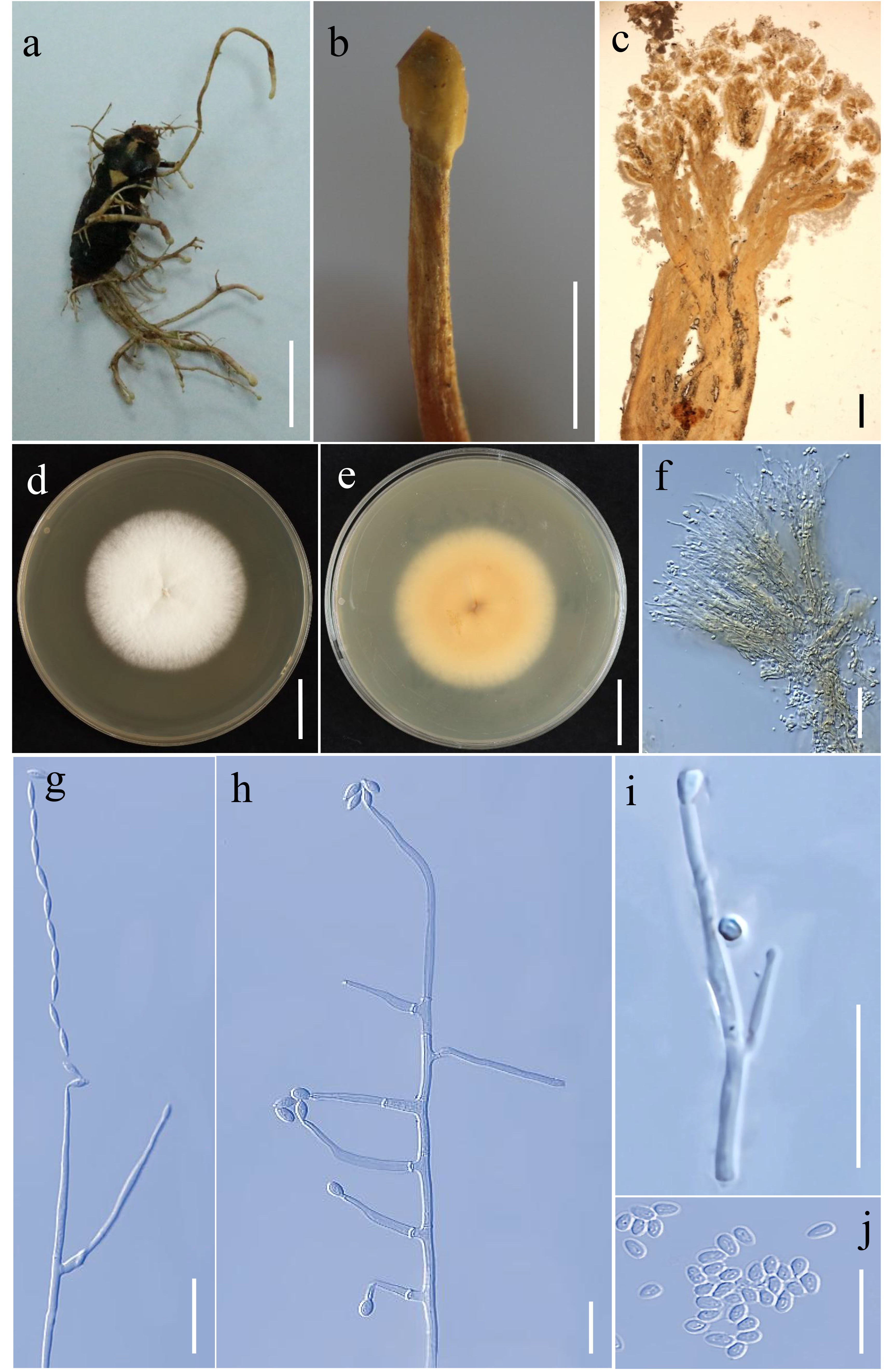
Parasitic on adult of Cicada buried in soil. Sexual morph: Undetermined. Asexual morph: Hyphomycetous. Synnemata 1–25 mm long, 0.1–1 mm wide, scattered or gathered on the Cicada, stipitate, solitary or forming 2–4 branches, white to pale yellow, cylindrical or rhizoid with or without an enlarged globose fertile head at the apex. Fertile heads 0.1–0.3 mm wide, globose to subglobose, white to pale yellow, coved with masses conidia. Stipes 0.5–5 mm long, 0.1–0.3 mm wide, cylindrical or rhizoid, white. Phialides 6–19× 0.9–2.3 µm ( = 16 × 1.8 µm, n = 30), tapering gradually from middle to the apex, narrowly cylindrical to subulate, 1.3–2 µm wide at the base and 1–1.6 µm wide at the apex, one-type, hyaline, smooth-walled. Conidia 1.7–3 × 1.2–2.1 µm ( = 2.5 × 1.6 µm, n = 30), one-celled, one-type, hyaline. The conidia on wild material were oval-shaped obovoid, and the conidia on cultured mycelium were oblong ellipsoid. Colonies derived from single spore isolation.
Culture characteristics: Colonies on PDA reaching 4 cm diam in 28 days at 25 °C, white mixed with some pale yellow, radial with dense mycelium, reverse yellow. Synnemata 1.5–4 cm long, 0.2–2 mm wide, stipitate, gathered, white, cylindrical or flaky, growing and spreading from one branch to multiple branches, conidia masses on the surface, without fertile head.
Material examined: CHINA, Yunnan Province, Baoshan City, on adult of Cicada buried in soil, 10 September 2023, X.M. Wang (HKAS 135019, culture KUNCC 10935), other collection: Ibid., X.M. Wang (HKAS 135018, culture KUNCC 10934).
Notes: The new collections clustered with other Pol. formosus sequences in the same clade with strong statistical support (Fig. 85). The morphology of the new collections is similar to Pol. formosus (Xiao et al. 2023). These specimens represent a new host record for Pol. formosus, with the host identified as adult of Cicada (Cicadidae, Hemiptera).
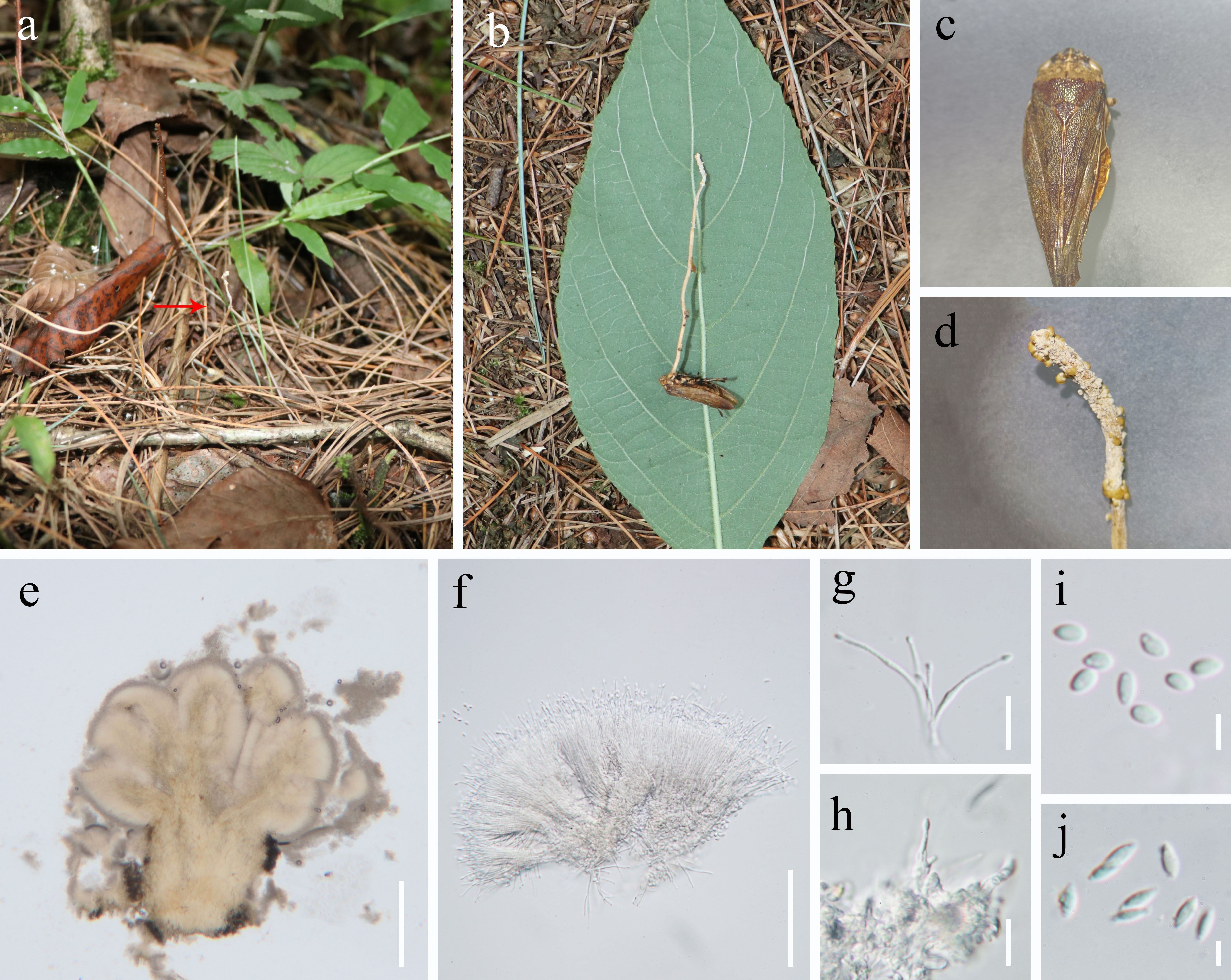
56. Polycephalomyces tengchongensis Y. Wang & T.C. Wen, in Wang, Wei, Peng, Kang, Li, Li, Zhang, Wang, Zhou, He, Chomnunti & Wen, MycoKeys 110: 195 (2024) Fig. 90
Chinese name:
Index Fungorum number: IF 901449; Facesoffungi number: FoF 19328
Parasitic on larva of Coleoptera buried in soil. Sexual morph: Undetermined. Asexual morph: Hyphae hyaline, smooth-walled, septate, 1.72–3.3 μm wide. Conidiophores cylindrical, 3–7 μm long, 1–2 μm wide at the base, and 1.69–2.46 μm at the neck. Phialides borne in whorls of 2–4 on the necks of conidiophores or terminal hyphae, flask-shaped, hyaline, smooth-walled, 8.95–14.38 μm long, 1.48–2.15 μm wide at the base, 1.51–2.46 μm at the middle, and 0.52–1.16 μm at the neck. Conidia hyaline, smooth-walled, aseptate, subellipsoidal to fusiform, 2.19–3.51 × 1.01–1.97 μm.
Culture characteristics: Colonies on PDA grew slowly, reaching 3.9 cm in diameter after 21 d at 25 °C. Colony center grey, expanding outward to pale yellow; surface pale yellow to milky white, producing highly branched, coral-like, flattened synnemata.
Material examined: CHINA, Yunnan Province, Baoshan City, on larva of Coleoptera buried in soil, 11 September 2023, X.M. Wang (HKAS 135025, culture KUNCC 10954), other collections: Ibid., X. M. Wang (culture KUNCC 10955).
Notes: The new collections cluster together with previously published sequences of Pol. tengchongensis with strong statistical support. Morphologically, it is also consistent with Pol. tengchongensis (Wang et al. 2024e) (Fig. 85). However, it differs in host species, which in this case is a larva of Coleoptera, whereas the original described specimens of Pol. tengchongensis were found on Lepidopteran hosts (Wang et al. 2024e). These specimens represent a new host record for Pol. tengchongensis, with the host identified as larvae of Coleoptera.
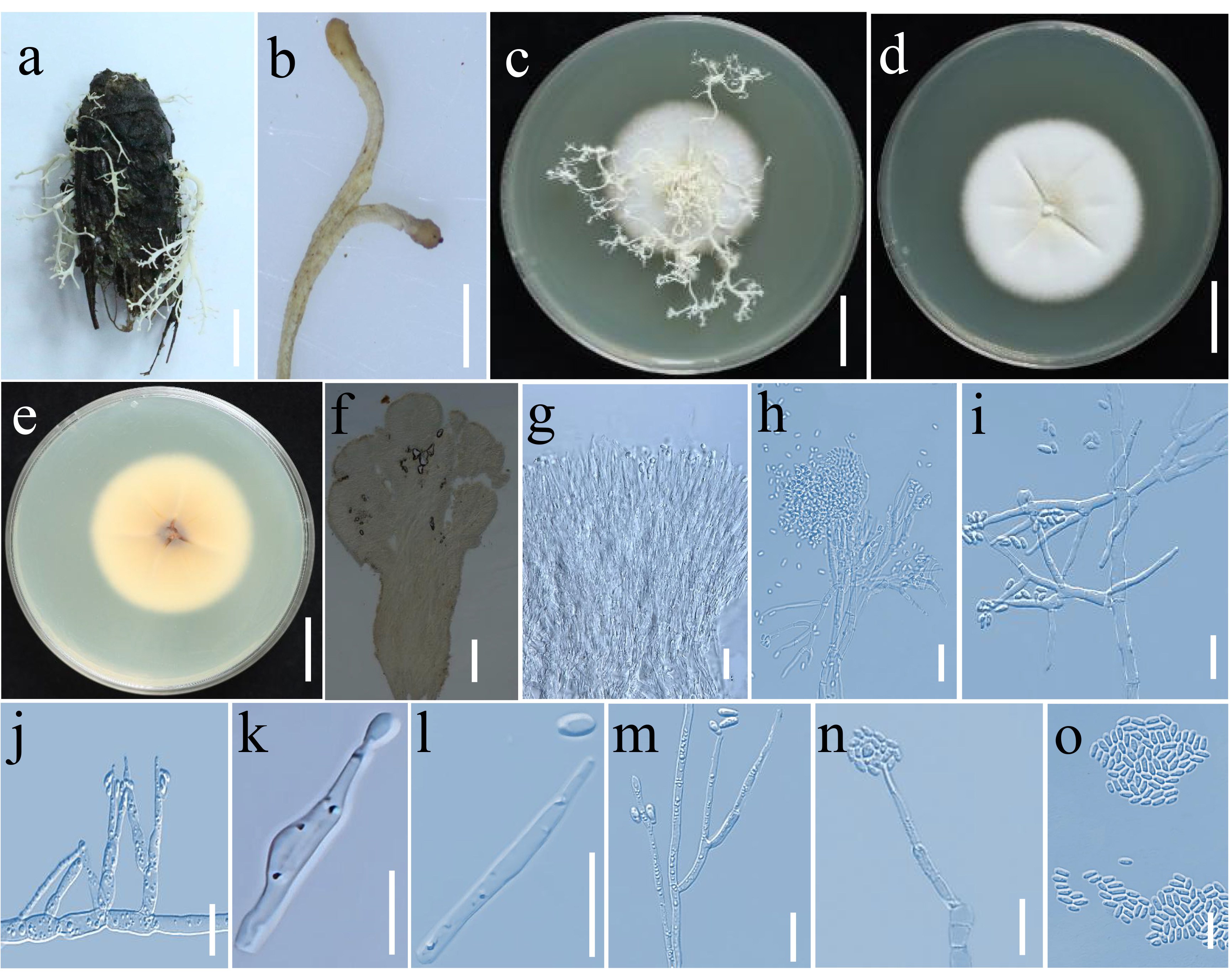
Microascales Luttr. ex Benny & Kimbr
Microascaceae Luttr. ex Malloch
Kernia Nieuwl
Kernia was established by Nieuwland (1916), with K. nitida as the type species. Malloch & Cain (1970) positioned Kernia within the Microascaceae, citing its absence of croziers, the presence of dextrinoid ascospores with germ pores, and the intricate annellophore-type conidial stages as key taxonomic features. Currently, the genus comprises 20 taxa recorded in Index Fungorum (Index Fungorum). Su et al. (2020) distinguished Kernia from Acaulium based on multigene (ITS, LSU, tub, tef1-α) phylogenetic analyses and morphological characteristics. Kernia is typically collected from animal dung, although K. retardata and K. peruviana have been isolated from soil (Su et al. 2020). The sexual morph of Kernia is distinguished by its superficial, dark cleistothecia, which can exhibit triangular, rectangular, or globose shapes, often adorned with appendages-either elongated or abbreviated-at the angles, resulting in a depressed morphology. These appendages display an olive, green to brown coloration, and can be straight or arcuate, occasionally undulating, terminating in one or two circinate or recurved tips. They are septate, with thick walls and a smooth surface. The asci are unitunicate, irregularly arranged within the fruiting body, pyriform or clavate in shape, possessing short stalks, and are characteristically 8-spored. The ascospores are hyaline to light olive-green, ellipsoidal, and exhibit either acute or rounded apices (Malloch & Cain 1970; Malloch 1971; Su et al. 2020). Some species of Kernia produce both sexual and asexual morphs in culture (Su et al. 2020). The genus has been reported in Canada, China (Beijing, Taiwan), France, Germany, South America, and Uganda (Nieuwland 1916; Malloch et al. 1971; Su et al. 2020).
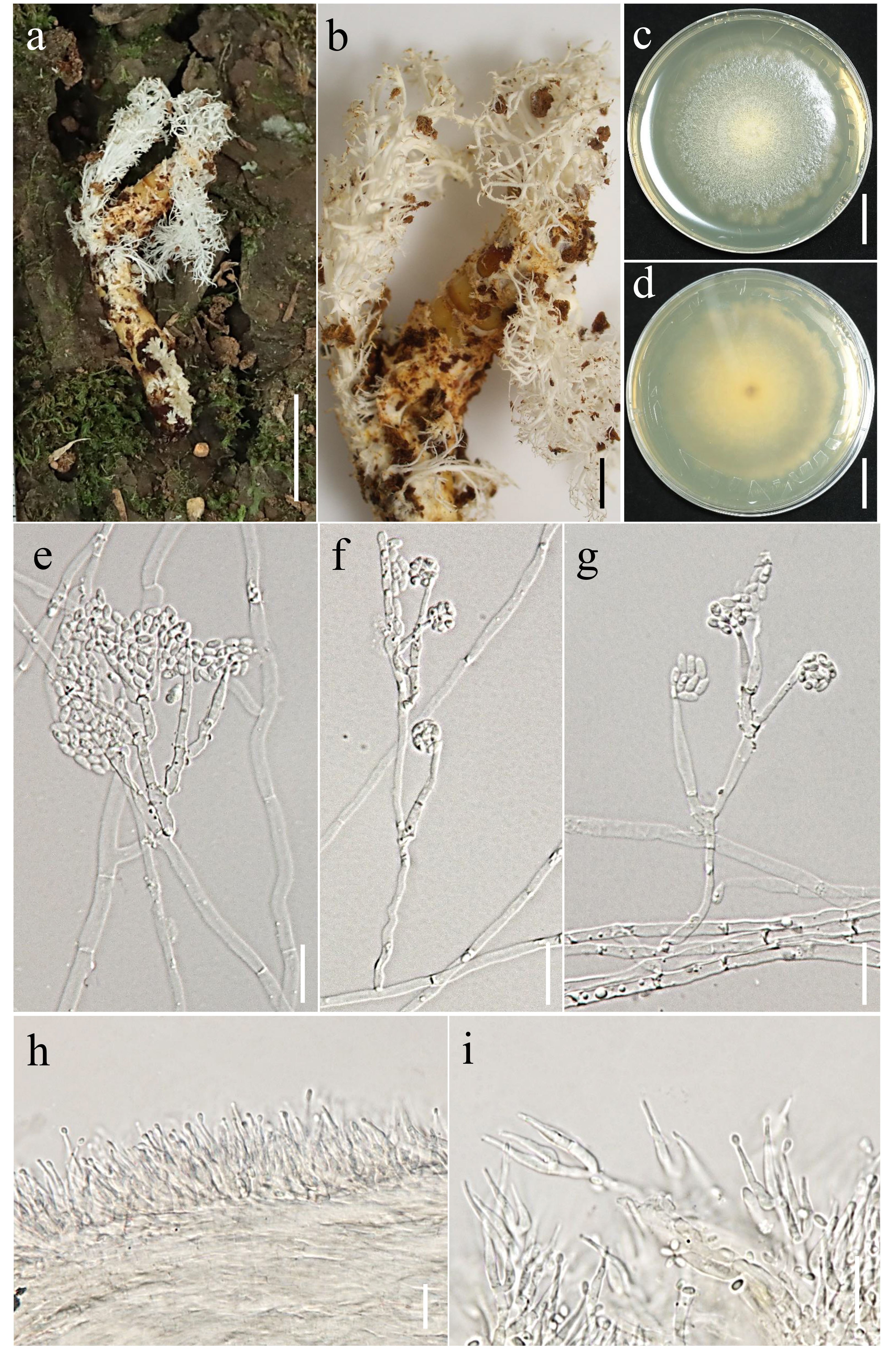
57. Kernia xizangensis R.J. Xu & Q. Zhao, sp. nov. Fig. 92
Chinese name
Index Fungorum number: IF 905280; Facesoffungi number: FoF 19329
Etymology: The species is named after the Xizang, where the type specimen was collected.
Holotype: HKAS 136196
Saprobic on decaying wood submerged in freshwater stream. Sexual morph: Undetermined. Asexual morph: Conidiomata on the natural substrate scattered or caespitose, with glistening conidial masses at apex. Synnemata 244–406 × 11–23 μm ( =322 × 15 μm, n = 20), septate, pale brown to subhyaline, smooth, penicillately branched at the apex. Conidiogenous cells polyblastic, hyaline to pale brown, percurrent, ampulliform, terminal. Conidia 5–7 × 3–5 μm ( = 6 × 4 μm, n = 30), obovoid, ellipsoidal to irregularly fusiform with a truncate base and rounded or bluntly pointed apex, hyaline, smooth, aseptate, collecting in wet masses at the apices of the conidiophores.
Culture characteristics: Conidia germinated on PDA media within 24 hours and germ tube were produced from base. Colonies grown on PDA reached 50 mm diam at room temperature in two weeks, circular, flat, sparse, dark brown mycelium, with radialized margin.
Material examined: CHINA, Xizang Autonomous Region, Linzhi City, on decaying wood submerged in a freshwater stream, 19 July 2021, R.J. Xu, MD-303 (HKAS 136196, holotype), ex-type culture (KUNCC 24-18037).
Notes: Phylogenetic analysis shows that our collection clusters within Kernia and is a sister group to K. anthracina and K. hipporrepida, with 96 ML/ 0.98 BYPP statistical support (Fig. 91). Morphologically, our collection resembles K. anthracina with a scopulariopsis-like branching pattern and the production of acrospores, and obovoid to ellipsoidal conidia (Su et al. 2020). However, our collection differs from K. anthracina by having pale brown to subhyaline synnemata, with penicillately branched at the apex and percurrent, ampulliform conidiogenous cells. In addition, our collection differs from K. hipporrepida in having longer synnemata (244–406 × 11–23 μm vs. 16–90 × 2.5–3.5 μm), and aseptate conidia (Malloch & Cain 1970).
Pleurotheciales Réblová & Seifert
Pleurotheciaceae Réblová & Seifert
Phaeoisaria Höhn. 1909
Phaeoisaria was introduced by Höhnel (1909) with P. clematidis as the type species and was distinguished in Pleurotheciaceae by Réblová et al. (2016b). Phaeoisaria is a widespread genus with 27 species currently reported (Li et al. 2025). Morphologically, Phaeoisaria asexual morph is characterized by macronematous, synnematous, brown to dark brown synnemata, polyblastic, hyaline to brown, sympodial, terminal conidiogenous cells, ellipsoidal to obovoid, hyaline, smooth-walled, aseptate conidia (Hyde et al. 2018; Luo et al. 2018, 2019; Xu et al. 2024a; Li et al. 2025), the sexual morph is characterized by black ascomata, paraphyses, branched, septate, smooth and asci with 8 spores. Ascospores are tapering at both ends, multi-septate, hyaline (Luo et al. 2019). Species of Phaeoisaria are commonly collected from freshwater habitats and decaying wood (Crous et al. 2015b; Luo et al. 2019; Xu et al. 2024a). Following reports of Phaeoisaria species in Xizang by Xu et al. (2024a) and Li et al. (2025). In this study, based on morphological and phylogenetic analyses, we describe a new species Phaeoisaria xizangensis from Xizang.
58. Phaeoisaria linzhiensis S.C. He, Q. Zhao & K.D. Hyde, sp. nov. Fig. 94
Chinese name
Index Fungorum number: IF 905281; Facesoffungi number: FoF 19330
Etymology: Referring to the location “Linzhi City” where the holotype of this fungus was collected.
Holotype: HKAS 145479
Saprobic on decaying stems of Lilium spp. Sexual morph: Undetermined. Asexual morph: Colonies effuse, solitary, brown, hairy. Mycelium immersed, superficial, branched, pale brown hyphae. Synnemata 328–462 × 8–15 μm ( = 390 × 12 μm, n = 10) µm, scattered, erect, brown to dark black, straight or flexuous, pale at the apex, composed of conidiophores, branched at the apex. Conidiophores 270–350 × 2.1–3.8 μm ( = 330 × 2.8 μm, n = 30), macronematous, synnematous, septate, cylindrical, branched, straight or flexuous, pale brown to brown, thick-walled, smooth-walled. Conidiogenous cells polyblastic, integrated, terminal and intercalary, rough, cylindrical, subhyaline to pale brown, with several small denticulate conidiogenous loci. Conidia 7–10 × 3–4 μm ( = 8 × 3.5 μm, n = 30) µm, amerospores solitary, acropleurogenous, simple, subglobose to obovoidal, smooth and guttulate, aseptate, hyaline to subhyaline.
Material examined: CHINA, Xizang Autonomous Region, Linzhi City, on Lilium spp, 22 August 2023, Shucheng He, ZYW74 (HKAS 145479, holotype).
Notes: Phaeoisaria linzhiensis formed a distinct clade within Phaeoisaria (Fig. 93). Its morphology fits well with the generic concept of Phaeoisaria with amerospores, acropleurogenous, subglobose to obovoidal, smooth, aseptate, hyaline (Xu et al. 2024a). Morphologically, P. linzhiensis resembles P. obovata in having macronematous, synnematous, septate, cylindrical conidiophores, terminal and intercalary, polyblastic, curved to recurved conidiogenous cells, and solitary, subglobose, hyaline, aseptate conidia (Wang et al. 2024a). However, P. obovata have bigger synnemata (727.4 × 14.6 μm vs. 390 × 12 μm). Based on morphological and phylogenetic analyses, we introduce a new species P. linzhiensis from Linzhi City, Xizang, China.
Sterigmatobotrys Oudem
Sterigmatobotrys comprises dematiaceous hyphomycetes commonly found on decaying wood in terrestrial and freshwater habitats. Initially described by Oudemans (1886), the genus included S. elata and S. papyrogena. Rabenhorst (1844) later classified Sterigmatobotrys as a subgenus of Stachybotrys, basing this placement on an illustration of Stachybotrys elata. However, subsequent studies re-established Sterigmatobotrys as a distinct genus, with S. elata lectotypified and S. macrocarpa designated as the type species. This reclassification was based on reassessments of historical type material by Hughes (1958) and Jong & Davis (1971). The teleomorph of S. macrocarpa, identified in the Czech Republic, is characterized by conical to subglobose, dark brown perithecia containing asci with eight hyaline, elongate-fusiform ascospores (Réblová & Seifert 2011b).
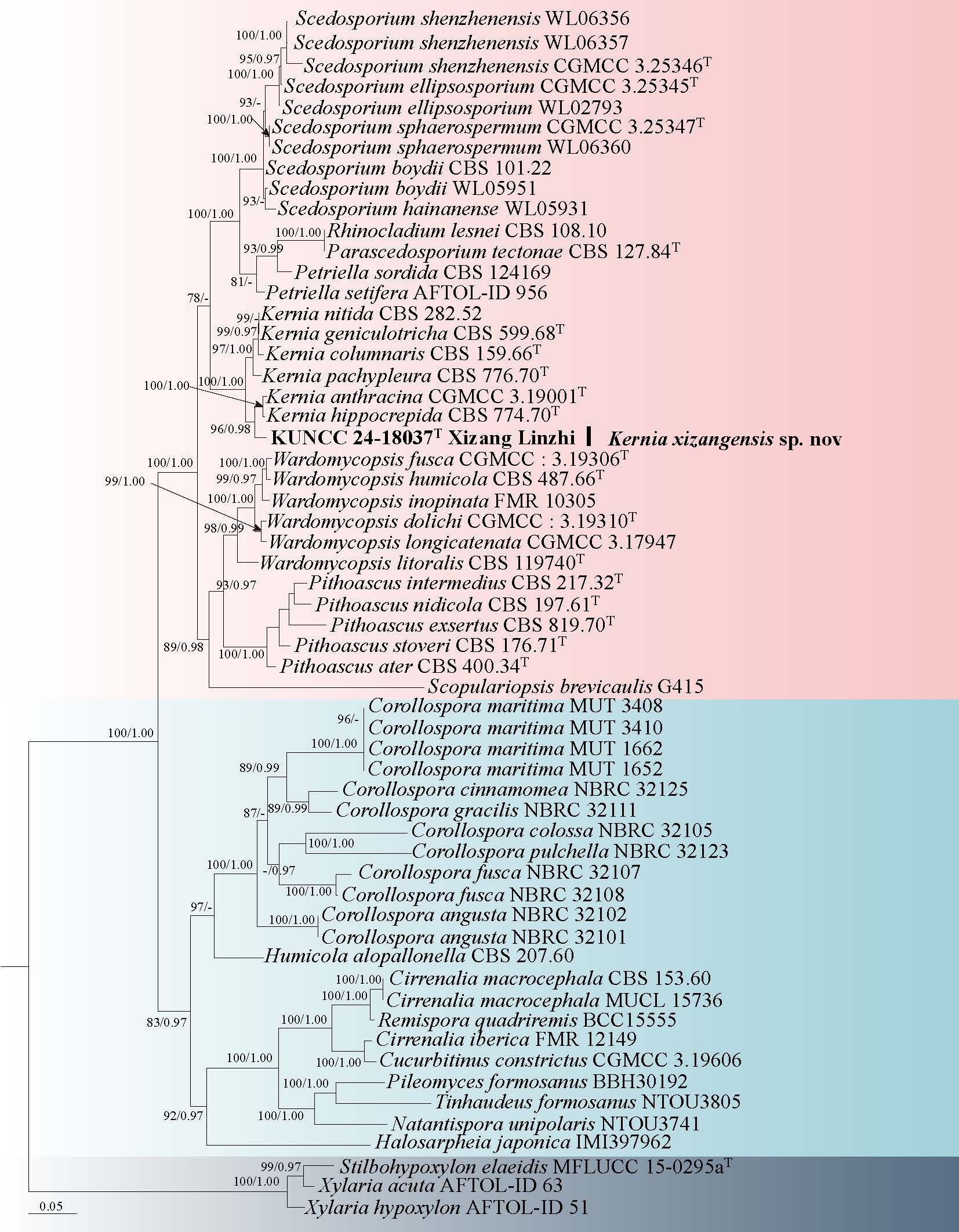
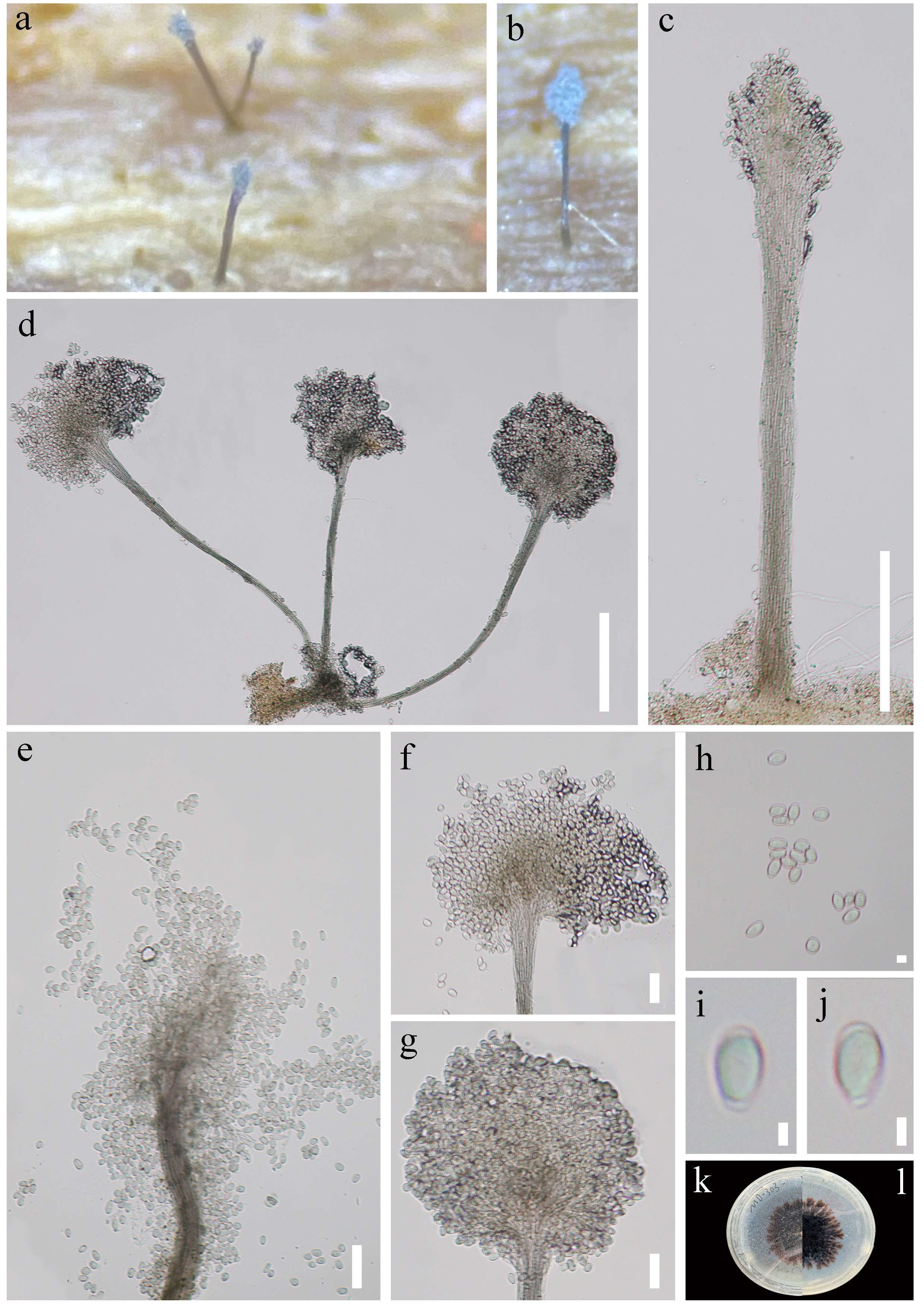
The asexual morph of Sterigmatobotrys is distinguished by macronematous, irregularly biverticillate to terverticillate conidiophores with stout, septate, and darkly pigmented stipes. These terminate in polyblastic conidiogenous cells bearing minute, sympodially arranged denticles. The hyaline, septate conidia mature to a brown color and are embedded in slime (Chang 1991; Réblová & Seifert 2011; Ertz et al. 2016; Luo et al. 2019). Currently, the genus includes three accepted species: S. macrocarpa, S. rudis, and S. uniseptate. S. macrocarpa is characterized by 2-septate, cylindrical to fusiform, hyaline conidia with a truncate base. In contrast, S. rudis features catenate, obclavate to fusiform, 3–12-septate conidia, while S. uniseptate is distinguished by its 1-septate, hyaline conidia (Réblová & Seifert 2011; Ertz et al. 2016; Luo et al. 2019; Yang et al. 2023).
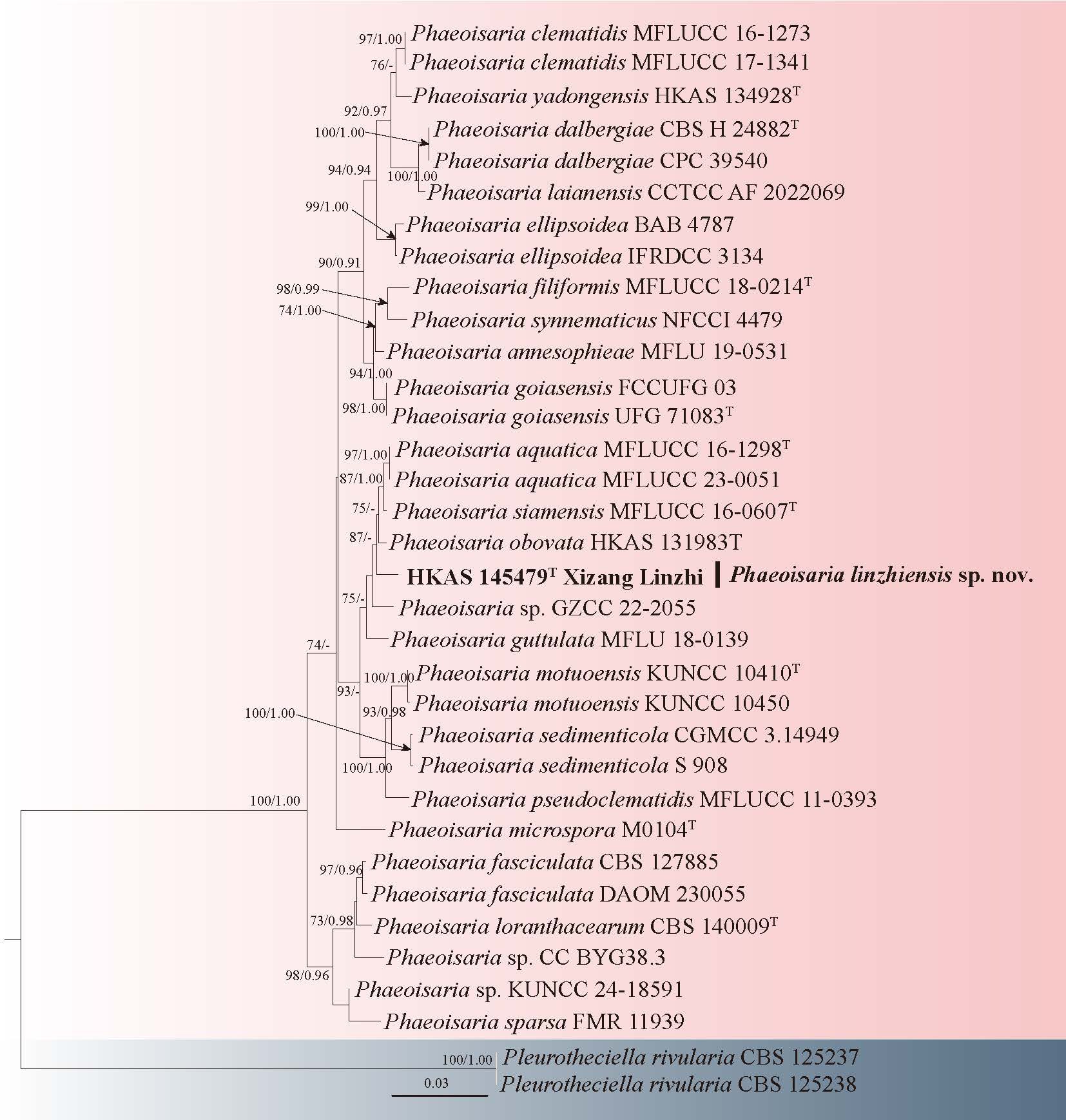
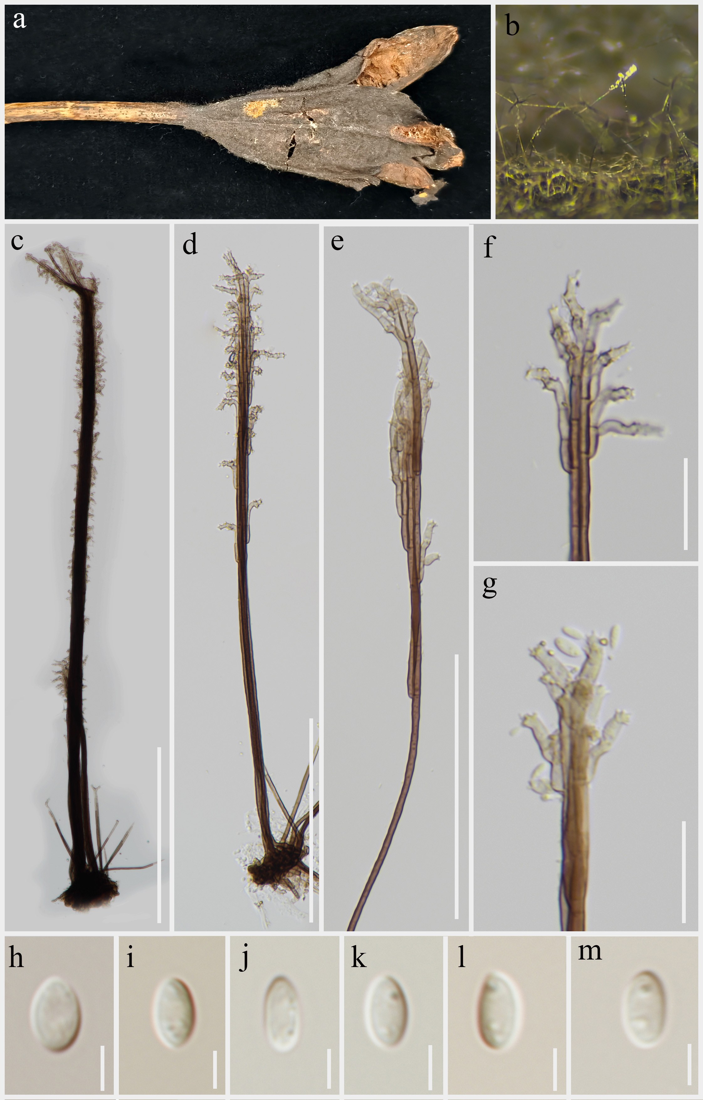
59. Sterigmatobotrys uniseptatus H.S. Chang [as 'uniseptata'], Mycol. Res. 95(9): 1142 (1991) Fig. 96
Chinese name
Index Fungorum number: IF 626943; Facesoffungi number: FoF 17432
Saprobic on submerged decaying wood. Sexual morph: Undetermined. Asexual morph: Colonies on the substratum superficial, effuse, with white, glistening conidial masses at apex. Mycelium immersed, subhyaline to pale brown, composed of branched, septate hyphae. Conidiophores 132–181 × 7–11 µm ( = 151 × 9 μm, n = 15), macronematous, mononematous, cylindrical, terminating in an irregularly biverticillate to terverticillate head, erect, straight or slightly flexouse, solitary, septate, smooth, unbranched, dark brown or brown. Conidiogenous cells terminal, parallel, polyblastic, smooth, cylindrical, hyaline, bearing multiple sympodially produced denticles from which conidia develop holoblastically. Conidia 14–19 × 4–6 µm ( = 16 × 5 μm, n = 20), aggregated in slimy masses, ellipsoidal to fusoid, 1-septate, smooth-walled, hyaline, sometimes constricted at the septa.
Culture characteristics: Conidia germinating on PDA medium within 48 h and germ tubes produced from both ends. Colonies grown on PDA reached 20 mm diam at room temperature in three weeks, circular, with velvety, dry, umbonate, light brown mycelium on the surface, from below dark brown with entire margin.
Material examined: CHINA, Yunnan Province, Nujiang, Gongshan County, Dizhengdang Village, 28°4′47.37" N, 98°19′32.06" E, 2030 m a.s.l., on decaying wood submerged in a freshwater stream, 20 October 2019, R.J. Xu, MD-214 (HKAS 136182), culture KUNCC 24-18026. Yunnan Province, Nujiang, Gongshan County, Dizhengdang Village, 28°4′47.37" N, 98°19′32.06" E, 2030 m a.s.l., on decaying wood submerged in a freshwater stream, 20 October 2019, R.J. Xu, MD-243 (HKAS 136208), culture KUNCC 10411. Xizang Autonomous Region, Shannan City, Longzi County, Zhari Township, 28°39′50.06" N, 93°22′25.27" E, 3023 m a.s.l., on decaying wood submerged in a freshwater stream, 05 August 2023, R.J. Xu, LJN-60 (HKAS 135979), culture KUNCC 24-17967. Yunnan Province, Shangri-La City, Bita Lake, 27°45′21.04" N, 99°56′46.56" E, 2306 m a.s.l., on decaying wood submerged in a freshwater river, 23 July 2020, R.J. Xu, MD-239 (HKAS 136161), culture KUNCC 24-18030.
Notes: Sterigmatobotrys uniseptata was described by Chang (1991), collected from unidentified twigs in Taiwan, and was later found on decaying wood submerged in freshwater in Yunnan Province, China (Luo et al. 2019). This species is characterized by its macronematous, mononematous conidiophores, which arise from stromatic cells. Each conidiophore features a well-defined stipe and a complex penicillate head composed of a series of penicillate branches, terminating in conidiogenous cells. These cells produce a head of slimy conidia. The conidiogenous cells are polyblastic and hyaline, while the conidia are solitary, slimy, thin-walled, hyaline, straight, cylindrical to subclavate, and 1-septate.
Phylogenetic analysis revealed that our collections (KUNCC 24-18026, KUNCC 10411, KUNCC 24-17967 and KUNCC 24-18030) clustered with Sterigmatobotrys uniseptate (MFLUCC 15-0358) (Fig. 95). The shape and size of conidiophores, conidiogenous cells, and conidia in our strains are identical to those described in strains of S. uniseptate by Chang (1991) and Luo et al. (2019). Therefore, we identify our collections as S. uniseptate.
Savoryellales Boonyuen, Suetrong, Sivichai, K.L. Pang & E.B.G. Jones
Savoryellaceae Jaklitsch & Réblová
Savoryella E.B.G. Jones & R.A. Eaton
Savoryella was established by Jones & Eaton (1969), with S. lignicola as the type species. The sexual morphs of Savoryella are defined by ascostromata that are immersed, partially immersed, or superficial, and globose, subglobose, or ellipsoidal in shape. These structures typically contain 8-spored, occasionally 2-spored, cylindrical or clavate, unitunicate asci, and ellipsoidal, three-septate ascospores. In contrast, the asexual morphs are distinguished by glistening, punctiform colonies; micronematous, mononematous conidiophores; holoblastic, determinate, integrated, terminal, or intercalary conidiogenous cells; and solitary or aggregated, pyriform to obovoid, septate conidia. Zhang et al. (2019) synonymized Trichocladium nypae with Savoryella nypae and introduced the asexual species S. sarushimana into the genus based on morphological and phylogenetic data. Subsequently, Tian et al. (2024) reported two more asexual species, S. cocois and S. chiangraiensis, from decaying leaves of Arecaceae, supported by phylogenetic analysis and morphological evidence. Savoryella, a holomorphic genus, predominantly inhabits submerged decaying woody debris in both aquatic and terrestrial ecosystems. The genus has been extensively studied and systematically illustrated by mycologists worldwide (Cai et al. 2002, 2003; Luo et al. 2004), and is widely distributed across diverse freshwater habitats globally, with particularly abundant occurrences in plateau lakes of Yunnan Province, China. Furthermore, all known genera in Savoryellales have been found to be distributed in the Q-X Plateau region (Wang et al. 2025a; Xu et al. 2025).
60. Savoryella submersa R.J. Xu, K.D. Hyde & Q. Zhao, sp. nov. Fig. 98
Chinese name
Index Fungorum number: IF 905046; Facesoffungi number: FoF 17439
Etymology: the species epithet "submersa" refers to the submerged wood where this species was collected.
Holotype: HKAS 136193
Saprobic on decaying stems of wood submerged in a freshwater stream habitat. Sexual morph: Undetermined. Asexual morph: Colonies effuse, scattered, black, glistening, punctiform. Mycelium immersed, subhyaline to pale brown, composed of branched, septate hyphae. Conidiophores inconspicuous or micronematous, mononematous, hyaline to pale brown, smooth. Conidiogenous cells holoblastic, determinate, integrated, pale brown to brown, cylindrical. Conidia 18–28 × 13–23 µm ( = 21× 19 µm, n = 30), acrogenous, solitary, obovoid to subspherical, 1-septate, distinct constricted at septa, dividing the conidium into two unequal cells, smooth, brown when mature, the basal cell pale brown.
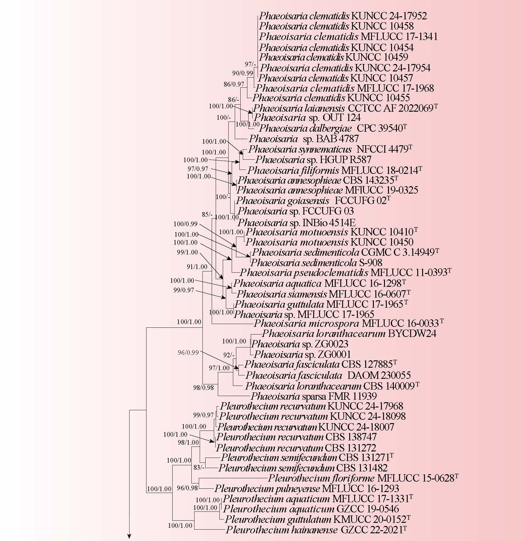
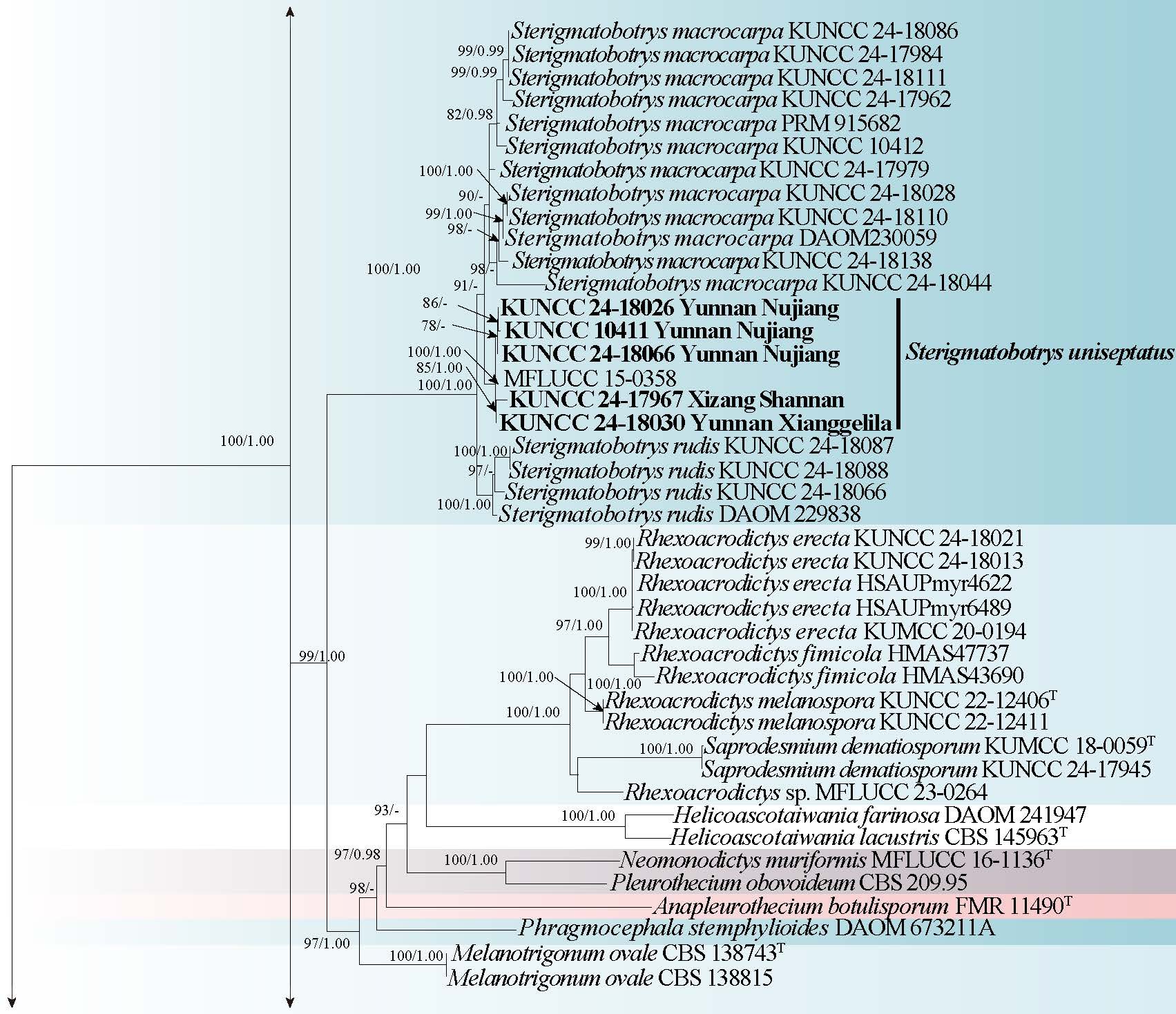
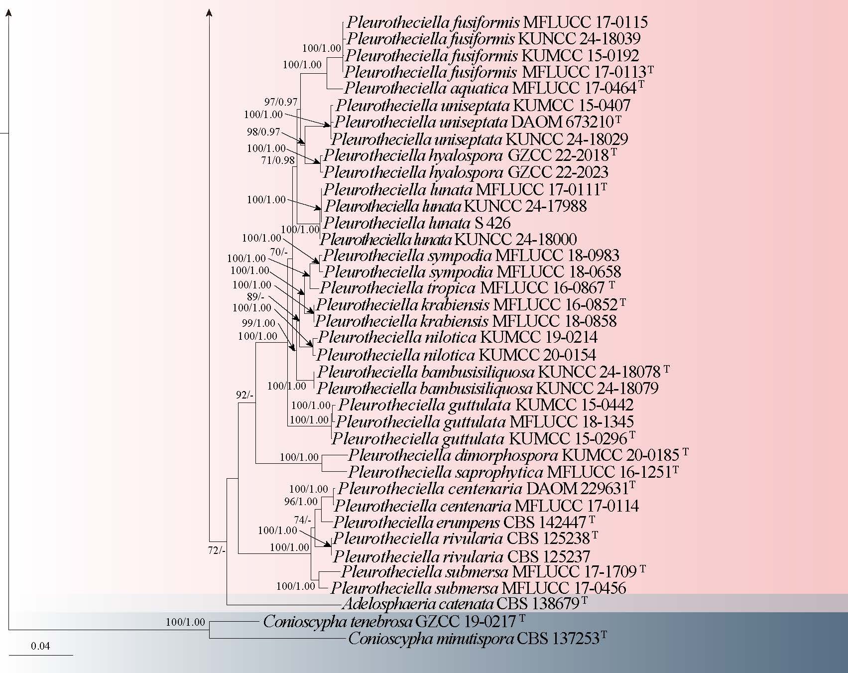
Culture characteristics: Conidia germinating on PDA medium within 24 h and germ tubes arising from terminal end of conidium. Colonies grown on PDA reached 30 mm diam at room temperature in two weeks, circular, with velvety, flat, dark brown mycelium on the surface, from below dark brown to black with entire margin.
Material examined: CHINA, Xizang Autonomous Region, Linzhi City, Motuo County, 29° 23' 36.31" N, 95° 35' 22.18" E, 667 m a.s.l., on decaying wood submerged in a freshwater stream, 13 July 2022, R.J. Xu, MD-99 (HKAS 136193, holotype), ex-type culture (KUNCC 24-18017).
Notes: Zhang et al. (2019) synonymized Trichocladium nypae with Savoryella nypae and introduced an asexual species, S. sarushimana, into the genus Savoryella, based on morphological and phylogenetic analyses. Subsequently, Tian et al. (2024) reported two additional asexual species, S. cocois, and S. chiangraiensis, collected from decaying leaves of the Arecaceae. Later, Xu et al. (2024f) introduced a unique asexual species, S. claviformis, which can be distinguished from all asexual species in Savoryella by its fusiform, claviform conidia. So far, five asexual species have been reported in Savoryella (Zhang et al. 2019; Tian et al. 2024b; Xu et al. 2024c).
Among the asexual species of Savoryella, S. submersa is distinguished by its obovoid to subspherical, 1-septate conidia, which are distinctly constricted at the septa (Zhang et al. 2019; Tian et al. 2024b; Xu et al. 2024c). Savoryella submersa differs from S. nypae in having 1-septate, obovoid to subspherical conidia, while the conidia of S. nypae In addition, in the phylogenetic analysis (Fig. 97), S. submersa (KUNCC 24-18017) formed a distinct branch within Savoryella, clearly separating it from other asexual species, and clustered as a sister group to S. aquatica, S. verrucosa, and S. yunnanensis with (100% ML/1.00 BYPP) statistical support.
Sordariales Chad. Ex D. Hawksw. & O.E. Erikss.
Bombardiaceae S.K. Huang & K.D. Hyde
Ramophialophora M. Calduch, Stchigel, Gené & Guarro
Ramophialophora was introduced by Calduch et al. (2004), with R. vesiculosa designated as the type species. The conidiophores of Ramophialophora resemble those of Gonytrichum, Dicyma, and Surculiseries, but the genus differs significantly in its conidiogenesis and the arrangement of conidiogenous cells on the conidiophore (Calduch et al. 2004). Ramophialophora is characterized by macronematous, mononematous, erect, cylindrical, septate, and branched conidiophores with subhyaline, subspherical to clavate vesicles. The conidiogenous cells are monophialidic or polyphialidic, discrete, terminal or lateral, lageniform, pale olivaceous, and smooth-walled, with conspicuous, slightly darker collarettes. The conidia are one-celled, spherical, and possess a protuberant basal hilum (Calduch et al. 2004).
61. Ramophialophora ramosa R.J. Xu, Q. Zhao & K.D. Hyde, sp. nov. Fig. 100
Chinese name
Index Fungorum number: IF 905047; Facesoffungi number: FoF 17440
Etymology: the species epithet "ramosa" refers to the branched conidiophores characteristic of this species.
Holotype: HKAS 136227
Saprobic on decaying wood submerged in freshwater habitats. Sexual morph: Undetermined. Asexual morph: Colonies on natural substrate effuse, hairy, velvety, pale brown. Mycelium superficial and immersed in the substrate, pale brown, septate, branched. Conidiophores macronematous, mononematous, erect, cylindrical, septate, branched, brown to dark brown, becoming paler towards the apex. Conidiogenous cells polyphialidic, discrete, terminal and lateral, lageniform or cylindrical, pale brown to subhyaline, cup-like collarette. Conidia 2–3 × 2–3 µm ( = 3 × 3 μm, n = 35), solitary, spherical, subglobose, one-celled at the base, brown to subhyaline, smooth.
Culture characteristics: Conidia germinating on PDA medium within 24h. Colonies grown on PDA reached 60 mm diam at room temperature in one week, circular, with velvety, flat, dark brown mycelium on the surface, from below dark brown with entire margin.
Material examined: CHINA, Xinjiang Autonomous Region, Yili Kazak Autonomous Prefecture, Gongliu County, Yuhu lake, 42°41′55.19" N, 81°4′51.31" E, 1955 m a.s.l., on decaying wood submerged in a freshwater lake, 05 August 2021, R.J. Xu, MD-318 (HKAS 136227, holotype), ex-type culture KUNCC 10414. Xinjiang Autonomous Region, Yili Kazak Autonomous Prefecture, Gongliu County, Kiziltar Village, 43°20′12.61" N, 82°30′56.54" E, 921 m a.s.l., on decaying wood submerged in a freshwater stream, 04 August 2021, R.J. Xu, MD-318-1 (HKAS 136226, paratype), ex-paratype culture KUNCC 24-18041.
Notes: Phylogenetic analysis showed that our collections (KUNCC 10414 and KUNCC 24-18041) clustered together and formed a sister group with Ramophialophora vesiculosa with (98% ML/1.00 BYPP) statistical support (Fig. 99). Ramophialophora ramosa resembles R. vesiculosa in having macronematous, mononematous, branched, brown conidiophores; monophialidic or polyphialidic, lageniform conidiogenous cells; and subhyaline conidia (Calduch et al. 2004). However, R. ramosa can be easily distinguished from R. vesiculosa by its lageniform or cylindrical conidiogenous cells with a cup-like collarette (Calduch et al. 2004). Ramophialophora globispora can be distinguished from R. ramosa by its unbranched conidiophores and chained conidia (Zhang et al. 2017b). In addition, they are located on different clade of the phylogenetic tree.
Chaetomiaceae G. Winter
Dichotomopilus X. Wei Wang, Samson & Crous
Wang et al. (2014) introduced Chaetomium indicum. Subsequently, based on the produces dichotomously-branched terminal hairs, Wang et al. (2016b) transferred Chaetomium indicum to Dichotomopilus and introduced Dichotomopilus indicus as the type species. There are currently 13 species reported in Dichotomopilus (Kedves et al. 2021; Wang et al. 2016b; Hyde et al. 2024a), mainly distributed in the China, Germany, Iran and USA, found in corn straw, soil, discarded sheep dung, straw of Triticum aestivum, and stored cotton (Wang et al. 2014). Morphologically, terminal hairs seta-like and the beginning, then developing into dichotomously or irregularly branched, lateral hairs unbranched, seta-like, tapering towards tips. Asci fasciculate, clavate, with 8 biseriate or irregularly arranged ascospores (Wang et al. 2016b).
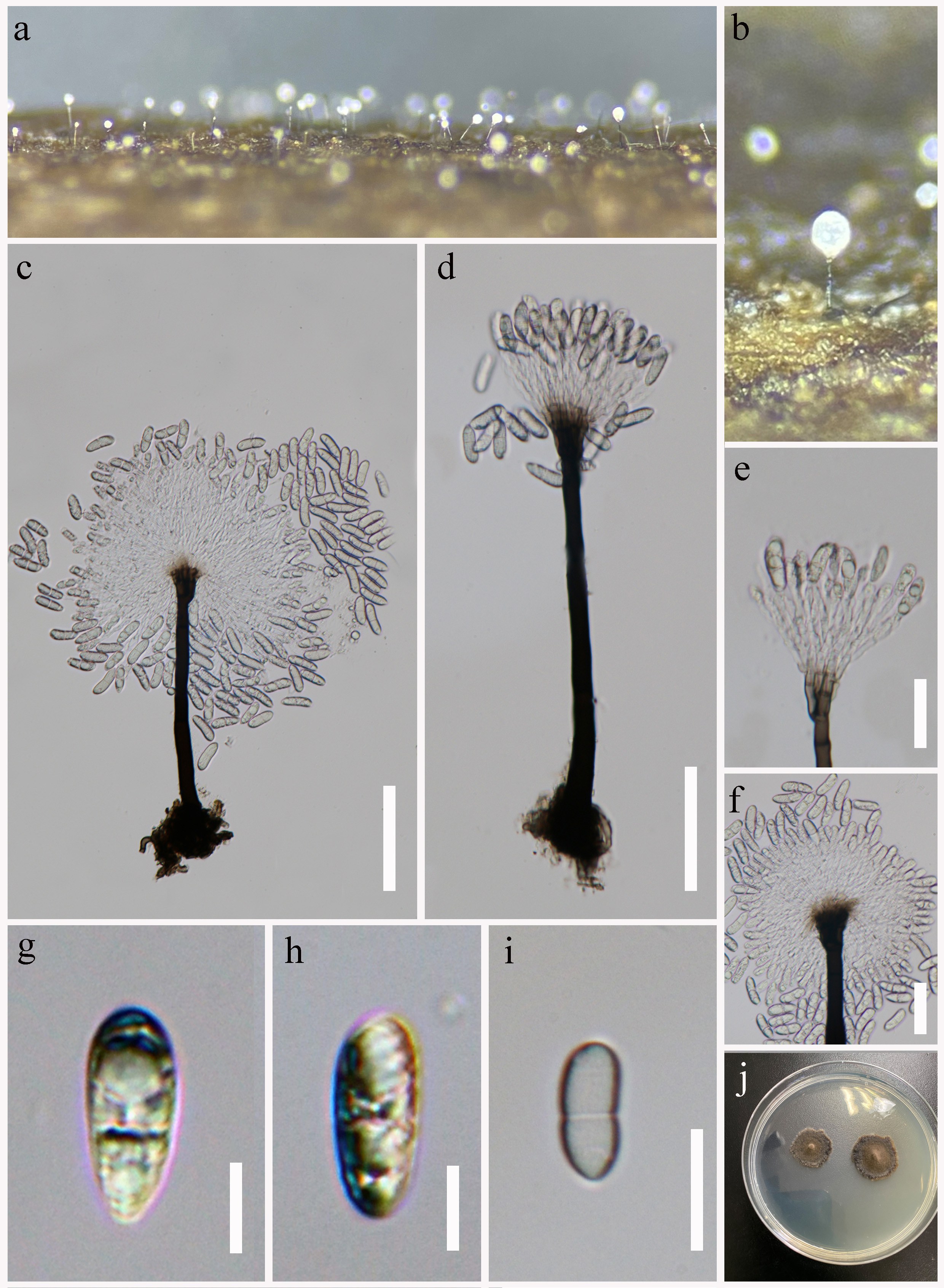
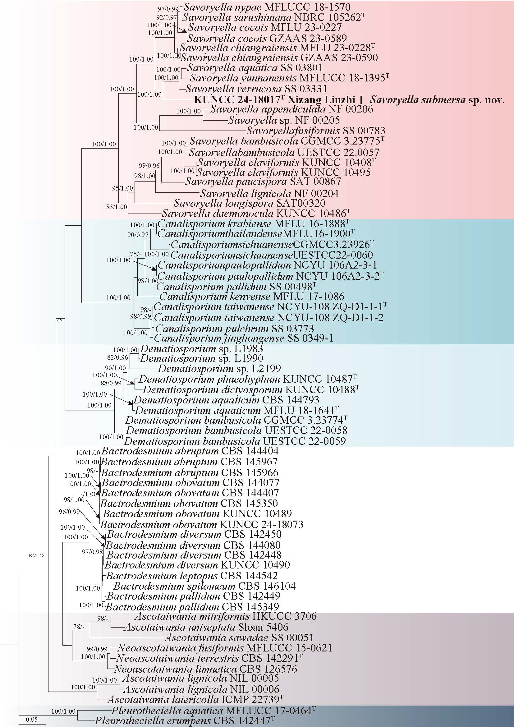
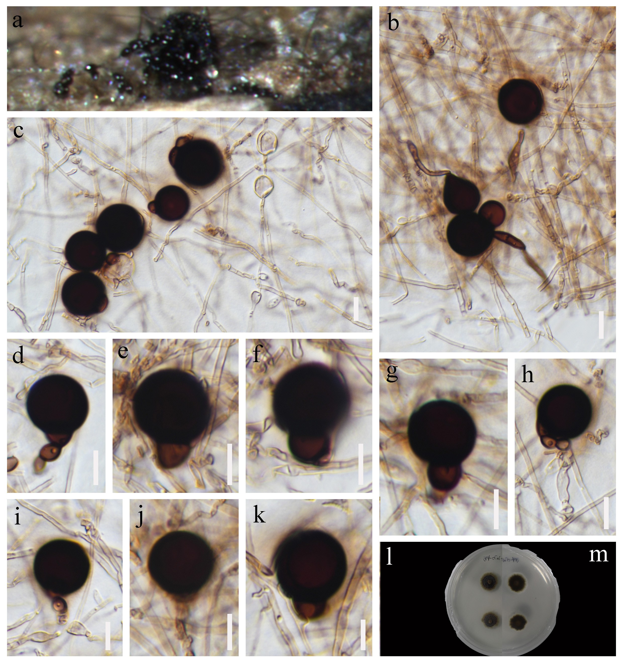
62. Dichotomopilus ramosissimus X. Wei Wang & Samson, Studies in Mycology 84, 217 (2016)
= Chaetomium ramosissimum X. Wei Wang & L. Cai, Mycological Progress 13, 725 (2014)
Chinese name: – 多分枝二叉孢 (duo fen zhi er cha bao)
Index Fungorum number: IF 818869; Facesoffungi number: FoF 07245
Saprobic on decaying stems of Chromolaena odorata. Sexual morph: Ascomata (on PDA) 700 × 500 μm diam, superficial, subglobose or ellipsoidal, brown to black, ostiolate. Peridium comprising brown, angular, or elongated cell structures that resemble hyphae. Terminal hairs erect, irregular in length, dark brown, dichotomously branched at wide to acute angles starting, verrucose. Thinner hairs resembling terminal setae, aseptate. Lateral hairs reduced to short spines. Asci already deliquescent. Ascospores 5–8 × 3–5 μm ( = 6.5 × 4, n = 30), pale brown when young, rather brown when mature, ovate to limoniform, thin-walled, smooth-walled, aseptate. Asexual morph: Undetermined.
Culture characteristics: Colonies on PDA, entire edge, about 50 mm diam. after 20 days of incubation at 25°C, white to pale yellow, sparse, floccose aerial hyphae, without pigment, reverse yellow, Ascomata is produced after 80 days.
Material examined: CHINA, Yunnan Province, Xishuangbanna, Jinghong City, on dead twigs of Chromolaena odorata, 7 September 2022, Shu-Cheng He, HSC1042 (HKAS 150298).
Known distribution: India, China, UK (Wang et al. 2016b; Chethana et al. 2021b).
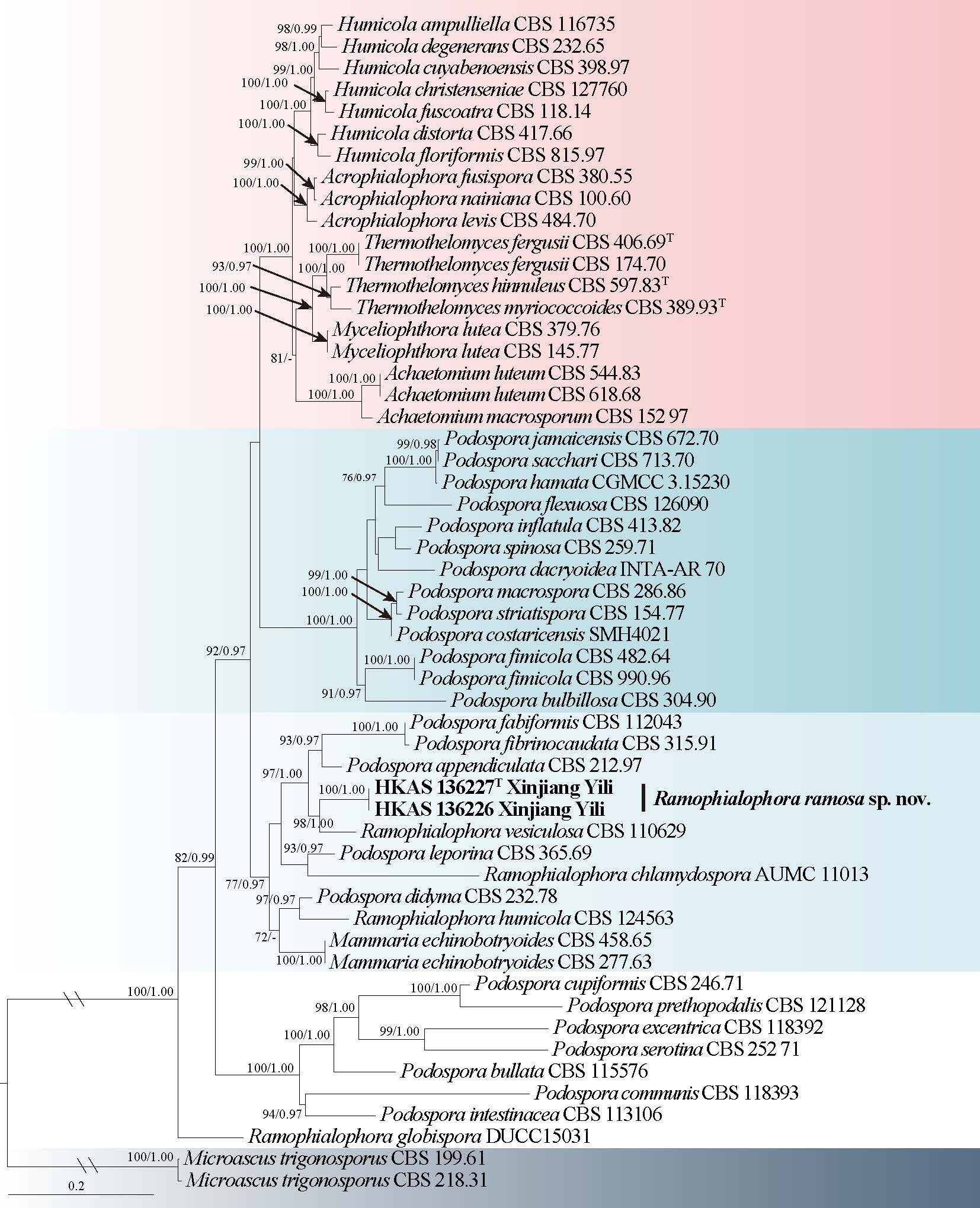
Known hosts: Clematis vitalba, Dolichandrone spathacea, Panax notoginseng (Wang et al. 2016b; Chethana et al. 2021b), Chromolaena odorata (this study).
Notes: Wang et al. (2014) introduced Chaetomium ramosissimum (Wang et al. 2014), which was subsequently reclassified as Dichotomopilus ramosissimus within the genus Dichotomopilus by Wang et al. (2016b). Based on multigene phylogenetic (ITS-LSU-rpb2-tub2) analysis, our isolate clusters with D. ramosissimus (Fig. 101). Morphologically, our isolate is similar to D. ramosissimus, both having dichotomously or irregularly branched. D. ramosissimus is by Wang et al. (2016b) first report (Wang et al. 2014, 2016). In addition, the sexual morph has been reported from China (Zhang et al. 2017a) and India (Chethana et al. 2021b). Thus, we report this collection as a new host record of D. ramosissimus from Yunnan Province, China, and provide additional molecular and morphological photoplates for this species.
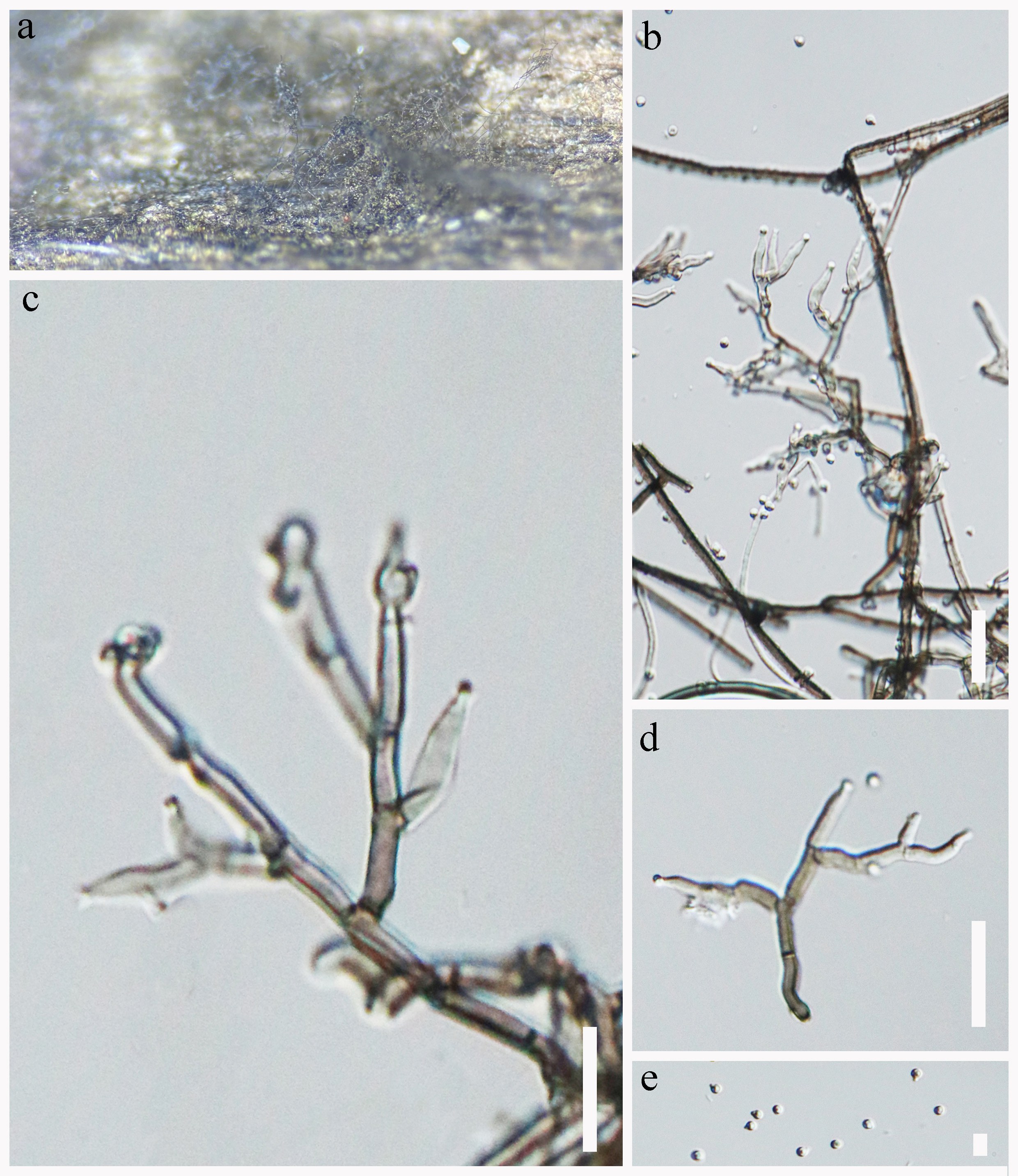
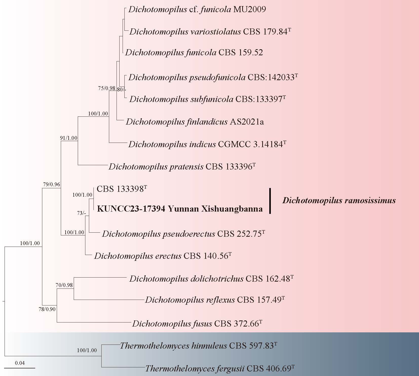
Sordariaceae G. Winter
Neodictyosporium Tennakoon, C.H. Kuo & K.D. Hyde
Neodictyosporium was established by Tennakoon et al. (2021) to accommodate a dictyosporium-like coelomycetous fungus that produces cheirosporous conidia. The asexual morph is characterized by pycnidial, superficial, subglobose conidiomata, holoblastic, determinate, terminal, cylindrical to subcylindrical, subhyaline to pale brown conidiogenous cells, and dimorphic conidia; the cheirosporous conidia are hyaline to pale brown, consisting of arms, euseptate, with a basal connecting cell; the clavate to obovoid conidia are hyaline, septate, guttulate, smooth-walled (Tennakoon et al. 2021; Crous et al. 2024; Yu et al. 2024). Phylogenetic analyses based on a multi-locus dataset (LSU, SSU, and rpb2) placed this genus within Sordariales genera incertae sedis (Tennakoon et al. 2021). Subsequent studies by Yu et al. (2024) on fungi associated with bamboo substrates revealed the sexual morph of Neodictyosporium, which is characterized by immersed to semi-immersed, subglobose ascomata, hyaline, septate paraphyses, 8-spored, unitunicate, cylindrical asci with long pedicellate, and an apical ring, and fusiform, hyaline, septate ascospores, with or without a mucilaginous sheath (Yu et al. 2024). Based on comprehensive phylogenetic analyses, Yu et al. (2024) elevated Neodictyosporium to familial rank, resulting in the establishment of Neodictyosporiaceae, a monotypic family that currently comprises only this genus. Species of Neodictyosporium are saprobic and inhabit terrestrial environments, typically occurring on decaying plant material such as Macaranga tanarius, Juncus maritimus, and various bamboo species (Tennakoon et al. 2021; Crous et al. 2024; Yu et al. 2024). To date, six species have been described within the genus, among which N. bambusae and N. sexuale represent the sexual morphs. These species have been recorded only from China and the Netherlands, suggesting a geographically restricted and potentially underexplored distribution (Tennakoon et al. 2021; Crous et al. 2024; Yu et al. 2024).
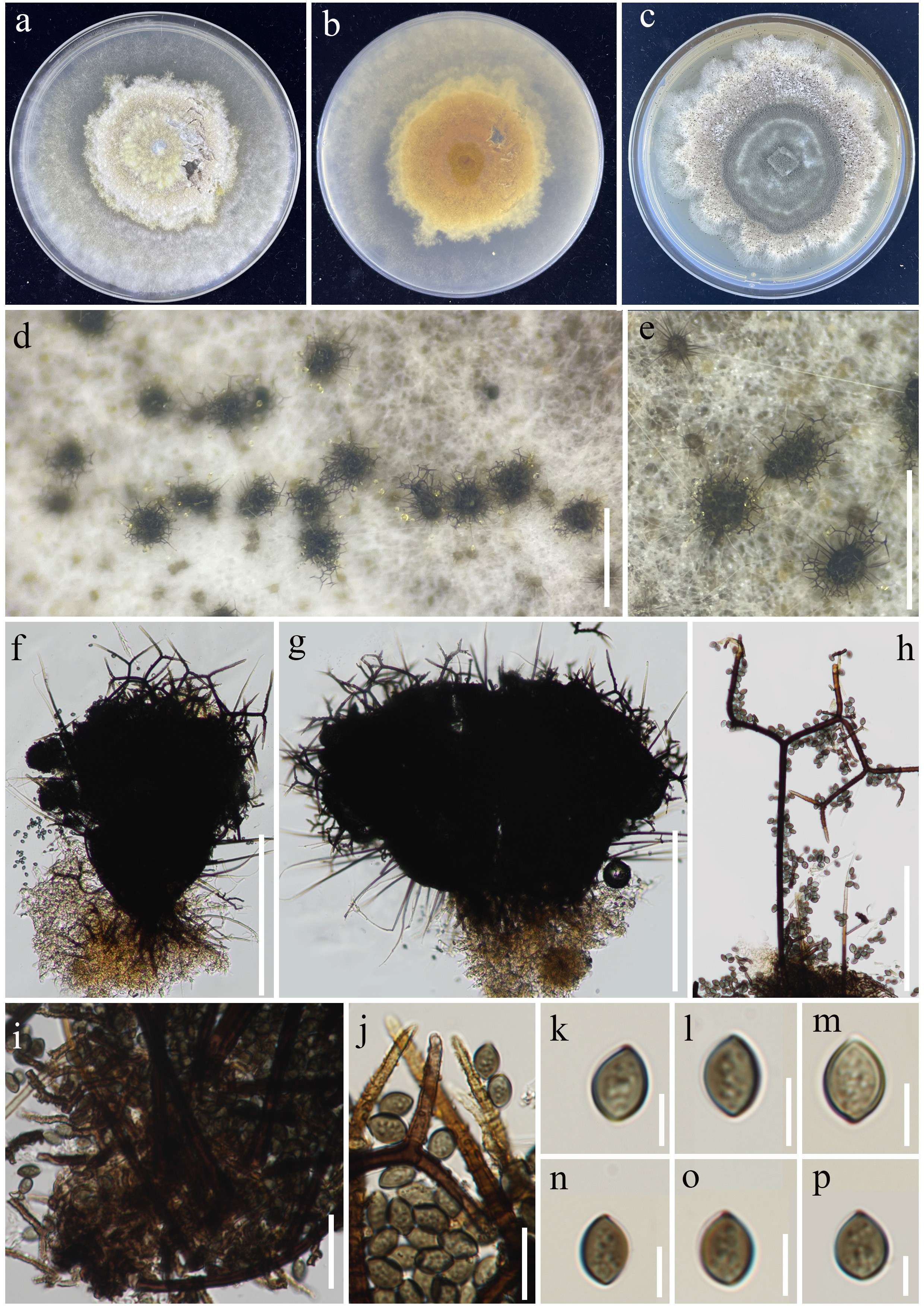
63. Neodictyosporium lacustre H.W. Shen, K.D. Hyde & Z.L. Luo, sp. nov. Fig. 104
Chinese name: – 杜鹃湖新砖格孢 (du juan hu xin zhuan ge bao )
Index Fungorum number: IF 905282; Facesoffungi number: FoF 19331
Etymology: “lacustre” refers to the Dujuanhu Lake in Yunnan Province, China, where the species was collected.
Holotype: HKAS 132049
Saprobic on submerged decaying wood in freshwater habitat. Sexual morph: Ascomata 354–382 µm high, 328–366 µm diam, semi-immersed to immersed, scattered, solitary, subglobose or ellipsoidal, dark brown to black, carbonaceous, uniloculate, biloculate, ostiolate. Ostiolar neck central or oblique, short,86–114 µm long, 138–168 µm wide, papillate, rounded, dark brown. Peridium 22–35 µm thick, composed of several layers of pale brown to dark brown, thick-walled cells of textura angularis. Paraphyses 2 µm wide, branched, septate, hyaline, numerous, hyphae-like, anastomosing above the asci. Asci 97–129 × 13–16 µm ( = 113 × 14 µm, n = 20) µm, 8-spored, unitunicate, inoperculate, cylindrical, straight or slightly curved, apex rounded, long pedicellate, with an apical annulus. Ascospores (20–)24–27 × 7–8 µm ( = 25 × 8 µm, n = 40), overlapping, uniseriate, fusiform, straight or slightly curved, hyaline, 3-septate, slightly constricted at the septa, guttulate, smooth or verrucous, without mucilaginous sheath. Asexual morph: Undetermined.
Culture characteristics: Conidia germinating on PDA within 12 h and germ tubes produced from both ends and around conidia. Colonies on PDA reaching 2–3 cm diameter after two months at room temperature (around 22 °C) in the dark, dense, velvety, pale brown to dark brown, smooth margins from above; dense, brown to dark brown, smooth margins from below.
Material examined: CHINA, Yunnan Province, Dujuanhu Lake, 24°32’15.6” N, 100°1′30.00″ E, 2500 m a.s.l., on the unidentified submerged decaying wood, 23 February 2023, H.W. Shen, L2085 (HKAS 132049, holotype), ex-type culture KUNCC 23-13778 = DLUCC 2090.
Notes: Phylogenetic analyses showed that Neodictyosporium lacustre (KUNCC 23-13778 = DLUCC 2090) formed a distinct clade at the base of the N. bambusae (CGMCC 3.27440 and ESTCC 24.0111), N. cheirosporum (CGMCC 3.27450 and UESTCC 24.0121), and N. sichuanense (CGMCC 3.27593) lineages, with 100% ML and 1.00 PP statistical support (Fig. 103). Among these, only N. bambusae is known to possess a sexual morph. Neodictyosporium lacustre is morphologically similar to N. bambusae in having semi-immersed, subglobose, uni- or multi-loculate ascomata, numerous, hyphae-like, unbranched, hyaline, septate paraphyses, 8-spored, unitunicate, cylindrical asci with long pedicellate and an apical ring, and fusiform, hyaline, septate ascospores (Yu et al. 2024). However, N. lacustre differs from N. bambusae in having larger ascospores and lacking a gelatinous sheath (Yu et al. 2024). Nucleotide comparisons revealed that N. lacustre differs from N. bambusae by 13 bp (including 5 gaps) in the ITS region, 2 bp in the LSU region, and 15 bp in the tef1-α region. In comparison to N. cheirosporum, N. lacustre shows 15 bp (including 6 gaps) differences in ITS, 3 bp in LSU, and 14 bp in the tef1-α, while differences between N. lacustre and N. sichuanense are 16 bp (including 7 gaps) bp in ITS, 3 bp in LSU, and 15 bp in the tef1-α. Based on both morphological characteristics and molecular evidence, we introduce N. lacustre as a novel species. Notably, it represents the first species of Neodictyosporium documented from freshwater habitats.
Sporidesmiales Crous
Sporidesmiaceae Fr.
Sporidesmium Link
Sporidesmiaceae was established by Fries (1849) and later redefined by Su et al. (2016) to delineate a monophyletic clade of Sporidesmium species, designated as Sporidesmiaceae sensu stricto. Members of this family are saprobic, predominantly found on woody plant material in both aquatic and terrestrial environments. They exhibit diverse morphologies but are primarily hyphomycetous fungi. Asexual morphs are characterized by brown, cylindrical conidiophores and conidia with distinctive septation. Sporidesmium was introduced by Link (1809) without the designation of a type species. Initially, the genus encompassed a vast array of species, reflected in nearly 500 epithets listed in Index Fungorum (2025). However, Shenoy et al. (2006) revealed that Sporidesmium sensu lato was polyphyletic within Ascomycota. Su et al. (2016) redefined the genus as a concise, monophyletic clade (Sporidesmium sensu stricto). Morphologically, Sporidesmium is distinguished by macronematous, mononematous, erect conidiophores, and monoblastic, holoblastic, percurrently proliferating conidiogenous cells. The conidia vary in shape, including ovoid, obpyriform, and rostrate forms, and are either euseptate or distoseptate, sometimes with an appendage or mucilaginous sheath (Su et al. 2016; Luo et al. 2019; Bao et al. 2021; Yang et al. 2018c, 2023a).
64. Sporidesmium fluviatile R.J. Xu, Q. Zhao & K.D. Hyde, sp. nov. Fig. 106
Chinese name
Index Fungorum number: IF 905049; Facesoffungi number: FoF 17442
Etymology: referring to the riverine habitat where the species is typically found
Holotype: HKAS 136180
Saprobic on submerged decayed wood in freshwater habitats. Sexual morph: Undetermined. Asexual morph: Colonies effuse, velvety, scattered or in small groups, pale brown, glistening. Mycelium partly immersed, mostly immersed, composed of septate, smooth. Conidiophores 38–54 × 3–5 µm ( = 48 × 4 μm, n = 15), macronematous, mononematous, erect, straight or slightly flexuous, solitary or 2-groups, cylindrical, smooth-walled, 2–4-septate, unbranched, pale brown to brown, truncate at the apex, often with hyphopodium at the base. Conidiogenous cells 17–19 × 3–4 µm ( = 18× 4 μm, n = 15), holoblastic, monoblastic, terminal, integrated, determinate, cylindrical, pale brown. Conidia 40–51 × 9–14 µm ( = 45× 10 μm, n = 20), acrogenous, solitary, lageniform, obclavate, rostrate, straight or slightly curved, 2–8-distoseptate, pale brown to brown, tapering at apex, truncate at the base, with a darkened scar at the base, smooth-walled, with a mucilaginous sheath over the apex.
Culture characteristics: Conidia germinating on PDA medium within 48 h and germ tubes arising from terminal end of conidium. Colonies grown on PDA reached 30 mm diam at room temperature in one month, irregular, with dense, wrinkled, viscous, pulvinate, grayish-white mycelium in the middle, sparse, mostly immersed mycelium which producing dark brown to black pigment at the edge, from below dark brown to black with entire margin.
Material examined: CHINA, Yunnan Province, Gongshan County, Dizhengdang Village, Dulong River, 28° 4' 47" N, 98° 19' 32" E, 1680 m a.s.l., on decaying wood submerged in a freshwater river, 10 July 2020, R.J. Xu, MD-271 (HKAS 136180, holotype), ex-type culture KUNCC 24-18035.
Notes: Morphologically, Sporidesmium fluviatile shares common characteristics with S. aquaticivaginatum and S. olivaceoconidium in having macronematous, mononematous, erect, straight or slightly, cylindrical conidiophores and conidia possess a mucilaginous sheath (Hyde et al. 2016; Luo et al. 2019; Bao et al. 2021). However, S. fluviatile differs from S. aquaticivaginatum in having smaller conidiophores (38–54 × 3–5 vs. 60–125 × 4–6 µm) and fewer conidia septate (2–8 vs. 6–10 distoseptate). It differs from S. olivaceoconidium in pale brown to brown, rostrate conidia. In addition, the phylogenetic analyses showed that S. fluviatile formed a distinct branch in Sporidesmium (Fig. 105).
Torpedosporales E.B.G. Jones, Abdel-Wahab & K.L. Pang
Juncigenaceae E.B.G. Jones, Abdel-Wahab & K.L. Pang
Juncigena Kohlm., Volkm. -Kohlm. & O.E. Erikss.
Juncigena was introduced by Kohlmeyer et al. (1997) based on the type species J. adarca, which was collected from senescent leaves of Juncus roemerianus from Atlantic coast in the USA. It is characterized by immersed, subglobose to pyriform, ostiolate, coriaceous, fuscous ascomata with cylindrical, periphysate neck, 8-spored, fusiform to cylindrical, short pedunculate, unitunicate asci with a non-amyloid apical ring, and uni- to biseriate, fusiform to ellipsoidal, 3-septate, hyaline ascospores (Kohlmeyer et al. 1997). Its asexual morph was described as Cirrenalia adarca with helicoid and brown conidia (Kohlmeyer et al. 1997). Abdel-Wahab et al. (2010) transferred Cirrenalia adarca to Moheitospora based on phylogenetic and morphological evidence. Subsequently, Réblová et al. (2016a) synonymized Moheitospora under Juncigena.
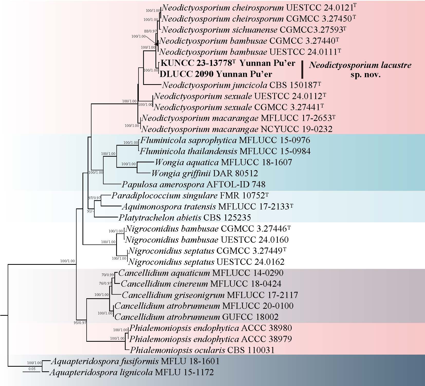
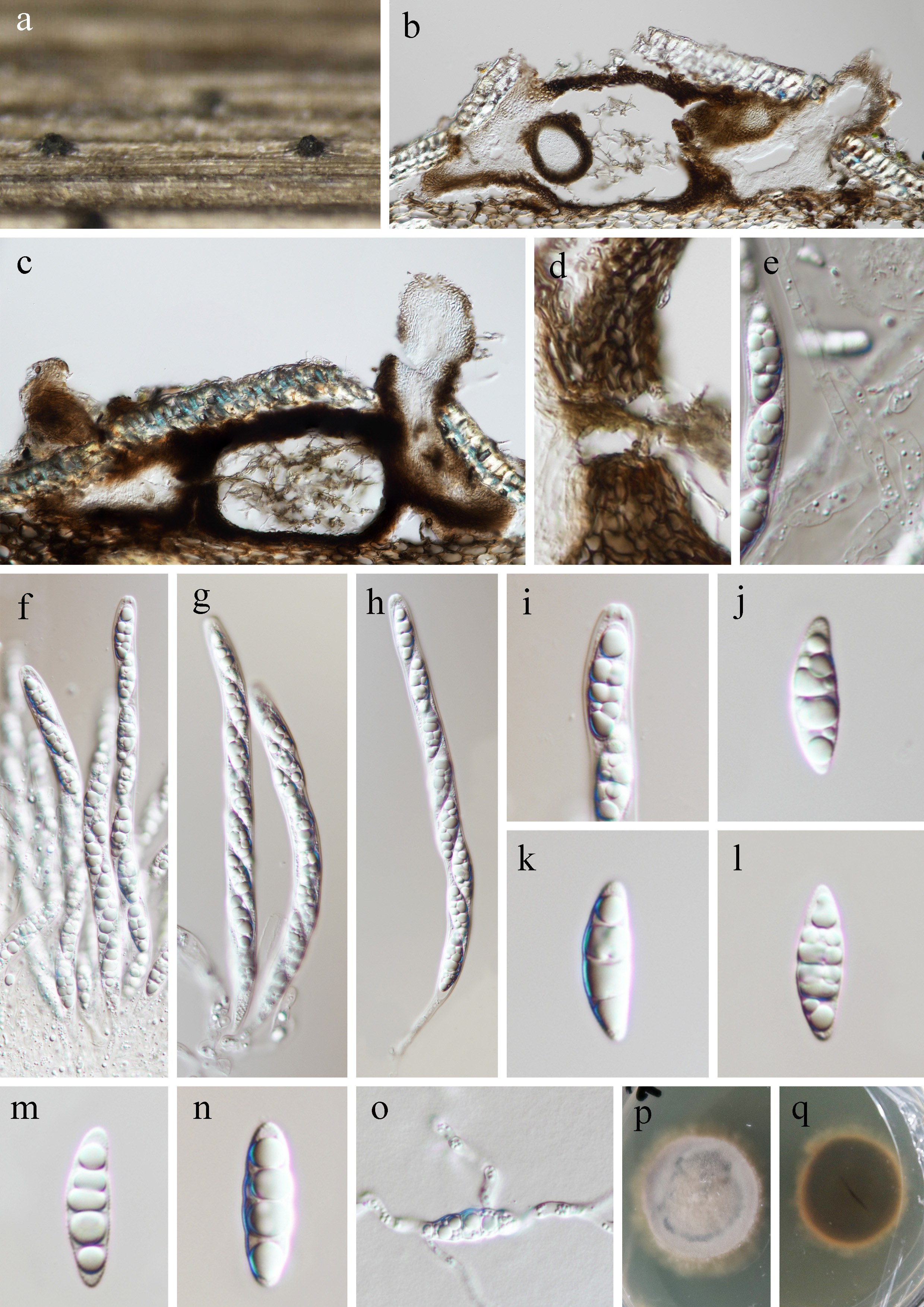
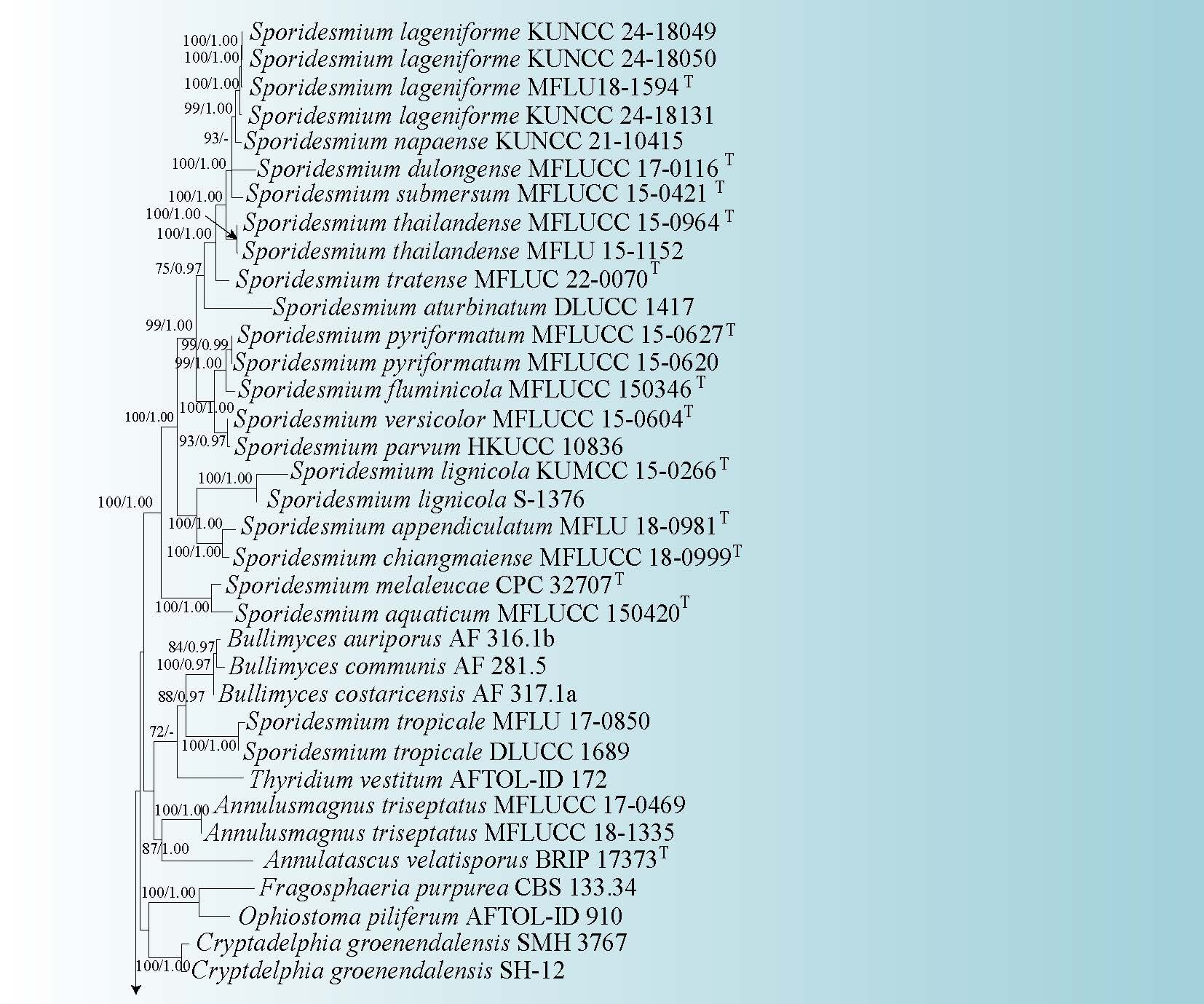
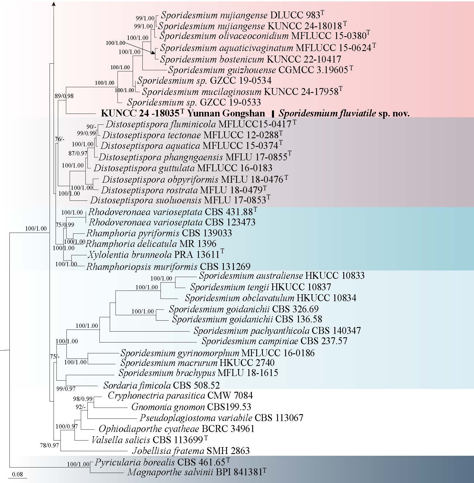
65. Juncigena hyalina J. Ma & Y.Z. Lu, sp. nov. Fig. 108
Chinese name
Fungal Names number: FN 573590; Facesoffungi number: FoF 19332
Etymology: the species epithet "hyalina" refers to its hyaline ascospores
Holotype: GZAAS 23-0032
Saprobic on submerged decaying twigs in freshwater habitats. Sexual morph: Ascomata 100–150 μm high, 110–210 μm wide, subglobose to pyriform, completely submersed under the cortex, ostiolate, papillate, coriaceous, fuscous, single. Neck cylindrical, curved, dark brown, ostiolar canal periphysate. Peridium 8–25 μm thick, comprising several layers of pale brown to brown cells of textura angularis. Hamathecium composed of numerous unbranched, septate pseudoparaphyses, attached at the top and bottom. Asci not observed. Ascospores 29–47 × 4.5–6.5 μm ( = 40.5 × 5.5 μm, n = 25), fusiform to elongate ellipsoidal, hyaline, 3-septate, slightly curved, guttulate, thin-walled, smooth. Asexual morph: Undetermined.
Culture characteristics: Ascospores germinated on PDA within 12 h, at room temperature and germ tubes produced from both ends of the ascospores. Colonies on PDA reaching 4 cm diam after four weeks. Colonies superficial, dense, circular, with slightly raised center and entire edge, white from above, pale yellow from below.
Material examined: CHINA, Xinjiang Autonomous Region, Bayingolin, Bohu County, 41°51′54″ N, 86°43′46″ E, 991 m a.s.l., on decaying wood submerged in a freshwater river, 22 July 2021, Y.Z. Lu, Lu 1 (GZAAS 23-0032, holotype), ex-type culture (GZCC 23-0483).
Notes: In the phylogenetic analyses (Fig. 107), our strain GZCC 23-0483 grouped with Juncigena adarca (JK5548A) and J. fruticosae (EF14) with 94% ML/1.00 BYPP support. Our collection resembles the type species J. adarca in having subglobose to pyriform, ostiolate ascomata, and fusiform to ellipsoidal, hyaline, 3-septate ascospores (Kohlmeyer et al. 1997). However, our collection has smaller ascomata (100–150 × 110–210 μm vs. 225–400 × 135–200 μm) and longer and thinner ascospores (29–47 × 4.5–6.5 μm vs. 26.5–34.5 × 6–7 μm) (Kohlmeyer et al. 1997). Juncigena fruticosae is characterized by its hyphomycetous asexual morph, thus, their morphology is incomparable. However, phylogenetic analyses show our collection is a distinct species.
Xylariaceae Tul. & C. Tul.
Digitodochium Tubaki & Kubono
Digitodochium was established by Tubaki & Kubono (1989) to accommodate an asexual species, D. rhodoleucum (the type species), originally described from corticated twigs of Fagus crenata in Japan. For over three decades, no additional reports of this genus were published until recently, when Voglmayr et al. (2020) linked the sexual and asexual morphs of Digitodochium. Subsequently, six additional species were introduced within Digitodochium, five from China and one from Australia (Dissanayake et al. 2024; Liu et al. 2025; Tian et al. 2025). Currently, eight species are accepted in the genus, they occurring as saprobes on decaying branches or leaves of Grevillea sessilis, Fagus sylvatica and various bamboo species (Voglmayr et al. 2020; Dissanayake et al. 2024; Liu et al. 2025; Tian et al. 2025). Both sexual and asexual morphs are known in the genus; the sexual morph is characterized by scattered or gregarious, solitary, immersed, subglobose, black ascomata, with a small black clypeus; papillate, central, black ostioles, with a conical neck; hyaline, septate, unbranched, cellular paraphyses; unitunicate, cylindrical to slightly fusiform asci, apically rounded, with a wedge-shaped, apical ring; uniseriate, aseptate, ellipsoid to fusiform ascospores, with or without sheath. And the asexual morph is characterized by sporodochial, immersed, cushion-shaped, subglobose to irregular conidiomata; aggregated, hyaline, branched conidiophores; integrated, holoblastic conidiogenous cells; subhyaline, staurosporous conidia, consisting of a cylindrical main axis with whorls of lateral branches arising at wide angles from the basal 1–3 cells of the main axis, septate, gradually tapering towards the top, narrowly rounded tips (Voglmayr et al. 2020; Dissanayake et al. 2024; Liu et al. 2025; Tian et al. 2025). In this study, we discuss a novel saprobic taxon collected on submerged decaying bamboo wood from freshwater habitat in Yunnan Province, China.
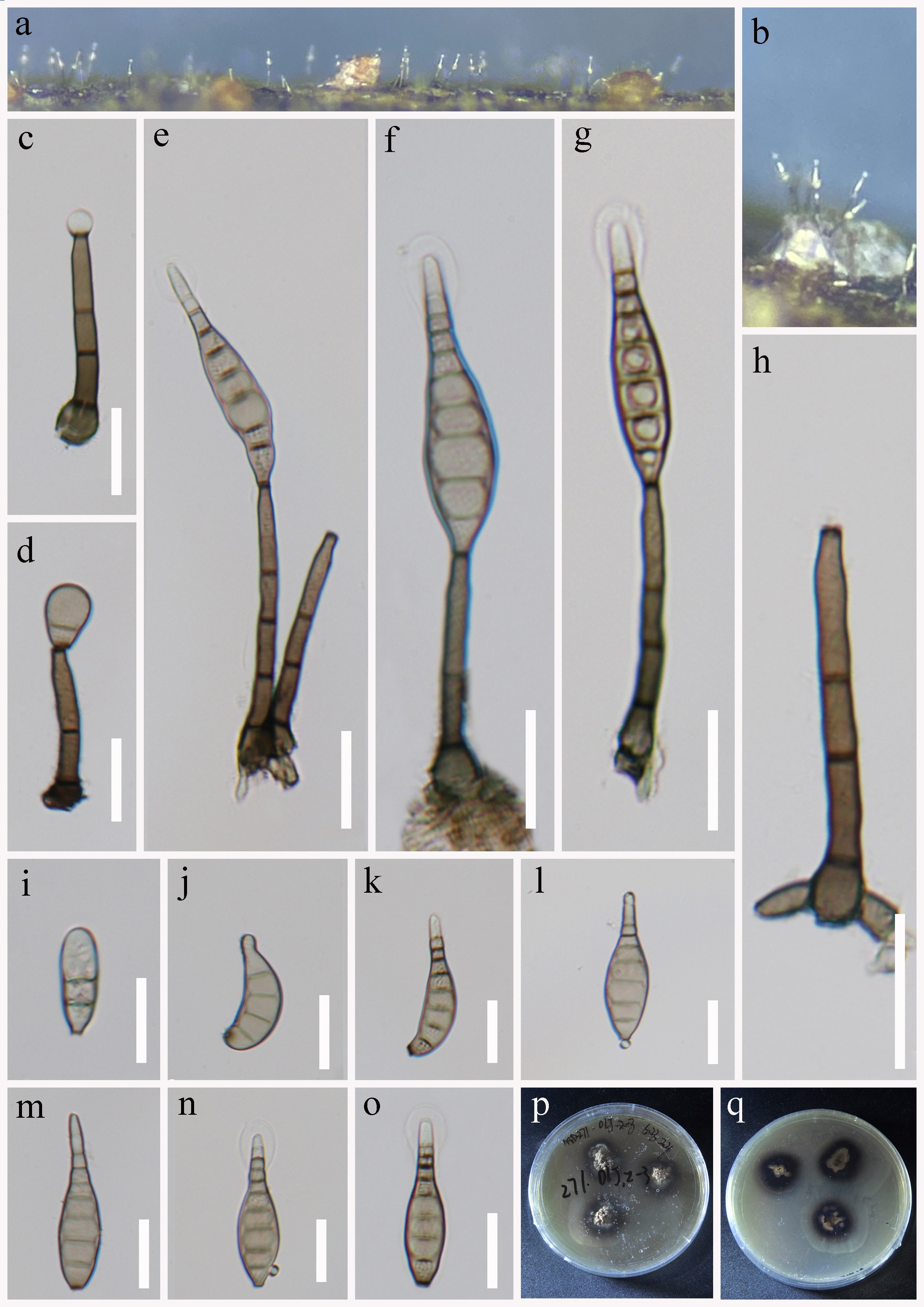
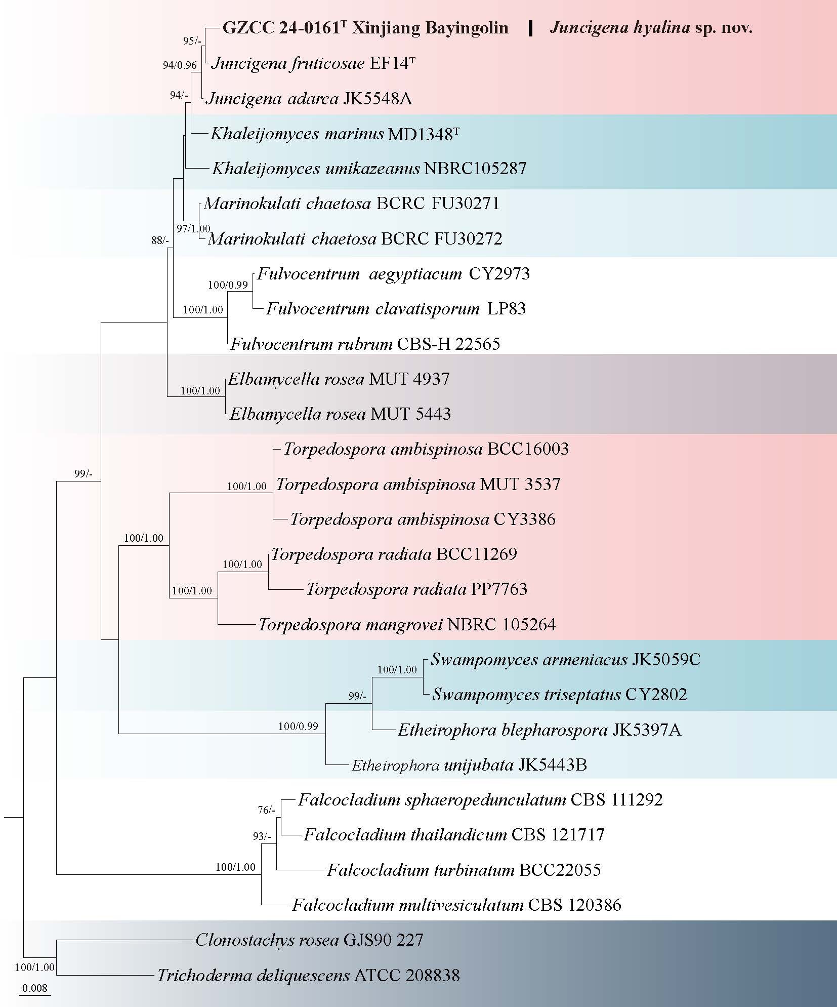
66. Digitodochium lacustre H.W. Shen, K.D. Hyde & Z.L. Luo, sp. nov. Fig. 110
Chinese name:
Index Fungorum number: IF 905283; Facesoffungi number: FoF 19887
Etymology: “lacustre” refers to the freshwater lake where the species was collected.
Holotype: HKAS 132103.
Saprobic on decaying submerged bamboo wood in a lake. Sexual morph: Ascomata 430–490 µm high, 580–670 µm diam, scattered or gregarious, solitary, immersed, black, subglobose, papillate ostioles. Ostioles papillate, central, black, with a conical neck. Peridium uneven in thickness, 30–60 μm wide, composed of several layers of thick, dark brown to black, irregular cells of textura angularis. Paraphyses 2–3 µm wide, composed of numerous, filamentous, cellular, septate, guttulate, embedded in a gelatinous matrix. Asci 130–187(–208) × 10–13 µm ( = 162 × 11 µm, n = 20), 8-spored, unitunicate, cylindrical to slightly fusiform, straight or slightly curved, short-pedicellate, rounded at the apex. Ascospores 13–17 × 4.5–6 µm ( = 15 × 5.3 µm, n = 40), overlapping uniseriate, aseptate, ellipsoid to narrowly ellipsoid, hyaline to dark brown, smooth-walled, usually malachite green, slightly rounded ends, with a small cell at the base, 2–4 guttules, mostly with 2 large guttules, surrounded by a mucilaginous sheath, 15–21 µm wide. Asexual morph: Undetermined.
Culture characteristics: Ascospore germinated on PDA within 12 h and germ tubes produced from the ends of the ascospore. Colonies on PDA reaching 2.8–3 cm diam. after six weeks at room temperature, colony circular, smooth edge, surface hyphae cream to pale brown, flocculent, dense; reverse side cream, smooth, dense, smooth.
Material examined: CHINA, Yunnan Province, Puer City, Dujuanhu Lake, 24°32'15.6" N, 100°1′30.00″ E, 2500 m a.s.l., on a rotten bamboo branch submerged in water, 19 February 2023, H.W. Shen, L2087 (HKAS 132103, holotype); ex-type culture (KUNCC 23-13817).
Notes: Multigene phylogenetic analysis revealed that Digitodochium lacustre clustered with D. zhangjiajieense with 99% ML/1.00 BYPP bootstrap support (Fig. 109). Morphologically, D. lacustre resembles D. ailaoshanense in having scattered or gregarious, subglobose, immersed, black ascomata; unitunicate, cylindrical to slightly fusiform asci and aseptate, hyaline to dark brown, guttulate ascospores, with a small cell at the base, and surrounded by a mucilaginous sheath. However, D. lacustre is distinguished from D. ailaoshanense by having larger asci (130–187 × 10–13 µm vs. 70–110 × 6–9 μm). Comparison of ITS and LSU sequences between D. lacustre (KUNCC 23-13817) and D. ailaoshanense (HKAS 130308) showed nucleotide differences of 25 bp (excluding gaps) and 16 bp, respectively. Similarly, comparisons of ITS, LSU and tub2 sequences between D. lacustre (KUNCC 23-13817) and D. zhangjiajieense (GMB5607) revealed nucleotide differences of 9 bp (including 2 gaps), 7 bp and 24 bp, respectively. Based on both morphological characteristics and molecular evidence, we introduce D. lacustre as a new species, marking the first record of a Digitodochium species from a freshwater habitat.
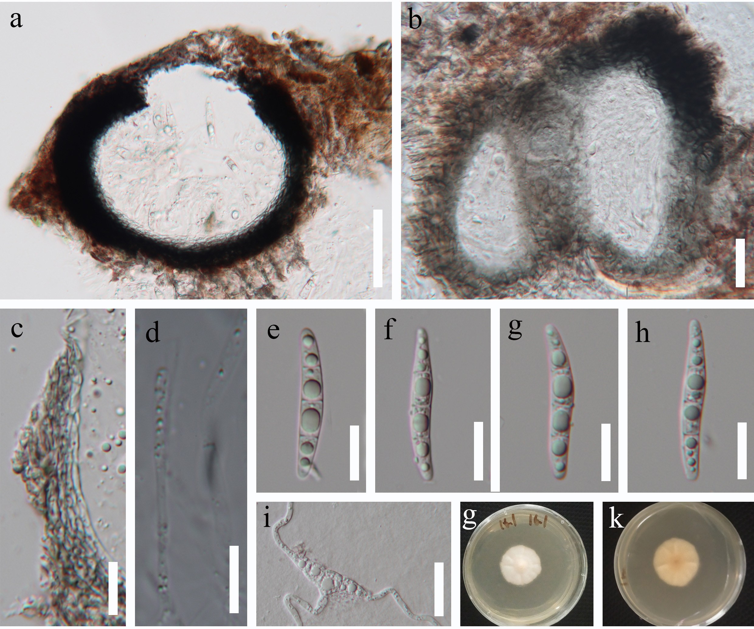
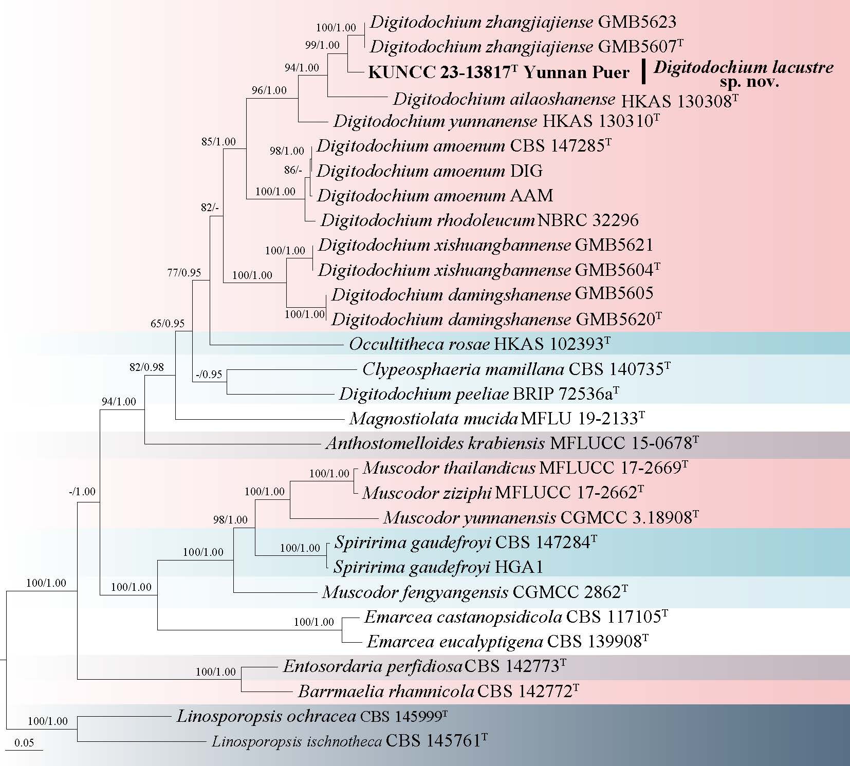
Sordariomycetes families incertae sedis
Junewangiaceae J.W. Xia & X.G. Zhang
Sporidesmiella P.M. Kirk
Sporidesmiella was established by Kirk (1982), with S. claviformis designated as the type species. It is characterized by clavate to obovoid or cuneate conidia with a few distosepta, rounded or coronate at the apex, which secede schizolytically from monoblastic, integrated, terminal, annellidic, or occasionally sympodially extending conidiogenous cells (Kirk 1982; Ma et al. 2012; Luo et al. 2019; Xiong et al. 2024). Sporidesmiella has been reported in various countries, including China, Mexico, Brazil, the Netherlands, and Thailand, and spans tropical and subtropical regions (Wang 2005; Monteiro et al. 2014; Heredia et al. 2015; Luo et al. 2019; Crous et al. 2021; Dong et al. 2021).
Based on multigene phylogenetic analysis, Luo et al. (2019) placed Sporidesmiella within Junewangiaceae. Hyde et al. (2020b) classified the genus as Diaporthomycetidae genera incertae sedis, observing that Sporidesmiella clustered in a separate clade with Junewangiaceae. However, further phylogenetic evidence from Dong et al. (2021a) strongly supported its placement within Junewangiaceae. Currently, Sporidesmiella has 54 records listed in Index Fungorum (2025), but only 13 species have corresponding DNA sequences in GenBank. While taxonomic revisions have been made for some species, the classification of many others remains uncertain due to insufficient sequence data.
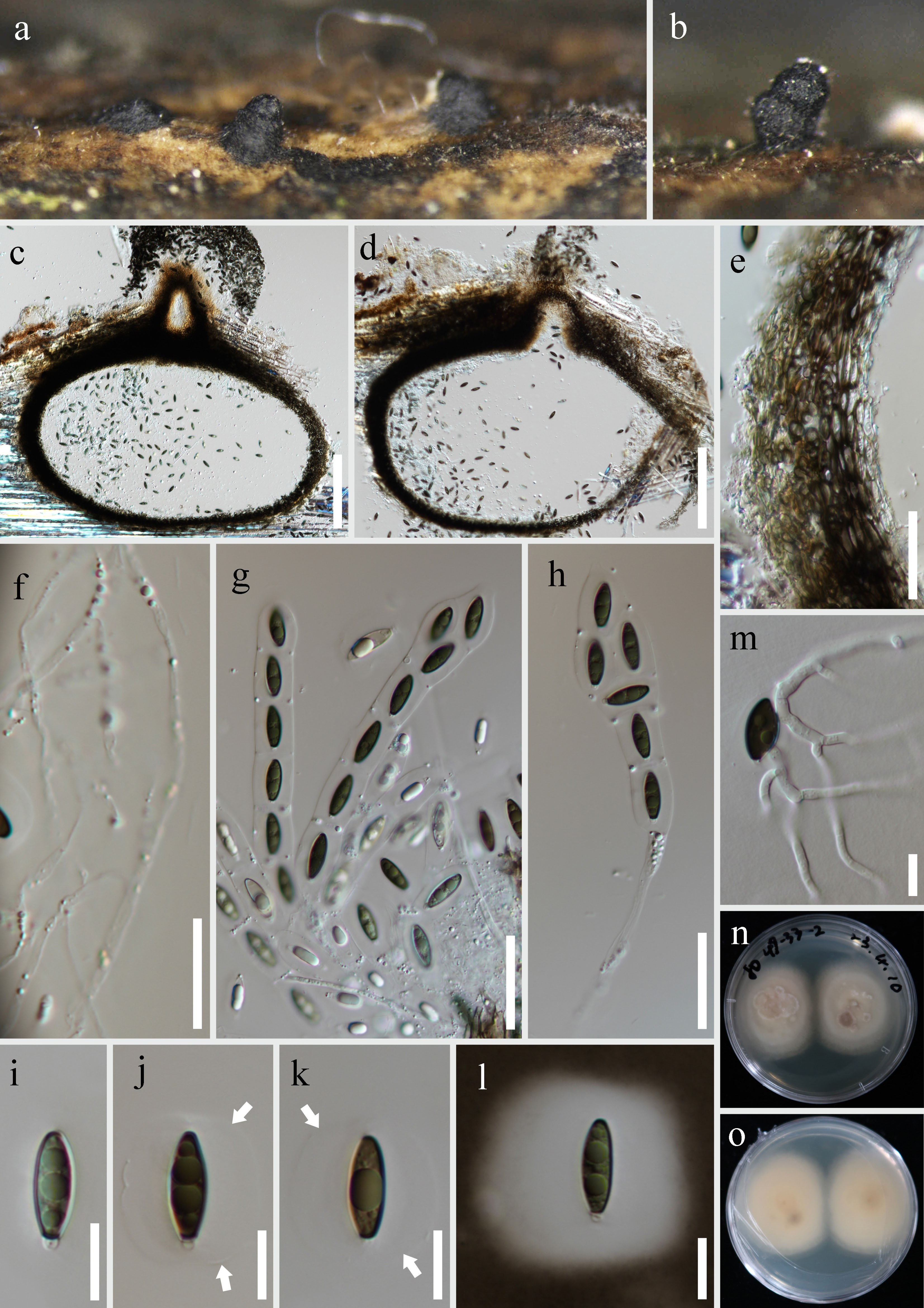
67. Sporidesmiella gezaensis R.J. Xu, Q. Zhao & K.D. Hyde, sp. nov. Fig. 112
Chinese name
Index Fungorum number: IF 905284; Facesoffungi number: FoF 19888
Etymology: It is named after Geza Township, where the type specimen was collected.
Holotype: HKAS 136093
Saprobic on submerged decaying wood in freshwater stream. Asexual morph: Colonies superficial, effuse, glitter, pale brown to brown. Mycelium superficial, partly immersed, consisting of branched, septate, smooth, hyaline to pale brown hyphae. Conidiophores 161–202 × 4–5 μm ( = 185 × 5 μm, n = 20), macronematous, mononematous, unbranched, single or 2–3-groups, erect, cylindrical, straight or slightly flexuous, slightly constricted at septa, brown, pale toward the apex, 4–8-septate, smooth. Conidiogenous cells 17–63 × 4–5 μm ( = 34 × 4 μm, n = 15), holoblastic, polyblastic, integrated, with sympodial proliferations, terminal and intercalary, with several sympodial proliferations, conspicuous denticles, cylindrical, hyaline, smooth. Conidia 18–25 × 9–15 μm ( = 21 × 11 μm, n = 25), acrogenous or intercalary, solitary, obovoid, rounded at the apex, narrow towards truncate at the base, hyaline to pale brown, becoming brown at maturity, 0–1- distoseptate when young, 3–4-euseptate when mature, guttulate, smooth-walled. Sexual morph: Undetermined.
Culture characteristics: Conidia germinating on PDA medium within 24 h and germ tubes arising from terminal end of conidium. Colonies grown on PDA reached 20 mm diam at room temperature in two weeks, irregular, dense, glistening, dry, flat, grayish-white mycelium on the surface, cream, butyrous, immersed mycelium on the surrounding, from below cream with entire margin.
Material examined: CHINA, Yunnan Province, Shangri-La City, Geza Township, 27°58′24.94" N, 99°46′39.32" E, 3307 m a.s.l., on decaying wood submerged in a freshwater stream, 19 June 2022, R.J. Xu, XK-33-1 (HKAS 136093, holotype); ex-type culture KUNCC 10426. CHINA, Yunnan Province, Shangri-La City, Geza Township, 27°58′24.94" N, 99°46′39.32" E, 3307 m a.s.l., on decaying wood submerged in a freshwater stream, 19 June 2022, R.J. Xu, XK-33-1-1 (HKAS 136092, paratype), ex-paratype culture KUNCC 24-18116.
Notes: Morphologically, the macronematous, mononematous, unbranched, cylindrical conidiophores, holoblastic, integrated, sympodial, cylindrical conidiogenous cells that with proliferating percurrently, and solitary, obovoid, distoseptate conidia are the typical characteristics in Sporidesmiella.
Sporidesmiella gezaensis was previously confused with S. aquatica, but the new species differs from the latter in having smaller conidia (18–25 × 9–15 μm vs. 51–59 × 18–22 μm) (Luo et al. 2019). Phylogenetic analyses showed that our collections form a distinct clade within Sporidesmiella, closely related to S. yadongensis (KUNCC 24-17996) (Fig. 111). However, a comparison of ITS nucleotide sequences reveals that S. gezaensis differs from S. yadongensis (KUNCC 24-17996) by 12/525 bp (2.3%, excluding gaps). Therefore, Sporidesmiella gezaensis is introduced as a new species based on morphological distinctiveness, phylogenetic evidence, and species delimitation criteria proposed by Jeewon and Hyde (2016) and Chethana et al. (2021a).
Discussion
The Q-X Plateau, renowned for its unique ecological and geographical characteristics, has long been an area of interest for researchers across various disciplines. However, the mycological diversity of this region has not been extensively documented until recent years. This paper, the third in a series dedicated to documenting novel fungal taxa in this region, focuses on filamentous-fungi and entomopathogenic fungi that are endemic to the Q-X Plateau. Based on molecular evidence and morphological characteristics, 4 classes, 19 orders, 35 families, and 52 genera, viz. Acrogenospora, Akanthomyces, Aquaphila, Beauveria, Chalara, Chloridium, Cladophialophora, Clonostachys, Conioscypha, Cordana, Cordyceps, Corynespora, Cylindrotrichum, Dichotomopilus, Dictyocheirospora, Digitodochium, Falholtia, Fusarium, Helminthosporium, Hermatomyces, Ilyonectria, Juncigena, Kernia, Kylindria, Mariannaea, Metarhizium, Moelleriella, Neodictyosporium, Ophiocordyceps, Papiliomyces, Perennicordyceps, Periconia, Petchia, Phaeoisaria, Phialocephala, Phialosporostilbe, Polycephalomyces, Ramophialophora, Samsoniella, Savoryella, Setophoma, Simplicillium, Sporidesmiella, Sporidesmium, Sporoschisma, Stanjehughesia, Sterigmatobotrys, Tetraploa, Thysanorea, Torula, Trichoderma and Zangmuomyces, are identified. Among these genera, some have garnered attention due to their cosmopolitan distribution, while others are notable for their unique presence in specific regions such as the Q-X Plateau.
Acrogenospora, Akanthomyces, Beauveria, Cladophialophora, Clonostachys, Cordyceps, Fusarium, Helminthosporium, Metarhizium, and Trichoderma, exhibit a remarkable ability to thrive in diverse environments, are characterized by their widespread occurrence, often facilitated by their adaptability to various ecological niches and their interactions with a wide range of hosts. For instance, Beauveria, Cladophialophora, Fusarium, and Trichoderma are renowned for their entomopathogenic properties, making them pivotal in biological control strategies against agricultural pests (Agrawal et al. 2014; Robène-Soustrade et al. 2015). Their taxonomic classification has been refined through molecular phylogenetic studies, which have elucidated their evolutionary relationships and underscored the importance of genetic diversity within these genera. In contrast, the Q-X Plateau, characterized by its distinctive climatic and geographical conditions, is home to several endemic fungal genera, such Zangmuomyces, that have evolved specific adaptations to thrive in this extreme environment. Notably, Cordyceps, along with its allied genera such as Ophiocordyceps and Metarhizium, are well-known for their entomopathogenic species, some of which are highly valued in traditional medicine, including the renowned caterpillar fungus, exhibit specialized traits that enable their survival in the high-altitude, low-oxygen milieu of the plateau (Xiao et al. 2023). The taxonomic investigation of these endemic genera is essential for elucidating their evolutionary trajectories and ecological functions. Advanced molecular techniques, including DNA sequencing and phylogenetic analysis, have proven pivotal in deciphering the intricate taxonomy of these fungi, thereby uncovering their unique genetic lineages and adaptive mechanisms.
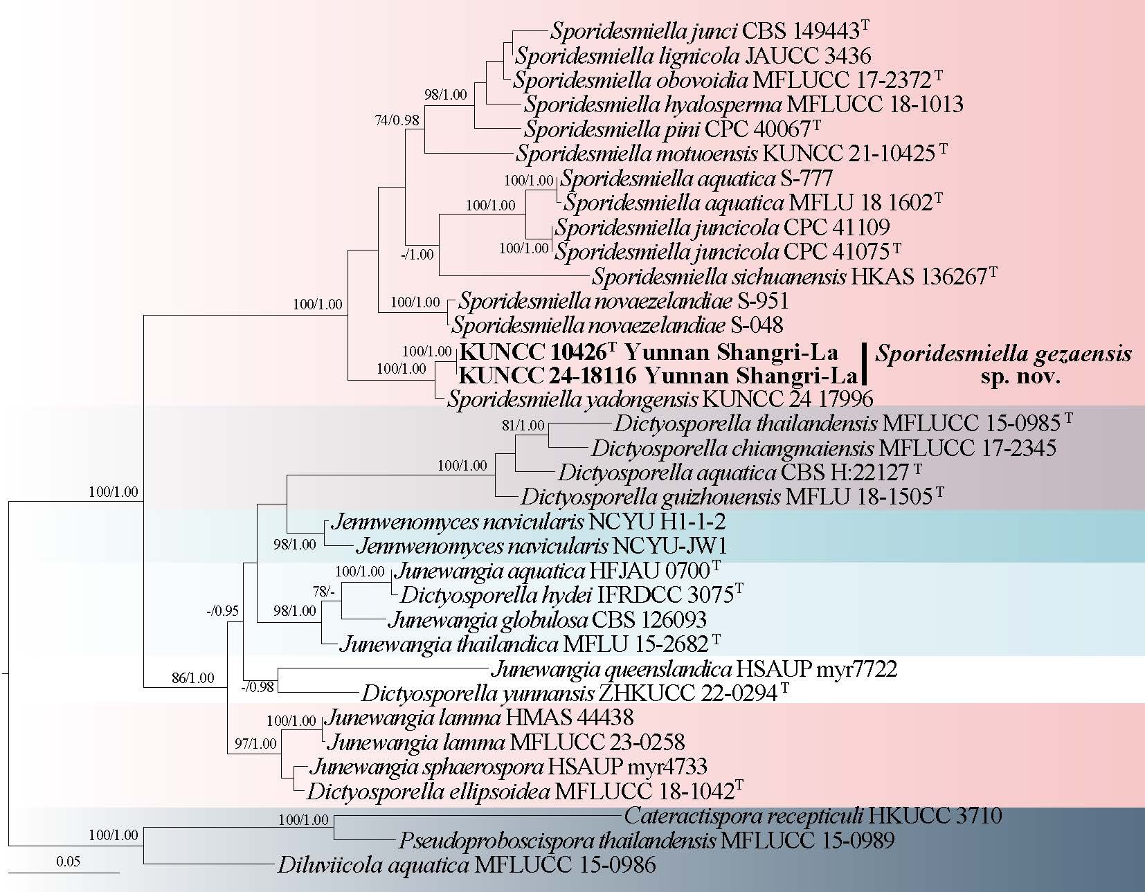
Entomopathogenic fungi, also known as insect-pathogenic fungi, play a crucial role in the natural regulation of insect populations. Among the 47 genera documented in this study, Akanthomyces, Beauveria, Cordyceps, Metarhizium, Ophiocordyceps, Papiliomyces, Perennicordyceps, Pleurocordyceps, Polycephalomyces and Samsoniella are recognized as typical entomopathogenic fungi, known for their ability to infect and kill insects (Wang et al. 2022; Xiao et al. 2023). Beauveria is one of the most well-known entomopathogenic fungi, particularly Beauveria bassiana. The discovery of new species, such as Cordyceps biclavata, Ophiocordyceps fusispora, O. hutiaoxiaensis, O. multiseptata, O. polyphialidica, O. yuhongii and O. sporangifera, highlights the region's potential as a reservoir of unexplored fungal diversity.
The pathogenicity of fungi is a topic of significant interest, particularly in the context of plant and animal health. While many fungi are benign or beneficial, some can cause diseases in plants, animals, or humans. In this study, the pathogenic potential of newly identified species was assessed, with particular attention given to those that may impact agriculture or natural ecosystems. For example, Fusarium altiplanum, F. rubellum and Corynespora xinjiangensis are known for their pathogenicity in plants, which could have implications for local agriculture and plant conservation efforts. Understanding the pathogenicity of these fungi is crucial for developing strategies to manage and mitigate their impacts. The newly identified fungi from this study exhibit a range of ecological types, including saprophytic, parasitic, and symbiotic relationships. Saprobic fungi, such as Trichoderma habaense and T. xinpingense, play a vital role in decomposing organic matter, thereby contributing to nutrient cycling within their ecosystems.
The classification and taxonomy of fungi are continually evolving fields, driven by advances in molecular biology and phylogenetic analyses. This study utilized robust phylogenetic analyses to confirm the taxonomic placements of the newly described species within the established fungal classification framework. These analyses not only validated the morphological assessments but also provided insights into the evolutionary relationships between different fungal taxa. Future studies could focus on the ecological roles of fungi in nutrient cycling and ecosystem functioning, the potential applications of fungi in biotechnology and medicine, and the impacts of climate change on fungal diversity and distribution. Additionally, there is a need for continued documentation and conservation efforts to protect the unique fungal communities of this region.
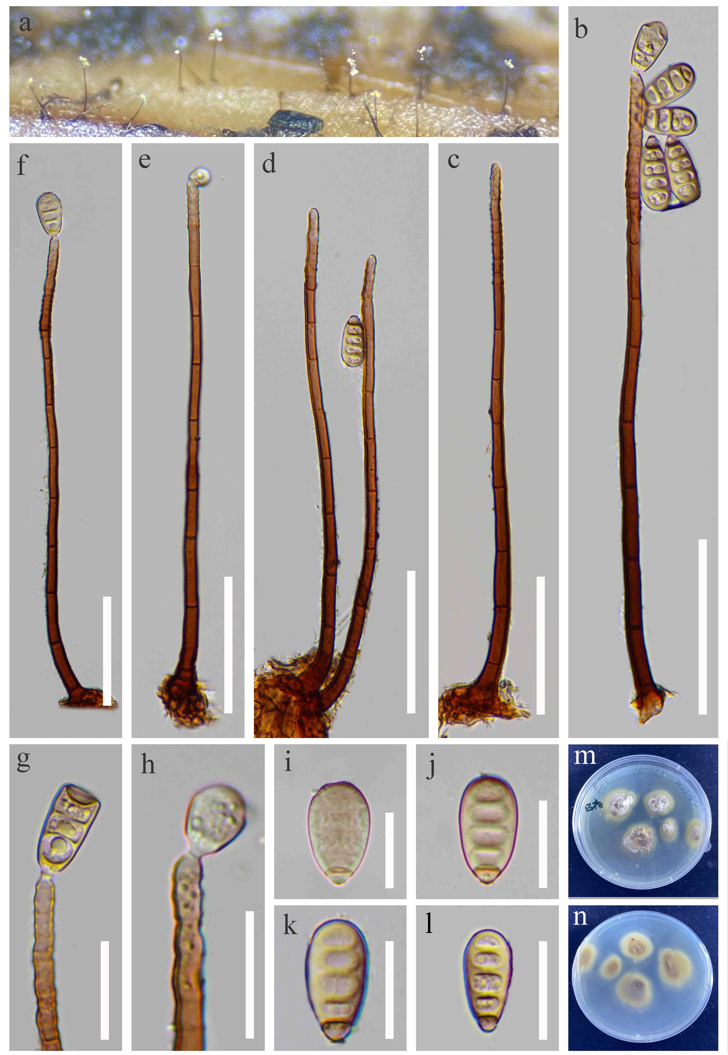
Acknowledgments
The whole work was funded by the Second Tibetan Plateau Scientific Expedition and Research Program (Grant No. 2024QZKK02010303), the National Natural Science Foundation of China (32470015, U2202205, 32060007), the Key project at central government level: the ability establishment of sustainable use for valuable Chinese medicine resources (2060302), the Major Science and Technology Special Project of Yunnan Province (202502AE090044), the Yunnan Basic Research Special Project (202401AS070030), the Technological Innovation Talent Cultivation Project Zhao Qi of Yunnan Province (202405AD350054), the Innovation Guidance and Technology-Based Enterprise Cultivation Program of Yunnan Province Science and Technology Department (No. 202504BI090008), the Yunnan Province Zhao Qi Expert Workstation (Yunnan Bofan Biotechnology Co., Ltd.), and the Yunnan Province Zhao Qi Expert Workstation (Chengjiang Green Field Agricultural Technology Service Co., Ltd.). Kevin D Hyde and Fatima Al-Otibi extend their appreciation to the ongoing Research Funding Program (ORF-2026-114), King Saud University, Riyadh, Saudi Arabia. We thank Mr. Peng Gao, Mr. Xue-Hua Zhang (Lasa, Xizang), Mr. Jia-Xi Pan (Shantou, Guangdong) for helping with the field trips in Xizang and Xinjiang, China.
Author contributions
Conceptualization, Xu RJ, Wang YB, Chen BZ and Zhao Q; methodology, Xu RJ, Wang YB, Chen BZ and Li JN; formal analysis, Hyde KD, Zhao Q and Yang ZL; resources, Xu RJ, Wang YB, Chen BZ, Li JN, He SC, Shen HW, Luo ZL, Lu YZ, Ma J, Xiao YP, Li Y, Yang HD, Zhao YW, Fan Q, Liao HF, Wei CY, Tang M, Wang XM and Al-Otibi F; data curation, Xu RJ, Wang YB, Chen BZ and Li JN; writing—original draft preparation, Xu RJ, Wang YB, Chen BZ and Zhao Q; writing—review and editing, Zhao Q, Wang YB, Hyde KD and Yang ZL; supervision, Zhao Q, Wang YB, Hyde KD and Yang ZL; project administration, Zhao Q, Hyde KD and Wang YB; funding acquisition, Zhao Q, Hyde KD and Wang YB. All authors have read and agreed to the published version of the manuscript.
ORCID
Rong-Ju Xu: https://orcid.org/0000-0002-3968-8442
Yuan-Bing Wang: https://orcid.org/0000-0002-3305-9418
Bao-Zheng Chen: https://orcid.org/0009-0005-7214-4913
Kevin D. Hyde: https://orcid.org/0000-0002-2191-0762
Conflict of interest statement
The author list includes members of the Editorial Board of Fungal Diversity. They were not involved in the journal’s review of, or decisions related to, this manuscript. The authors declare no competing interests.
Data availability
The datasets generated during and/or analyzed during the current study are available from the corresponding author on reasonable request.
Supplementary Information
The online version contains supplemental information available at https://doi.org/10.65390/fdiv.2026.136012.
Rights and permissions
The Author(s) 2026. Published by BioAcademic Press on behalf of Kunming Institute of Botany, Chinese Academy of Sciences (CAS) and Mushroom Research Foundation. This is an open access article under the Creative Commons Attribution license (http://creativecommons.org/licenses/by/4.0), which permits use, distribution and reproduction in any medium, provided the original work is properly cited.
References
- Abdel-Wahab MA, Pang KL, Nagahama T, Abdel-Aziz FA, Jones EBG (2010) Phylogenetic evaluation of anamorphic species of Cirrenalia and Cumulospora with the description of eight new genera and four new species. Mycological Progress 9: 537–558.
https://doi.org/10.1007/s11557-010-0661-x - Abdollahzadeh J, Groenewald JZ, Coetzee MPA, Wingfield MJ, Crous PW (2020) Evolution of lifestyles in Capnodiales. Studies in Mycology 95: 381–414.
https://doi.org/10.1016/j.simyco.2020.02.004 - Agrawal Y, Mual P, Shenoy B (2014) Multi-gene genealogies reveal cryptic species Beauveria rudraprayagi sp. nov. from India. Mycosphere 5: 719–736.
https://doi.org/10.5943/mycosphere/5/6/3 - Ai CC, Ma J, Zhang K, Castañeda-Ruíz RF, Zhang XG (2019) Cordana meilingensis and C. lushanensis spp. nov. from Jiangxi, China. Mycotaxon 134: 329–334.
https://doi.org/10.5248/134.329 - Alcorn JL (1988) The taxonomy of “Helminthosporium” species. Annual Review of Phytopathology 26: 37–56.
https://doi.org/10.1146/annurev.py.26.090188.000345 - Ando K (1992) A study of terrestrial aquatic hyphomycetes. Nippon Kingakkai Kaiho 33: 415–425.
- Ariyawansa HA, Hyde KD, Jayasiri SC, Buyck B, Chethana KWT, Dai DQ, Dai YC, Daranagama DA, Jayawardena RS, Lücking R et al (2015) Fungal diversity notes 111–252—taxonomic and phylogenetic contributions to fungal taxa. Fungal Diversity 75: 27–274.
https://doi.org/10.1007/s13225-015-0346-5 - Arzanlou M, Groenewald J, Gams W, Braun U, Shin HD, Crous PW (2007) Phylogenetic and morphotaxonomic revision of Ramichloridium and allied genera. Studies in Mycology 58: 57–93.
https://doi.org/10.3114/sim.2007.58.03 - Badali H, Gueidan C, Najafzadeh MJ, Bonifaz A, van den Ende AHG, de Hoog GS (2008) Biodiversity of the genus Cladophialophora. Studies in Mycology 61: 175–191.
https://doi.org/10.3114/sim.2008.61.18 - Ban S, Sakane T, Toyama K, Nakagiri A (2009) Teleomorph–anamorph relationships and reclassification of Cordyceps cuboidea and its allied species. Mycoscience 50: 261–272.
https://doi.org/10.1007/S10267-008-0480-Y - Bánki O, Roskov Y, Döring M, Ower G, Hernández Robles DR, Plata Corredor CA, Stjernegaard Jeppesen T, Örn A, Pape T, Hobern D et al (2025) Catalogue of Life (Annual Checklist 2025). Catalogue of Life Foundation, Amsterdam, Netherlands.
https://doi.org/10.48580/dgr6n - Bao DF, Hyde KD, Maharachchikumbura SSN, Perera RH, Thiyagaraja V, Hongsanan S, Wanasinghe DN, Shen HW, Tian XG, Yang LQ et al (2023) Taxonomy, phylogeny and evolution of freshwater Hypocreomycetidae (Sordariomycetes). Fungal Diversity 121: 1–94.
https://doi.org/10.1007/s13225-023-00521-8 - Bao DF, Hyde KD, McKenzie EHC, Jeewon R, Su HY, Nalumpang S, Luo ZL (2021) Biodiversity of lignicolous freshwater hyphomycetes from China and Thailand and description of sixteen species. Journal of Fungi 7: 669.
https://doi.org/10.3390/jof7080669 - Bao DF, McKenzie EH, Bhat DJ, Hyde KD, Luo ZL, Shen HW, Su HY (2020) Acrogenospora (Acrogenosporaceae, Minutisphaerales) appears to be a very diverse genus. Frontiers in Microbiology 11: 1606.
https://doi.org/10.3389/fmicb.2020.01606 - Berkeley MJ (1847) Gardeners’ Chronicle. London, 540 p (footnote)
- Berkeley MJ, Broome CE (1850) Notices of British fungi (438–501). Ann. Mag. Nat. Hist 5: 455–466.
https://doi.org/10.1080/03745486009494947 - Bhat DJ, Kendrick B (1993) Twenty-five new conidial fungi from the Western Ghats and the Andaman Islands (India). Mycotaxon 49: 19–90. https://doi.org/10.5962/p.416484
- Boonmee S, Calabon MS, Phookamsak R, Elgorban AM, Hyde KD (2020) Triseptata sexualis gen. et sp. nov. in Latoruaceae (Pleosporales). Phytotaxa 447: 252–264.
https://doi.org/10.11646/phytotaxa.447.4.3 - Boonmee S, D’souza MJ, Luo ZL, Pinruan U, Tanaka K, Su HY, Bhat DJ, McKenzie EHC, Jones EBG, Taylor JE et al (2016) Dictyosporiaceae fam. nov. Fungal Diversity 80: 457–482.
https://doi.org/10.1007/s13225-016-0363-z - Boonmee S, Wanasinghe DN, Calabon MS, Huanraluek N, Chandrasiri SKU, Jones GEB, Rossi W, Leonardi M, Singh SK, Rana S et al (2021) Fungal diversity notes 1387–1511: Taxonomic and phylogenetic contributions on genera and species of fungal taxa. Fungal Diversity 111: 1–335.
https://doi.org/10.1007/s13225-021-00489-3 - Booth C (1959) Studies of pyrenomycetes: IV. Nectria (part I). Mycological Papers 73: 1–115
- Booth C (1966) The genus Cylindrocarpon. Mycological Papers 104: 1–56.
- Borelli D (1980) Causal agents of chromoblastomycosis (chromomycetes). In: Proceedings of the Vth International Conference on Mycoses (Pan American Health Organization Scientific Publication 396). Pan American Health Organization, Washington, D.C. pp 334–335.
- Bresadola I (1897) Genus Mölleria Bres. critice disquisitum. Bolletino della Società Botanica Italiana 1897: 291–292.
- Bucher VVC, Hyde KD, Pointing SB, Reddy CA (2004) Production of wood decay enzymes, loss of mass, and lignin solubilization in wood by diverse tropical freshwater fungi. Fungal Diversity 15: 1–14.
https://doi.org/10.1007/s00248-003-0132-x - Buyck B, Zoller S, Hofstetter V (2018) Walking the thin lin ten years later: the dilemma of above- versus below-ground features to support phylogenies in the Russulaceae (Basidiomycota). Fungal Diversity 89: 267–292.
https://doi.org/10.1007/s13225-018-0397-5 - Cabral A, Groenewald JZ, Rego C, Oliveira H, Crous PW (2012a) Cylindrocarpon root rot: multi-gene analysis reveals novel species within the Ilyonectria radicicola species complex. Mycological Progress 11: 655–688.
https://doi.org/10.1007/s11557-011-0777-7 - Cabral A, Rego C, Nascimento T, Oliveira H, Groenewald JZ, Crous PW (2012b) Multi-gene analysis and morphology reveal novel Ilyonectria species associated with black foot disease of grapevines. Fungal Biology 116: 62–80.
https://doi.org/10.1016/j.funbio.2011.09.010 - Cai F, Druzhinina IS (2021) In honor of John Bissett: authoritative guidelines on molecular identification of Trichoderma. Fungal Diversity 107: 1–69.
https://doi.org/10.1007/s13225-020-00464-4 - Cai L (2004) New species of Cordana and Spadicoides from decaying bamboo culms in China. Sydowia 56: 222–228.
- Cai L, Tsui CKM, Zhang KQ, Hyde KD (2002) Aquatic fungi from Lake Fuxian, Yunnan, China. Fungal Diversity 9: 57–70.
- Cai L, Zhang KQ, McKenzie EHC, Hyde KD (2003) Freshwater fungi from bamboo and wood submerged in the Liput River in the Philippines. Fungal Diversity 13: 1–12.
- Calduch M, Gené J, Stchigel AM, Cano JF, Guarro J (2004) Ramophialophora, a new anamorphic genus of Sordariales. Studies in Mycology 50: 83–88.
- Castañeda RRF, Heredia G (2000) Two new dematiaceous hyphomycetes on Cyathea from Mexico. Cryptogamie Mycologie 21: 221–228.
https://doi.org/10.1016/S0181-1584(00)01047-2 - Castañeda RRF, Iturriaga T, Guarro J (1999) A new species of Cordana from Venezuela. Mycotaxon 73: 1–8. https://doi.org/10.5962/p.418611
- Chang HS (1991) Sterigmatobotrys uniseptata sp. nov. from Taiwan. Mycological Research 95: 1142–1144.
https://doi.org/10.1016/S0953-7562(09)80563-7 - Chang RL, Wang YC, Liu YY, Wang YR, Li SG, Zhao GY, Zhang SS, Dai MX, Zheng XX, Bose T et al (2023) Nine new species of black lichenicolous fungi from the genus Cladophialophora (Chaetothyriales) from two different climatic zones of China. Frontiers in Microbiology 14: 1191818.
https://doi.org/10.3389/fmicb.2023.1191818 - Chaverri P, Liu M, Hodge KT (2008) A monograph of the entomopathogenic genera Hypocrella, Moelleriella, and Samuelsia gen. nov. (Ascomycota, Hypocreales, Clavicipitaceae), and their Aschersonia-like anamorphs in the Neotropics. Studies in Mycology 60: 1–66.
https://doi.org/10.3114/sim.2008.60.01 - Chaverri P, Salgado C, Hirooka Y, Rossman AY, Samuels GJ (2011) Delimitation of Neonectria and Cylindrocarpon (Nectriaceae, Hypocreales, Ascomycota) and related genera with Cylindrocarpon-like anamorphs. Studies in Mycology 68: 57–78.
https://doi.org/10.3114/sim.2011.68.03 - Chen JL, Tzean SS (2000) Conioscypha taiwaniana sp. nov. and several new records of the genus from Taiwan. Botanical Bulletin of Academia Sinica 41: 315–322.
- Chen QT, Xiao SR, Shi ZY (1984) Paecilomyces sinensis sp. nov. and its connection with Cordyceps sinensis. Acta Mycologica Sinica 3: 24–28.
- Chen WH, Liu C, Han YF, Liang JD, Tian WY, Liang ZQ (2019) Three novel insect-associated species of Simplicillium (Cordycipitaceae, Hypocreales) from Southwest China. MycoKeys 58: 83–102.
https://doi.org/10.3897/mycokeys.58.37176 - Chen YP, Tian WH, Guo YB, Madrid H, Maharachchikumbura SSN (2022) Synhelminthosporium gen. et sp. nov. and two new species of Helminthosporium (Massarinaceae, Pleosporales) from Sichuan Province, China. Journal of Fungi 8: 712.
https://doi.org/10.3390/jof8070712 - Chethana KWT, Manawasinghe IS, Hurdeal VG, Bhunjun CS, Appadoo MA, Gentekaki E, Raspé O, Promputtha I, Hyde KD (2021a) What are fungal species and how to delineate them? Fungal Diversity 109: 1–25.
https://doi.org/10.1007/s13225-021-00483-9 - Chethana KWT, Niranjan M, Dong W, Samarakoon MC, Bao DF, Calabon MS, Chuankid B, Dayarathne MC, de Silva NI, Devadatha B et al (2021b) AJOM new records and collections of fungi: 101–150. Asian Journal of Mycology 4: 113–260.
https://doi.org/10.5943/ajom/4/1/8 - Christiansen MS (1993) Chalara lichenicola n. sp. (Deuteromycotina), a lichenicolous hyphomyete from Svalbard. Nordic Journal of Botany 13: 309–312.
https://doi.org/10.1111/j.1756-1051.1993.tb00054.x - Chuang WY, Lin YC, Shrestha B, Luangsa-Ard JJ, Stadler M, Tzean SS, Wu S, Ko CC, Hsieh SY, Wu ML et al (2024) Phylogenetic diversity and morphological characterization of cordycipitaceous species in Taiwan. Studies in Mycology 109: 1–56.
https://doi.org/10.3114/sim.2024.109.01 - Chuaseeharonnachai C, Somrithipol S, Suetrong S, Klaysuban A, Pornputtapong N, Jones EBG, Boonyuen N (2017) Conioscypha nakagirii, a new species from naturally submerged wood in Thailand based on morphological and molecular data. Mycoscience 58: 424–431.
https://doi.org/10.1016/j.myc.2017.06.003 - Cometto A, Leavitt SD, Grube M, De Hoog S, Muggia L (2023) Tackling fungal diversity in lichen symbioses: molecular and morphological data recognize new lineages in Chaetothyriales (Eurotiomycetes, Ascomycota). Mycological Progress 22: 53.
https://doi.org/10.1007/s11557-023-01901-9 - Cooke M (1889) New Australian fungi. Grevillea 18: 1–8
- Corda AC (1837) Icones fungorum hucusque cognitorum. JG Calve, Prague.
- Crous PW, Carris LM, Giraldo A, Groenewald JZ, Hawksworth DL, Hemández-Restrepo M, Jaklitsch WM, Lebrun MH, Schumacher RK, Stielow JB et al (2015a) The genera of fungi-fixing the application of the type species of generic names-G 2: Allantophomopsis, Latorua, Macrodiplodiopsis, Macrohilum, Milospium, Protostegia, Pyricularia, Robillarda, Rotula, Septoriella, Torula, and Wojnowicia. IMA Fungus 6: 163–198.
https://doi.org/10.5598/imafungus.2015.06.01.11 - Crous PW, Osieck ER, Jurjević Ž, Boers J, Iperen van AL, Starink-Willemse M, Dima B, Balashov S, Bulgakov TS, Johnston PR et al (2021) Fungal Planet description sheets: 1284–1382. Persoonia 47: 178–374.
https://doi.org/10.3767/persoonia.2021.47.06 - Crous PW, Schumacher RK, Wingfield MJ, Lombard L, Giraldo A, Christensen M, Gardiennet A, Nakashima C, Pereira OL, Smith AJ et al (2015b) Fungal systematics and evolution: FUSE 1. Sydowia 67: 81–118.
https://doi.org/10.12905/0380.sydowia67-2015-0081 - Crous PW, Shivas RG, Quaedvlieg W, van der Bank M, Zhang Y, Summerell BA, Guarro J, Wingfield MJ, Wood AR, Alfenas AC et al (2014) Fungal Planet description sheets: 214–280. Persoonia 32: 184–306.
https://doi.org/10.3767/003158514X682395 - Crous PW, Wingfield MJ, Jurjevic Z, Balashov S, Osieck ER, Marin-Felix Y, Luangsa-Ard JJ, Mejia LC, Cappelli A, Parra LA et al (2024) Fungal Planet description sheets: 1697-1780. Fungal Systematics and Evolution 14: 325–577.
https://doi.org/10.3114/fuse.2024.14.19. - Crous PW, Wingfield MJ, Richardson DM, Le Roux JJ, Strasberg D, Edwards J, Roets F, Hubka V, Taylor PWJ, Heykoop M et al (2016) Fungal Planet description sheets: 400–468. Persoonia 36: 316–458.
https://doi.org/10.3767/003158516X692185 - Crous PW, Wingfield MJ, Schumacher RK, Akulov A, Bulgakov TS, Carnegie AJ, Jurjević Ž, Decock C, Denman S, Lombard L et al (2020) New and interesting fungi. 3. Fungal Systematics and Evolution 6: 157–231.
https://doi.org/10.3114/fuse.2020.06.09 - Damm U, Fourie P, Crous PW (2010) Coniochaeta (Lecythophora), Collophora gen. nov. and Phaeomoniella species associated with wood necroses of Prunus trees. Persoonia 24: 60–80.
https://doi.org/10.3767/003158510X500705 - de Hoog GS, Guarro J, Gené J, Figueras MJ (2000) Atlas of clinical fungi, 2nd edn. Centraalbureau voor Schimmelcultures, Utrecht.
- de Hoog GS, van Oorschot C, Hijwegen T (1983) Taxonomy of the Dactylaria complex II. Dissoconium gen. nov. and Cordana preuss. Proc. Kon. Ned. Akad. Wet. Series C 86: 197–206.
- DiCosmo F, Berch S, Kendrick B (1983) Cylindrotrichum, Chaetopsis, and two new genera of Hyphomycetes, Kylindria and Xenokylindria. Mycologia 75: 949–973.
https://doi.org/10.1080/00275514.1983.12023781 - Dissanayake LS, Samarakoon MC, Maharachchikumbura SSN, Hyde KD, Tang X, Li QR, Mortimer PE, Faraj T, Xu JC, Kang JC et al (2024) Exploring the taxonomy and phylogeny of Sordariomycetes taxa emphasizing Xylariomycetidae in Southwestern China. Mycosphere 15: 1675–1793.
https://doi.org/10.5943/mycosphere/15/1/15 - Doilom M, Dissanayake AJ, Wanasinghe DN, Boonmee S, Liu JK, Bhat DJ, Taylor JE, Bahkali AH, McKenzie EH, Hyde KD (2017) Microfungi on Tectona grandis (teak) in Northern Thailand. Fungal Diversity 82: 107–182.
https://doi.org/10.1007/s13225-016-0368-7 - Dong W, Hyde KD, Jeewon R, Doilom M, Yu XD, Wang GN, Liu NG, Hu DM, Nalumpang S, Zhang H (2021) Towards a natural classification of annulatascaceae-like taxa Ⅱ: Introducing five new genera and eighteen new species from freshwater. Mycosphere 12: 1–88.
https://doi.org/10.5943/mycosphere/12/1/1 - Dong W, Wang B, Hyde KD, McKenzie EH, Raja HA, Tanaka K, Abdel-Wahab MA, Abdel-Aziz FA, Doilom M, Phookamsak R (2020) Freshwater Dothideomycetes. Fungal Diversity 105: 319–575.
https://doi.org/10.1007/s13225-020-00463-5 - Earle FS (1901) Collections of Alabama fungi. In: Mohr C (ed) Plant life of Alabama. Contributions from the United States National Herbarium, pp 10–23.
- Ellis MB (1971a) Dematiaceous hyphomycetes. Commonwealth Mycological Institute, Kew, Surrey, England.
- Ellis MB (1971b) Dematiaceous hyphomycetes. Dematiaceous hyphomycetes.
https://doi.org/10.1079/9780851986180.0000 - Ertz D, Heuchert B, Braun U, Freebury CE, Common RS, Diederich P (2016) Contribution to the phylogeny and taxonomy of the genus Taeniolella, with a focus on lichenicolous taxa. Fungal Biology 120: 1416–1447.
https://doi.org/10.1016/j.funbio.2016.05.008 - Fries EM (1849) Sectio posterior. In: Fries EM (ed) Summa vegetabilium Scandinaviae. A. Bonnier, Holmiae, pp 259–572.
- García D, Stchigel AM, Cano J, Calduch M, Hawksworth DL, Guarro J (2006) Molecular phylogeny of Coniochaetales. Mycological Research 110: 1271–1289.
https://doi.org/10.1016/j.mycres.2006.07.007 - Goga N (2000) Periconia circinata a new pathogen of roots and stem base of wheat in northwestern Romania. Analele Institutului de Cercetări pentru Cereale și Plante Tehnice, Fundulea 67: 205–214
- Goh TK, Ho WH, Hyde KD, Umali T (1997) New records and species of Sporoschisma and Sporoschismopsis from submerged wood in the tropics. Mycological Research 101: 1295–1307.
https://doi.org/10.1017/S0953756297003973 - Goh TK, Hyde KD (1998a) A new hyphomycete genus, Conioscyphopsis, from wood submerged in a freshwater stream and a review of Conioscypha. Mycological Research 102: 308–312.
https://doi.org/10.1017/S0953756297004942 - Goh TK, Hyde KD, Ho WH (1998b) Aquaphila albicans gen. et sp. nov., a hyphomycete from submerged wood in the tropics. Mycological Research 102: 587–592.
https://doi.org/10.1017/S0953756297005303 - Goh TK, Hyde KD, Ho WH (1999) A revision of the genus Dictyosporium, with descriptions of three new species. Fungal Diversity 2: 65–100.
- Goh TK, Hyde KD, Tsui KM (1998c) The hyphomycete genus Acrogenospora, with two new species and two new combinations. Mycological Research 102: 1309–1315.
https://doi.org/10.1017/S0953756298006790 - Gordon TR (2017) Fusarium oxysporum and the Fusarium wilt syndrome. Annual Review of Phytopathology 55: 23–39.
https://doi.org/10.1146/annurev-phyto-080615-095919 - Halleen F, Schroers HJ, Groenewald JZ, Rego C, Oliveira H, Crous PW (2006) Neonectria liriodendri sp. nov., the main causal agent of black foot disease of grapevines. Studies in Mycology 55: 227–234.
https://doi.org/10.3114/sim.55.1.227 - Harrington AH, del Olmo-Ruiz M, U’Ren JM, Garcia K, Pignatta D, Wespe N, Sandberg DC, Huang YL, Hoffman MT, Arnold AE (2019) Coniochaeta endophytica sp. nov., a foliar endophyte associated with healthy photosynthetic tissue of Platycladus orientalis (Cupressaceae). Plant and Fungal Systematics 64: 65–79.
https://doi.org/10.2478/pfs-2019-0008 - Harrington AH, Sarmiento C, Zalamea PC, Dalling JW, Davis AS, Arnold AE (2022) Acrogenospora terricola sp. nov., a fungal species associated with seeds of pioneer trees in the soil seed bank of a lowland forest in Panama. International Journal of Systematic and Evolutionary Microbiology 72, 005558.
https://doi.org/10.1099/ijsem.0.005558 - Hashimoto AH, Matsumura M, Hirayama K, Tanaka K (2017) Revision of Lophiotremataceae (Pleosporales, Dothideomycetes): Aquasubmersaceae, Cryptocoryneaceae, and Hermatomycetaceae fam. nov. Persoonia 39: 51–73.
https://doi.org/10.3767/persoonia.2017.39.03 - He SC, Hyde KD, Jayawardena RS, Thiyagaraja V, Wanasinghe DN, Zhao YW, Wang ZY, Cai T, Yang YY, Al-Otibi F et al (2025) Taxonomic contributions to Pleosporales and Kirschsteiniotheliales from the Xizang Autonomous Region, China. Mycology 1–38.
https://doi.org/10.1080/21501203.2025.2493072 - He SC, Thiyagaraja V, Bhunjun CS, Jayawardena RS, Chomnunti P, Al-Otibi F, Zhao YW, Zhao Q, Hyde KD (2024c) A new Myxospora (Hypocreales, Sordariomycetes) species from the Tibetan Plateau, China. New Zealand Journal of Botany 1–17.
https://doi.org/10.1080/0028825X.2024.2438411 - He SC, Wei DP, Bhunjun CS, Apurillo CCS, Thiyagaraja V, Jayawardena RS, Zhao YW, Zhao Q (2024a) A new species of Neotorula (Pleosporales) from the Southwest, China. Phytotaxa 662: 251–261.
https://doi.org/10.11646/phytotaxa.662.3.4 - He SC, Wei DP, Bhunjun CS, Jayawardena RS, Thiyagaraja V, Zhao Q, Fatimah Al-Otibi, Hyde KD (2024b) Morphology and multi-gene phylogeny reveal a new species of family Torulaceae from Yunnan Province, China. Diversity 16: 551.
https://doi.org/10.3390/d16090551. - Heredia G, Gamboa-Angulo M, Arias RM, Castañeda-Ruiz RF (2015) Two new species of Spadicoides and Sporidesmiella from Yucatán, Mexico. Mycotaxon 130: 511–516.
https://doi.org/10.5248/130.511 - Hernández-Gutiérrez A, Sutton BC (1997) Imimyces and Linkosia, two new genera segregated from Sporidesmium sensu lato, and redescription of Polydesmus. Mycological Research 101: 201–209.
https://doi.org/10.1017/S0953756296002419 - Hernández-Restrepo M, Gené J, Mena-Portales J, Cano J, Madrid H, Castañeda-Ruiz RF, Guarro J (2014) New species of Cordana and epitypification of the genus. Mycologia 106: 723–734.
https://doi.org/10.3852/13-122 - Hernández-Restrepo M, Giraldo A, Van Doorn R, Wingfield M, Groenewald J, Barreto R, Colmán A, Mansur P, Crous P (2020) The Genera of Fungi G6: Arthrographis, Kramasamuha, Melnikomyces, Thysanorea, and Verruconis. Fungal Systematics and Evolution 6: 1–24.
https://doi.org/10.3114/fuse.2020.06.01 - Höhnel F (1909) Fragmente zur Mykologie VI. Mitteilung, Nr. 182 bis 288. Sitzungsber. Kaiserliche Akademie der Wissenschaften in Wien, Mathematisch-Naturwissenschaftliche Klasse. Abteilung I 118: 275–452.
- Hongsanan S, Hyde KD, Phookamsak R, Wanasinghe DN, McKenzie EHC, Sarma VV, Boonmee S, Lücking R, Bhat DJ, Liu NG et al (2020) Refined families of Dothideomycetes: Orders and families incertae sedis in Dothideomycetidae. Mycosphere 11: 1553–2107.
https://doi.org/10.5943/mycosphere/11/1/13. - Hsieh SY, Goh TK, Kuo CH (2021) A taxonomic revision of Stanjehughesia (Chaetosphaeriaceae, Sordariomycetes), with a novel species S. kaohsiungensis from Taiwan. Phytotaxa 484: 261–280.
https://doi.org/10.11646/phytotaxa.484.3.2 - Hu DM, Wang M, Cai L (2017) Phylogenetic assessment and taxonomic revision of Mariannaea. Mycological Progress 16: 271–283.
https://doi.org/10.1007/s11557-016-1252-2 - Hu JJ, Zhao GP, Tuo YL, Dai D, Guo DZ, Rao G, Qi ZX, Zhang ZH, Li Y, Zhang B (2021) Morphology and molecular study of three new Cordycipitoid fungi and its related species collected from Jilin Province, northeast China. MycoKeys 83: 161–180.
https://doi.org/10.3897/mycokeys.83.72325 - Huang ZY, Shen HW, Su XJ, Luo ZL (2024) Phyllosticta cangshanensis sp. nov., an endophytic fungus from Rhododendron decorum in Cangshan Mountain, Yunnan Province, China. Phytotaxa 678: 146–166.
https://doi.org/10.11646/phytotaxa.678.3.1 - Hughes SJ (1951) Stachylidium, Gonytrichum, Mesobotrys, Chaetopsis and Chaetopsella. Transactions of the British Mycological Society 34: 551–576.
https://doi.org/10.1016/s0007-1536(51)80041-x - Hughes SJ (1958) Revisiones hyphomycetum aliquot cum appendice de nominibus rejiciendis. Canadian Journal of Botany 36: 727–836.
https://doi.org/10.1139/b58-067 - Hughes SJ (1966) 6. Sporoschisma Berk. and Br. New Zealand Journal of Botany 4: 77–85.
https://doi.org/10.1080/0028825X.1966.10443955 - Hughes SJ (1978) New Zealand fungi 25. Miscellaneous species. New Zealand Journal of Botany 16: 311–370.
https://doi.org/10.1080/0028825X.1978.10425143 - Huhndorf SM, Miller AN, Fernández FA (2004) Molecular systematics of the Sordariales: the order and the family Lasiosphaeriaceae redefined. Mycologia 96: 368–387.
https://doi.org/10.1080/15572536.2005.11832982 - Hyde KD, Chaiwan N, Norphanphoun C, Boonmee S, Camporesi E, Chethana KWT, Dayarathne MC, de Silva NI, Dissanayake AJ, Ekanayaka AH et al (2018) Mycosphere notes 169-224. Mycosphere 9: 271–430.
https://doi.org/10.5943/mycosphere/9/2/8 - Hyde KD, de Silva NI, Jeewon R, Bhat DJ, Phookamsak R, Doilom M, Boonmee S, Jayawardena RS, Maharachchikumbura SSN, Senanayake IC et al (2020a) AJOM new records and collections of fungi: 1–100. Asian Journal of Mycology 3: 22–294.
https://doi.org/10.5943/ajom/3/1/3 - Hyde KD, Dong Y, Phookamsak R, Jeewon R, Bhat DJ, Jones EBG, Liu NG, Abeywickrama PD, Mapook A, Wei DP et al (2020b) Fungal diversity notes 1151–1276: taxonomic and phylogenetic contributions on genera and species of fungal taxa. Fungal Diversity 100: 5–277.
https://doi.org/10.1007/s13225-020-00439-5 - Hyde KD, Hongsanan S, Jeewon R, Bhat DJ, McKenzie EHC, Jones EBG, Phookamsak R, Ariyawansa HA, Boonmee S, Zhao Q et al (2016) Fungal diversity notes 367-490: taxonomic and phylogenetic contributions to fungal taxa. Fungal Diversity 80: 1–270.
https://doi.org/10.1007/s13225-016-0373-x - Hyde KD, Jones EBG, Liu JK, Ariyawansa HA, Boehm E, Boonmee S, Braun U, Chomnunti P, Crous PW, Dai DQ et al (2013) Families of Dothideomycetes. Fungal Diversity 63: 1–313.
https://doi.org/10.1007/s13225-013-0263-4 - Hyde KD, Noorabadi MT, Thiyagaraja V, He MQ, Johnston PR, Wijesinghe SN, Armand A, Biketova AY, Chethana KWT, Erdoğdu M et al (2024a) The 2024 Outline of Fungi and fungus-like taxa. Mycosphere 15: 5146–6239.
https://doi.org/10.5943/mycosphere/15/1/25 - Hyde KD, Norphanphoun C, Abreu VP, Bazzicalupo A, Thilini Chethana K, Clericuzio M, Dayarathne MC, Dissanayake AJ, Ekanayaka AH, He MQ et al (2017) Fungal diversity notes 603–708: taxonomic and phylogenetic notes on genera and species. Fungal Diversity 87: 1–235.
https://doi.org/10.1007/s13225-017-0391-3 - Hyde KD, Norphanphoun C, Ma J, Yang HD, Zhang JY, Du TY, Gao Y, Gomes de Farias A, Gui H, He SC et al (2023) Mycosphere notes 387-412 novel species of fungal taxa from around the world. Mycosphere 14: 663–744.
https://doi.org/10.5943/mycosphere/14/1/8 - Hyde KD, Norphanphoun C, Maharachchikumbura SSN, Bhat DJ, Jones EBG, Bundhun D, Chen YJ, Bao DF, Boonmee S, Calabon MS et al (2020c) Refined families of Sordariomycetes. Mycosphere 11: 305–1059.
https://doi.org/10.5943/mycosphere/11/1/7 - Hyde KD, Tennakoon DS, Jeewon R, Bhat DJ, Maharachchikumbura SSN, Rossi W, Leonardi M, Lee HB, Mun HY, Houbraken J et al (2019) Fungal diversity notes 1036–1150: taxonomic and phylogenetic contributions on genera and species of fungal taxa. Fungal Diversity 96: 1–242.
https://doi.org/10.1007/s13225-019-00429-2 - Hyde KD, Wijesinghe SN, Afshari N, Aumentado HD, Bhunjun CS, Boonmee S, Camporesi E, Chethana KWT, Doilom M, Dong W et al (2024b) Mycosphere notes 469–520. Mycosphere 15: 1294–1454.
https://doi.org/10.5943/mycosphere/15/1/11 - Imoulan A, Wu HJ, Lu WL, Li Y, Li BB, Yang RH, Wang WJ, Wang XL, Kirk PM, Yao YJ (2016) Beauveria medogensis sp. nov., a new fungus of the entomopathogenic genus from China. Journal of Invertebrate Pathology 139: 74–81.
https://doi.org/10.1016/j.jip.2016.07.006 - Isola D, Zucconi L, Onofri S, Caneva G, De Hoog GS, Selbmann L (2016) Extremotolerant rock inhabiting black fungi from Italian monumental sites. Fungal Diversity 76: 75–96.
https://doi.org/10.1007/s13225-015-0342-9 - Jaklitsch WM (2011) European species of Hypocrea part II: species with hyaline ascospores. Fungal Diversity 48: 1–250.
https://doi.org/10.1007/s13225-011-0088-y - Jaklitsch WM, Voglmayr H (2015) Biodiversity of Trichoderma (Hypocreaceae) in Southern Europe and Macaronesia. Studies in Mycology 80: 1–87.
https://doi.org/10.1016/j.simyco.2014.11.001 - Japanese Cordyceps Society (2018) Field Guide to the Natural World of Cordyceps. Human Think Tank Publishing, Japan. https://doi.org/10.56021/9781421412030
- Jayasiri SC, Hyde KD, Ariyawansa HA, Bhat DJ, Buyck B, Cai L, Dai YC, Abd-Elsalam KA, Ertz D, Hidayat I et al (2015) The Faces of Fungi database: fungal names linked with morphology, phylogeny and human impacts. Fungal Diversity 74: 3–18.
https://doi.org/10.1007/s13225-015-0351-8 - Jayasiri SC, Hyde KD, Jones EBG, McKenzie EHC, Jeewon R, Phillips AJL, Bhat DJ, Wanasinghe DN, Liu JK, Lu YZ et al (2019) Diversity, morphology and molecular phylogeny of Dothideomycetes on decaying wild seed pods and fruits. Mycosphere 10: 1–186.
https://doi.org/10.5943/mycosphere/10/1/1 - Jayasiri SC, Hyde KD, Jones EBG, Peršoh D, Camporesi E, Kang JC (2018) Taxonomic novelties of hysteriform Dothideomycetes. Mycosphere 9: 803–837.
https://doi.org/10.5943/mycosphere/9/4/8 - Jayawardena RS, Bhunjun CS, Hyde KD, Gentekaki E, Itthayakorn P (2021) Colletotrichum: lifestyles, biology, morpho-species, species complexes and accepted species. Mycosphere 12: 519–669.
https://doi.org/10.5943/mycosphere/12/1/7 - Jayawardena RS, Hyde KD, Jeewon R, Ghobad-Nejhad M, Wanasinghe DN, Liu NG, Phillips AJL, Oliveira-Filho JRC, da Silva GA, Gibertoni TB et al (2019) One stop shop II: taxonomic update with molecular phylogeny for important phytopathogenic genera: 26–50 (2018). Fungal Diversity 94: 41–129.
https://doi.org/10.1007/s13225-019-00418-5 - Jayawardena RS, Hyde KD, Wang S, Sun YR, Suwannarach N, Sysouphanthong P, Abdel-Wahab MA, Abdel-Aziz FA, Abeywickrama PD, Abreu VP et al (2022) Fungal diversity notes 1512–1610: Taxonomic and phylogenetic contributions on genera and species of fungal taxa. Fungal Diversity 117: 1–272.
https://doi.org/10.1007/s13225-022-00513-0 - Jeewon R, Hyde KD (2016) Establishing species boundaries and new taxa among fungi: recommendations to resolve taxonomic ambiguities. Mycosphere 7: 1669–1677.
https://doi.org/10.5943/mycosphere/7/11/4 - Jong S, Davis E (1971) The genus Sterigmatobotrys. Norwegian Journal of Botany 18: 177–181.
- Kedves O, Kocsubé S, Bata T, Andersson MA, Salo JM, Mikkola R, Salonen H, Szűcs A, Kedves A, Kónya Z et al (2021) Chaetomium and chaetomium-like species from European indoor environments include Dichotomopilus finlandicus sp. nov. Pathogens 10: 1133.
https://doi.org/10.3390/pathogens10091133 - Kendrick WB (1961) The Leptographium complex. Phialocephala gen. nov. Canadian Journal of Botany 39: 1079–1085.
https://doi.org/10.1139/b61-094 - Kepler RM, Humber RA, Bischoff JF, Rehner SA (2014) Clarification of generic and species boundaries for Metarhizium and related fungi through multigene phylogenetics Mycologia 106: 811–829.
https://doi.org/10.3852/13-319 - Kepler RM, Sung GH, Ban S, Nakagiri A, Chen MJ, Huang B, Li Z, Spatafora JW (2012) New teleomorph combinations in the entomopathogenic genus Metacordyceps. Mycologia 104: 182–197.
https://doi.org/10.3852/11-070 - Khao-Ngam S, Mongkolsamrit S, Rungjindamai N, Noisripoom W, Pooissarakul W, Duangthisan J, Himaman W, Luangsa-ard JJ (2021) Ophiocordyceps asiana and Ophiocordyceps tessaratomidarum (Ophiocordycipitaceae, Hypocreales), two new species on stink bugs from Thailand. Mycological Progress 20: 341–353.
https://doi.org/10.1007/s11557-021-01684-x - Khonsanit A, Noisripoom W, Mongkolsamrit S, Phosrithong N, Luangsa-ard JJ (2021) Five new species of Moelleriella infecting scale insects (Coccidae) in Thailand. Mycological Progress 20: 847–867.
https://doi.org/10.1007/s11557-021-01709-5 - Kirk PM (1982) New or interesting microfungi VI. Sporidesmiella gen. nov. (Hyphomycetes). Transactions of the British Mycological Society 79: 479–489.
https://doi.org/10.1016/S0007-1536(82)80040-5 - Kirk PM (1986) New or interesting microfungi. XV. Miscellaneous hyphomycetes from the British Isles. Transactions of the British Mycological Society 86: 409–428. https://doi.org/10.5962/p.419001
- Kiyuna T, An KD, Kigawa R, Sano C, Sugiyama J (2017) Two new Cladophialophora species, C. tumbae sp. nov. and C. tumulicola sp. nov., and chaetothyrialean fungi from biodeteriorated samples in the Takamatsuzuka and Kitora Tumuli. Mycoscience 59: 75–84.
https://doi.org/10.1016/j.myc.2017.08.008 - Kobayasi Y (1939) On the genus Cordyceps and its allies on cicadae from Japan. Bulletin of the Biogeographical Society of Japan 9: 145–176.
- Kobayasi Y (1941) The genus Cordyceps and its allies. Science Reports of the Tokyo Bunrika Daigaku 5: 53–260.
- Kobayasi Y, Shimizu D (1983) Cordyceps species from Japan. 6. Bulletin of the National Science Museum Tokyo 9: 1–21.
- Kohlmeyer J, Volkmann-Kohlmeyer B, Eriksson OE (1997) Fungi on Juncus roemerianus. 9. New obligate and facultative marine Ascomycotina. Botanica Marina 40: 291–300.
https://doi.org/10.1515/botm.1997.40.1-6.291 - Konta S, Hyde KD, Karunarathna SC, Mapook A, Senwanna C, Dauner LAP, Nanayakkara CM, Xu, JC, Tibpromma S, Lumyong S (2021) Multi-gene phylogeny and morphology reveal Haplohelminthosporium gen. nov. and Helminthosporiella gen. nov. associated with palms in Thailand and a checklist for Helminthosporium reported worldwide. Life 11: 454.
https://doi.org/10.3390/life11050454 - Konta S, Tibpromma S, Karunarathna SC, Samarakoon MC, Steven LS, Mapook A, Boonmee S, Senwanna C, Balasuriya A, Eungwanichayapant PD et al (2023) Morphology and multigene phylogeny reveal ten novel taxa in Ascomycota from terrestrial palm substrates (Arecaceae) in Thailand. Mycosphere 14: 107–152.
https://doi.org/10.5943/mycosphere/14/1/2 - Koukol O, Delgado G, Hofmann TA, Piepenbring M (2018) Panama, a hot spot for Hermatomyces (Hermatomycetaceae, Pleosporales) with five new species, and a critical synopsis of the genus. IMA Fungus 9: 107–141.
https://doi.org/10.5598/imafungus.2018.09.01.08 - Latgé JP, Chamilos G (2019) Aspergillus fumigatus and Aspergillosis in 2019. Clinical Microbiology Reviews 33: 10–1128.
https://doi.org/10.1128/CMR.00140-18. - Leslie JF, Summerell BA (2006) Species descriptions. In: The Fusarium Laboratory Manual. Blackwell Publishing, Ames, Iowa, pp 121–278.
https://doi.org/10.1002/9780470278376.ch13. - Li JF, Phookamsak R, Jeewon R, Bhat DJ, Mapook A, Camporesi E, Shang QJ, Chukeatirote E, Bahkali AH, Hyde KD (2017a) Molecular taxonomy and morphological characterization reveal new species and new host records of Torula species (Torulaceae, Pleosporales). Mycological Progress 16: 447–461.
https://doi.org/10.1007/s11557-017-1292-2 - Li JN, Wang SY, Xu RJ, Xu KE, Ma CH, Zhao Q, Zhu YA (2023) Tetraploa lignicola, a new freshwater fungal species from Yunnan Province, China. Phytotaxa 629: 245–254.
https://doi.org/10.11646/phytotaxa.629.3.6 - Li JN, Xu RJ, Xu K, Zhao Q, Zhu YA (2024a) Distoseptispora motuoensis (Distoseptisporaceae), a new freshwater hyphomycetous species from Xizang Autonomous Prefecture, China. Phytotaxa 675: 122–134.
https://doi.org/10.11646/phytotaxa.675.2.3 - Li L, Du HZ, Thiyagaraja V, Bhat DJ, Phookamsak R, Cheewangkoon R (2024b) Two novel freshwater hyphomycetes, in Acrogenospora (Minutisphaerales, Dothideomycetes) and Conioscypha (Conioscyphales, Sordariomycetes) from Southwestern China. MycoKeys 101: 249–273.
https://doi.org/10.3897/mycokeys.101.115209 - Li WL, Luo ZL, Liu JK, Bhat DJ, Bao DF, Su HY, Hyde KD (2017b) Lignicolous freshwater fungi from China I: Aquadictyospora lignicola gen. et sp. nov. and new record of Pseudodictyosporium wauense from northwestern Yunnan Province. Mycosphere 8: 1587–1597.
https://doi.org/10.5943/mycosphere/8/10/1 - Li XH, Liu YL, Song HY, Hu DM, Gao Y, Hu HJ, Zhou JP (2021) Sporidesmiella lignicola sp. nov., a new hyphomycetous fungus from freshwater habitats in China. Biodiversity Data Journal 9: e77414.
https://doi.org/10.3897/BDJ.9.e77414 - Li Y, He SC, Li CJ, Yu FM, Luangharn T, Zhao Q (2025) Phaeoisaria yadongensis sp. nov. (Pleurotheciaceae) from Xizang Autonomous Region, China. Phytotaxa 710: 249–260.
https://doi.org/10.11646/phytotaxa.710.3.2 - Liao CF, Doilom M, Jeewon R, Hyde KD, Manawasinghe IS, Chethana KWT, Balasuriya A, Thakshila SAD, Luo M, Mapook A et al (2025) Challenges and update on fungal endophytes: classification, definition, diversity, ecology, evolution and functions. Fungal Diversity 131: 301–367.
https://doi.org/10.1007/s13225-025-00550-5 - Liao CF, Hyde KD, Thilini Chethana KW, Dong W, Yang YH, Doilom M (2024a) Three New Periconia Species Isolated from Wurfbainia villosa in Guangdong, China: A Discussion on the Doubtful Taxa Clustering in this Genus. Diversity 16: 141.
https://doi.org/10.3390/d16030141 - Liao CF, Yang YH, Dong W, Tangtrakulwanich K, Zhang YX, Khan S, Nadir S, Chethana KWT, Doilom M (2024b) Tetraploa wurfbainiae sp. nov. (Tetraplosphaeriaceae, Pleosporales) isolated from Wurfbainia villosa in Guangdong, China. New Zealand Journal of Botany 62: 303–316.
https://doi.org/10.1080/0028825X.2024.2311964 - Liao HF, Su PP, Chen ZM, Huang LD, Fan Q, Yang QL, Yang ZL, Hu WH, Wang YB (2025) Ophiocordyceps polystromata, a new entomopathogenic fungus from the Gaoligong Mountains in western Yunnan, China. New Zealand Journal of Botany 63: 1657–1667.
https://doi.org/10.1080/0028825X.2025.2526189 - Link HF (1809) Observationes in ordines plantarum naturales. Dissertatio I. Magazin der Gesellschaft Naturforschender Freunde zu Berlin 3: 3–42
- Link HF (1816) Observationes in ordines plantarum naturales. Dissertatio II, sistens nuperas de Mucedinum et Gastromycorum ordinibus observationes. Der Gesellschaft naturforschender Freunde zu Berlin Magazin für die neuesten Entdeckungen in der gesammten Naturkunde 7: 25–45.
- Liu F, Wang J, Li H, Wang W, Cai L (2019a) Setophoma spp. on Camellia sinensis. Fungal Systematics and Evolution 4: 43–57.
https://doi.org/10.3114/fuse.2019.04.05 - Liu JK, Hyde KD, Jones EBG, Ariyawansa HA, Bhat DJ, Boonmee S, Maharachchikumbura SSN, McKenzie EHC, Phookamsak, R, Phukhamsakda C et al (2015a) Fungal diversity notes 1–110: taxonomic and phylogenetic contributions to fungal species. Fungal Diversity 72: 1–197.
https://doi.org/10.1007/s13225-015-0324-y - Liu JW, Hu YF, Luo XX, Castañeda-Ruíz RF, Ma J (2022) Three novel species of Helminthosporium (Massarinaceae, Pleosporales) from China. MycoKeys 94: 73–89.
https://doi.org/10.3897/mycokeys.94.95888 - Liu JW, Hu YF, Luo XX, Castañeda-Ruíz RF, Xia JW, Xu ZH, Cui RQ, Shi XG, Zhang LH, Ma J (2023a) Molecular phylogeny and morphology reveal four novel species of Corynespora and Kirschsteiniothelia (Dothideomycetes, Ascomycota) from China: A checklist for Corynespora reported worldwide. Journal of Fungi 9: 107.
https://doi.org/10.3390/jof9010107 - Liu LL, Song LC, Gu XF, Wei QQ, Zhang M, Liu ZY, Gou JL (2023b) Dictyosporium duliujiangense sp. nov. (Dictyosporiaceae, Pleosporales) from freshwater habitat in Guizhou Province, China. Phytotaxa 606: 259–272.
https://doi.org/10.11646/phytotaxa.606.4.2 - Liu NG, Bhat DJ, Hyde KD, Liu JK (2019b) Conioscypha tenebrosa sp. nov. (Conioscyphaceae) from China and notes on Conioscypha species. Phytotaxa 413: 159–171.
https://doi.org/10.11646/phytotaxa.413.2.5 - Liu NG, Hongsanan S, Yang J, Bhat DJ, Liu JK, Jumpathong J, Liu ZY (2017) Periconia thailandica (Periconiaceae), a new species from Thailand. Phytotaxa 323: 253–263.
https://doi.org/10.11646/phytotaxa.323.3.4 - Liu NG, Hyde KD, Sun YR, Bhat DJ, Jones EBG, Jumpathong J, Lin CG, Lu YZ, Yang J, Liu LL et al (2024a) Notes, outline, taxonomy and phylogeny of brown-spored hyphomycetes. Fungal Diversity 129: 1–281.
https://doi.org/10.1007/s13225-024-00539-6 - Liu SL, Wang XW, Li GJ, Deng CY, Rossi W, Leonardi M, Liimatainen K, Kekki T, Niskanen T, Smith ME et al (2024b) Fungal diversity notes 1717–1817: taxonomic and phylogenetic contributions on genera and species of fungal taxa. Fungal Diversity 124: 1–216.
https://doi.org/10.1007/s13225-023-00529-0 - Liu XY, Udayanga D, Luo ZL, Chen LJ, Zhou DQ, Su HY, Hyde KD (2015b) Backbone tree for Chaetothyriales with four new species of Minimelanolocus from aquatic habitats. Fungal Biology 119: 1046–1062.
https://doi.org/10.1016/j.funbio.2015.08.005 - Liu ZH, Tang DX, Lu YL, Zhu JY, Luo LJ, Sun T, Yu H (2024c) Morphology and phylogeny of four new species within Polycephalomycetaceae (Hypocreales) parasitising Ophiocordyceps species. MycoKeys 105: 179–202.
https://doi.org/10.3897/mycokeys.105.119893 - Locquin M (1984) Mycologie générale et structurale. Elsevier Masson, Paris, pp. 1–551.
- Lombard L, van Der Merwe A, Groenewald JZ, Crous PW (2014) Lineages in Nectriaceae: re-evaluating the generic status of Ilyonectria and allied genera. Phytopathologia Mediterranea 53: 515–532.
https://doi.org/10.14601/Phytopathol_Mediterr-14976 - Lu YZ, Liu JK, Hyde KD, Jeewon R, Kang JC, Fan C, Boonmee S, Bhat DJ, Luo ZL, Lin CG et al (2018) A taxonomic reassessment of Tubeufiales based on multi-locus phylogeny and morphology. Fungal Diversity 92: 131–344.
https://doi.org/10.1007/s13225-018-0411-y - Luo J, Yin JF, Cai L, Zhang KQ, Hyde KD (2004) Freshwater fungi in Lake Dianchi, a heavily polluted lake in Yunnan, China. Fungal Diversity 16: 93–112.
- Luo ZL, Hyde KD, Bhat DJ, Jeewon R, Maharachchikumbura SSN, Bao DF, Li WL, Su XJ, Yang XY, Su HY (2018) Morphological and molecular taxonomy of novel species Pleurotheciaceae from freshwater habitats in Yunnan, China. Mycological Progress 17: 511–530.
https://doi.org/10.1007/s11557-018-1377-6 - Luo ZL, Hyde KD, Liu JK, Maharachchikumbura SSN, Jeewon R, Bao DF, Bhat DJ, Lin CG, Li WL, Yang J et al (2019) Freshwater Sordariomycetes. Fungal Diversity 99: 451–660.
https://doi.org/10.1007/s13225-019-00438-1 - Luttrell ES (1964) Systematics of Helminthosporium and related genera. Mycologia 56: 119–132.
- Ma J, Zhang YD, Ma LG, Castañeda-Ruíz RF, Zhang XG (2012) Three new species of Sporidesmiella from southern China. Mycoscience 53: 187–193.
https://doi.org/10.1007/s10267-011-0152-1 - Maharachchikumbura SSN, Chen YP, Ariyawansa HA, Hyde KD, Haelewaters D, Perera RH, Samarakoon MC, Wanasinghe DN, Bustamante DE, Liu JK et al (2021) Integrative approaches for species delimitation in Ascomycota. Fungal Diversity 109: 155–179.
https://doi.org/10.1007/s13225-021-00486-6 - Maharachchikumbura SSN, Jones EBG, McKenzie EHC, Stadler M, Lee HB, Samarakoon MC, Ekanayaka AH, Camporesi E, Liu JK, Liu ZY (2020) Fungi on wild seeds and fruits. Mycosphere 11: 2108–2480.
https://doi.org/10.5943/mycosphere/11/1/14 - Maharachchikumbura SSN, Luo ZL, Su HY, Al-Sadi AM, Cheewangkoon R (2018) Reticulascaceae hyphomycetes from submerged wood in Yunnan, China. Phytotaxa 348: 187–198.
https://doi.org/10.11646/phytotaxa.348.3.2 - Malloch D, Cain RF (1970) The genus Kernia. Canadian Journal of Botany 49: 855–867.
https://doi.org/10.1139/b71-126 - Malloch D, Cain RF (1971) New cleistothecial Sordariaceae and a new family, Coniochaetaceae. Canadian Journal of Botany 49: 869–880.
https://doi.org/10.1139/b71-127 - Mao XL, Jiang CP, Ouzhu CW (1993) Economic Macrofungi in Tibet. Beijing: Beijing Science and Technology Press.
- Matočec N, Kušan I, Ozimec R (2014) The genus Polycephalomyces (Hypocreales) in the frame of monitoring Veternica cave (Croatia) with a new segregate genus Perennicordyceps. Ascomyceteorg 6: 125–133.
https://doi.org/10.25664/art-0114 - Matsushima T (1975) Icones Microfungorum a Matsushima Lectorum. Published by the author, Kobe, Japan.
- McKenzie EHC, Pinnoi A, Wong MKM, Hyde KD, Jones EBG (2002) Two new hyaline Chalara species and a key to species described since 1975. Fungal Diversity 11: 129–139.
- Miller MA, Pfeiffer W, Schwartz T (2012) The CIPRES science gateway: enabling high-impact science for phylogenetics researchers with limited resources. In: Proceedings of the 1st Conference of the Extreme Science and Engineering Discovery Environment: Bridging from the extreme to the campus and beyond pp. 1–8.
https://doi.org/10.1145/2335755.2335836 - Mongkolsamrit S, Khonsanit A, Noisripoom W, Luangsa-ard JJ (2015) Two new entomogenous species of Moelleriella with perithecia in tubercles from Thailand. Mycoscience 56: 66–74.
https://doi.org/10.1016/j.myc.2014.03.002 - Mongkolsamrit S, Khonsanit A, Thanakitpipattana D, Tasanathai K, Noisripoom W, Lamlertthon S, Himaman W, Houbraken J, Samson RA, Luangsa-ard J (2020) Revisiting Metarhizium and the description of new species from Thailand. Studies in Mycology 95: 171–251.
https://doi.org/10.1016/j.simyco.2020.04.001 - Monteiro JS, Carmo LTd, Fiúza PO, Ottoni BMdP, Gusmão LFP (2014) New species of microfungi from Brazilian Amazon rainforests. Mycotaxon 127: 81–87.
https://doi.org/10.5248/127.81 - Nag Raj TR, Kendrick B (1975) A monograph of Chalara and allied genera. Wilfrid Laurier University Press, Waterloo
- Nannizzi A (1934) Repertorio sistematico dei miceti dell'uomo e degli animali. SA Poligrafica Meini
- Niego AGT, Lambert C, Mortimer P, Thongklang N, Rapior S, Grosse M, Schrey H, CharriaGirón E, Walker A, Hyde KD et al (2023a) The contribution of fungi to the global economy. Fungal Diversity 121: 95–137.
https://doi.org/10.1007/s13225-023-00520-9 - Niego AGT, Rapior S, Thongklang N, Raspé O, Hyde KD, Mortimer P (2023b) Reviewing the contributions of macrofungi to forest ecosystem processes and services. Fungal Biology Reviews 44: 100294.
https://doi.org/10.1016/j.fbr.2022.11.002 - Nieuwland JA (1916) Critical notes on new and old genera of plants. VIII. The American Midland Naturalist 4: 379–386.
https://doi.org/10.2307/2992735 - Norphanphoun C, Hongsanan S, Doilom M, Bhat DJ, Wen TC, Senanayake IC, Bulgakov TS, Hyde KD (2016) Lamproconiaceae fam. nov. to accommodate Lamproconium desmazieri. Phytotaxa 270: 89–102.
https://doi.org/10.11646/phytotaxa.270.2.2 - Obase K, Douhan GW, Matsuda Y, Smith ME (2015) Cladophialophora floridana and Cladophialophora tortuosa, new species isolated from sclerotia of Cenococcum geophilum in forest soils of Florida, USA. Mycoscience 57: 26–34.
https://doi.org/10.1016/j.myc.2015.07.005 - Onofri S, Pagano S, Zucconi L (1994) Conidiogenesis in Phialocephala humicola. Mycological Research 98: 745–748.
https://doi.org/10.1016/S0953-7562(09)81048-4 - Oudemans CAJA (1886) Contribution à la flore mycologique des Pay-Bas. XI. Nederlandsch Kruidkundig Archief serie 2 4: 502–562.
- Pem D, Hyde KD, McKenzie EHC, Hongsanan S, Wanasinghe DN, Boonmee S, Darmostuk V, Bhat JD, Tian Q, Htet ZH et al (2023) A comprehensive overview of genera in Dothideomycetes. Mycosphere 15: 2175–4568.
https://doi.org/10.5943/mycosphere/15/1/18 - Perdomo H, García D, Gené J, Cano J, Sutton DA, Summerbell R, Guarro J (2013) Phialemoniopsis, a new genus of Sordariomycetes, and new species of Phialemonium and Lecythophora. Mycologia 105: 398–421.
https://doi.org/10.3852/12-137 - Perera RH, Hyde KD, Jones EBG, Maharachchikumbura SSN, Bundhun D, Camporesi E, Akulov A, Liu JK, Liu ZY (2023) Profile of Bionectriaceae, Calcarisporiaceae, Hypocreaceae, Nectriaceae, Tilachlidiaceae, Ijuhyaceae fam. nov., Stromatonectriaceae fam. nov. and Xanthonectriaceae fam. nov. Fungal Diversity 118: 95–271.
https://doi.org/10.1007/s13225-022-00512-1 - Persoon CH (1795) Neuer versuch einer systematischen eintheilung der schwaümme. Neues Mag. Für Die Bot. Ihrem Ganzen Umfange 1: 63–128.
- Petch T (1933) Notes on entomogenous fungi. Vol XVIII. Part I. Transactions of the British Mycological Society 16: 55–75.
- Phookamsak R, Hyde KD, Jeewon R, Bhat DJ, Jones EBG, Maharachchikumbura SSN, Raspé O, Karunarathna SC, Wanasinghe DN, Hongsanan S et al (2019) Fungal diversity notes 929–1035: taxonomic and phylogenetic contributions on genera and species of fungi. Fungal Diversity 95: 1–273.
https://doi.org/10.1007/s13225-019-00421-w - Phookamsak R, Jiang H, Suwannarach N, Lumyong S, Xu JC, Xu S, Liao CF, Chomnunti P (2022) Bambusicolous fungi in Pleosporales: Introducing four novel taxa and a new habitat record for Anastomitrabeculia didymospora. Journal of Fungi 8: 1–35.
https://doi.org/10.3390/jof8060630 - Phookamsak R, Liu JK, McKenzie EHC, Manamgoda DS, Ariyawansa H, Thambugala KM, Dai DQ, Camporesi E, Chukeatirote E, Wijayawardene NN et al (2014) Revision of Phaeosphaeriaceae. Fungal Diversity 68: 159–238.
https://doi.org/10.1007/s13225-014-0308-3 - Phukhamsakda C, McKenzie EH, Phillips AJ, Jones EBG, Bhat DJ, Marc S, Bhunjun CS, Wanasinghe DN, Thongbai B, Camporesi E et al (2020) Microfungi associated with Clematis (Ranunculaceae) with an integrated approach to delimiting species boundaries. Fungal Diversity 102: 1–203.
https://doi.org/10.1007/s13225-020-00448-4 - Pratibha SJ, Gawas P, Hyde KD, Bhat DJ (2005) Chalara indica sp. nov. and Sorocybe indicus sp. nov. from India. Cryptogamie Mycologie 26: 97–103.
- Preuss CGT (1851) Uebersicht untersuchter Pilze besonders aus der Umgegend von Hoyerswerda. Linnaea 24: 99–153.
- Quaedvlieg W, Binder M, Groenewald J, Summerell B, Carnegie AJ, Burgess TI, Crous PW (2014) Introducing the consolidated species concept to resolve species in the Teratosphaeriaceae. Persoonia 33: 1–40.
https://doi.org/10.3767/003158514X681981 - Quaedvlieg W, Verkley GJM, Shin HD, Barreto RW, Alfenas AC, Swart WJ, Groenewald JZ, Crous PW (2013) Sizing up Septoria. Studies in Mycology 75: 307–390.
https://doi.org/10.3114/sim0017 - Quandt CA, Kepler RM, Gams W, Araújo JP, Ban S, Evans HC, Hughes D, Humber R, Hywel-Jones N, Li ZZ et al (2014) Phylogenetic-based nomenclatural proposals for Ophiocordycipitaceae (Hypocreales) with new combinations in Tolypocladium. IMA Fungus 5: 121–134.
https://doi.org/10.5598/imafungus.2014.05.01.12 - Rabenhorst GL (1844) Deutschlands Kryptogamen-Flora. 1: 1–61.
- Rabenhorst L (1889) Kryptogamen-Flora von Deutschland, Oesterreich und der Schweiz, vol 3. E. Kummer, Leipzig.
- Réblová M (1999a) Studies in Chaetosphaeria sensu lato I. The genera Chaetosphaerella and Tengiomyces gen. nov. of the Helminthosphaeriaceae. Mycotaxon 70: 387–420.
https://doi.org/10.5962/p.415033 - Réblová M (1999b) Studies in Chaetosphaeria sensu lato III. Umbrinosphaeria gen. nov. and Miyoshiella with Sporidesmium anamorphs. Mycotaxon 71: 13–43.
https://doi.org/10.5962/p.415088 - Réblová M, Gams W, Seifert K (2011a) Monilochaetes and allied genera of the Glomerellales, and a reconsideration of families in the Microascales. Studies in Mycology 68: 163–191.
https://doi.org/10.3114/sim.2011.68.07 - Réblová M, Hernández-Restrepo M, Sklenář F, Nekvindová J, Réblová K, Kolařík M (2022) Consolidation of Chloridium: new classification into eight sections with 37 species and reinstatement of the genera Gongromeriza and Psilobotrys. Studies in Mycology 103: 87–212.
https://doi.org/10.3114/sim.2022.103.04 - Réblová M, Miller AN, Rossman AY, Seifert KA, Crous PW, Hawksworth DL, Abdel-Wahab MA, Cannon PF, Daranagama DA, Beer ZWD et al (2016a) Recommendations for competing sexual-asexually typified generic names in Sordariomycetes (except Diaporthales, Hypocreales, and Magnaporthales). IMA Fungus 7: 131–153.
https://doi.org/10.5598/imafungus.2016.07.01.08 - Réblová M, Nekvindová J, Hernández-Restrepo M (2021) Reflections on Menisporopsis, Multiguttulispora and Tainosphaeria using molecular and morphological data. Journal of Fungi 7: 1–36.
https://doi.org/10.3390/jof7060438 - Réblová M, Nekvindová J, Miller AN, Hernández-Restrepo M (2024) Re-evaluation of Exserticlava and other genera in Chaetosphaeriaceae with chalara-, phaeostalagmus-, phialocephala-and stanjehughesia-like morphotypes. Persoonia 53: 62–99.
https://doi.org/10.3767/persoonia.2024.53.03 - Réblová M, Seifert K (2011b) Discovery of the teleomorph of the hyphomycete, Sterigmatobotrys macrocarpa, and epitypification of the genus to holomorphic status. Studies in Mycology 68: 193–202.
https://doi.org/10.3114/sim.2011.68.08 - Réblová M, Seifert K, Fournier J, Štěpánek V (2016b) Newly recognised lineages of perithecial ascomycetes: the new orders Conioscyphales and Pleurotheciales. Persoonia 37: 57–81.
https://doi.org/10.3767/003158516X689819 - Réblová M, Seifert KA (2004) Conioscyphascus, a new ascomycetous genus for holomorphs with Conioscypha anamorphs. Studies in Mycology 50: 95–108.
- Rehner SA, Minnis AM, Sung GH, Luangsa-ard JJ, Devotto L, Humber RA (2011) Phylogeny and systematics of the anamorphic, entomopathogenic genus Beauveria. Mycologia 103: 1055–1073.
https://doi.org/10.3852/10-302 - Ren GC, Wanasinghe DN, Monkai J, Mortimer PE, Hyde KD, Xu JC, Pang A, Gui H (2021) Novel saprobic Hermatomyces species (Hermatomycetaceae, Pleosporales) from China (Yunnan Province) and Thailand. MycoKeys 82: 57–79.
https://doi.org/10.3897/mycokeys.82.67973 - Robène-Soustrade I, Jouen E, Pastou D, Payet-Hoarau M, Goble T (2015) Description and phylogenetic placement of Beauveria hoplocheli sp. nov. used in the biological control of the sugarcane white grub, Hoplochelus marginalis, on Reunion Island. Mycologia 107: 1221–1232.
https://doi.org/10.3852/14-344 - Ronquist F, Teslenko M, Van Der Mark P, Ayres DL, Darling A, Höhna S, Larget B, Liu L, Suchard MA, Huelsenbeck JP (2012) MrBayes 3.2: efficient Bayesian phylogenetic inference and model choice across a large model space. Systematic Biology 61: 539–542.
https://doi.org/10.1093/sysbio/sys029 - Saccardo PA (1880) Conspectus generum fungorum Italiae inferorium. Michelia 2: 1–38.
- Samarakoon MC, Gafforov Y, Liu N, Maharachchikumbura SS, Bhat JD, Liu JK, Promputtha I, Hyde KD (2018) Combined multi-gene backbone tree for the genus Coniochaeta with two new species from Uzbekistan. Phytotaxa 336: 43–58.
https://doi.org/10.11646/phytotaxa.336.1.3 - Samson RA (1974) Paecilomyces and some allied Hyphomycetes. Studies in Mycology 6: 1–119.
- Samson RA, Bigg WL (1988) A new species of Mariannaea from California. Mycologia 80: 131–134.
https://doi.org/10.1080/00275514.1988.12025512 - Sangdee A, Sangdee K, Seephonkai P, Jaihan P, Kanyaphum T (2017) Colony characteristics, nucleoside analog profiles, and genetic variations of medicinal fungus Polycephalomyces nipponicus (ascomycetes) isolates from northeast Thailand. International Journal of Medicinal Mushrooms 19: 445–455.
https://doi.org/10.1615/IntJMedMushrooms.v19.i5.60 - Schroers HJ (2002) A monograph of Bionectria (Ascomycota, Hypocreales, Bionectriaceae) and its Clonostachys anamorph. Studies in Mycology 46: 1–211.
https://doi.org/10.1017/S0269915X03272177 - Seifert KA, Gams W (2011) The genera of Hyphomycetes–2011 update. Persoonia-Molecular Phylogeny and Evolution of Fungi 27: 119–129.
https://doi.org/10.3767/003158511X617435 - Senanayake IC, Rossi W, Leonardi M, Weir A, McHugh M, Rajeshkumar KC, Verma RK, Karunarathna SC, Tibpromma S, Ashtekar N et al (2023) Fungal diversity notes 1611–1716: taxonomic and phylogenetic contributions on fungal genera and species emphasis in south China. Fungal Diversity 122: 161–403.
https://doi.org/10.1007/s13225-023-00523-6 - Sharma R, Sharma R, Crous PW (2015) Matsushimamyces, a new genus of keratinophilic fungi from soil in central India. IMA fungus 6: 337–343.
https://doi.org/10.5598/imafungus.2015.06.02.05 - Shearer C, Motta J (1973) Ultrastructure and conidiogenesis in Conioscypha (Hyphomycetes). Canadian Journal of Botany 51: 1747–1751.
https://doi.org/10.1139/b73-226 - Shen HW, Bao DF, Luan S, Wanasinghe DN, Du TY, Hongsanan S, Yang J, Zhang JY, Tang X, Bhat JD et al (2025) Taxonomy and phylogeny of lignicolous freshwater fungi from plateau lakes in Yunnan Province, China. Fungal Diversity 134: 635–899.
https://doi.org/10.1007/s13225-025-00564-z - Shen HW, Bao DF, Wanasinghe DN, Boonmee S, Liu JK, Luo ZL (2022) Novel species and records of Dictyosporiaceae from freshwater habitats in China and Thailand. Journal of Fungi 8: 1200.
https://doi.org/10.3390/jof8111200 - Shen HW, Luo ZL, Bao DF, Luan S, Bhat DJ, Boonmee S, Wang WP, Su XJ, Li YX, Al-Otibi F et al (2024) Lignicolous freshwater fungi from China IV: Morphology and phylogeny reveal new species of Pleosporales from plateau lakes in Yunnan Province, China. Mycosphere 15: 6439–6524.
https://doi.org/10.5943/mycosphere/15/1/28 - Shenoy BD, Jeewon R, Wu WP, Bhat DJ, Hyde KD (2006) Ribosomal and RPB2 DNA sequence analyses suggest that Sporidesmium and morphologically similar genera are polyphyletic. Mycological Research 110: 916–928.
https://doi.org/10.1016/j.mycres.2006.06.004 - Shirouzu T, Harada Y (2004) Bambusicolous fungi in Japan (2): Phialosporostilbe gregariclava, a new anamorphic fungus from Sasa. Mycoscience 45: 390–394. https://doi.org/10.1007/s10267-004-0200-1
- Shrestha B (2011) Diversity of Cordyceps Fungi in Nepal. Nepal Journal of Science and Technology 12: 103–110.
https://doi.org/10.3126/njst.v12i0.6487 - Shrestha B, Sung JM (2005). Notes on Cordyceps species collected from the central region of Nepal. Mycobiology 33: 235–239.
https://doi.org/10.4489/MYCO.2005.33.4.235 - Sierra ÁM, Portales JM (1985) Nuevo género de hifomicete fialídico de Cuba. Revista del Jardín Botánico Nacional 6: 57–60.
- Silvestro D, Michalak I (2012) raxmlGUI: a graphical front-end for RAxML. Organisms Diversity & Evolution 12: 335–337.
https://doi.org/10.1007/s13127-011-0056-0 - Sivanesan A (1996) Corynesporasca caryotae gen. et sp. nov. with a Corynespora anamorph, and the family Corynesporascaceae. Mycological Research 100: 783–788.
https://doi.org/10.1016/S0953-7562(96)80022-0 - Sivanesan A, Sutton BC (1985) Microfungi on Xanthorrhoea. Transactions of the British Mycological Society 85: 239–255.
https://doi.org/10.1016/S0007-1536(85)80186-8 - Smith LJ, Datnoff L, Pernezny K, Schlub R (2007) Phylogenetic and pathogenic characterization of Corynespora cassiicola isolates. In II International Symposium on Tomato Diseases 808: 51–56.
https://doi.org/10.17660/ActaHortic.2009.808.6 - Soares DJ, Nechet KL, Barreto RW (2005) Cordana versicolor sp. nov. (Dematiaceous hyphomycete) causing leaf-spot on Canna denudata (Cannaceae) in Brazil, with observations on Cordana musae. Fungal Diversity 18: 147–155.
- Spatafora JW, Blackwell M (1993) Molecular systematics of unitunicate perithecial ascomycetes: the Clavicipitales–Hypocreales connection. Mycologia 85: 912–922.
https://doi.org/10.1080/00275514.1993.12026353 - Spegazzini C (1910) Mycetes Argentinenses (Series V). Anales del Museo Nacional de Historia Natural Buenos Aires 3: 329–467.
- Stamatakis A (2014) RAxML version 8: a tool for phylogenetic analysis and post-analysis of large phylogenies. Bioinformatics 30: 1312–1313.
https://doi.org/10.1093/bioinformatics/btu033 - Su HL, Hyde KD, Luo L, Zhao Q, Kandawatte WTC (2025) Four new species of Chlorociboria from Yunnan, China. Mycological Progress 24: 27.
https://doi.org/10.1007/s11557-025-02046-7 - Su HY, Hyde KD, Maharachchikumbura SSN, Ariyawansa HA, Luo ZL, Promputtha I, Tian Q, Lin CG, Shang QJ, Zhao YC et al (2016) The families Distoseptisporaceae fam. nov., Kirschsteiniotheliaceae, Sporormiaceae and Torulaceae, with new species from freshwater in Yunnan Province, China. Fungal Diversity 80: 375–409.
https://doi.org/10.1007/s13225-016-0362-0 - Su L, Zhu H, Niu YC, Guo YX, Du XP, Guo JG, Zhang L, Qin C (2020) Phylogeny and taxonomic revision of Kernia and Acaulium. Scientific Reports 10: 10302.
https://doi.org/10.1038/s41598-020-67347-1 - Su XJ, Luo ZL, Jeewon R, Bhat DJ, Bao DF, Li WL, Hao YE, Su HY, Hyde KD (2018) Morphology and multigene phylogeny reveal new genus and species of Torulaceae from freshwater habitats in northwestern Yunnan, China. Mycological Progress 17: 531–545.
https://doi.org/10.1007/s11557-018-1388-3. - Subramanian CV (1992) A reassessment of Sporidesmium (hyphomycetes) and some related taxa. Proc Indian Natn Sci Acad B58: 179–190.
- Sun T, Chen Y, Wang D, Dai Y, Zou W, Luo R, Dong QY, Yu, H (2024) Mitogenomics, phylogeny and morphology reveal two new entomopathogenic species of Ophiocordyceps (Ophiocordycipitaceae, Hypocreales) from south-western China. MycoKeys 109: 49–72.
https://doi.org/10.3897/mycokeys.109.124975 - Sun T, Zou W, Dong Q, Huang O, Tang D, Yu H (2022) Morphology, phylogeny, mitogenomics and metagenomics reveal a new entomopathogenic fungus Ophiocordyceps nujiangensis (Hypocreales, Ophiocordycipitaceae) from Southwestern China. MycoKeys 94: 91–108.
https://doi.org/10.3897/mycokeys.94.89425 - Sun W, Su L, Yang S, Sun JZ, Liu BJ, Fu R, Wu B, Liu XZ, Cai L, Xiang MC (2020) Unveiling the hidden diversity of rock-inhabiting fungi: Chaetothyriales from China. Journal of Fungi 6: 187.
https://doi.org/10.3390/jof6040187 - Sun YR, Hyde KD, Liu NG, Jayawardena RS, Wijayawardene NN, Ma J, Zhang Q, Al-Otibi F, Wang Y (2025) Micro-fungi in southern China and northern Thailand: emphasis on medicinal plants. Fungal Diversity 131: 99–299.
https://doi.org/10.1007/s13225-024-00549-4 - Sung GH, Sung JM, Hywel-Jones NL, Spatafora JW (2007) A multi-gene phylogeny of Clavicipitaceae (Ascomycota, Fungi): identification of localized incongruence using a combinational bootstrap approach. Molecular Phylogenetics and Evolution 44: 1204–1223.
https://doi.org/10.1016/j.ympev.2007.03.011 - Sureshkumar G, Sharath Babu K, Kunwar IK, Manoharachary C (2005) Two new hyphomycetous fungal species from India. Mycotaxon 92: 279–283.
https://doi.org/10.5962/p.414634 - Sutton DA, Rinaldi MG, Sanchez SE (2009) Dematiaceous fungi. In: Anaissie EJ, McGinnis MR, Pfaller MA (eds) Clinical Mycology, 2nd edn. Elsevier, Philadelphia, pp 329–354.
- Swofford DL, Sullivan J (2003) Phylogeny inference based on parsimony and other methods using PAUP*. In: Salemi M, Vandamme AM (eds) The Phylogenetic Handbook: A Practical Approach to DNA and Protein Phylogeny. Cambridge University Press, Cambridge, pp 160–206.
https://doi.org/10.1017/cbo9780511819049.010 - Tan YP, Bishop-Hurley SL, Marney TS, Shivas RG (2025) Index of Australian Fungi no. 53. Zenodo 1–14.
https://doi.org/10.5281/zenodo.15080534 - Tanaka K, Hirayama K, Yonezawa H, Hatakeyama S, Harada Y, Sano T, Shirouzu T, Hosoya T (2009) Molecular taxonomy of bambusicolous fungi: Tetraplosphaeriaceae, a new pleosporalean family with tetraploa-like anamorphs. Studies in Mycology 64: 175–209.
https://doi.org/10.3114/sim.2009.64.10 - Tanaka K, Hirayama K, Yonezawa H, Sato G, Toriyabe A, Kudo K, Hashimoto A, Matsumura M, Harada Y, Kurihara Y et al (2015) Revision of the Massarineae (Pleosporales, Dothideomycetes). Studies in Mycology 82: 75–136.
https://doi.org/10.1016/j.simyco.2015.10.002 - Tang J, Zhuang K, Ran X, Dai Y, Ran Y (2017) Chromoblastomycosis caused by Cladophialophora carrionii. Indian Journal of Dermatology, Venereology and Leprology 83: 482.
https://doi.org/10.4103/ijdvl.ijdvl_707_16 - Tang X, Jeewon R, Lu YZ, Alrefaei AF, Jayawardena RS, Xu RJ, Ma J, Chen XM, Kang JC (2023) Morphophylogenetic evidence reveals four new fungal species within Tetraplosphaeriaceae (Pleosporales, Ascomycota) from tropical and subtropical forest in China. MycoKeys 100: 171–204.
http://doi.org/10.3897/mycokeys.100.113141 - Tanney JB, Douglas B, Seifert KA (2016) Sexual and asexual states of some endophytic Phialocephala species of Picea. Mycologia 108: 255–280.
https://doi.org/10.3852/15-136 - Tanney JB, Seifert KA (2020) Mollisiaceae: An overlooked lineage of diverse endophytes. Studies in Mycology 95: 293–380.
https://doi.org/10.1016/j.simyco.2020.02.005 - Tennakoon DS, de Silva NI, Maharachchikumbura SS, Bhat DJ, Kumla J, Suwannarach N, Lumyong S (2023) Exploring More on Dictyosporiaceae: The Species Geographical Distribution and Intriguing Novel Additions from Plant Litter. Diversity 15: 410.
https://doi.org/10.3390/d15030410 - Tennakoon DS, Kuo CH, Maharachchikumbura SSN, Thambugala KM, Gentekaki E, Phillips AJL, Bhat DJ, Wanasinghe DN, de Silva NI, Promputtha I et al (2021) Taxonomic and phylogenetic contributions to Celtis formosana, Ficus ampelas, F. septica, Macaranga tanarius and Morus australis leaf litter inhabiting microfungi. Fungal Diversity 108: 1–215.
https://doi.org/10.1007/s13225-021-00474-w - Thanakitpipattana D, Tasanathai K, Mongkolsamrit S, Khonsanit A, Lamlertthon S, Luangsa-ard JJ (2020) Fungal pathogens occurring on Orthopterida in Thailand. Persoonia 44: 140–160.
https://doi.org/10.3767/persoonia.2020.44.06 - Thitla T, Kumla J, Hongsanan S, Senwanna C, Khuna S, Lumyong S, Suwannarach N (2023) Exploring diversity rock-inhabiting fungi from northern Thailand: a new genus and three new species belonged to the family Herpotrichiellaceae. Frontiers in Cellular and Infection Microbiology 13: 1252482.
https://doi.org/10.3389/fcimb.2023.1252482 - Thiyagaraja V, Hyde KD, Piepenbring M, Davydov EA, Dai DQ, Abdollahzadeh J, Bundhun D, Chethana KWT, Crous PW, Gajanayake AJ et al (2025) Orders of Ascomycota. Mycosphere 16: 536–1411.
https://doi.org/10.5943/mycosphere/16/1/8 - Tian WH, Liu JW, Jin Y, Chen YP, Zhou YF, Wu K, Su PW, Guo XY, Wanasinghe DN, Hyde KD et al (2024a) Morphological and phylogenetic studies of Ascomycota from gymnosperms in Sichuan Province, China. Mycosphere 15: 1794–1900.
https://doi.org/10.5943/mycosphere/15/1/16 - Tian XG, Bao DF, Karunarathna S, Jayawardena R, Hyde KD, Bhat DJ, Luo ZL, Elgorban AM, Hongsanan S, Rajeshkumar KC et al (2024b) Taxonomy and phylogeny of ascomycetes associated with selected economically important monocotyledons in China and Thailand. Mycosphere 15: 1–274.
https://doi.org/10.5943/mycosphere/15/1/1 - Tibpromma S, Bhat J, Doilom M, Lumyong S, Nontachaiyapoom S, Yang JB, Hyde KD (2016) Three new Hermatomyces species (Lophiotremataceae) on Pandanus odorifer from Southern Thailand. Phytotaxa 275: 127–139.
https://doi.org/10.11646/phytotaxa.275.2.4 - Tibpromma S, Hyde KD, Jeewon R, Maharachchikumbura SSN, Liu JK, Bhat DJ, Jones EBG, McKenzie EHC, Camporesi E, Bulgakov TS et al (2017) Fungal diversity notes 491–602: taxonomic and phylogenetic contributions to fungal taxa. Fungal Diversity 83: 1–261.
https://doi.org/10.1007/s13225-017-0378-0 - Tibpromma S, Hyde KD, McKenzie EH, Bhat DJ, Phillips AJL, Wanasinghe DN, Samarakoon MC, Jayawardena RS, Dissanayake AJ, Tennakoon DS et al (2018) Fungal diversity notes 840–928: micro-fungi associated with Pandanaceae. Fungal Diversity 93: 1–160.
https://doi.org/10.1007/s13225-018-0408-6 - Tode HJ (1790) Fungi mecklenburgenses selecti, vol 1. Apud Ioh. Fried. Guil. Lemke
https://doi.org/10.5962/bhl.title.148599 - Torbati M, Arzanlou M, Sandoval-Denis M, Crous PW (2019) Multigene phylogeny reveals new fungicolous species in the Fusarium tricinctum species complex and novel hosts in the genus Fusarium from Iran. Mycological Progress 18: 119–133.
https://doi.org/10.1007/s11557-018-1422-5 - Troy GC, Panciera DL, Pickett JP, Sutton DA, Gene J, Cano JF, Guarro J, Thompson EH, Wickes BL (2013) Mixed infection caused by Lecythophora canina sp. nov. and Plectosphaerella cucumerina in a German shepherd dog. Medical Mycology 51: 455–460.
https://doi.org/10.3109/13693786.2012.754998 - Tsui CK, Sivichai S, Rossman AY, Berbee ML (2007) Tubeufia asiana, the teleomorph of Aquaphila albicans in the Tubeufiaceae, Pleosporales, based on cultural and molecular data. Mycologia 99: 884–894.
https://doi.org/10.1080/15572536.2007.11832520 - Tubaki K, Kubono T (1989) Digitodochium, a new staurosporous anamorph genus. Sydowia 41: 344–348.
- Vázquez-Campos X, Kinsela AS, Waite TD, Collins RN, Neilan BA (2014) Fodinomyces uranophilus gen. nov. sp. nov. and Coniochaeta fodinicola sp. nov., two uranium mine-inhabiting Ascomycota fungi from northern Australia. Mycologia 106: 1073–1089.
https://doi.org/10.3852/14-013 - Visagie CM, Yilmaz N, Allison JD, Barreto RW, Boekhout T, Boers J, Delgado MA, Dewing C, Fitza KNE, Furtado ECA et al (2024) New and Interesting Fungi. 7. Fungal Systematics and Evolution 13: 441–494.
https://doi.org/10.3114/fuse.2024.13.12 - Voglmayr H, Jaklitsch WM (2017) Corynespora, Exosporium and Helminthosporium revisited-new species and generic reclassification. Studies in Mycology 87: 43–76.
https://doi.org/10.1016/j.simyco.2017.05.001 - Voglmayr H, Tello S, Jaklitsch WM, Friebes G, Baral HO, Fournier J (2022) About spirals and pores: Xylariaceae with remarkable germ loci. Persoonia 49: 58–98.
https://doi.org/10.3767/persoonia.2022.49.02 - von Höhnel FXR (1904) Mycologische Fragmente. Mycologische Fragmente 2: 38–60.
- Wan YL, Bao DF, Luo ZL, Bhat DJ, Xu YX, Su HY, Hao YE (2021) Two new species of Minimelanolocus (Herpotrichiellaceae, Chaetothyriales) from submerged wood in Yunnan, China. Phytotaxa 480: 45–56.
https://doi.org/10.11646/phytotaxa.480.1.4 - Wanasinghe DN, Maharachchikumbura SSN (2023) Exploring the diversity and systematics of Phaeosphaeriaceae: Taxonomic novelties from ecologically diverse habitats and their phylogenetic resolution. Journal of Fungi 9: 853.
https://doi.org/10.3390/jof9080853 - Wang RX, Luo ZL, Hyde KD, Bhat DJ, Su XJ, Su HY (2016a) New species and records of Dictyocheirospora from submerged wood in north-western Yunnan, China. Mycosphere 7: 1357–1367.
https://doi.org/10.5943/mycosphere/7/9/9 - Wang WP, Bhat DJ, Yang L, Shen HW, Luo ZL (2024a) New Species and Records of Pleurotheciaceae from Karst Landscapes in Yunnan Province, China. Journal of Fungi 10: 516.
https://doi.org/10.3390/jof10080516 - Wang WP, Hyde KD, Bao DF, Wanasinghe DN, Lin CG, Shen HW, Luo ZL (2024b) Lignicolous freshwater fungi from karst landscapes in Yunnan Province, China. Mycosphere 15: 6525–6640.
https://doi.org/10.5943/mycosphere/15/1/29 - Wang WP, Luan S, Shen HW, Liu NG, Li YX, Luo ZL (2024c) Two new freshwater dematiaceous hyphomycetes of Pleosporales from Yunnan Province, China. Phytotaxa 676: 63–74.
https://doi.org/10.11646/phytotaxa.676.1.4 - Wang WP, Shen HW, Bao DF, Jeewon R, Zhang ZQ, Yang LQ, Luo ZL (2025a) Biogeography and species diversity of freshwater Savoryellomycetidae (Sordariomycetes) fungi. Mycology 1–70.
https://doi.org/10.1080/21501203.2025.2509809. - Wang XH, Cai Q, Yu FM, Yang ZL, Zhou SY, Wang ZR, Cui YY, Wang Y, Zhang P, Cao SQ et al (2025b) New and notable taxa of Basidiomycota on the Qinghai-Xizang Plateau and its surrounding areas. Fungal Diversity 133: 235–437.
https://doi.org/10.1007/s13225-025-00558-x - Wang XW, Houbraken J, Groenewald JZ, Meijer M, Andersen B, Nielsen KF, Crous PW, Samson RA (2016b) Diversity and taxonomy of Chaetomium and chaetomium-like fungi from indoor environments. Studies in Mycology 84: 145–224.
https://doi.org/10.1016/j.simyco.2016.11.005 - Wang XW, Wang XL, Liu FJ, Zhao XM, Li J, Cai L (2014) Phylogenetic assessment of Chaetomium indicum and allied species, with the introduction of three new species and epitypification of C. funicola and C. indicum. Mycological Progress 13: 719–732.
https://doi.org/10.1007/s11557-013-0955-x - Wang Y, Bao DF, Shen HW, Yang L, Luo ZL (2024d) Appendopyricularia guttulata sp. nov., a lignicolous freshwater fungus from Dianchi Lake in Yunnan, China. Phytotaxa 660: 55–64.
https://doi.org/10.11646/phytotaxa.660.1.5 - Wang Y, Bhat DJ, Bao DF, Shen HW, Feng Y, Luo ZL (2025c) Morpho-molecular analyses reveal two novel species and two new records of Dictyosporiaceae (Pleosporales) from Dulongjiang River in northwestern Yunnan Province, China. MycoKeys 117: 1–27.
https://doi.org/10.3897/mycokeys.117.145587 - Wang Y, Dai YD, Yang ZL, Guo R, Wang YB, ZL Y, Ding L, Yu H (2021) Morphological and molecular phylogenetic data of the Chinese medicinal fungus Cordyceps liangshanensis reveal its new systematic position in the family Ophiocordycipitaceae. Mycobiology 9:1–11.
https://doi.org/10.1080/12298093.2021.1923388 - Wang Y, Fan Q, Wang D, Zou WQ, Tang DX, Hongthong P, Yu H (2022) Species Diversity and Virulence Potential of the Beauveria bassiana Complex and Beauveria scarabaeidicola Complex. Frontiers in Microbiology 13: 841604.
https://doi.org/10.3389/fmicb.2022.841604 - Wang Y, Wei DP, Peng XC, Kang JC, Li ZZ, Li CR, Zhang X, Wang GY, Zhou Y, He XS et al (2024e) Interesting mycoparasites and Paradingleyomyces lepidopterorum gen. et sp. nov. (Hypocreales, Polycephalomycetaceae) from Yunnan Province, China. MycoKeys 110: 185–210.
https://doi.org/10.3897/mycokeys.110.134132 - Wang YB, Nguyen TT, Dai YD, Yu H, Zeng WB, Wu CK (2018) Molecular phylogeny and morphology of Ophiocordyceps unituberculata sp. nov. (Ophiocordycipitaceae), a pathogen of caterpillars (Noctuidae, Lepidoptera) from Yunnan, China. Mycological Progress 17: 745–753.
https://doi.org/10.1007/s11557-017-1370-5 - Wang YB, Wang Y, Fan Q, Duan DE, Zhang GD, Dai RQ, Dai YD, Zeng WB, Chen ZH, Li DD et al (2020a) Multigene phylogeny of the family Cordycipitaceae (Hypocreales): new taxa and the new systematic position of the Chinese Cordycipitoid fungus Paecilomyces hepiali. Fungal Diversity 103: 1–46.
https://doi.org/10.1007/s13225-020-00457-3 - Wang YB, Yu H, Dai YD, Chen ZH, Zeng WB, Yuan F, Liang ZQ (2015) Polycephalomyces yunnanensis (Hypocreales), a new species of Polycephalomyces parasitizing Ophiocordyceps nutans and stink bugs (hemipteran adults). Phytotaxa 208: 34–44.
https://doi.org/10.11646/phytotaxa.208.1.3 - Wang YH, Ban S, Wang WJ, Li Y, Wang K, Kirk PM, Bushley KE, Dong CH, Hawksworth DL, Yao YJ (2020b) Pleurocordyceps gen. nov. for a clade of fungi previously included in Polycephalomyces based on molecular phylogeny and morphology. Journal of Systematics and Evolution 59: 1065–1080.
https://doi.org/10.1111/jse.12705 - Wang YH, Wang WJ, Wang K, Dong CH, Hao JR, Kirk PM, Yao YJ (2023) Akanthomyces zaquensis (Cordycipitaceae, Hypocreales), a new species isolated from both the stroma and the sclerotium of Ophiocordyceps sinensis in Qinghai, China. Phytotaxa 579: 198–208.
https://doi.org/10.11646/phytotaxa.579.3.5 - Wang YZ, Zang M (1983) Fungi of Xizang. Science Press, Beijing.
- Wijayawardene NN, Hyde KD, Dai DQ, Sánchez-García M, Goto BT, Saxena RK, Erdoğdu M, Selçuk F, Rajeshkumar KC, Aptroot A et al (2022) Outline of fungi and fungus-like taxa–2021. Mycosphere 13: 53–453.
https:// doi.org/10.5943/mycosphere/13/1/2 - Wu WP (2004) Chalara and allied genera from China: new species and new records of Chalara from China. Mycosystema 23: 313–323.
- Wu WP, Diao YZ (2022) Anamorphic chaetosphaeriaceous fungi from China. Fungal Diversity 116: 1–546.
https://doi.org/10.1007/s13225-022-00509-w - Wu WP & Diao YZ (2023) The chalara-like anamorphs of Leotiomycetes. Fungal Diversity 119: 213–490.
https://doi.org/10.1007/s13225-023-00515-6 - Wu WP, Zhuang WY (2005) Sporidesmium, Endophragmiella and related genera from China. Fungal Diversity Research Series 15: 1–351. https://doi.org/10.1127/nova_hedwigia/2016/0356
- Wu YM, Zhang TY (2009) New species of Phialosporostilbe and Pleurothecium from soil. 1150 Mycotaxon 110: 1–4. https://doi.org/10.5248/110.1
- Xiao YP, Hongsanan S, Hyde KD, Brooks S, Xie N, Long FY, Wen TC (2019). Two new entomopathogenic species of Ophiocordyceps in Thailand. MycoKeys 47: 53–74.
https://doi.org/10.3897/mycokeys.47.2989 - Xiao YP, Wang YB, Hyde KD, Eleni G, Sun JZ, Yang Y, Meng J, Yu H, Wen TC (2023) Polycephalomycetaceae, a new family of clavicipitoid fungi segregates from Ophiocordycipitaceae. Fungal Diversity 120: 1–76.
https://doi.org/10.1007/s13225-023-00517-4 - Xiao YP, Wen TC, Hongsanan S, Jeewon R, Luangsa-ard JJ, Brooks S, Wanasinghe DN, Long FY, Hyde KD (2018) Multigene phylogenetics of Polycephalomyces (Ophiocordycipitaceae, Hypocreales), with two new species from Thailand. Scientific Reports 8: 18087.
https://doi.org/10.1038/s41598-018-36792-4 - Xiao YP, Yang Y, Jayawardena RS, Gentekaki E, Peng XC, Luo ZL, Lu YZ (2024) Four novel Pleurocordyceps (Polycephalomycetaceae) species from China. Frontiers in Microbiology 14: 1256967.
https://doi.org/10.3389/fmicb.2023.1256967 - Xiong YC, Xu RJ, Luo ZL, Gao Q, Zhao Q (2024) Sporidesmiella motuoensis, a new freshwater fungus from Tibetan Plateau, China. Phytotaxa 635: 105–112.
https://doi.org/10.11646/phytotaxa.635.1.7 - Xu K, Xu RJ, Li JN, Zhao Q, Zhu YA (2024a) A novel species of Phaeoisaria (Pleurotheciaceae, Sordariomycetes) from the Tibetan Plateau, China. Phytotaxa 642: 61–72.
https://doi.org/10.11646/phytotaxa.642.1.5 - Xu RF, Karunarathna SC, Phukhamsakda C, Dai DQ, Elgorban AM, Suwannarach N, Tibpromma S (2024b) Four new species of Dothideomycetes (Ascomycota) from Pará Rubber (Hevea brasiliensis) in Yunnan Province, China. MycoKeys 103: 71–95.
https://doi.org/10.3897/mycokeys.103.117580 - Xu RJ, Boonmee S, Dong W, Guo YY, Yang QY, Hyde KD, Zhao Q (2024c) Savoryella claviformis (Savoryellaceae), a new freshwater hyphomycetous species from the Tibetan Plateau, China. Studies in Fungi 9.
https://doi.org/10.48130/sif-0024-0009 - Xu RJ, Dong W, Wei DP, Zhao Q, Boonmee S (2023e) Two new species in Neomyrmecridium and two new records in Myrmecridium (Myrmecridiaceae, Myrmecridiales) from the Tibetan Plateau, China. Current Research in Environmental and Applied Mycology 13: 489–504.
https://doi.org/10.5943/cream/13/1/18 - Xu RJ, Hyde KD, Li JN, Boonmee S, Liu NG, Yang J, Li Y, Bao DF, Shen HW, Zhu XT et al (2025) Lignicolous freshwater fungi of the pan Qinghai-Xizang Plateau, China. Fungal Diversity 133: 23–234.
https://doi.org/10.1007/s13225-025-00555-0 - Xu RJ, Li JF, Zhou DQ, Boonmee S, Zhao Q, Chen YY (2024d) Three novel species of Aquapteridospora (Distoseptisporales, Aquapteridosporaceae) from freshwater habitats in Tibetan Plateau, China. MycoKeys 102: 183–200.
https://doi.org/10.3897/mycokeys.102.112905 - Xu RJ, Thiyagaraja V, Li Y, Zhou DQ, Boonmee S, Zhao Q (2024e) Two novel lignicolous freshwater fungi, Conioscypha xizangensis and Cordana linzhiensis, from the Tibetan Plateau, China. New Zealand Journal of Botany 62: 426–442.
https://doi.org/10.1080/0028825X.2024.2336044 - Xu RJ, Zhou DQ, Yang ZL, Li Y, Zhao Q (2024f) Two new freshwater hyphomycetous species of Sporoschisma Berk. & Broome (Chaetosphaeriales) from Tibetan plateau, China. Cryptogamie Mycology 45: 29–37.
https://doi.org/10.5252/cryptogamie-mycologie2024v45a3 - Xu RJ, Zhu YA, Liu NG, Boonmee S, Zhou DQ, Zhao Q (2023b) Taxonomy and phylogeny of hyphomycetous muriform conidial taxa from the Tibetan Plateau, China. Journal of Fungi 9: 560.
https://doi.org/10.3390/jof9050560 - Yahagi N, Yahagi R, Takano F, Fushiya S, Tanaka T, Murakami K, & Ohta T (2004) Growth of ascocarps from cultured Cordyceps militaris (L.: Fr.) Fr. and Cordyceps formicarum Kobayasi in an agar medium. Transactions of the Mycological Society of Japan (Japan) 45: 15–19.
https://doi.org/10.18962/jjom.jjom.H15-15 - Yan QH, Ni QR, Gu WJ, Liu HW, Yuan XY, Sun JZ (2023) Simplicillium sinense sp. nov., a novel potential pathogen of tinea faciei. Frontiers in Microbiology 14: 1156027.
https://doi.org/10.3389/fmicb.2023.1156027 - Yang EF, Phookamsak R, Jiang HB, Tibpromma S, Bhat DJ, Karunarathna SC, Dai DQ, Xu JC, Promputtha I (2022a) Taxonomic reappraisal of Periconiaceae with the description of three new Periconia species from China. Journal of Fungi 8: 243.
https://doi.org/10.3390/jof8030243 - Yang H, Wijayawardene NN, Dai DQ, Tang LZ, Zhang H (2016a) Notes Worthy Aquatic Hyphomycetes from Thailand–I. Fungal Diversity 77: 1–316.
- Yang J, Liu JK, Hyde KD, Bhat DJ, Jones EBG, Liu ZY (2016b) New species of Sporoschisma (Chaetosphaeriaceae) from aquatic habitats in Thailand. Phytotaxa 289: 147–157.
https://doi.org/10.11646/phytotaxa.289.2.4 - Yang J, Liu JK, Hyde KD, Jones EBG, Liu ZY (2018a) New species in Dictyosporium, new combinations in Dictyocheirospora and an updated backbone tree for Dictyosporiaceae. MycoKeys 36: 83–105.
https://doi.org/10.3897/mycokeys.36.27051 - Yang J, Liu LL, Jones EBG, Hyde KD, Liu ZY, Bao DF, Liu NG, Li WL, Shen HW, Yu XD et al (2023) Freshwater fungi from karst landscapes in China and Thailand. Fungal Diversity 119: 1–212.
https://doi.org/10.1007/s13225-023-00514-7 - Yang J, Liu NG, Liu JK, Hyde KD, Jones EBG, Liu ZY (2018b) Phylogenetic placement of Cryptophiale, Cryptophialoidea, Nawawia, Neonawawia gen. nov. and Phialosporostilbe. Mycosphere 9: 1132–1150.
https://doi.org/10.5943/mycosphere/9/6/5 - Yang J, Maharachchikumbura SSN, Liu JK, Hyde KD, Jones EBG, Al-Sadi AM, Liu ZY (2018c) Pseudostanjehughesia aquitropica gen. et sp. nov. and Sporidesmium sensu lato species from freshwater habitats. Mycological Progress 17: 591–616.
https://doi.org/10.1007/s11557-017-1339-4 - Yang XQ, Lv R, Yu ZF, Li JY, Qiao M (2022b) Cordana yunnanensis sp. nov., Isolated from Desertified Rocky Soil in Southwest China. Current Microbiology 79: 183.
https://doi.org/10.1007/s00284-022-02871-z - Yang Y, Xiao YP, Yu GJ, Wen TC, Deng CY, Meng J, Lu ZH (2021) Ophiocordyceps aphrophoridarum sp. nov., a new entomopathogenic species from Guizhou, China. Biodivers Data J. 9: e66115.
https://doi.org/10.3897/BDJ.9.e66115 - Yang ZL (1997) Die Amanita-Arten von Südwestchina. Bibliotheca Mycologica 170: 1–240.
- Ying JZ, Zang M (1994) Economic Macrofungi from Southwestern China. Beijing: Science Press.
https://doi.org/10.5962/p.415683 - Yu XD, Zhang SN, Liang XD, Zhu JT, Hyde KD, Liu JK (2024) Bambusicolous Fungi from Southwestern China. Mycosphere 15: 5038–5145
https://doi.org/10.5943/mycosphere/15/1/24. - Yuan HS, Lu X, Dai YC, Hyde KD, Kan YH, Kušan I, He SH, Liu NG, Sarma VV, Zhao CL et al (2020) Fungal diversity notes 1277–1386: taxonomic and phylogenetic contributions to fungal taxa. Fungal Diversity 104: 1–266.
https://doi.org/10.1007/s13225-020-00461-7 - Zang M (1979) Some new species of higher fungi from Xizang (Tibet) of China. Acta Botanica Yunnanica 1: 101–104.
- Zang M (1980) Some new species of Basidiomycetes from the Xizang Autonomous Region of China. Acta Microbiologica Sinica 20: 29–34.
https://doi.org/10.11646/zootaxa.2336.1.4 - Zang M, Li B, Xi J, Zhang D (1996) Fungi of Hengduan mountains. Science Press, Beijing.
- Zang M, Xia Y (1989) Notes on the fungi from western Kunlun Mountains. Acta Botanica Yunnanica 11: 397–406.
- Zelski SE, Raja HA, Miller AN, Shearer CA (2014) Conioscypha peruviana sp. nov., its phylogenetic placement based on 28S rRNA gene, and a report of Conioscypha gracilis comb. nov. from Peru. Mycoscience 56: 319–325.
https://doi.org/10.1016/j.myc.2014.09.002 - Zhang JY, Hyde KD, Bao DF, Hongsanan S, Liu YX, Kang JC, Luo ZL, Liu JK, Lu YZ (2025) A worldwide checklist and morpho-molecular systematics of fungi associated with pteridophytes. Fungal Diversity 132: 151–423.
https://doi.org/10.1007/s13225-025-00554-1 - Zhang SN, Abdel-Wahab MA, Jones EBG, Hyde KD, Liu JK (2019) Additions to the genus Savoryella (Savoryellaceae), with the asexual morphs Savoryella nypae comb. nov. and S. sarushimana sp. nov. Phytotaxa 408: 195–207.
https://doi.org/10.11646/phytotaxa.408.3.4 - Zhang Y, Wu WP, Cai L (2017a) Polyphasic characterisation of Chaetomium species from soil and compost revealed high number of undescribed species. Fungal Biology 121: 21–43.
https://doi.org/10.1016/j.funbio.2016.08.012 - Zhang YD, Ma J, Ma LG, Zhang XG (2010) Two new species of Kylindria from Fujian, China. Mycotaxon 114: 367–371.
https://doi.org/10.5248/114.367 - Zhang ZF, Liu F, Zhou X, Liu XZ, Liu SJ, Cai L (2017b) Culturable mycobiota from Karst caves in China, with descriptions of 20 new species. Persoonia 39: 1–31.
https://doi.org/10.3767/persoonia.2017.39.01 - Zhao L, Groenewald JZ, Hernández-Restrepo M, Schroers HJ, Crous PW (2023) Revising Clonostachys and allied genera in Bionectriaceae. Studies in Mycology 105: 205–266.
https://doi.org/10.3114/sim.2023.105.03 - Zhao P, Li BW, Han SL, Ren LL, Tsering Y, Liu F, Gong WF, Cai L (2025) Fusarium and allied genera from cropland in Xizang, China. Fungal Diversity 135: 1–56.
https://doi.org/10.1007/s13225-025-00567-w - Zhou J, Li A, Jiang N (2025) Morphology and Phylogeny Reveal New Species and Records of Diplodia, Dothiorella, and Phaeobotryon Associated with Tree Cankers in Xizang, China. Journal of Fungi 11: 331.
https://doi.org/10.3390/jof11050331