Abstract
Introduction
Fungi of the Tricholomataceae family hold significant ecological, commercial, and scientific importance. Most species form ectomycorrhizal symbiosis with various trees (e.g., Fagaceae, Pinaceae, Salicaceae, and Nothofagaceae), and play important ecological roles in forest ecosystems (Christensen & Heilmann-Clausen 2013; Sánchez-García & Matheny 2017; Vaario et al. 2017; Cosme 2023). In addition, the family comprises numerous edible fungi that are culturally and economically valued in different regions, such as Tricholoma populinum J.E. Lange, T. magnivelare (Peck) Redhead, and T. portentosum (Fr.) Quél. are consumed in Europe and North America (Bessette et al. 2013; Christensen & Heilmann-Clausen 2013), as well as the highly prized T. matsutake (S. Ito & S. Imai) Singer and T. bakamatsutake Hongo in East Asia (Vaario et al. 2017; Wang et al. 2017). Notably, T. matsutake is currently listed as Vulnerable by the International Union for Conservation of Nature (IUCN) due to substantial population declines driven by habitat loss, pine forest diseases, and land-use change (Brandrud 2020). Conversely, Tricholomataceae also includes several toxic species (e.g. T. equestre (L.) P. Kumm, T. highlandense Zhu L. Yang et al., T. pardinum (Pers.) Quél., and T. saponaceum (Fr.) P. Kumm.), of which can cause poisoning in humans, highlighting the importance of accurate taxonomy understanding within this family (Anand et al. 2009; Christensen & Heilmann-Clausen 2013; Chen et al. 2016; Laubner & Mikulevičienė 2016; Wan et al. 2023).
Taxonomically, Tricholomataceae sensu Singer (1986) was broadly circumscribed to include 98 genera, characterized by pale spore prints, various types of lamellar attachment and trama structures, amyloid or inamyloid basidiospores, and a wide range of ecological habitats. Due to its morphologically diverse characters and broad delimitation, this family has long been considered one of the most problematic groups within Agaricales. The application of molecular phylogenetics has profoundly reshaped the circumscription of this family. Based on multi-gene phylogenetic analyses, Sánchez-García et al. (2014) redefined Tricholomataceae s.str. and demonstrated that the family comprises seven genera, including Leucopaxillus Boursier, Tricholoma (Fr.) Staude, Pseudotricholoma (Singer) Sánchez-García & Matheny, Porpoloma Singer, Dennisiomyces Singer, Corneriella Sánchez-García, and Albomagister Sánchez-García, Birkebak & Matheny. This narrow circumscription of Tricholomataceae is followed in the present study. Notably, only four of these genera (Tricholoma, Porpoloma, Dennisiomyces, and Leucopaxillus) are congruent with Tricholomataceae sensu Singer (1986). Subsequent studies have further refined the systematic framework of Tricholomataceae s.str. Pseudoporpoloma Vizzini & Consiglio was described as a new genus within Tricholomataceae s.str. (Vizzini et al. 2016). Dermoloma J.E. Lange ex Herink and Pseudobaeospora Singer were later supported as members of the family based on multi-gene phylogenetic evidence (Sánchez-García et al. 2021). The placement of Albomagister within Tricholomataceae s.str. was further corroborated and defined by the description of four new species from eastern North America (Matheny et al. 2024). More recently, Vizzini et al. (2025) proposed a subdivision of the family into two subfamilies, Tricholomatoideae and Pseudobaeosporoideae, and the new genus Neodermoloma Sánchez-García, Matheny & Adamčík was established shortly thereafter (Adamčíková et al. 2025). At present, Tricholomataceae s.str. comprises two subfamilies (Tricholomatoideae and Pseudobaeosporoideae) and eleven genera (Albomagister, Corneriella, Dennisiomyces, Dermoloma, Leucopaxillus, Neodermoloma, Porpoloma, Pseudobaeospora, Pseudoporpoloma, Pseudotricholoma, and Tricholoma) (Sánchez-García et al. 2014, 2021; Vizzini et al. 2016; Matheny et al. 2024; Adamčíková et al. 2025; Vizzini et al. 2025). However, to date, only two genera (Tricholoma and Pseudobaeospora) have been documented from China with molecular support (Wu et al. 2017; Ding et al. 2023), and the actual generic diversity of Tricholomataceae s. str. in China remains poorly understood due to the lack of comprehensive systematic studies of this family.
With respect to species diversity, more than 1,000 published species names have been listed under Tricholomataceae sensu Singer (1986). However, under the recent circumscription of Tricholomataceae s.str., only approximately half of these names are currently accepted as belonging to the family (Fungal Names; Index Fungorum). Historically, investigations of species diversity within this group have focused primarily on Europe and North America, resulting in numerous papers and monographs (Ovrebo 1973, 1980; Bon 1984; Riva 1988, 2003; Christensen & Noordeloos 1999; Bessette et al. 2013; Christensen & Heilmanmm-Clausen 2013; Landry et al. 2022). In China, early taxonomic studies of the family relied predominantly on morphological data, with molecular evidence incorporated more recently (Zang 1990; Cao 2003; Deng 2005; Wu et al. 2017; Yu et al. 2006; Yu 2007, 2011; Hosen et al. 2016; Yang et al. 2017; Reschke et al. 2018; Xu et al. 2020; Cui et al. 2022; Ding et al. 2022; Zhao et al. 2022; Yang et al. 2023). To date, more than 100 species names of Tricholomataceae have been reported from China (Zang 1990; Cao 2003; Deng 2005; Yu 2011). Nevertheless, most of these records represent misapplications of European or North American taxa that do not actually occur in the region, and only 21 species (approximately 20%) were originally described from Chinese collections (Wu et al. 2017; Ding et al. 2023). Some species, such as T. virgatum (Fr.) P. Kumm., T. portentosum, and T. focale (Fr.) Ricken, are widely distributed across the Northern Hemisphere (Ding et al. 2023). In contrast, other species have a more restricted distribution in East Asia, including T. sinoacerbum Li et al., T. highlandense, and T. nigrosquamosum (P.G. Liu) Zhu L. Yang & G.S. Wang (Hosen et al. 2016; Yang et al. 2017; Wang et al. 2024). Moreover, recent studies in the region have shown that many species of this family, especially in the genus Tricholoma have not yet been described scientifically (Ding et al. 2023). These findings highlight the need for comprehensive species delimitation of Chinese Tricholomataceae s.str. based on integrated molecular, morphological, and ecological evidence.
Within Tricholomataceae s.str., Tricholoma represents one of the most species-rich and taxonomically complex genera, and it has therefore been the primary focus of taxonomically systemic studies within the family. Various classification systems have been proposed, including those by Bon (1984, 1991), Riva (1988), Singer (1986), and Noordeloos & Christensen (1999) prior to the molecular era. Molecular phylogenetic studies, such as those by Heilmann-Clausen et al. (2017) and Reschke et al. (2018), have significantly advanced the taxonomy of this genus. Recently, Ding et al. (2023) inferred the phylogeny of Tricholoma using a combined dataset of 50 loci generated through extensive PCR experiments, and proposed a taxonomic framework comprising four subgenera and 11 sections. However, this approach is labor-intensive, relies on limited analysis tools, and is generally best suited to fresh specimens, which restricts the number of genes that can be obtained. As a result, the phylogenetic relationships among several major clades of the genus remain unresolved. In this context, the application of next-generation sequencing (NGS) to retrieve single-copy orthologous genes (SOGs) directly from genome data has opened new opportunities for resolving both deep and shallow phylogenetic relationships in macrofungi (Dentinger et al. 2016; He et al. 2023; Kraisitudomsook et al. 2024; Wang et al. 2024; Qu et al. 2025). This genome-based approach is more efficient, supported by a wide range of analytical tools, applicable to older herbarium specimens, and capable of generating large numbers of genes, thereby offering further avenues for research on Tricholoma evolution and taxonomy.
The objectives of this study are: (1) to identify and delimit the genera of Tricholomataceae occurring in China using four-gene phylogenetic analyses, morphological characters and ecological data; (2) to construct a robust phylogenomic framework of Tricholoma based on numerous SOGs and to define diagnostic characteristics for its infrageneric classification, particularly at the levels of subgenera and sections; and (3) to assess and delimit the species diversity of Tricholomataceae in China based on multi-locus phylogenetic analyses, ITS data, and morphological evidence.
Materials and methods
Abbreviations of generic names used in this study
The generic names of Tricholomataceae s.str. used in this paper are abbreviated as follows: A. = Albomagister, C. = Corneriella, D. = Dennisiomyces, De. = Dermoloma, L. = Leucopaxillus, N. = Neodermoloma, Po. = Porpoloma, P. = Pseudobaeospora, Ps. = Pseudoporpoloma, Pse. = Pseudotricholoma, and T. = Tricholoma.
Sample collection
In total, 371 specimens of Tricholomataceae s. str. from China were examined. Among these, 352 specimens were collected between 1999 and 2023 for this study, and 19 specimens were borrowed from fungaria and collected between 1972 and 2017. In addition, six specimens from other parts of the world (Belarus, Canada, and Germany) were included. All specimens were deposited in the Herbarium of Cryptogams at the Kunming Institute of Botany, Chinese Academy of Sciences (KUN-HKAS), the Fungarium of Institute of Microbiology, Chinese Academy of Sciences (HMAS), the Herbarium of Mycology, Jilin Agricultural University, Changchun, Jilin, China (HMJAU), Alpine Fungarium, Tibet Plateau Institute of Biology (AF), the Herbarium Marburgense, Philipps-Universität Marburg, Marburg, Germany (MB), and the Mycological Herbarium of Tarim University, Aral, China (HMUT).
DNA extraction, sequencing, and data processing
Total DNA was extracted from silica gel-dried material or air-dried specimens using the Ezup Column Fungi Genomic DNA Purification Kit (Sangon Biotech, Shanghai, China). Seven gene fragments were employed in the phylogenetic analyses, including the internal transcribed spacers 1 and 2 with the 5.8S rDNA (ITS), the large and small subunit of nuclear ribosomal RNA (nrLSU and nrSSU), the mitochondrial small subunit rDNA (mtSSU), partial sequences of the second-largest subunit of RNA polymerase (RPB2), the translation elongation factor 1-α (EF1-α), and the minichromosome maintenance protein 7 (MCM7). The primer pairs ITS1F and ITS4 (White et al. 1990; Gardes & Bruns 1993) were employed to amplify the ITS sequences, LR0R/LR5 (Vilgalys & Hester 1990) were used for the nrLSU region, PNS1/NS8, PNS1/NS41 and NS51/NS8NS19 (White et al. 1990; Hibbett 1996) for nrSSU, MS1 and MS2 (White et al. 1990) for mtSSU, bRPB2-6F and bRPB2-7.1R (Matheny et al. 2007) for RPB2, 983F/1567R (Rehner & Buckley 2005) for EF1-α, as well as bMCM7-709F/bMCM7-1348R and MCM7-T1F/MCM7-T1R for MCM7 (Ding et al. 2023). The PCR amplification and sequencing protocols followed those employed by Ding et al. (2023). Sequences newly generated in this study and their GenBank (http://www.ncbi.nlm.nih.gov) accession numbers are shown in Supplementary Tables S1–3.
Using a genome skimming approach, 45 samples representing 38 species of Tricholoma and five species of Albomagister, Leucopaxillus, Pseudotricholoma, and Pseudobaespora were sequenced on an Illumina Hi-seq platform. The raw sequence data of each sample were deposited in the Genome Sequence Archive in National Genomics Data Center, China National Center for Bioinformation/Beijing Institute of Genomics, Chinese Academy of Sciences (Accession no. CRA022587). Protocols for DNA extraction, library preparation, and library pooling for genome sequencing followed those described by Zeng et al. (2018). The raw sequencing reads were preprocessed using Trimmomatic v0.33 (Bolger et al. 2014), which involved trimming adapters and removing low-quality bases. The processed reads were then assembled de novo using SPAdes (Bankevich et al. 2012) with automatic K selection based on read length. The completeness of the genome assembly was evaluated using Benchmarking Universal Single-Copy Orthologs (BUSCO v3) (Manni et al. 2021), with the Agaricales OrthoDB v10 database (Zdobnov et al. 2021) as a reference. Thereafter, SOGs for each sample were retrieved from the genome using the results from the BUSCO analysis. Of these gene fragments, 1,652 shared by more than 95% of the representative samples were employed for further phylogenetic analyses. They were also blasted to the genome of T. matsutake in JGI to ascertain that they were single-copy genes.
Four datasets were built from sequences generated in this study and retrieved from GenBank (Supplementary Tables S1–3). Dataset I (nrLSU, ITS, nrSSU, and RPB2) aimed to investigate the genus and species diversity of Tricholomataceae in China. Pseudolaccaria pachyphylla (Fr.) Vizzini & Contu, Xerophorus olivascens (Boud.) Vizzini et al., Macrocybe titans (H.E. Bigelow & Kimbr.) Pegler et al, Callistosporium elegans Desjardin & B.A. Perry of Callistosporiaceae were employed as outgroups according to previous phylogenetic studies (Sánchez-García et al. 2014; Vizzini et al. 2020a; He & Yang 2022). Dataset II (single-copy orthologous genes) aimed to resolve the phylogeny of Tricholoma and to investigate the potential evolutionary patterns of key morphological traits within the genus. Five species of the other four genera of Tricholomataceae, Albomagister, Leucopaxillus, Pseudotricholoma, and Pseudobaespora, were selected as outgroups. The SOGs were applied exclusively to Tricholoma rather than the entire Tricholomataceae due to the lack of available genomic data from Corneriella, Dennisiomyces, Dermoloma, Neodermoloma, Porpoloma, and Pseudoporpoloma. Dataset III (ITS, EF1-α, RPB2, MCMC7, and mtSSU) was constructed to explore the species diversity of Tricholoma and the phylogenetic relationships among them. The dataset mainly included collections of the genus from China because most species of the genus described from other areas are only represented by ITS sequence data in GenBank. Two species of Leucopaxillus were employed as outgroups. Dataset IV (ITS) aimed to uncover the species diversity of Tricholoma and to elucidate the phylogenetic relationships between the species from China and those from other parts of the world. In the dataset, two species of Leucopaxillus were selected as outgroups.
Phylogenetic analyses
For Datasets I, III, and IV, the sequences of each gene fragment were aligned with MAFFT 7.402 using the E-INS-I strategy, manually optimized with BioEdit 7.0.9.0, and then concatenated with Geneious Prime 2020.0.5 (Hall 1999; Kearse et al. 201; Katoh & Standley 2013). The three datasets were then analyzed with both Maximum Likelihood (ML) and Bayesian Inference (BI) methods using RAxML v8.2.4 (Stamatakis 2014) and MrBayes v3.2.6 (Ronquist et al. 2012), respectively. The optimal substitution models for each gene fragment of the three datasets were determined with MrModeltest 2.3 based on the Akaike Information Criterion (AIC). The selected substitution models for each gene fragment were as follows: GTR + I + G for nrLSU, ITS, nrSSU and RPB2 in Dataset I, GTR + I + G for ITS, mtSSU, and MCMC7 in Dataset III, SYM+ I + G for EF1-α and RPB2 in Dataset III, GTR + I + G for ITS in Dataset IV. In the ML analyses, the statistical supports were obtained using rapid bootstrapping with 1,000 replicates, and the other parameters were set to the default settings. Some of the selected models could not be implemented in RAxML; thus, the GTR + I + G model, which included all the parameters of the chosen model, was used for all datasets. For BI analyses, four Markov Chain Monte Carlo (MCMC) chains were run simultaneously for 5 to 20 million generations under the best-fit models (Dataset I: 5 million generations; Dataset III: 20 million generations; Dataset IV: 20 million generations). The trees were sampled every 1,000 generations. Chain convergence was determined using Tracer v1.5 (http://tree.bio.ed.ac.uk/software/tracer/) to confirm sufficiently large ESS values (>200). Subsequently, the sampled trees were summarized, and posterior probabilities (PP) were obtained by discarding the first 25% of generations as burn-in.
For Dataset II, the 1,652 SOGs shared by more than 95% of samples were aligned using MAFFT v7.505 (Katoh & Standley 2013) under the L-INS-I setting. Three of them, with a length of less than 100 aligned amino acids, were excluded from further phylogenetic analyses. The ambiguously aligned regions of the remaining alignments were then trimmed using trimAl v1.4.rev15 (Capella-Gutiérrez et al. 2009). To investigate the potential effects of sequence biases in the dataset on the phylogenetic reconstruction, several related tests were performed using TreSpEx (Struck 2014), which was recently developed to detect possible sequence biases, including long branch attraction (LBA) and saturation. A total of 1,477 SOGs were included in the final analyses. Phylogenetic analyses were then performed based on the concatenated dataset of these SOGs using both concatenation and coalescence methods. For the concatenation analysis, the SOGs were concatenated using PhyKIT (Steenwyk et al. 2021) and then subjected to the ML analysis using IQ-TREE v.2.0-rc1 with automatic detection for the best-fit model with the “−MFP” option (Nguyen et al. 2015). Branch supports were generated with 1,000 UFBoot (ultrafast bootstrap) replicates (Hoang et al. 2018). For the coalescent analysis, sequences with more than 60% gap characters were removed from each SOG to improve the accuracy of individual gene trees (Sayyari et al. 2017). Individual gene trees were then generated by IQ-TREE with 1,000 UFBoot replicates under the best substitution model. We also contracted branches with very low support (less than 20% UFBoot support) from each gene tree to improve accuracy (Zhang et al. 2017; Zhang et al. 2018). Based on these individual gene trees, the species tree was estimated using Astral v.5.7.3 (Zhang et al. 2018) with local posterior probabilities (LPP) (Sayyari & Mirarab 2016).
Morphological studies
Macroscopic characteristics were recorded from fresh basidioma, detailed field notes, and photographs. Particular attention was paid to traits, such as pileus size, color, and surface texture, lamellae attachment and spacing, stipe size and features, and color changes or reactions to bruising or exposure. Color codes (e.g. 1A1) of each species were obtained from Kornerup & Wanscher (1981). The microscopic features were sectioned under a stereomicroscope (ZEISS Stemi 305), and then observed under a light microscope (ZEISS Axioi Scope A1) after mounting in 5% KOH solution. Congo red was applied for dyeing when necessary. Melzer’s reagent was applied to test the amyloidity of the basidiospores. In addition to basidiospores, other microscopic structures, including basidia, pleurocystidia, cheilocystidia, and pileipellis, were carefully examined under a light microscope with a phase contrast objective at 1,000× magnification. These characters were documented based on repeated observations and illustrated by freehand drawings, and their diagnostic features were described qualitatively following standard taxonomic practice. Quantitative measurements and statistical analyses were applied only to basidiospores. In the descriptions of basidiospores, the abbreviation (n/m/p) means n basidiospores measured from m basidioma of p collections; the dimensions of basidiospores were presented in the form of (a–) b–c (–d), the range b–c indicates a minimum of 90% of the measured values, a and d in parentheses stands for extreme values if exists; Q means “length/width ratio” of a basidiospore in side view; Qm represents the mean Q of all basidiospores ± sample standard deviation. SigmaPlot 10.0 (Systat Software, San Jose, California) was applied to analyze these values. The scanning electron microscopy (ZEISS Sigma 300 scanning electron microscope, Oberkochen, Germany) was also applied to observe the basidiospores of some species.
Results
Phylogenetic analyses
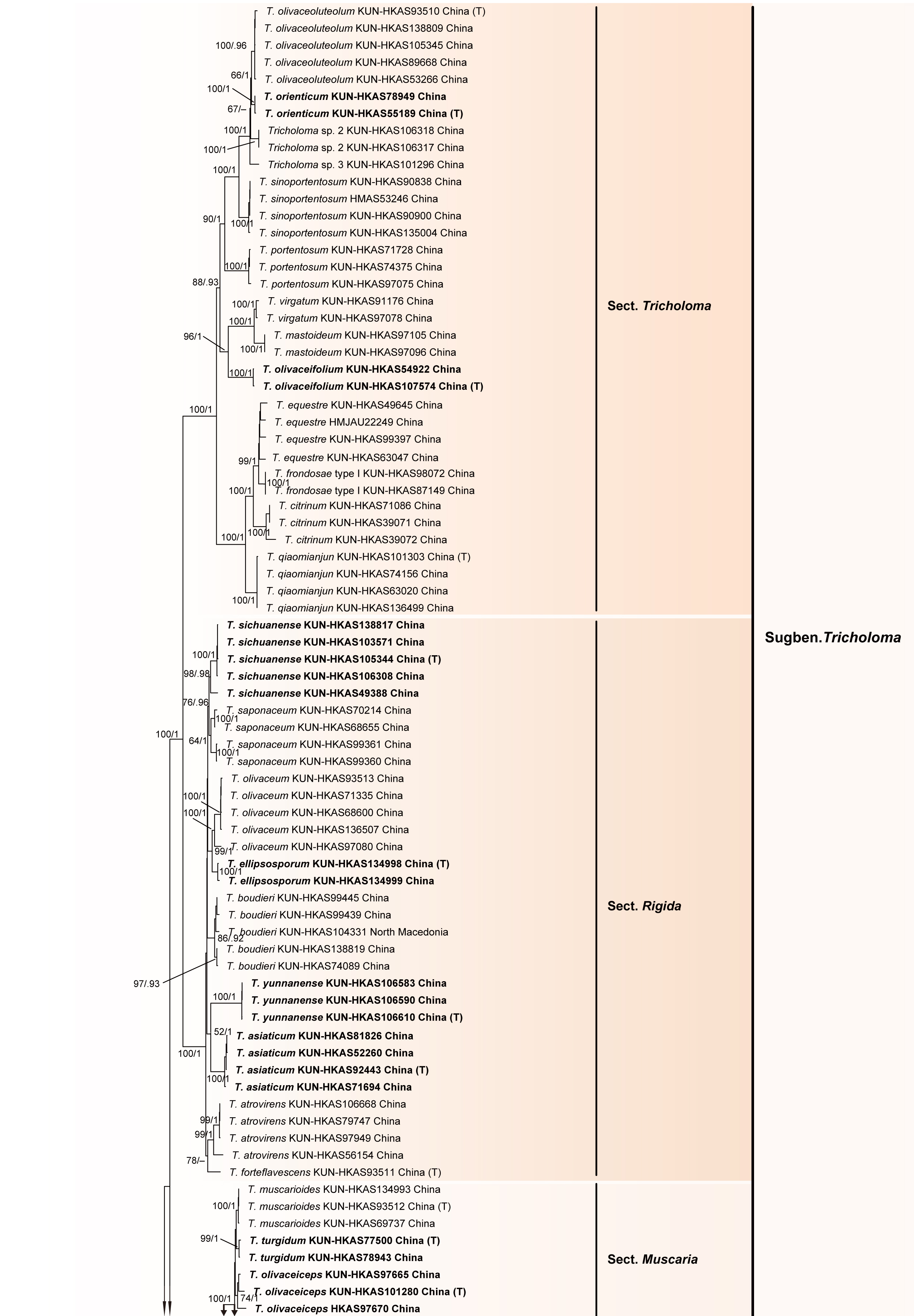
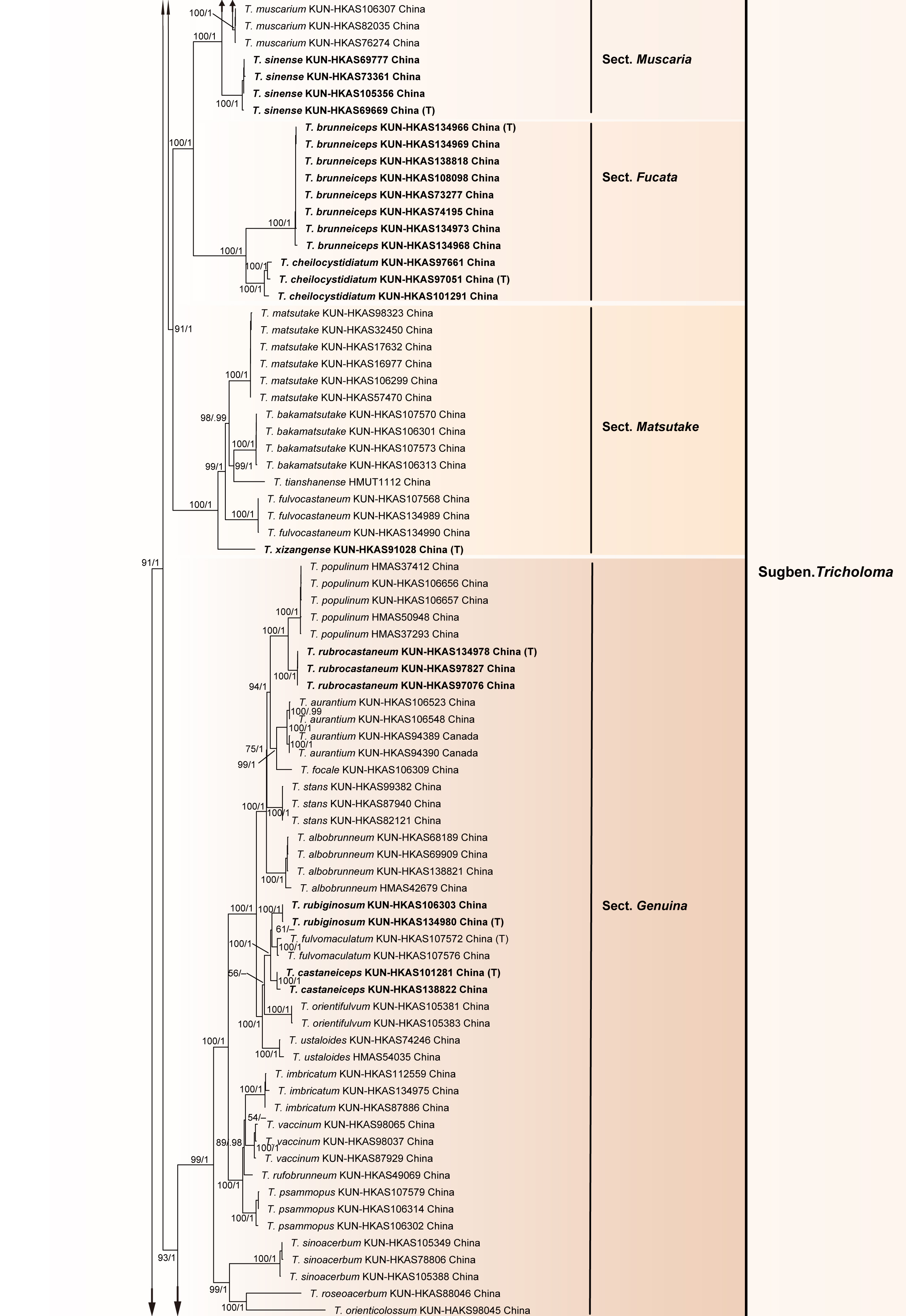
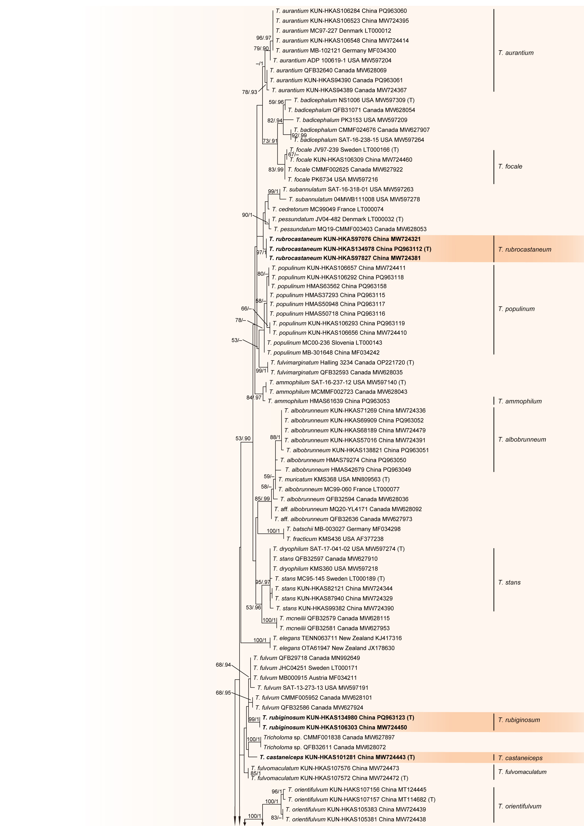
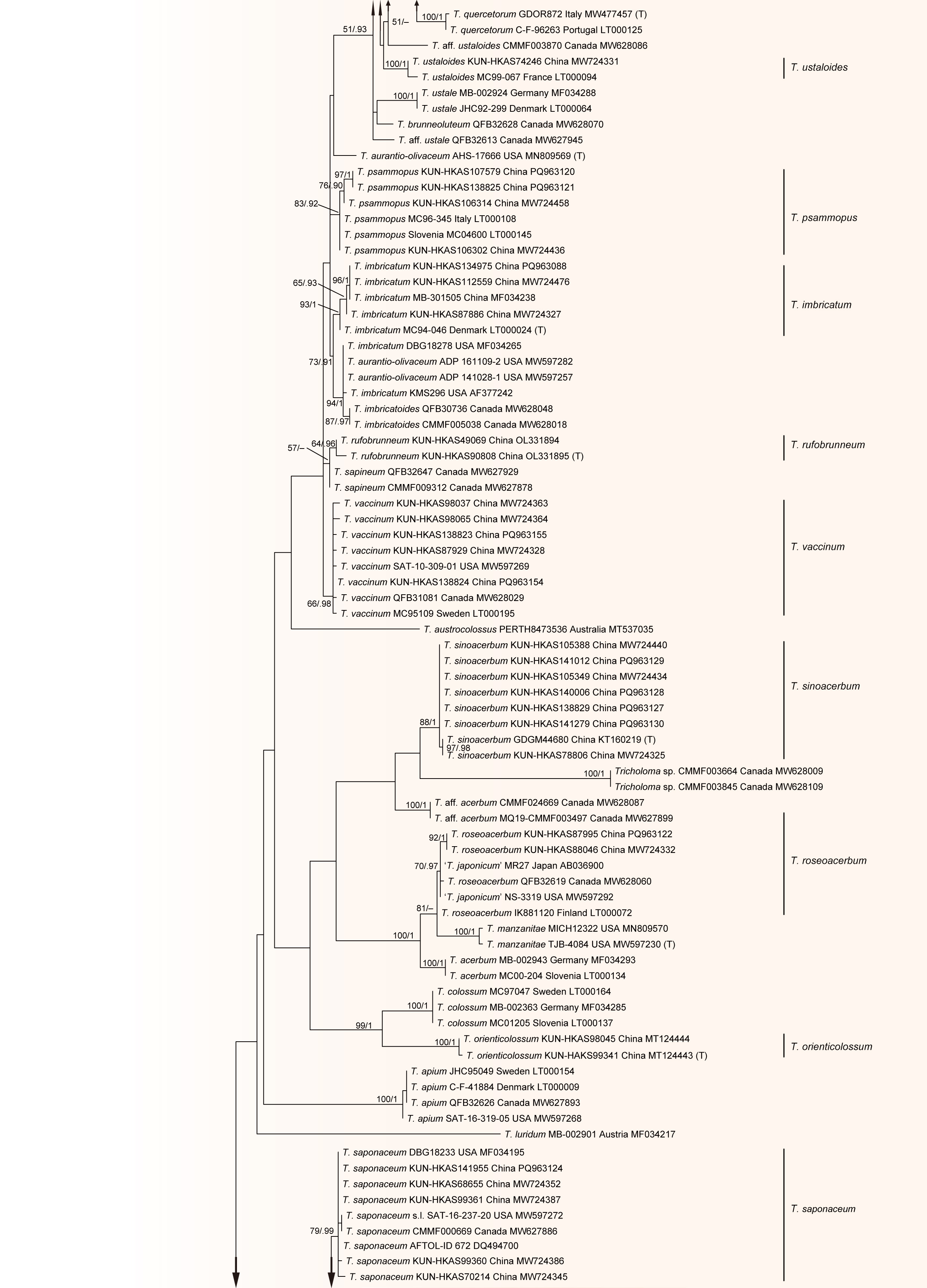
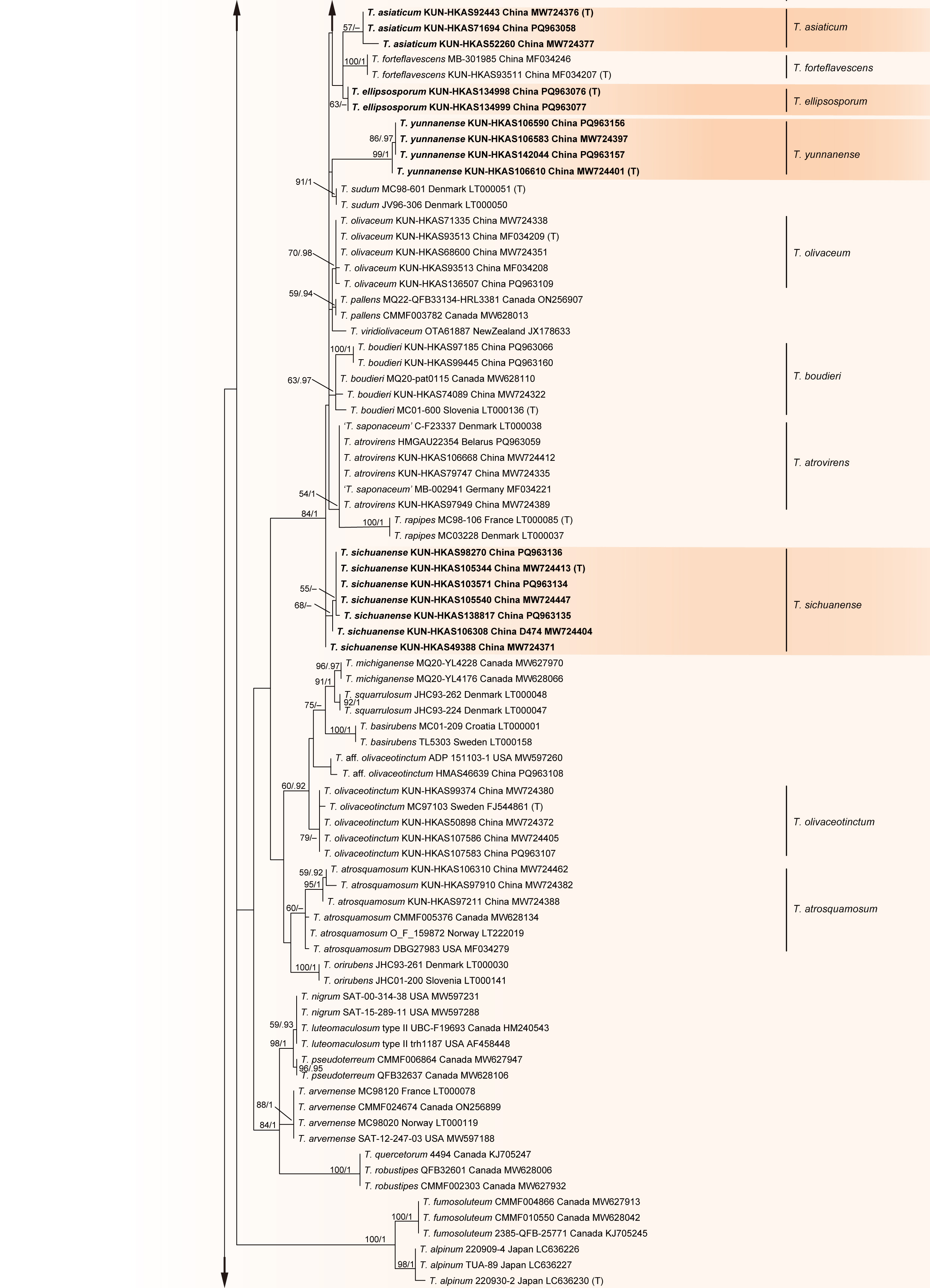
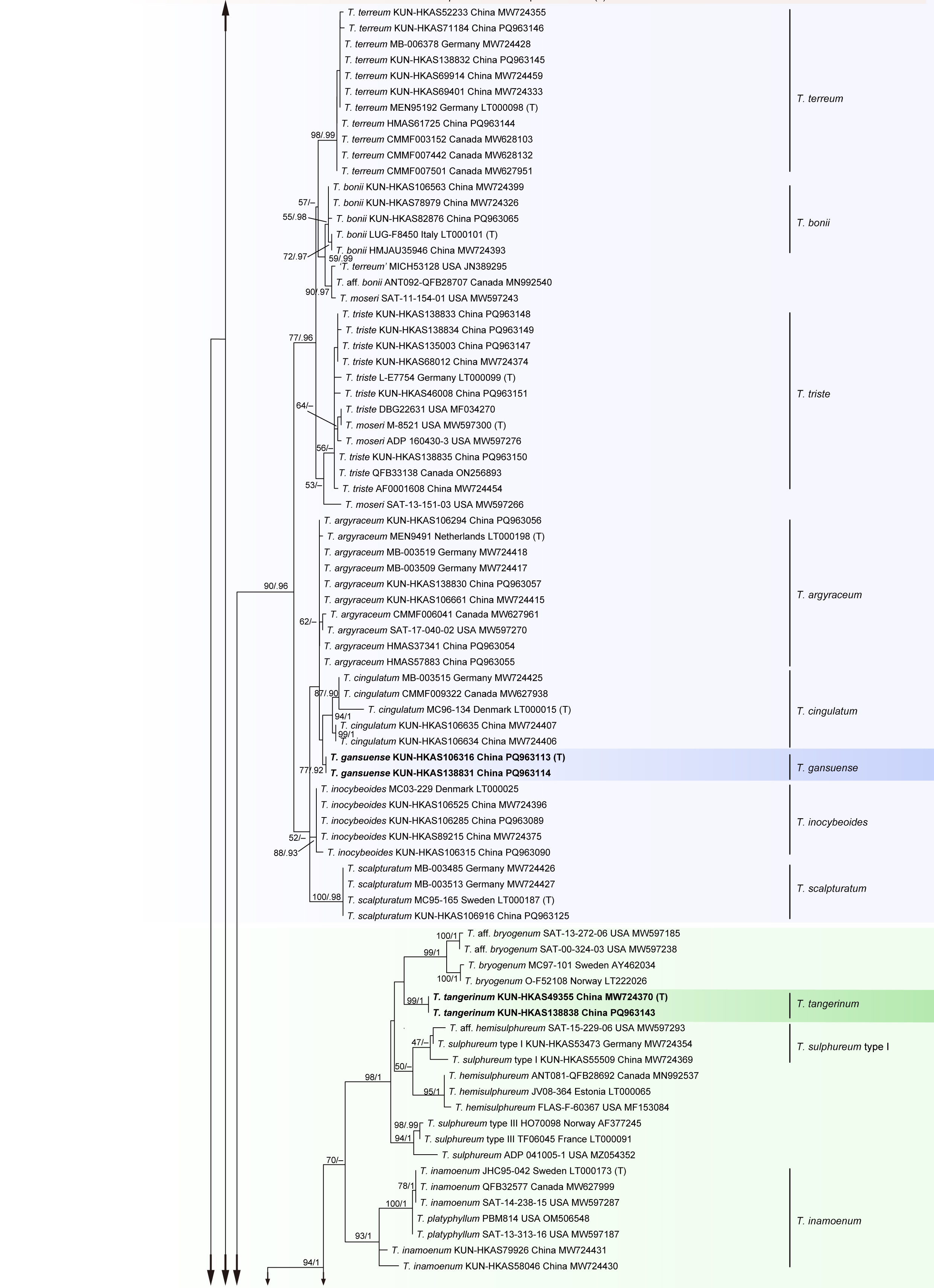
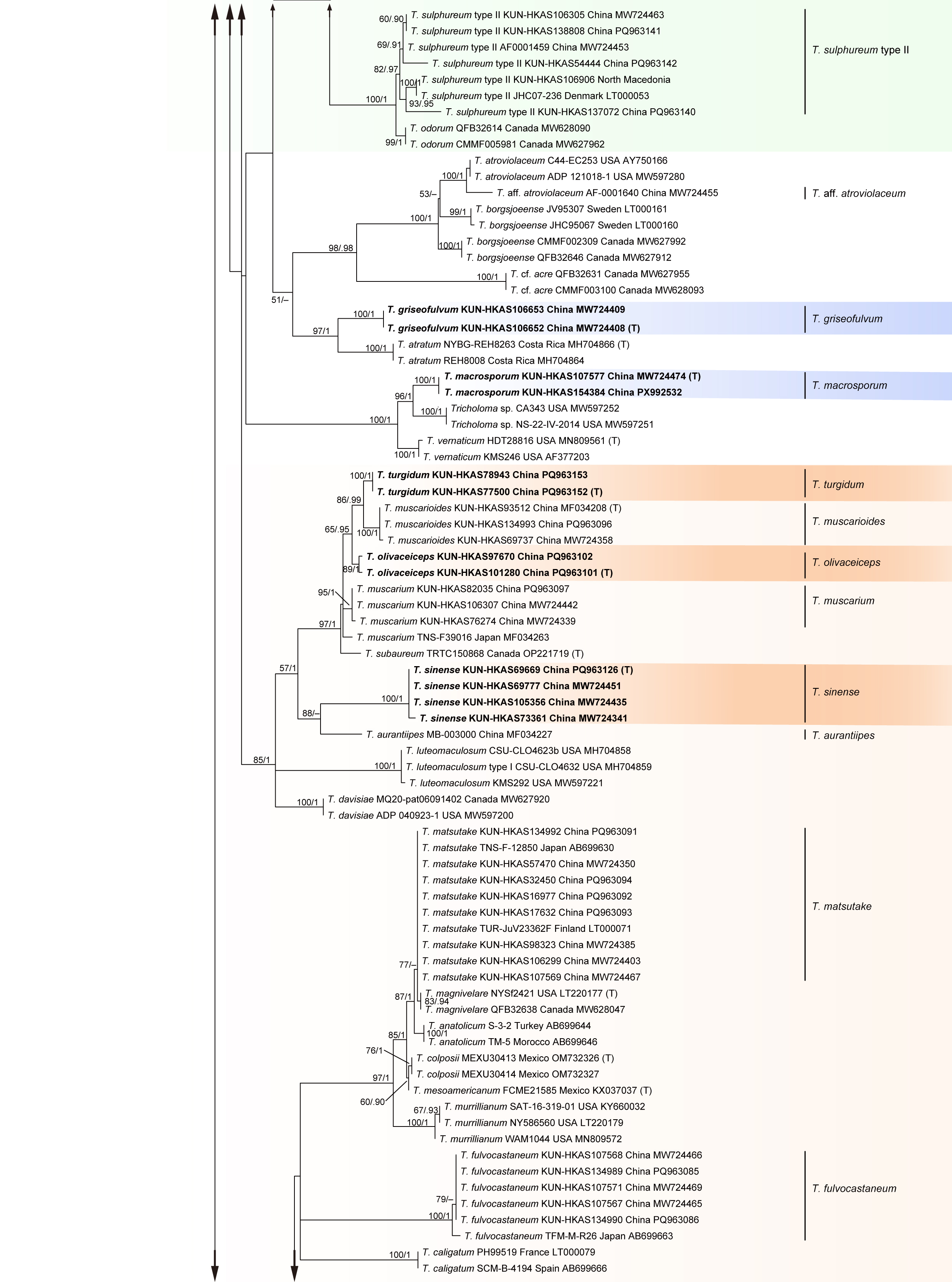
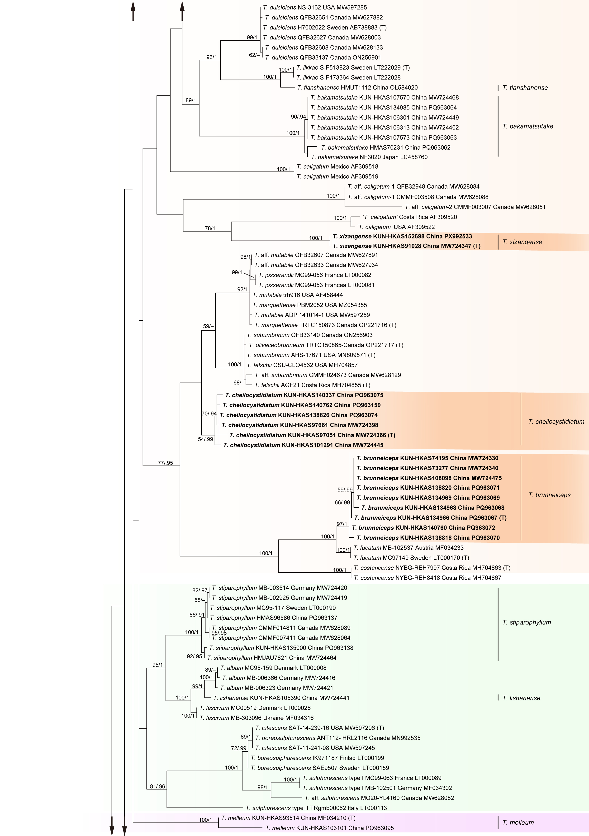
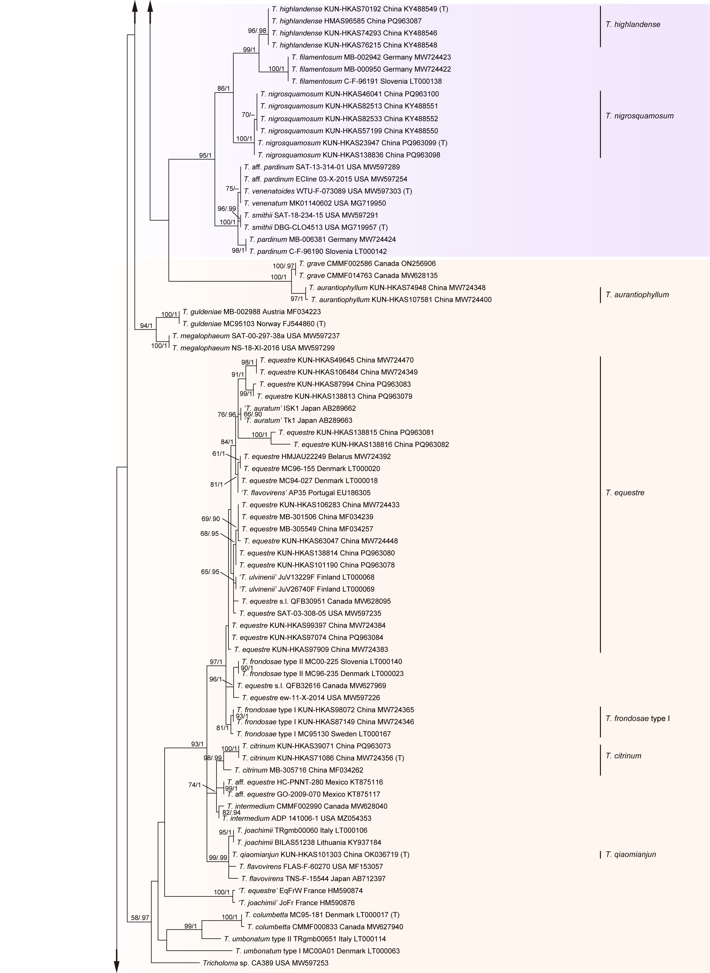
Dataset I comprised 92 nrLSU sequences (928 bp), 119 ITS sequences (1,046 bp), 35 nrSSU sequences (1,743 bp) and 66 RPB2 sequences (749 bp), which were derived from 117 representative samples of Tricholomataceae and four outgroup species of Callistosporiaceae. Among these, 18 samples were collected from China with 59 sequences. The dataset comprised 1,347 parsimony informative sites with a total length of 4,466 positions. Dataset III included 215 ITS sequences (714 bp), 180 EF1-α sequences (764 bp), 159 RPB2 sequences (655 bp), 142 MCMC7 sequences (602 bp) and 169 mtSSU sequences (871 bp) from 228 representative collections of Tricholoma and two outgroup species of Leucopaxillus. The total length of the dataset is 3,606 bp with 1,541 parsimony informative sites. Dataset IV consisted of 639 ITS sequences (775 bp), with 478 parsimony informative sites. In the three datasets, 373 sequences were newly generated in this study. Dataset II included 45 collections representing 43 species with a total length of 593,898 amino acid positions from 1,477 single-copy orthologous gene fragments. The phylogenetic trees inferred from the ML, BI, or coalescent-based analyses were similar in topology. Therefore, only the trees obtained from the ML analyses were presented (Figs. 1–4).
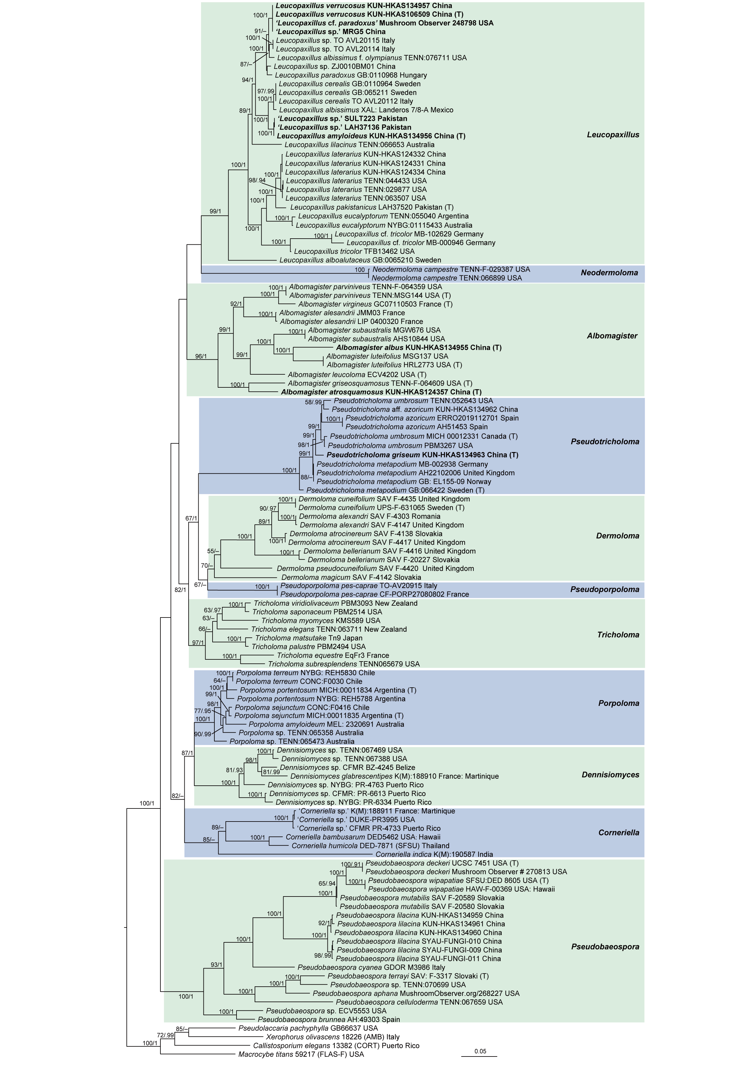
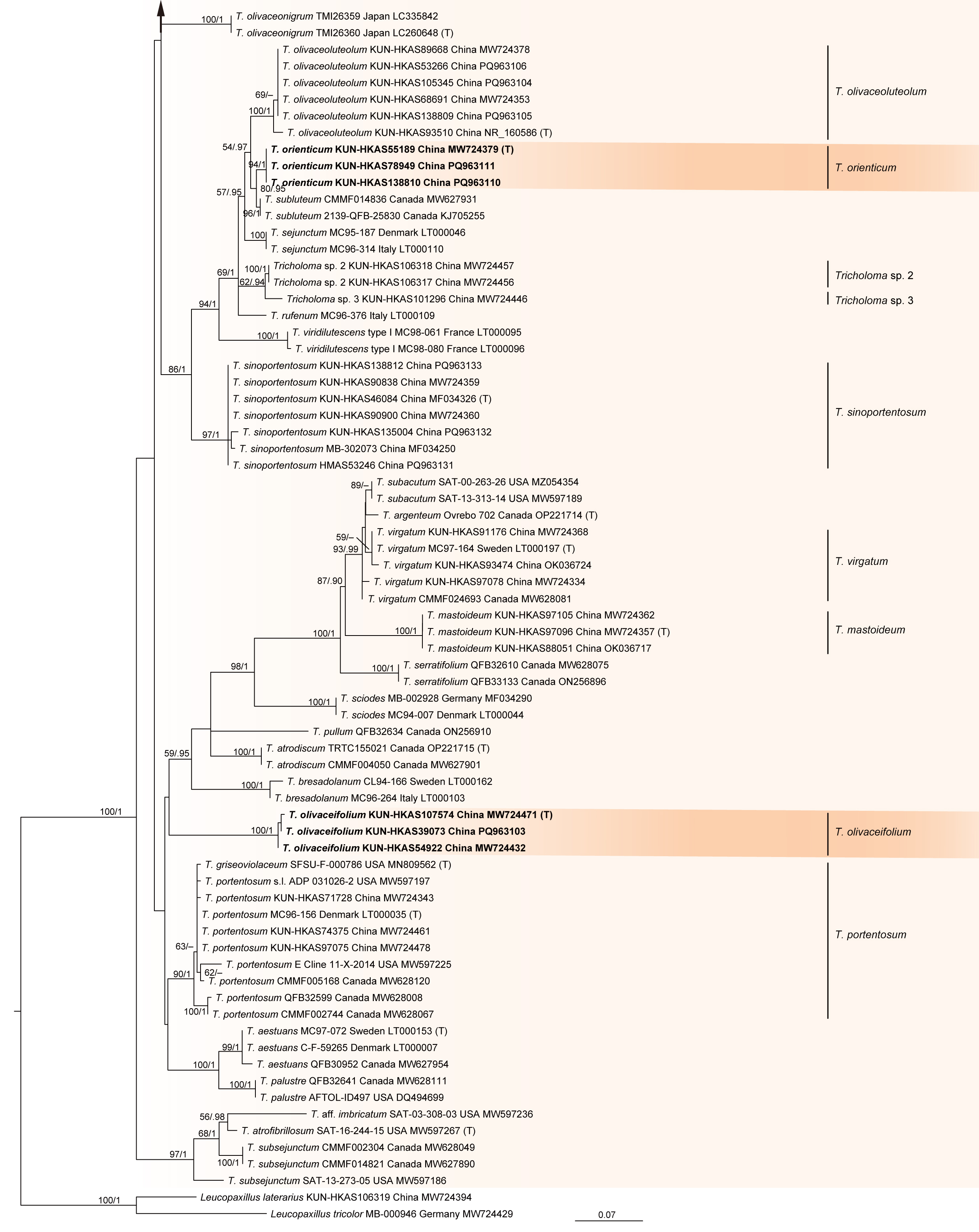
In the phylogenetic tree inferred from Dataset I (nrLSU/ITS/nrSSU/RPB2), Tricholomataceae s.str. was supported to be a monophyletic group, and 11 genus-level clades were included in the family (Fig. 1). All clades corresponded to the 11 well-circumscribed genera of the family. Among these genera, Dermoloma was sister to Pseudoporpoloma (MLB=67%). The two genera were closely related to Pseudotricholoma, forming a clade that was then sister to Tricholoma (MLB=82%, PP=1.0). Corneriella was closely related to the clade formed by Porpoloma and Dennisiomyces (Fig. 1). The collections from China, except Tricholoma, were clustered in Leucopaxillus, Albomagister, Pseudobaesopra, and Pseudotricholoma. Based on the results combined with morphological studies, these samples represent eight species, with five of them being described as new species in this study (Fig. 1).
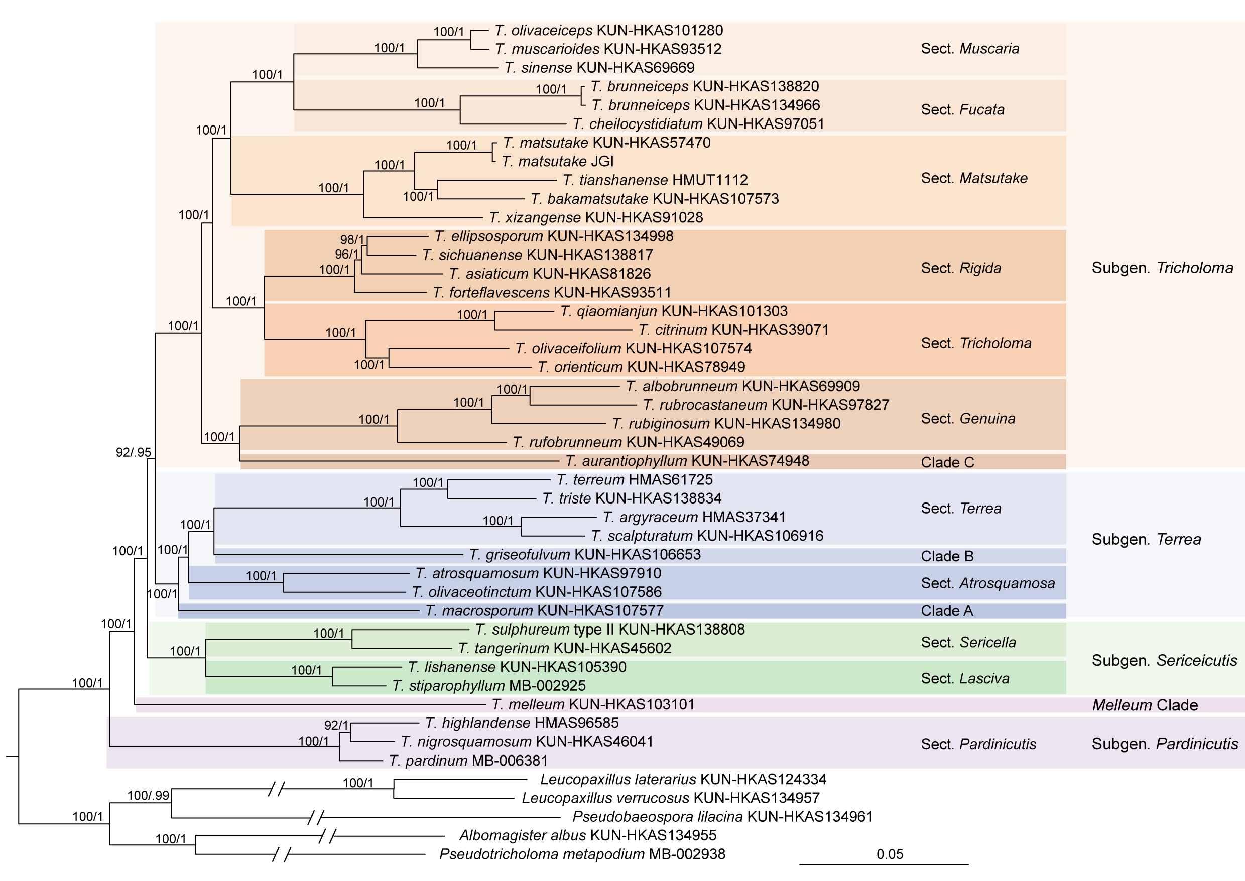
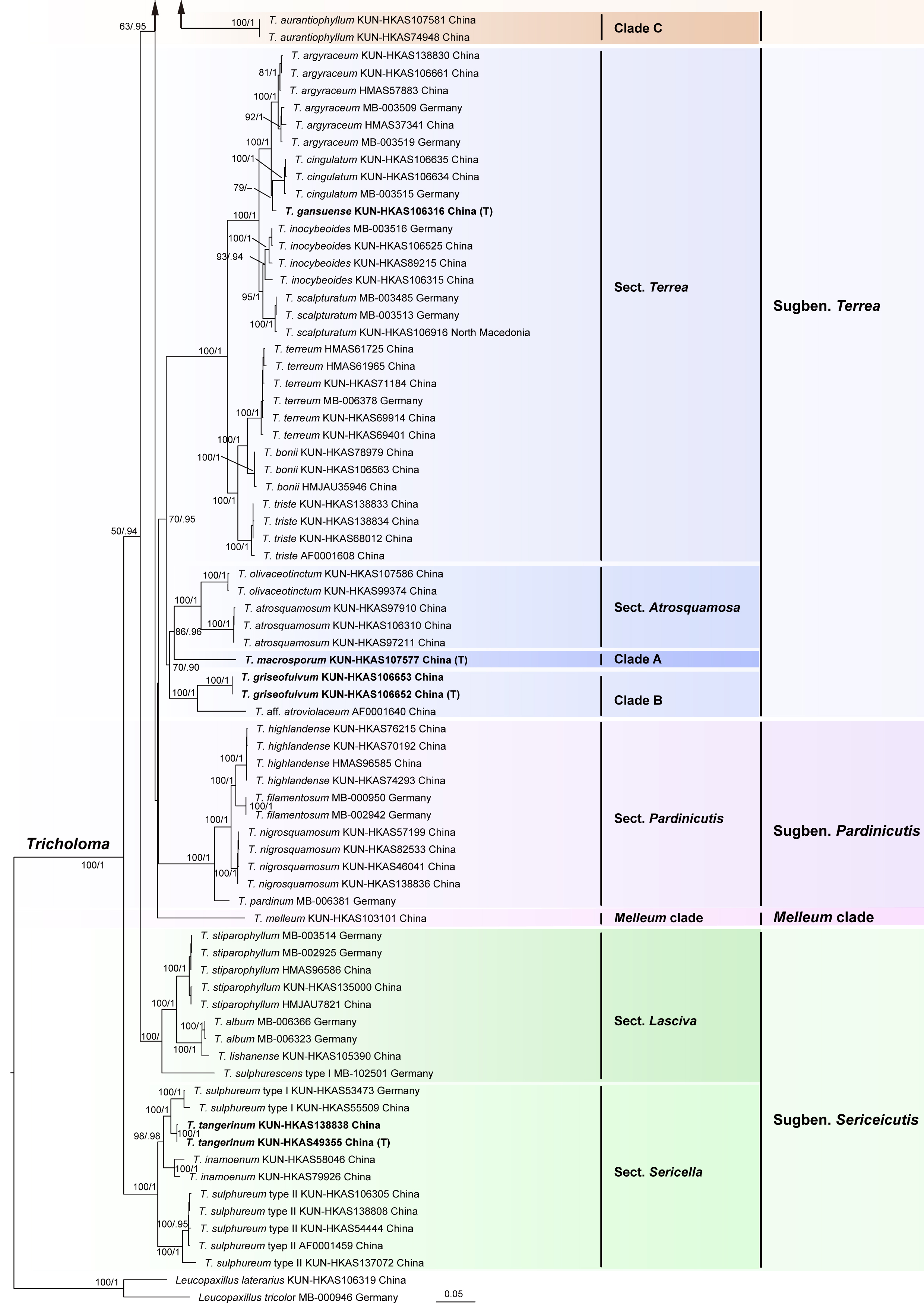
Based on molecular phylogenetic analyses of Dataset II (SOGs) and Dataset III (ITS/RPB2/MCMC/EF1-α/mtSSU), along with morphological observations, Tricholoma, the most species-rich genus of the family, was divided into five subgenus-level clades and 15 section-level clades (Figs. 2, 3). The phylogenetic relationships among these subgenera and sections were highly resolved in the tree inferred from Dataset II (Fig. 2). Tricholoma sugben. Tricholoma was closely related to T. subgen. Terrea Ding et al. The two subgenera were then sister to T. subgen. Sericeicutis Singer, followed by Melleum Clade and T. subgen. Pardinicutis Singer (Fig. 2). In the subgenus Tricholoma, the seven section-level clades were grouped into three monophyletic groups with high support values. Tricholoma sect. Muscaria Reschke was closely related to sect. Fucata Ding et al., and the two sections were then sister to sect. Matsutake Ding et al. The group formed by the three sections was sister to the group formed by sect. Rigida (Fr.) Quél. and sect. Tricholoma, followed by the group consisting of sect. Genuina (Fr.) Sacc. and Clade C. The phylogeny of the subgenus was also supported in the tree generated from Dataset III (Fig. 3). In the subgenus Terrea, sect. Terrea was sister to Clade B, followed by sect. Atrosquamosa Kühner ex Bon and Clade A (Fig. 2). However, the phylogenetic relationships among them were not supported with strong supports in the tree based on Dataset III (Fig. 3). The sister relationship between sect. Sericella (Fr.) Quél. and sect. Lasciva Bon, which was strongly supported in the tree generated from Dataset II, was also not elucidated in the tree based on Dataset III (Figs. 2, 3). There were 286 collections from China included in the tree generated from Datasets III and IV (Figs. 3, 4). Based on these results, along with morphological studies, these samples represented 76 Tricholoma species from China, and 19 of them were described as new species in this study.
Morphological observations
The 371 Chinese specimens examined in this study represented 82 species of Tricholomataceae s.str. Of these 58 are known species, while 24 taxa are new to science. Most of the taxa (74) belong to the genus Tricholoma, while the remaining species are allocated to Albomagister (2 species), Leucopaxillus (3 species), Pseudobaeospora (1 species), and Pseudotricholoma (2 species).
Among the Chinese representatives of Tricholomataceae s. str., Tricholoma includes the largest and most well-represented genus of species. Therefore, we have provided a summary of the morphological features of each structure within Tricholoma, as detailed below.
Basidioma: Most basidioma of Tricholoma species are small to medium-sized, robust, and fleshy; some are large to very large (e.g., T. orienticolossum X. Xu et al.), and others are very small to small (e.g., T. triste (Scop.) Quél.). According to Bas (1969), the size of basidioma is categorized based on pileus diameter: very small (≤3 cm), small (3–5 cm), medium-sized (5–9 cm), large (9–15 cm), and very large (≥15 cm). These categories were applied throughout this study.
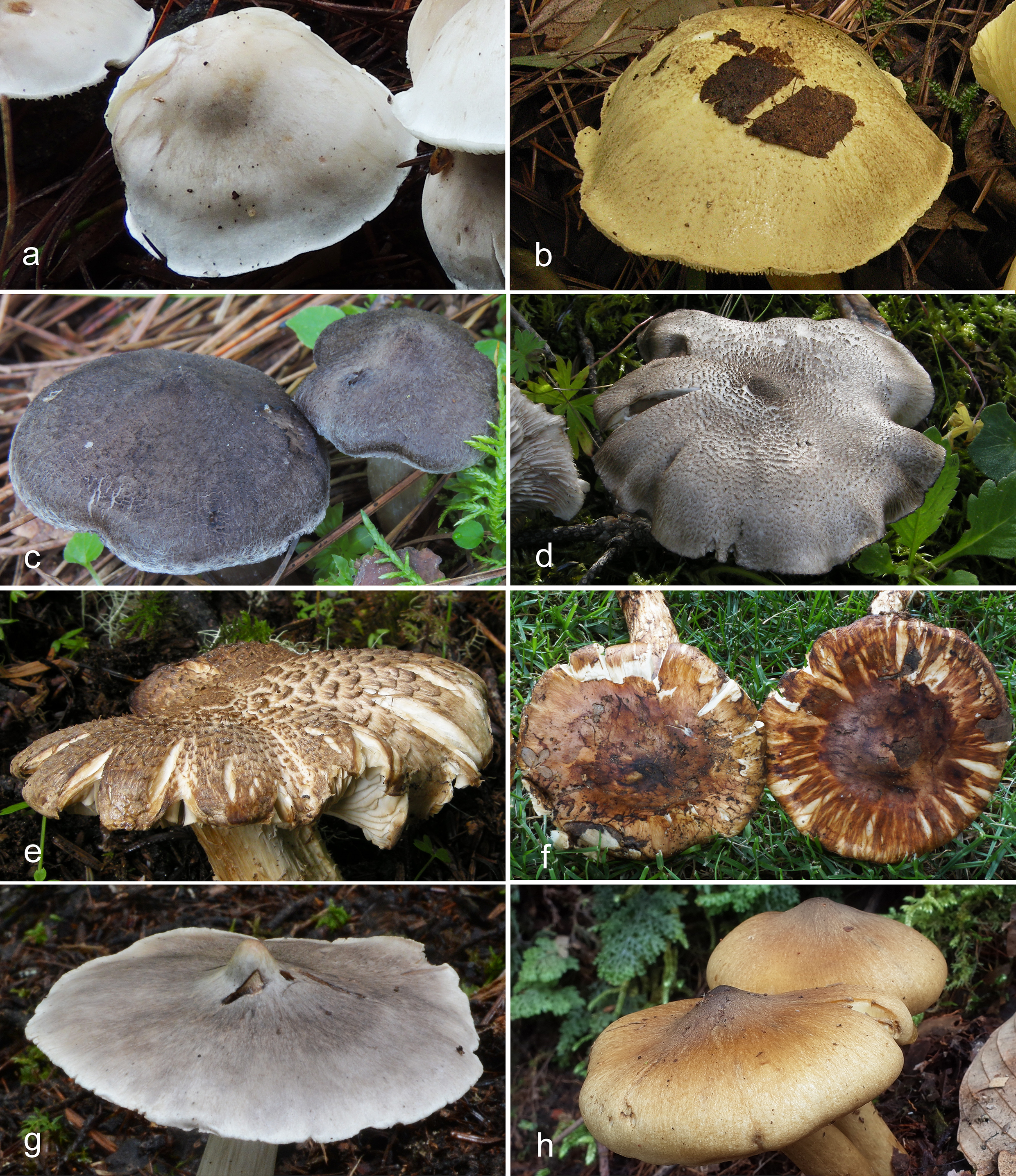
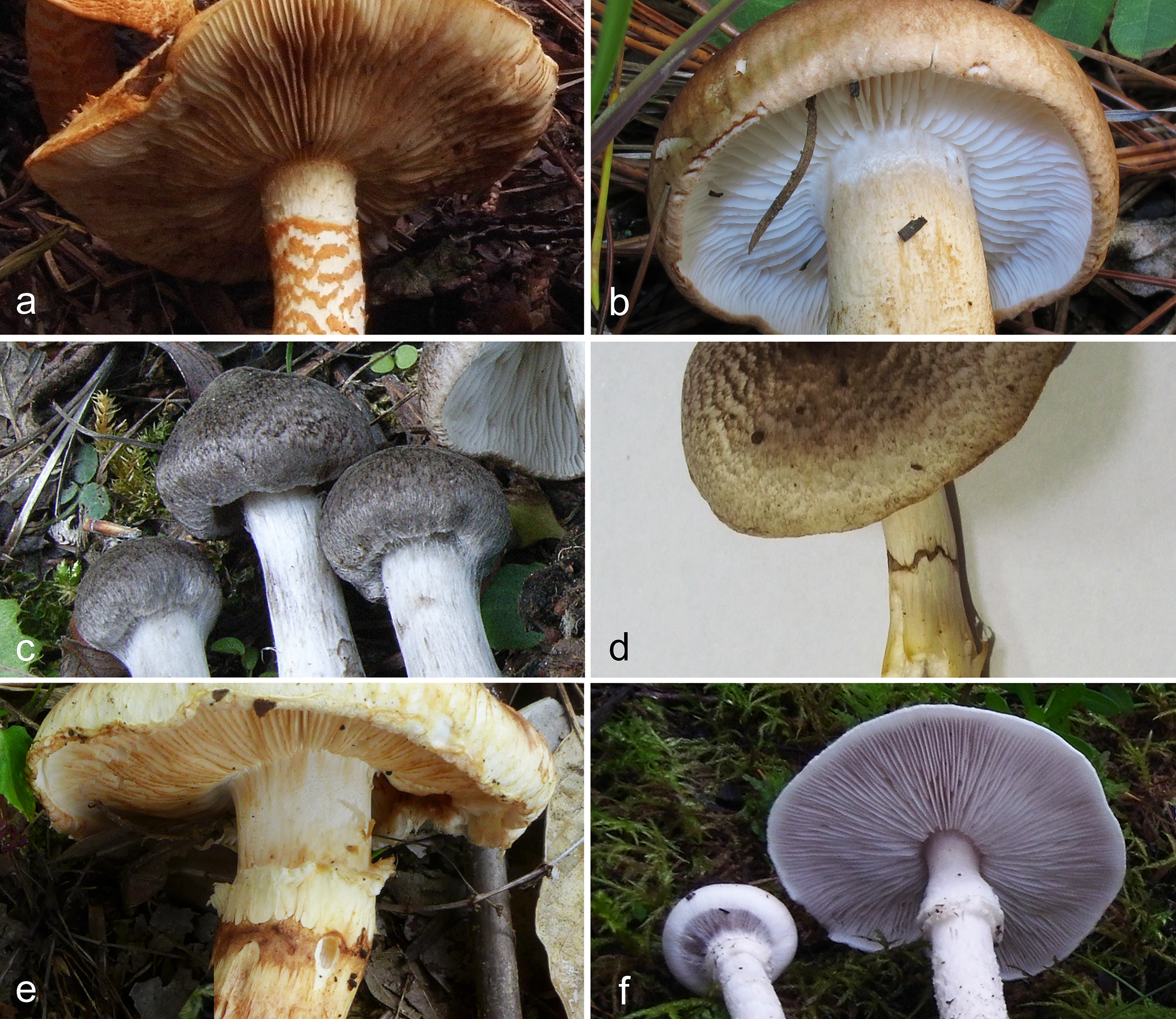
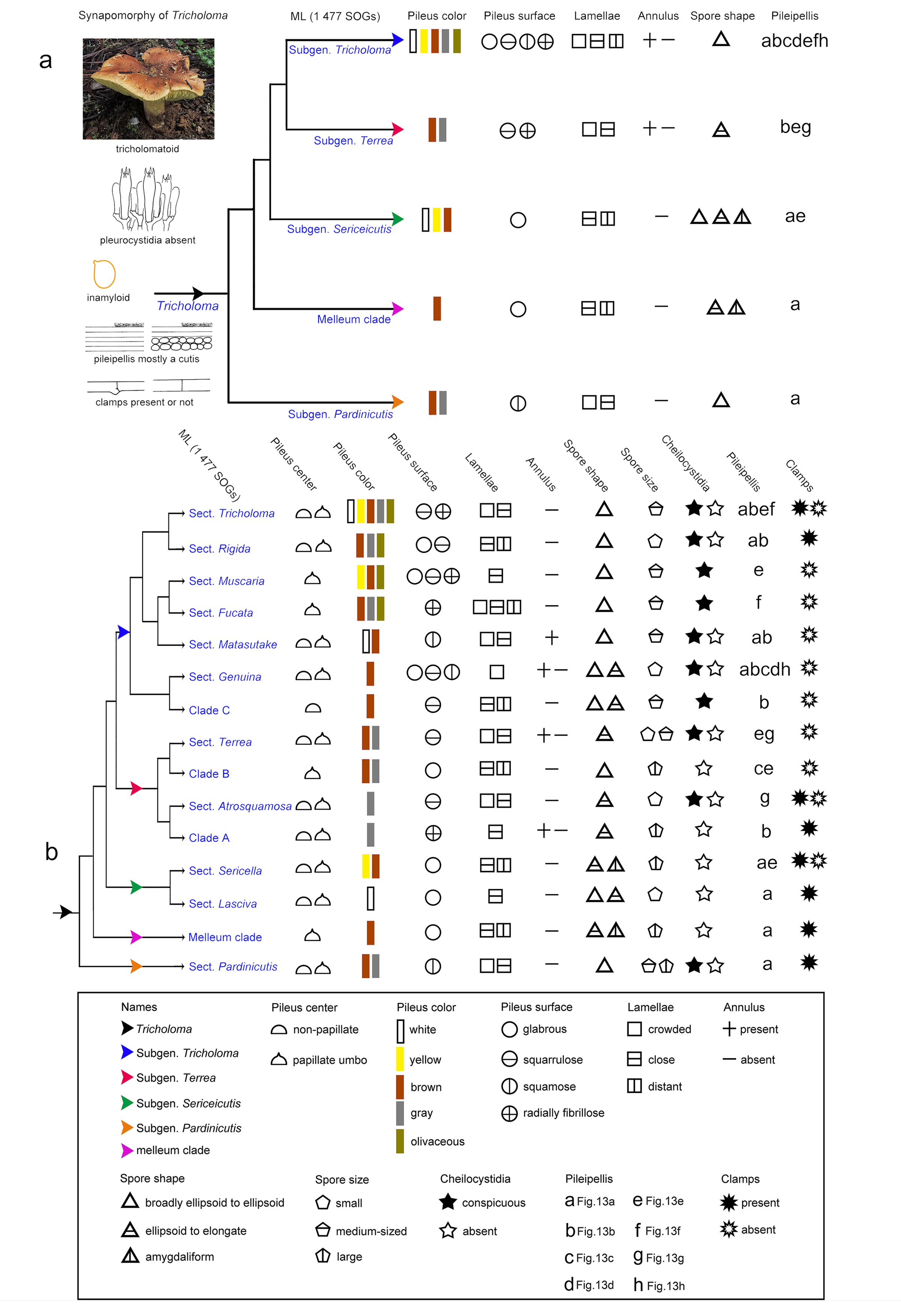
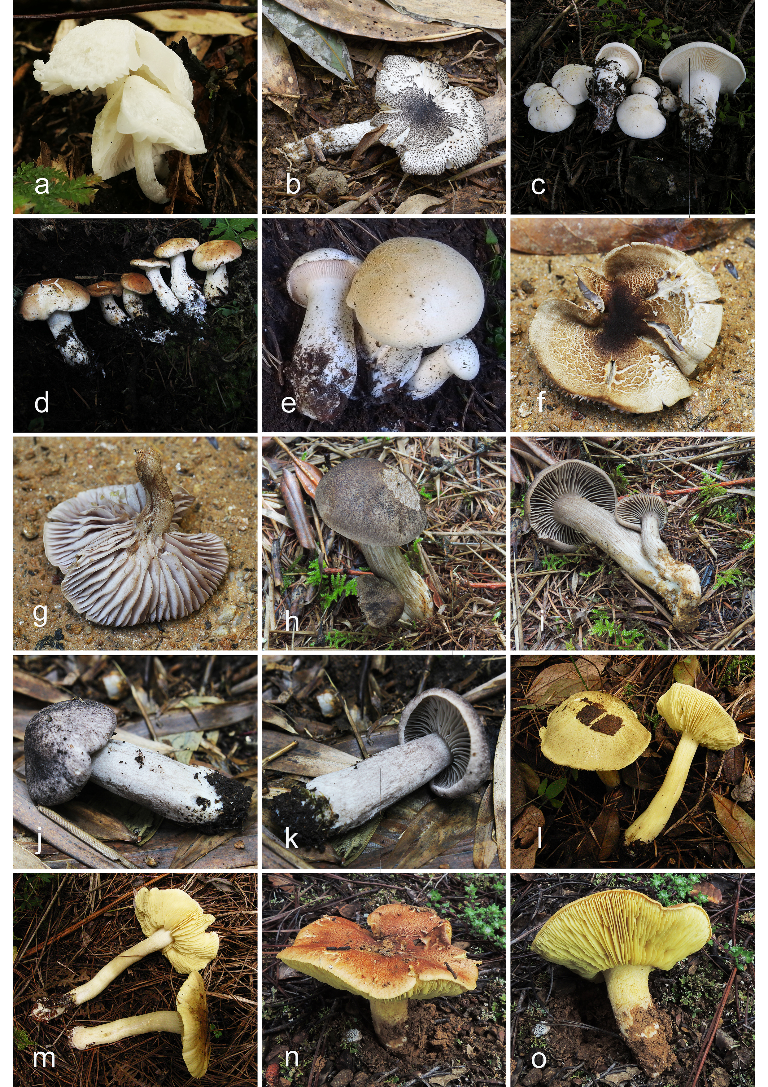
Pileus: The pileus of Tricholoma species is initially conical to convex, later becoming plano-convex to applanate, often irregular with a wavy appearance. Some species possess a distinct papillate umbo at the center of the pileus, a feature significant for section divisions within the genus (Fig. 14b). For example, species in sect. Muscaria and sect. Fucata often have papillate-umbonate pilei (Fig. 14b). The pileal margin is involute when young, with most species becoming straight at maturity. Pileus is dry or viscid, and its coloration is highly variable—white, yellow, brown, grey, or olivaceous. The surface can be glabrous (Fig. 5a), squarrulose (Fig. 5b–d), squamose (Fig. 5e, f), or radially fibrillose (Fig. 5g, h), features valuable for subgenus division (Fig. 14a).
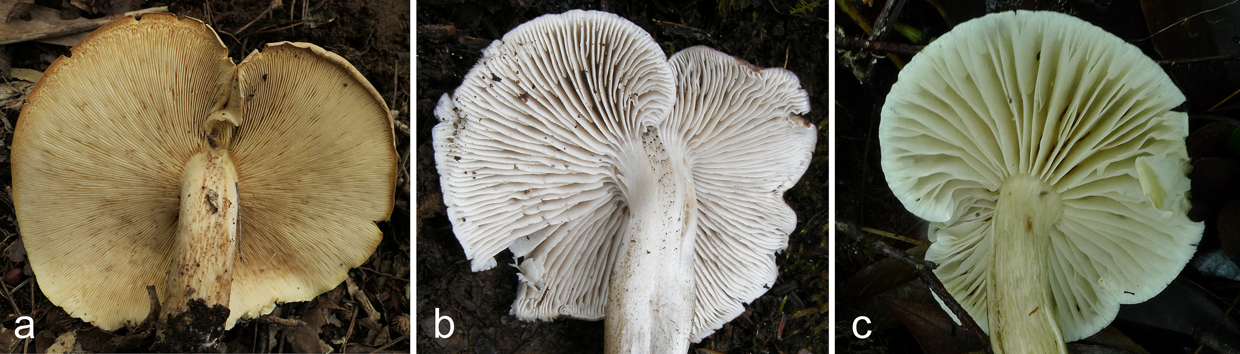
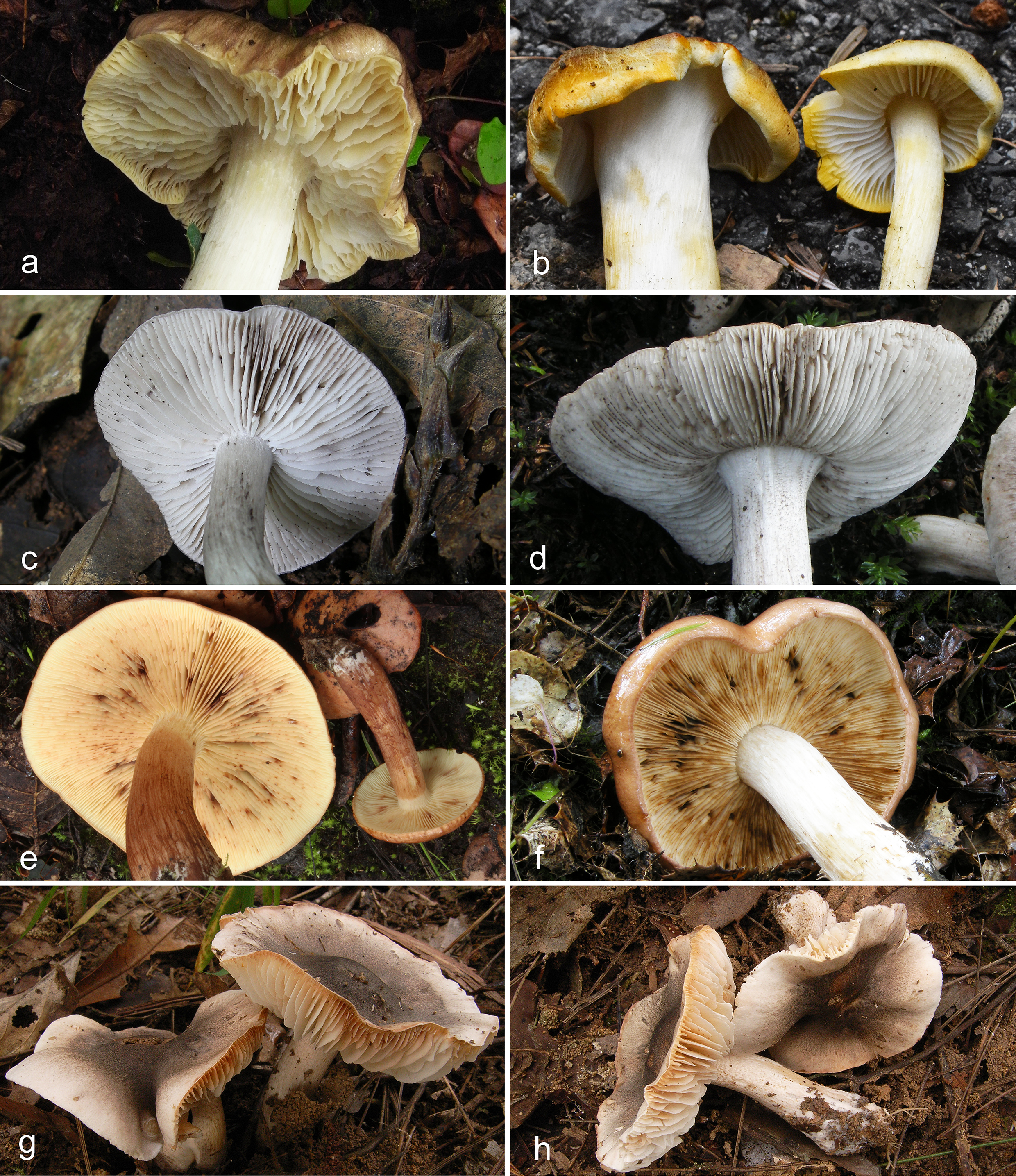
Lamellae: The lamellae of Tricholoma species are sinuate with lamellulae. The edge may be entire or not and is often easily broken. The spacing between them is crowded (Fig. 6a), close (Fig. 6b) to distant (Fig. 6c), which have played a certain role in the classification of subgenera and sections of Tricholoma (Fig. 14). For instance, species in subgen. Sericeicutis generally have close to distant lamellae, while sect. Rigida features close to distant lamellae and sect. Genuina has crowded lamellae (Fig. 14). Lamellae coloration ranges from white, cream-colored, yellow, grey to brown, with some species exhibiting color changes upon aging or bruising, including turning yellow (Fig. 7a, b), grey or with dark-grey to grey spots (Fig. 7c, d), brown or with brown spots (Fig. 7e, f), or red to reddish (Fig. 7g, h).
Stipe: The stipe is cylindrical, subcylindrical, clavate, or subclavate, with the center being solid, stuffed, or hollow. While stipe shape appears to have limited taxonomic value, stipe ornamentation can be informative for recognition in some species. For example, Tricholoma orienticolossum possesses brownish to red-brown, fibrillose squamules on the stipe surface.
Annulus: Most Tricholoma species lack an annulus, but some possess one near the top of the stipe. Annuli can be categorized into three types based on texture: (1) a pseudoannular zone—a white zone at the stipe apex that may be sharply delimited, unevenly or faintly delimited, or occasionally relieved and prominent, particularly in sect. Genuina (Fig. 8a, b), as exemplified by T. aurantium (Schaeff.) Ricken and T. albobrunneum (Pers.) P. Kumm.; (2) a cobwebby zone—cobweb-like cortina or remnants at the stipe apex (Fig. 8c, d), observed in T. argyraceum (Bull.) Gillet and T. scalpturatum (Fr.) Quél. (sect. Terrea); and (3) a distinct annulus—a large, cottony to woolly structure at the stipe apex (Fig. 8e, f), as found in species of sect. Matsutake, T. cingulatum (Almfelt ex Fr.) Jacobasch (sect. Terrea), and T. focole (sect. Genuina).
Taste and odour: Species of Tricholoma often exhibit distinctive tastes and odors, which are critical for species identification. The most common odor and taste is farinaceous. Unusual odors have been reported. For example, T. bakamatsutake has a slightly pungent, tongue-numbing taste and sweet odor; T. sulphureum (Bull.) P. Kumm. has an unpleasant taste and a strong gas or almond odor; and T. mastoideum Y.Y. Cui & Zhu L. Yang has a mild taste with an oak-like odor. However, organoleptic data are lacking for most Chinese collections, largely because such characters were insufficiently documented in earlier field studies. This highlights the necessity of systematically recording taste and odor in future field investigations.
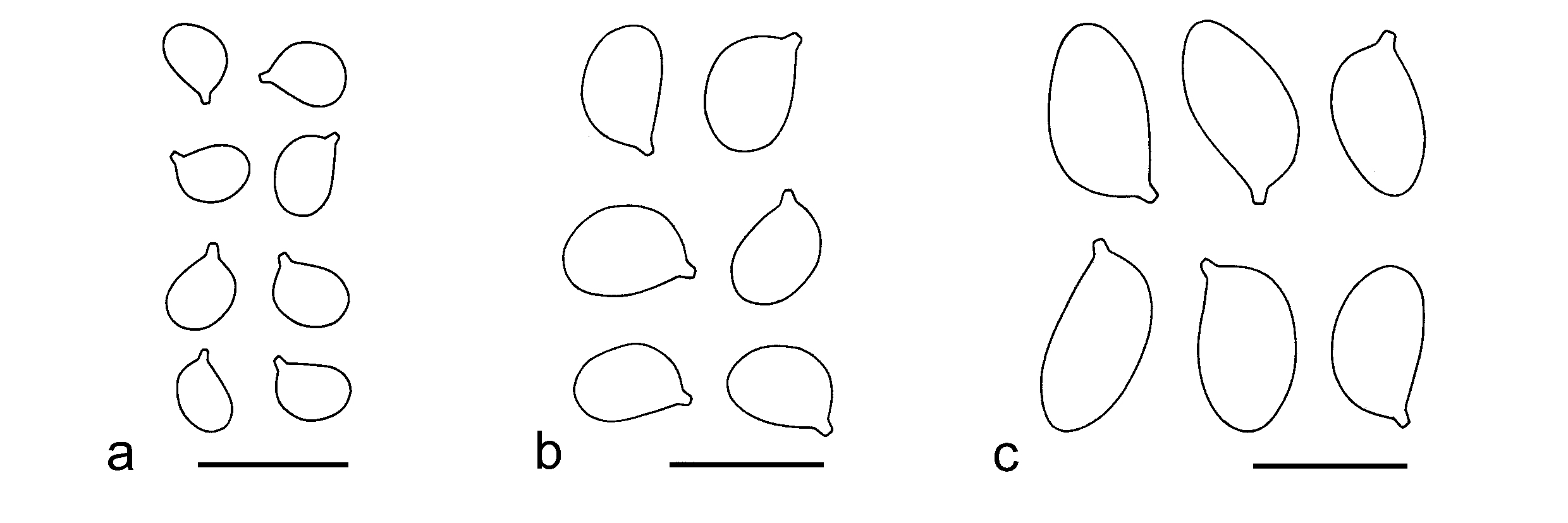
Basidiospores: The size of basidiospores is a critical character in the division of Tricholoma sections and the definition of some species (Fig. 14b). In this study, basidiospores were divided into three groups based on their mean length and mean width: (1) small (mean length ≤ 6.5 μm, mean width ≤ 4.5 μm) (Fig. 9a), characteristic of most species in sect. Rigida, sect. Genuina, sect. Atrosquamosa and sect. Lasciva; (2) medium-sized (6.5 μm < mean length ≤ 9 μm, 4.5 μm < mean width ≤ 5.5 μm) (Fig. 9b), typical of most species in sect. Tricholoma, sect. Muscaria, sect. Fucata and sect. Matsutake; and (3) large (mean length > 9 μm, mean width > 5.5 μm) (Fig. 9c), such as most species in sect. Sericella.
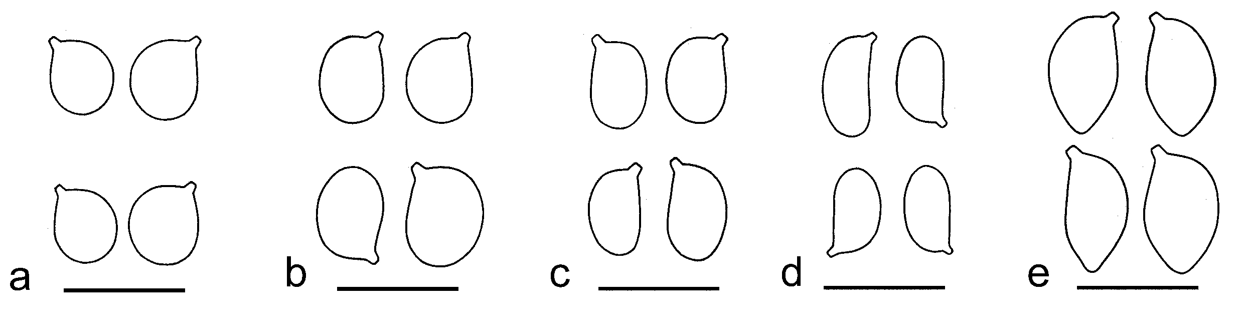
Bas (1969) categorized basidiospore shapes into six types: (1) globose (Q = 1.0–1.05); (2) subglobose (Q = 1.05–1.15); (3) broadly ellipsoid (Q = 1.05–1.3); (4) ellipsoid (Q = 1.3–1.6); (5) elongate (Q = 1.6–2.0); and (6) cylindrical (Q = 2.0–3.0). The basidiospores of Tricholoma species are predominantly broadly ellipsoid (Fig. 10b), ellipsoid (Fig. 10c), or elongate (Fig. 10d), occasionally subglobose (Fig. 10a). Additionally, amygdaliform spores (Fig. 10e) are present in species of sect. Sericella. Spore shape plays a pivotal role in distinguishing subgenera, sections, and certain species within Tricholoma (Fig. 14).
The basidiospores of this genus are inamyloid (Lebeuf et al. 2024; this study), a diagnostic feature that distinguishes Tricholoma from other genera within Tricholomataceae.
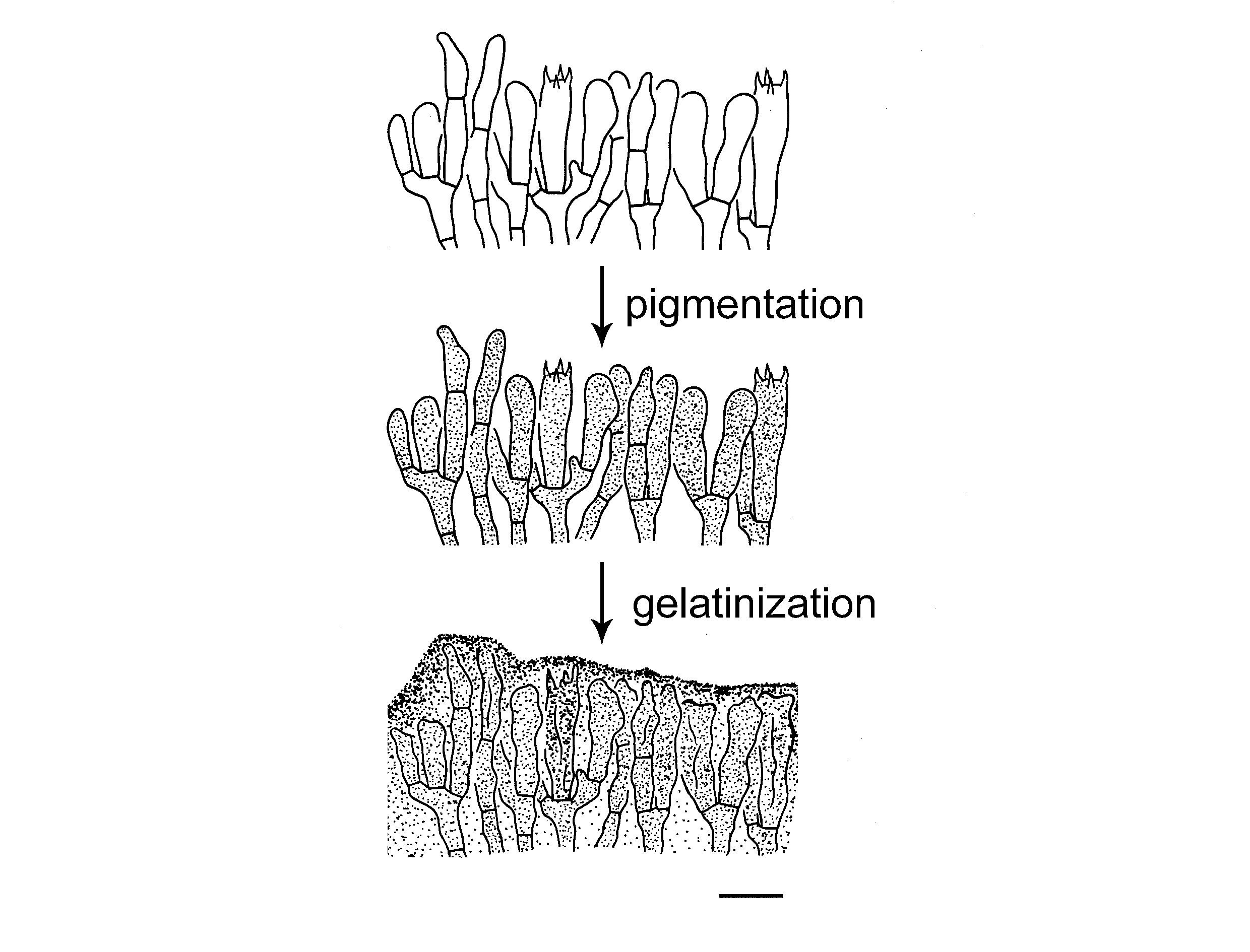
Discoloration of hymenophoral surface: In most species of sect. Genuina, the lamellae develop brown spots, eventually turning entirely brown with age or bruising. Microscopically, this discoloration results from pigment formation in hymenial cells, followed by cell gelatinization and degradation (Fig. 11).
Basidia: The basidia in Tricholoma are clavate to narrowly
clavate, predominantly 4-spored, occasionally mixed with 1-, 2-, or 3-spored ones. They are typically 20–50 μm long and 5–12 μm wide, with limited taxonomic significance.
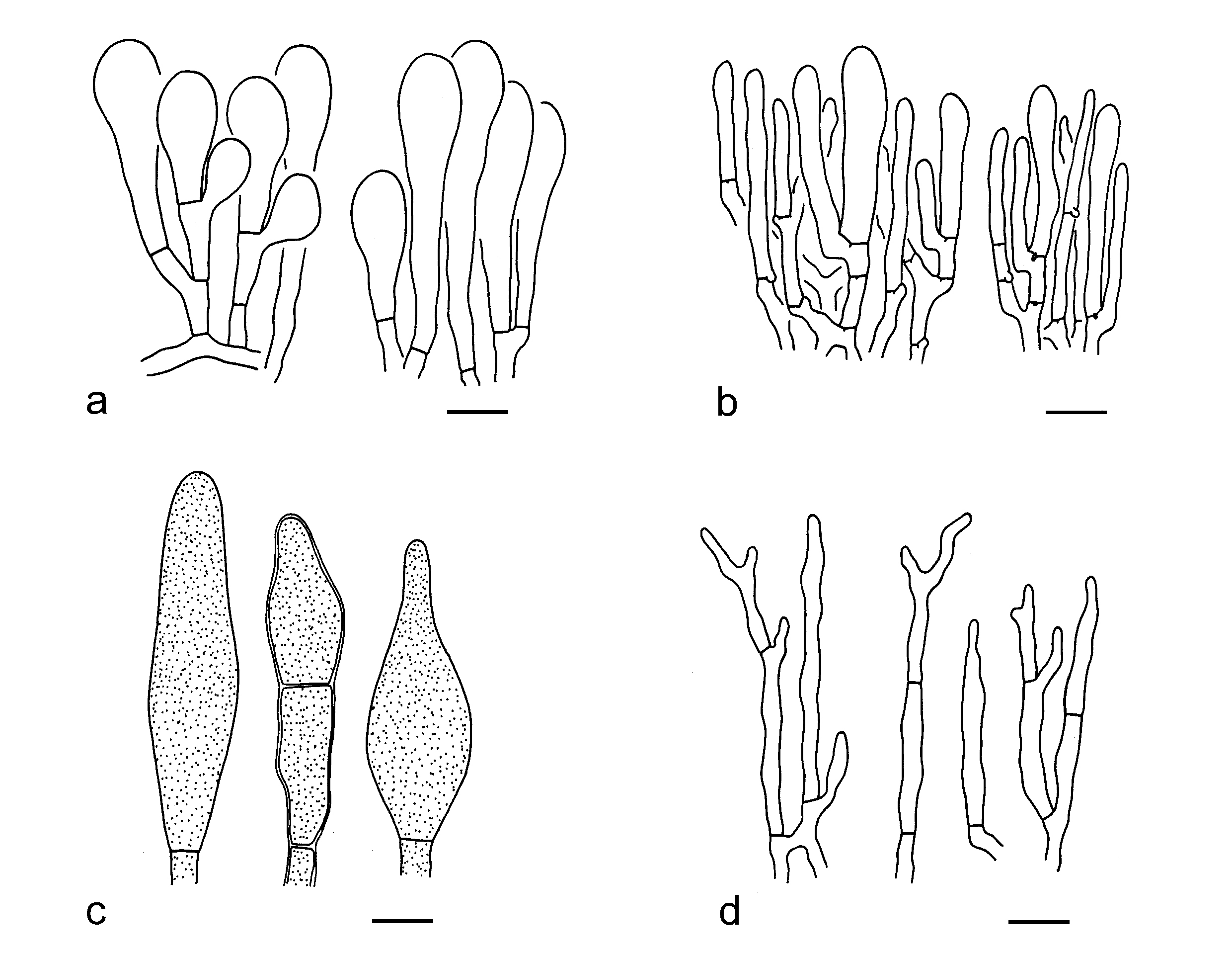
Cystidia: Pleurocystidia are usually absent in Tricholoma. For cheilocystidia, in most cases they are poorly differentiated and may often merely represent marginal cells emerging from the underlying gill trama. Sometimes the cheilocystidia are conspicuous and range in shape from broadly clavate (Fig. 12a), cylindrical (Fig. 12b), fusiform (Fig. 12c) to filamentous (Fig. 12d). Although the cheilocystidia may vary within some species, they are somewhat useful for species recognition in the genus.
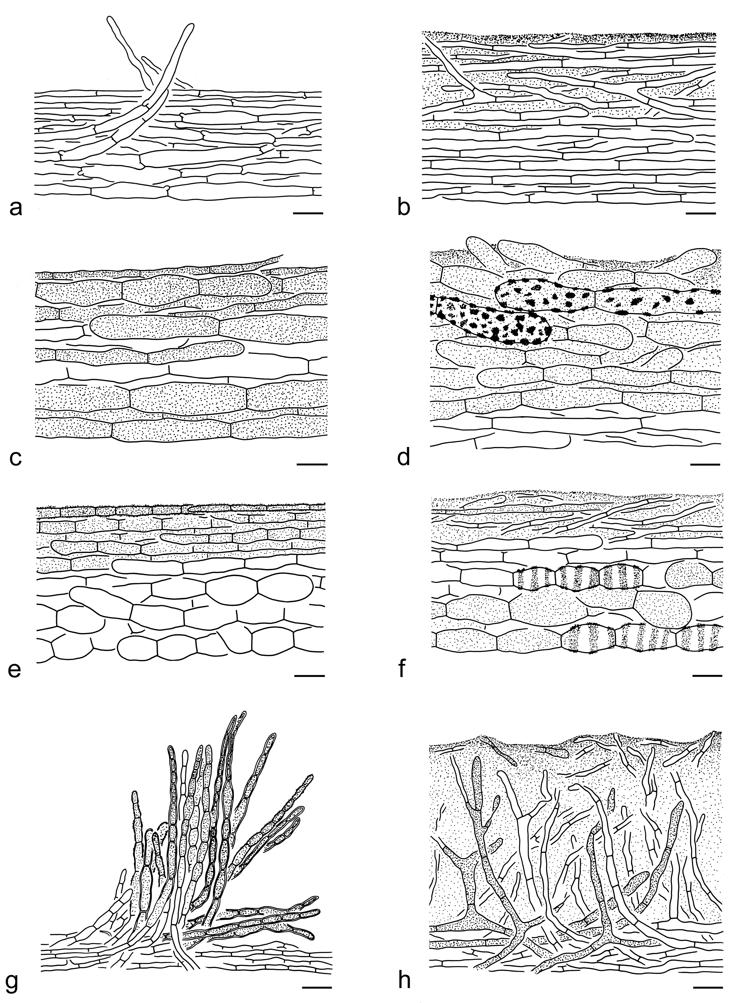
Pileipellis: The pileipellis structure is a crucial diagnostic feature for identifying subgenera, sections, and species within Tricholoma (Fig. 14). Hyphae in the pileipellis are mostly radially arranged, occasionally somewhat erect. The pileipellis can be categorized into eight structural types (Fig. 13).
Clamps: Clamps may be absent or present in Tricholoma species. When present, they are typically abundant at the bases of basidia but may occur less frequently elsewhere in the basidioma. The presence or absence of clamps among different collections in the same species is consistent. This feature holds significant value for distinguishing sections and species within the genus (Fig. 14b).
Results
Taxonomy
The taxonomic system of Tricholomataceae
Building on findings of previous studies (Sánchez-García et al. 2014, 2021; Vizzini et al. 2016; Ding et al. 2023; Adamčíková et al. 2025) combined with our phylogenomic, morphological and ecological data, we hereby present the taxonomic system of Tricholomataceae in alphabetic order:
Tricholomataceae R. Heim ex Pouzar
Type: Tricholoma (Fr.) Staude
I. Albomagister Sánchez-García et al.
Type: A. subaustralis (A.H. Sm. & Hesler) Sánchez-García et al.
II. Corneriella Sánchez-García
Type: C. bambusarum (Desjardin & Hemmes) Sánchez-García
III. Dennisiomyces Singer
Type: D. glabrescentipes Singer
IV. Dermoloma J.E. Lange ex Herink
Type: De. cuneifolium (Fr.) Singer ex Bon
i. Subgen. Dermoloma
Type: De. cuneifolium (Fr.) Singer ex Bon
1. Sect. Dermoloma
Type: De. cuneifolium (Fr.) Singer ex Bon
2. Sect. Conica Adamčík
Type: De. bellerianum Bon
ii. Subgen. Amylospora Adamčík
Type: De. phaeopodium P.D. Orton
3. Sect. Atrobrunnea Singer ex Contu
Type: De. atrobrunneum (Dennis) Singer ex Bon
4. Sect. Nigrescentia Adamčík
Type: De. magicum Arnolds
V. Leucopaxillus Boursier
Type: L. paradoxus (Costantin & L.M. Dufour) Boursier
VI. Neodermoloma Sánchez-García, Matheny & Adamčík
Type: N. campestre Sánchez-García, Matheny & Adamčík
VII. Porpoloma Singer
Type: Po. sejunctum Singer
VIII. Pseudobaeospora Singer
Type: P. oligophylla (Singer) Singer
IX. Pseudoporpoloma Vizzini & Consiglio
Type: Ps. pes-caprae (Fr.) Vizzini & Consiglio
X. Pseudotricholoma (Singer) Sánchez-García & Matheny
Type: Pse. umbrosum (A.H. Sm. & M.B. Walters) Sánchez-García & Matheny
XI. Tricholoma (Fr.) Staude
i. Subgen. Tricholoma
Type: T. equestre (L.) P. Kumm.
1. Sect. Tricholoma
Type: T. equestre (L.) P. Kumm.
2. Sect. Fucata X.X. Ding et al.
Type: T. fucatum (Fr.) P. Kumm.
3. Sect. Genuina (Fr.) Sacc.
Type: T. vaccinum (Schaeff.) P. Kumm.
4. Sect. Matsutake X.X. Ding et al.
Type: T. matsutake (S. Ito & S. Imai) Singer
5. Sect. Muscaria Reschke
Type: T. muscarium Kawam. ex Hongo
6. Sect. Rigida (Fr.) Quél.
Type: T. saponaceum (Fr.) P. Kumm.
ii. Subgen. Pardinicutis Singer
Type: T. pardinum (Pers.) Quél.
7. Sect. Pardinicutis
Type: T. pardinum (Pers.) Quél.
iii. Subgen. Sericeicutis Singer
Type: T. sulphureum (Bull.) P. Kumm.
8. Sect. Lasciva Bon
Type: T. lascivum (Fr.) Gillet
9. Sect. Sericella (Fr.) Quél.
Type: T. sulphureum (Bull.) P. Kumm.
iv. Subgen. Terrea Ding et al.
Type: T. terreum (Schaeff.) P. Kumm.
10. Sect. Atrosquamosa Kühner ex Bon
Type: T. atrosquamosum Sacc.
11. Sect. Terrea
Type: T. terreum (Schaeff.) P. Kumm.
Infrageneric classification of Tricholoma
The taxonomy of the genera and species of Tricholomataceae from China
Below we provided the taxonomy of the genera and species diversity of Tricholomataceae s.str. from China in alphabetic order:
Tricholomataceae R. Heim ex Pouzar, Česká Mykol. 37(3): 175 (1983)
Type: Tricholoma (Fr.) Staude, Schwämme Mitteldeutschl. 1: xxviii, 125 (1857).
Ecology: Ectomycorrhizal or saprotrophic on soil in forests or in grasslands.
Known distribution: Almost cosmopolitan. Most in temperate areas, and some in tropical areas.
Genera included: Albomagister, Corneriella, Dennisiomyces, Dermoloma, Leucopaxillus, Neodermoloma, Porpoloma, Pseudobaeospora, Pseudoporpoloma, Pseudotricholoma and Tricholoma.
Notes: Among the 11 genera (Fig. 1), Albomagister, Leucopaxillus, Pseudobaeospora, Pseudotricholoma and Tricholoma are found in China.
Key to the genera of Tricholomataceae
1. Basidioma large with thick flesh; basidiospores verrucose; pleurocystidia absent; cheilocystidia present Leucopaxillus
1. Basidioma small to large without obviously thick flesh; basidiospores smooth; pleurocystidia present or absent; cheilocystidia present or absent 2
2. Basidiospores inamyloid to indistinctly amyloid, sometimes dextrinoid 3
2. Basidiospores distinctly amyloid 6
3. Pleurocystidia absent; basidiospores at first thin-walled, then becoming thick-walled and more or less dextrinoid when mature Pseudobaeospora
3. Pleurocystidia absent or present; basidiospores thin-walled, non-dextrinoid 4
4. Pileipellis a hymeniderm, pluristratous hymeniderm or transitional to an epithelium, composed of one or more layers of inflated cells; context becoming black or without color change when bruised; clamps present Dermoloma
4. Pileipellis mostly a cutis or ixo-cutis; context without distinct color change when bruised; clamps present or absent 5
5. Basidioma small with thin flesh, white or grey; pleurocystidia present and conspicuous in most species, sometimes absent; cheilocystidia present; clamps present Albomagister
5. Basidioma medium-sized to large with thick flesh, various in color, white, yellow, brown, grey to olivaceous; pleurocystidia absent; cheilocystidia present or absent; clamps present or absent Tricholoma
6. Pileipellis a hymeniderm, pluristratous hymeniderm or transitional to an epithelium, composed of one or more layers of inflated cells 7
6. Pileipellis not a hymeniderm 8
7. Basidioma grey-brown; pileipellis with relatively larger inflated elements Dermoloma
7. Basidioma pale grey; pileipellis with relatively smaller inflated elements Neodermoloma
8. Lamellae becoming red then blacking when bruised; pleurocystidia absent Pseudotricholoma
8. Lamellae without color change when bruised; pleurocystidia present or absent 9
9. Cheilocystidia rare and absent Pseudoporpoloma
9. Cheilocystidia present and conspicuous 10
10. Pleurocystidia present Dennisiomyces
10. Pleurocystidia absent 11
11. Basidioma distributed in tropical regions Corneriella
11. Basidioma mainly distributed in the Southern Hemisphere, but also recorded in Europe Porpoloma
Albomagister Sánchez-García et al., Taxon 63(5): 1000 (2014)
Type: Albomagister subaustralis (A.H. Sm. & Hesler) Sánchez-García et al., Taxon 63(5): 1000 (2014).
Description: Basidioma very small to small, with pileus ca. 2–4 cm in diam. Pileus mostly white, sometimes grey and scaly. Lamellae white. Basidiospores small, thin-walled, smooth and inamyloid. Pleurocystidia present and conspicuous in most species, sometimes absent. Cheilocystidia present and conspicuous. Clamps present.
Ecology: Putatively ectomycorrhizal on soil in forests with Quercus, Tsuga, Pinus, and Eucalyptus, but European species occur among non-ectomycorrhizal plants (Sánchez-García et al. 2014; Moreau et al. 2015; Sánchez-García & Matheny 2017; Matheny et al. 2024). Also collected in bamboo forests in China.
Known distribution: Known from Europe (Moreau et al. 2015; Corriol & Jargeat 2018), North America (Sánchez-García et al. 2014; Matheny et al. 2024) and Eastern China.
Notes: To date, seven species were described in this genus, only A. albus and A. atrosquamosus are found in China (see below).
Key to the species of Albomagister from China
1. Basidioma white; pleurocystidia fusiform to subfusiform; lamellar edge fertile with scattered cheilocystidia; caulocystidia colorless, narrowly clavate to clavate A. albus
1. Basidioma white but densely covered with dark to grey, fibrillose squamules; pleurocystidia broadly clavate with a mucronate apex; lamellar edge sterile with clustered cheilocystidia; caulocystidia grey-brown, fusiform, subfusiform to ventricose A. atrosquamosus
1. Albomagister albus Y.Y. Cui, T. Guo & Zhu L. Yang, sp. nov. Figs. 15a, 16
Fungal Names: FN 572328
Etymology: albus = white, referring to its white basidioma.
Diagnosis: Similar to A. subaustralis (A.H. Sm. & Hesler) Sánchez-García et al., but differs in having slightly broader basidiospores and is found only in China.
Type: CHINA. Anhui Province, Huangshan, in a subtropical evergreen broad-leaved forest, altitude 540 m, 21 Oct. 2020, Ting Guo 2213 (holotype, KUN-HKAS134955).
Description: Basidioma very small. Pileus 1–2 cm in diam., convex; surface dry, white (1A1), covered with white (1A1), fibrillose-silky squamules; margin slightly revolute, sometimes splitting. Lamellae emarginate, white (1A1), distant. Stipe 1.5–3 0.1–0.3 cm, subcylindrical to cylindrical, white (1A1), glabrous, exannulate. Taste and odour not recorded.
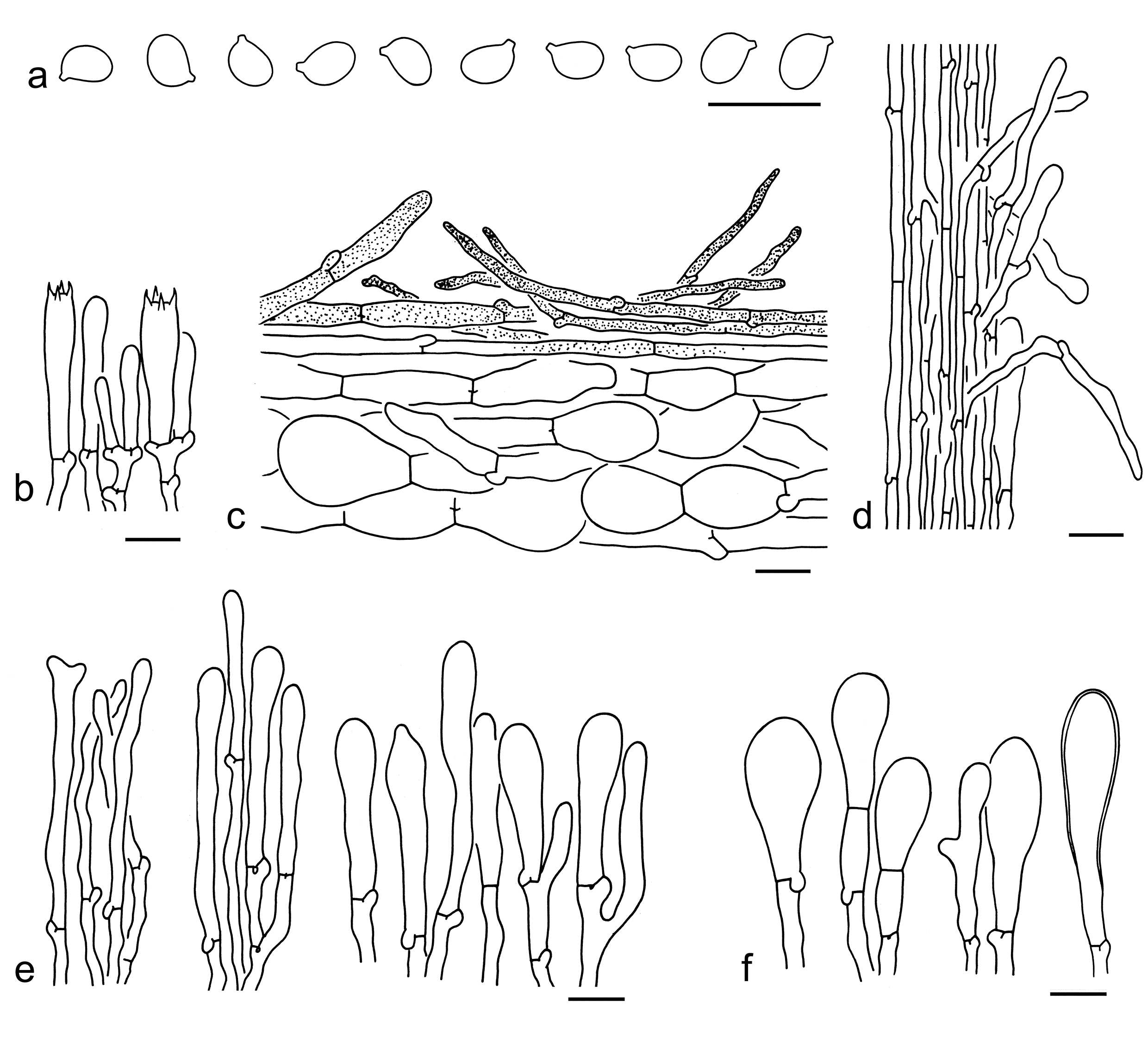
Basidiospores [30/1/1] 4.5–6 4–5 µm (mean length = 5.2 µm, mean width = 4.4 µm), Q = (1.06–) 1.15–1.26 (–1.32), Qm = 1.2 ± 0.06, broadly ellipsoid, non-amyloid, colorless, hyaline, thin-walled, smooth; apiculus relatively large. Basidia 20–30 5–8 µm, clavate, 4-spored, hyaline; sterigmata 3–5 µm long; basal septa with clamps. Pleurocystidia 55–85 10–15 µm, abundant, scattered, fusiform to subfusiform, slightly thick-walled (0.5–1 µm). Cheilocystidia 45–70 8–15 µm, conspicuous, abundant, scattered, fusiform to subfusiform, slightly thick-walled (0.5–1 µm). Subhymenium composed of 2–4 µm wide filamentous hyphae. Lamellar trama regular, composed of colorless, cylindrical to filamentous hyphae 5–20 µm wide. Pileipellis a cutis, composed of radially arranged, 2–20 µm wide, colorless hyphae; terminal cells in the upper layer fusiform to ventricose, often thick-walled (ca. 1 µm). Caulocystidia 40–100 8–10 µm, narrowly clavate to clavate, thin- to thick-walled up to 1 µm. Clamps present in all parts of basidioma.
Ecology: On soil in a subtropical evergreen broad-leaved forest.
Known distribution: Currently only known from Eastern China.
Notes: Albomagister albus is characterized by its very small, white basidioma with a fibrillose pileus, distant lamellae, broadly ellipsoid basidiospores, and fusiform, slightly thick-walled pleuro- and cheilocystidia. This species is related to A. luteifolius Lebeuf et al. (Fig. 1), but the latter has a stipitipellis without caulocystidia and is currently only found in eastern North America (Matheny et al. 2024).
Albomagister subaustralis, A. alesandrii P.A. Moreau et al. and A. virgineus G. Corriol & P. Jargeat are similar to the new species. However, A. subaustralis has slightly narrower basidiospores (5–6.5 3–4 µm) and is currently only found in North America (Hesler & Smith 1963; Sánchez-García et al. 2014). The European A. alesandrii differs from A. albus by its faintly amyloid basidiospores sometimes thick-walled, and thin-walled pleuro- and cheilocystidia (Moreau et al. 2015). Albomagister virgineus differs by its slightly larger basidiospores (5.8–6.7 4.9–6 µm) with few thick-walled, the absence of pleurocystidia and is currently only collected from Europe to date (Corriol & Jargeat 2018).
2. Albomagister atrosquamosus Y.Y. Cui, L.K. Jia & Zhu L. Yang, sp. nov. Figs. 15b, 17
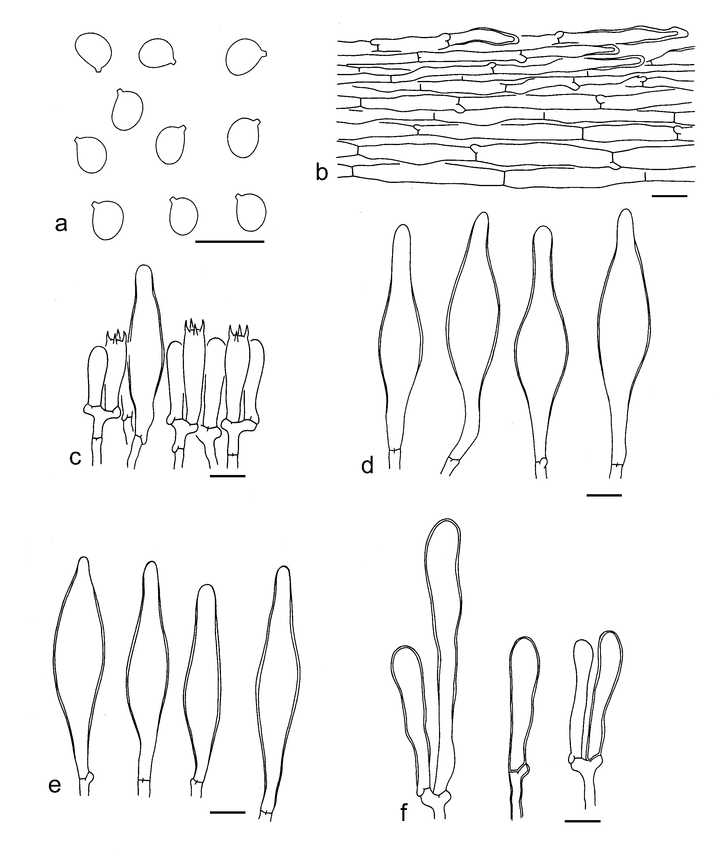
Etymology: atrosquamosus = black squamules, referring to its basidioma densely covered with black squamules.
Diagnosis: Related to A. griseosquamosus Lebeuf et al., but differs by having smaller basidiospores (4.5–6 3.5–5 µm).
Type: CHINA. Zhejiang Province, Lishui, Jingning She Autonomous County, in a subtropical forest with bamboos, altitude 687 m, 24 Jun. 2021, 331127MF-JLK1608 (holotype, KUN-HKAS124357).
Description: Basidioma small. Pileus ca. 3.5 cm in diam., applanate; surface dry, background white (1A1), densely covered with black (1F1–4) to grey (2F1–3), fibrillose squamules over the entire disk; margin straight. Lamellae sinuate to adnate, white (1A1) to dirty white (1B1–2), subclose to close; lamellar edges white (1A1) to dirty white (1B1–2), sometimes with grey (1C1–3) stains. Stipe ca. 5 0.9 cm, subcylindrical, white (1A1) background covered with black (1F1–4) to grey (2F1–3), fibrillose squamules, exannulate. Taste and odour not recorded.
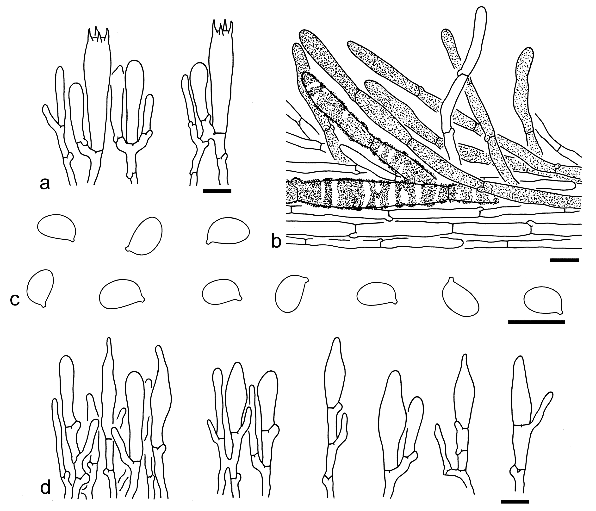
Basidiospores [20/1/1] 4.5–6 3.5–5 µm (mean length = 5.1 µm, mean width = 4.2 µm), Q = (1.1–) 1.15–1.29 (–1.37), Qm = 1.22 ± 0.07, broadly ellipsoid, occasionally subglobose or ellipsoid, inamyloid, colorless, hyaline, thin-walled, smooth; apiculus relatively large. Basidia 20–30 5–7 µm, clavate, 4-spored, hyaline; sterigmata 3–5 µm long; basal septa with clamps. Pleurocystidia 50–60 18–23 µm, abundant, scattered, broadly clavate with a mucronate apex, thick-walled (ca. 1 µm), colorless, hyaline. Cheilocystidia 55–65 10–15 µm, abundant, clustered, fusiform, broadly fusiform to ventricose, sometimes broadly clavate with a mucronate apex, thin- to thick-walled (0.5–2 µm), colorless, hyaline. Subhymenium composed of 2–4 µm wide filamentous hyphae. Lamellar trama regular, composed of colorless, ellipsoid, cylindrical, clavate to filamentous hyphae 5–35 µm wide. Pileipellis a cutis, composed of radially arranged, colorless or grey-brown to grey, 8–15 µm wide, slightly thick-walled (0.5–1 µm), cylindrical hyphae with lanceolate to cylindrical terminal cells; subpellis composed of radially arranged, colorless, 5–10 µm wide, thin-walled, cylindrical to filamentous hyphae. Stipitipellis a trichoderm-like layer, composed of erected arranged, clavate to subclavate, thin- to slightly thick-walled (0.5–1 µm), grey-brown, septate elements 5–20 µm wide, mixed with scattered, thick-walled (ca. 1 µm), grey-brown, lanceolate, subfusiform to ventricose caulocystidia. Clamps present in all parts of basidioma.
Ecology: On soil in a subtropical forest with bamboos.
Known distribution: Currently only known from Eastern China.
Notes: Albomagister atrosquamosus is well delimitated by its grey scaly basidioma, broadly ellipsoid basidiospores (4.5–6 3.5–5 µm), thick-walled, broadly clavate pleurocystidia with a mucronate apex, clustered, thin- to thick-walled, fusiform, broadly fusiform to ventricose cheilocystidia, and the presence of clamps. This species is closely related to A. griseosquamosus (Fig. 1), but the latter species has larger basidiospores (6–7.1–8 4.5–5.6–6.7 µm) and is currently distributed in eastern North America (Matheny et al. 2024).
Albomagister atrosquamosus can be easily separated from A. subaustralis, A. alesandrii and A. virgineus by its grey scaly basidioma. In addition, A. alesandrii has faintly amyloid, sometimes thick-walled basidiospores, thin-walled pleurocystidia, and thin-walled, sparse cheilocystidia (Moreau et al. 2015). Albomagister virgineus has relatively larger, few thick-walled basidiospores (5.8–6.7 4.9–6 µm), and no pleurocystidia (Corriol & Jargeat 2018). Albomagister albus has fusiform pleurocystidia, fertile lamellar edges with scattered cheilocystidia, and narrowly clavate to clavate caulocystidia. Tricholoma atrosquamosum Sacc. is somewhat similar to this species but can be easily separated by its larger basidioma with a glabrous stipe, ellipsoid to elongate basidiospores (5.5–7 3.5–4.5), and the absence of pleuro- and cheilocystidia, and clamps (this study). Pogonoloma spinulosum (Kühner & Romagn.) Sánchez-García is comparable to A. atrosquamosus. However, the former species can be easily separated by its amyloid basidiospores (Sánchez-García et al. 2014; Læssøe & Petersen 2019).
Corneriella Sánchez-García, Taxon 63(5): 1000 (2014)
Type: Corneriella bambusarum (Desjardin & Hemmes) Sánchez-García, Taxon 63(5): 1000 (2014).
Description: Lamellae white when young, then becoming darker. Basidiospores smooth, thin-walled, and amyloid. Cheilocystidia present and conspicuous. Pleurocystidia absent. Pileipellis a cutis with suberect to erect terminal cells. Clamps present.
Ecology: Saprotrophic (Sánchez-García et al. 2014; Raj et al. 2015). On soil and humus in tropical regions (Sánchez-García et al. 2014; Raj et al. 2015).
Known distribution: The known species of this genus is distributed in India, Malaysia, Thailand, and USA (Sánchez-García et al. 2014; Raj et al. 2015).
Notes: Only three species are recorded in Corneriella (Sánchez-García et al. 2014; Raj et al. 2015). Currently no species of this genus has been found in China. The description above is a summary of Sánchez-García et al. (2014) and Raj et al. (2015).
Dennisiomyces Singer, Anais Soc. Biol. Pernambuco 13(1): 225 (1955)
Type: Dennisiomyces glabrescentipes Singer, Anais Soc. Biol. Pernambuco 13(1): 225 (1955).
Description: Basidiospores smooth, thin-walled, and amyloid. Pleurocystidia present. Cheilocystidia present and conspicuous. Clamps present.
Ecology: Saprotrophic, on soil with various trees (Singer 1989; Sánchez-García & Matheny 2017; Corriol & Jargeat 2019).
Known distribution: Known from Europe, Caribbean Basin, Central America, Eastern U.S., and South America (Singer 1955a, b, 1989; Sánchez-García et al. 2014; Corriol & Jargeat 2019; Fig. 1)
Notes: To date, five species, including D. fibrillosus G. Corriol & P. Jargeat, D. fuscoalbus Singer, D. glabrescentipes Singer, D. griseus (Dennis) Singer and D. rionegrensis Singer, are recorded under Dennisiomyces (Singer 1955a, b, 1956, 1989; Corriol & Jargeat 2019). Only three of them (D. fibrillosus, D. glabrescentipes and D. griseus) are confirmed with molecular evidence (Sánchez-García et al. 2014; Corriol & Jargeat 2019), and the descriptions of this genus are a summary on these three species (Singer 1955a, b, 1956; Corriol & Jargeat 2019). No species of Dennisiomyces are found in China.
Dermoloma J.E. Lange ex Herink, Sb. severočesk. Mus., Hist. Nat. 1: 62 (1958)
Type: Dermoloma cuneifolium (Fr.) Singer ex Bon, Docums Mycol. 17(no. 65): 51 (1986).
Description: Context becoming black or without color change when bruised. Basidiospores smooth, thin-walled, amyloid or inamyloid. Pileipellis a hymeniderm, pluristratous hymeniderm or transitional to an epithelium, composed of one or more layers of inflated cells. Clamps present.
Ecology: Reported as a member of CHEGD (acronym of Clavariaceae, Hygrocybe s.l., Entoloma, Geoglossaceae, Dermoloma) fungi, which have a biotrophic lifestyle and probably form an unspecified symbiosis with vascular plants (Halbwachs et al. 2013, 2018; Adamčíková et al. 2025; Caboňová et al. 2025).
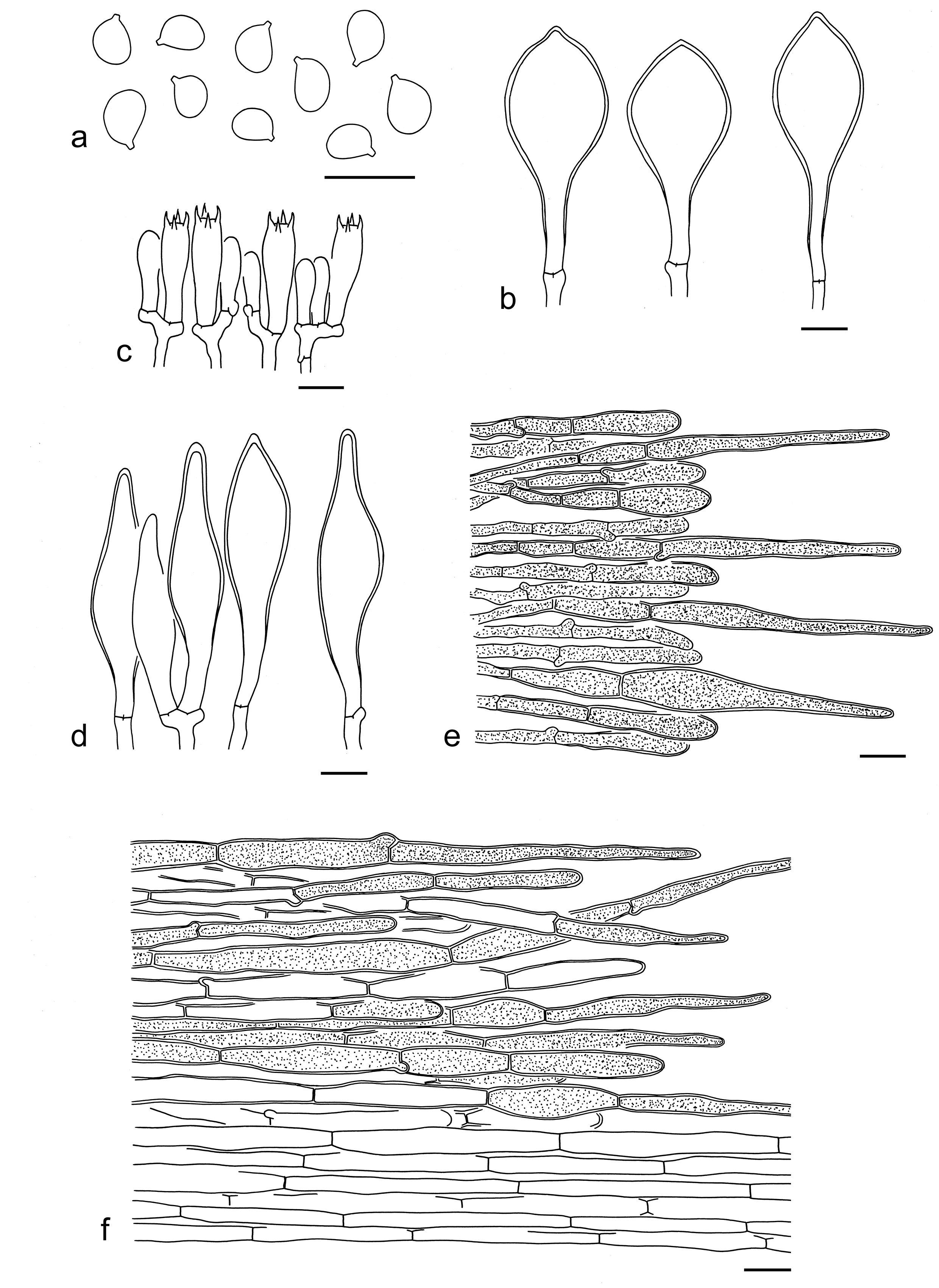
Known distribution: Almost cosmopolitan. Known from Africa, Asia, Australia and Oceania, Europe, and North America (Sánchez-García et al. 2021; Adamčíková et al. 2025; Caboňová et al. 2025).
Notes: Many mycologists have contributed to the study of Dermoloma based on morphological characters since its publishment (Singer 1962a, 1975, 1986; Bon 1979, 1986, 1998; Arnolds 1992, 1993; Contu et al. 2008; Vesterholt 2008, 2012). Sánchez-García et al. (2021) provided the first phylogenetic study of Dermoloma based on DNA sequence analysis of four gene fragments and supported the subdivision of this genus into two subgenera (subgen. Dermoloma and subgen. Amylospora) and four sections (sect. Dermoloma, sect. Conica, sect. Atrobrunnea and sect. Nigrescentia). Later, Adamčíková et al. (2025) validated this finding, however the subgenera splitting was not retrieved with molecular evidence, implying that the delimitation of subgenera was mainly based on morphological data and required more investigations. Here, we follow the studies of Sánchez-García et al. (2021) and Adamčíková et al. (2025). So far, no species of Dermoloma has been found in China.
Leucopaxillus Boursier, Bull. trimest. Soc. mycol. Fr. 41(3): 393 (1925)
Type: Leucopaxillus paradoxus (Costantin & L.M. Dufour) Boursier [as 'paradoxa'], Bull. trimest. Soc. mycol. Fr. 41(3): 393 (1925).
Description: Basidioma robust and fleshy. Pileus white, beige to brown, finely velvety. Lamellae crowded and narrow, often forked. Basidiospores verrucose and amyloid. Pleurocystidia absent. Cheilocystidia present. Pileipellis a cutis composed of radially arranged hyphae mixed with interwoven to more or less erected hyphae. Clamps present.
Ecology: Probably saprotrophic (Sánchez-García & Matheny 2017). Solitary to scattered on soil with various trees.
Known distribution: Almost cosmopolitan. Known from Asia, Europe, North America, Oceania, and South America (Singer & Smith 1943; Bougher 1987; Hubregtse & Hubregtse 2012; Vizzini et al. 2012).
Notes: Leucopaxillus, typified by L. paradoxus (Costantin & L.M. Dufour) Boursier, belongs to Tricholomataceae (Sánchez-García et al. 2014). There were 110 names recorded under this genus (Index Fungorum; Singer & Smith 1943, 1947; Bougher 1987; Kibby et al. 2009; Işık & Türkekul 2018), but many species were subsequently moved into other genera, such as Aspropaxillus Kühner & Maire, Giacomia Vizzini & Contu, Notholepista Vizzini & Contu and Pseudoclitopilus Vizzini & Contu (Vizzini et al. 2012; He & Yang 2022). Recent molecular phylogenetic analyses indicated that Leucopaxillus is a well-supported monophyletic group (Sánchez-García et al. 2014, 2021). Species in this genus can be separated from the other members of Tricholomataceae by its verrucose and amyloid basidiospores. To date, three species are found in China (see below).
Key to the species of Leucopaxillus from China
1. Pileus brown; basidiospores smaller, with length less than 5.5 µm L. laterarius
1. Pileus white, cream to brownish; basidiospores larger, with length most more than 5.5 µm 2
2. Pileus white, often with cream tinge; basidiospores ellipsoid to elongate, Q = 1.35–1.69, Qm = 1.52 ± 0.11 ………L. amyloideus
2. Pileus white to brownish; basidiospores broadly ellipsoid to ellipsoid, Q = 1.17–1.48, Qm = 1.32 ± 0.12 L. verrucosus
3. Leucopaxillus amyloideus Y.Y. Cui & Zhu L. Yang, sp. nov. Figs. 15c, 18, 19
Fungal Names: FN 572330
Etymology: amyloideus = amyloid, referring to its amyloid basidiospores.
Diagnosis: Similar to L. cerealis (Lasch) Singer, but differs by its bulbous stipe base.
Type: CHINA. Xinjiang, Tacheng, Shawan, altitude ca. 1700 m, Jian-Wei Liu 645 (holotype, KUN-HKAS134956).
Description: Basidioma medium-sized. Pileus ca. 6 cm in diam., hemispherical, convex to plano-convex; surface dry, white (1A1), often with cream (1A2–4) to brownish (3B2–4) tints, glabrous or slightly fibrillose; margin incurved, often ribbed in ages; context white (1A1). Lamellae subdecurrent, crowded, narrow, often forked, white (1A1) to beige white (1A2); edges entire. Stipe ca. 6 1.5–2.5 cm, firm, solid, cylindrical, often slightly tapering downwards, white (1A1), exannulate; basal mycelium white (1A1). Taste and odour not recorded.
Basidiospores [60/3/1] 6–8 (–8.5) 4–5.5 µm (mean length = 7.2 µm, mean width = 4.7 µm), Q = (1.24–) 1.35–1.69 (–1.89), Qm = 1.52 ± 0.11, ellipsoid to elongate, occasionally broadly ellipsoid, amyloid, colorless, hyaline, thin-walled, verrucose, sometimes glabrous. Basidia 30–45 10–12 µm, clavate, 4-spored, thin- to slightly thick-walled, hyaline; sterigmata ca. 4 µm long; basal septa with clamps. Pleurocystidia absent. Cheilocystidia 30–55 3–7 µm, scattered, irregularly fusiform to filiform, thin-walled, colorless, hyaline. Subhymenium composed of 2–4 µm wide filamentous hyphae, clamps present. Lamellar trama regular, composed of colorless; thin-walled hyphae 3–8 µm wide. Pileipellis a cutis, composed of radially arranged, 2–8 µm wide, thin-walled, colorless hyphae, often mixed with more or less erected hyphae. Clamps present in all parts of basidioma.
Ecology: On soil in a temperate forest with trees of Picea.
Known distribution: Currently known from Northwestern China and probably Pakistan.
Additional specimen examined: CHINA. Xinjiang, Tacheng, Shawan, altitude ca. 1800 m, Jian-Wei Liu (KUN-HKAS152699).
Notes: Leucopaxillus amyloideus is characterized by its white and fleshy basidioma, amyloid, verrucose basidiospores and the presence of clamps. This species is similar to L. cerealis, L. monticola (Singer & A.H. Sm.) Bon and L. paradoxus. Leucopaxillus cerealis can be separated by its white stipe often with a bulbous base (Singer & Smith 1943; Singer 1962b; Hubregtse & Hubregtse 2012). Leucopaxillus monticola differs from L. amyloideus by its collybioid or clitocyboid habit, fairly crowded lamellae and abundant cheilocystidia (Singer & Smith 1947; Vizzini & Contu 2009). Leucopaxillus paradoxus possesses slightly smaller basidiospores (5–7.5 3.3–4.8 µm) and no cheilocystidia (Singer & Smith 1943). Molecular phylogenetic analysis indicated that L. amyloideus also occur in Pakistan (Fig. 1).
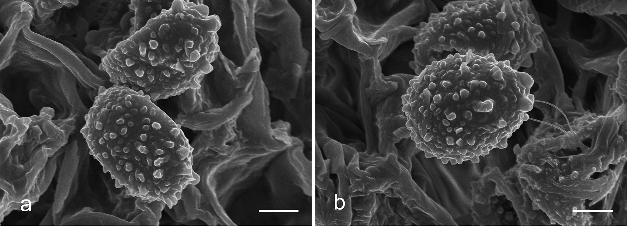
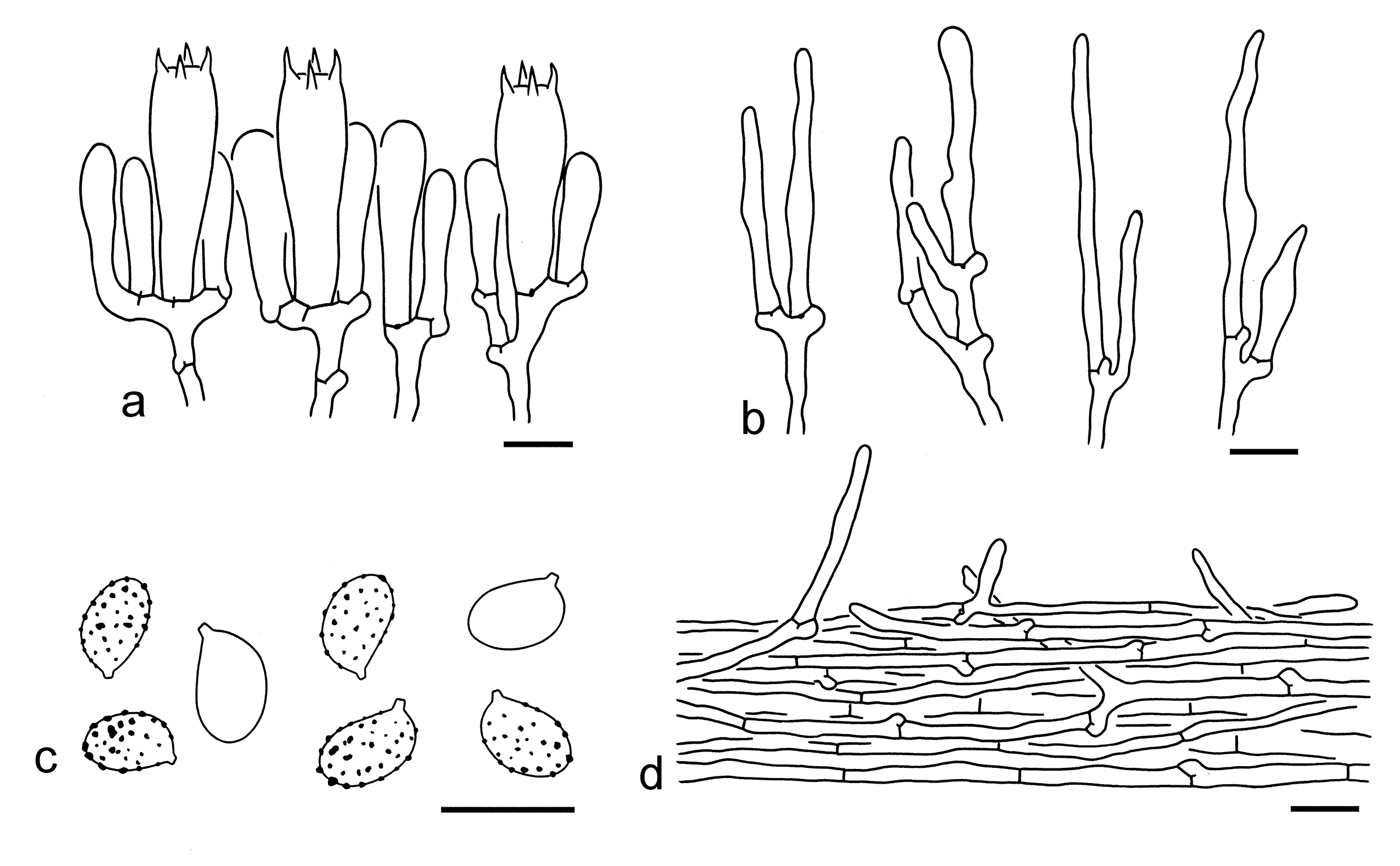
4. Leucopaxillus laterarius (Peck) Singer & A.H. Sm., Pap. Mich. Acad. Sci. 28: 117 (1943) [1942]. Figs. 15d, 20, 21
Basionym: Agaricus laterarius Peck, Bull. Buffalo Soc. nat. Sci. 1(2): 43 (1873) [1873-1874].
Description: Basidioma small to medium-sized, clustered, and robust. Pileus 2–5 cm in diam., convex to applanate, sometimes obtusely umbonate; surface dry, brown (5D4–6), matted-fibrillose, in ages sometimes becoming scurfy or cracking into small pieces around the disc; margin incurved, often ribbed or grooved; context white (1A1). Lamellae adnate, sinuate to decurrent, very crowded, narrow, often forked, white (1A1), with brownish (5A2–5) color in age; edges entire, concolor. Stipe 6–8 0.8–1.2 cm, very firm, solid, clavate, often inflated at base, white (1A1), sometimes with brown (5A2–5, 5B3–5) color, exannulate; basal mycelium white (1A1). Taste and odour not recorded.
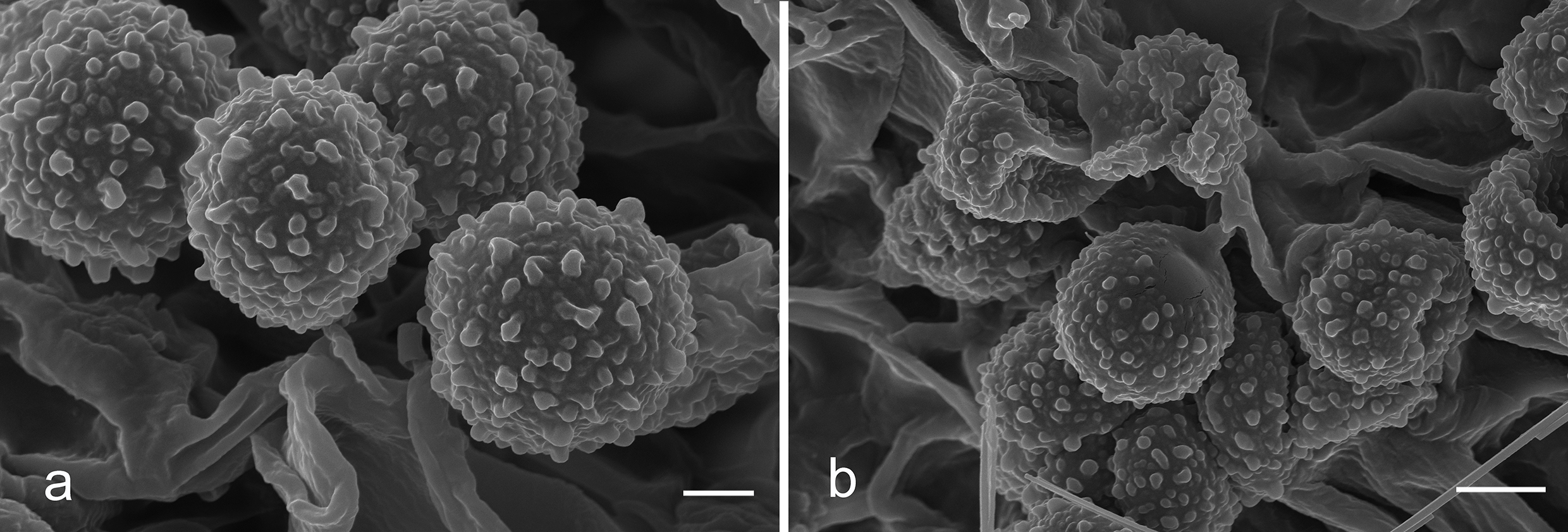
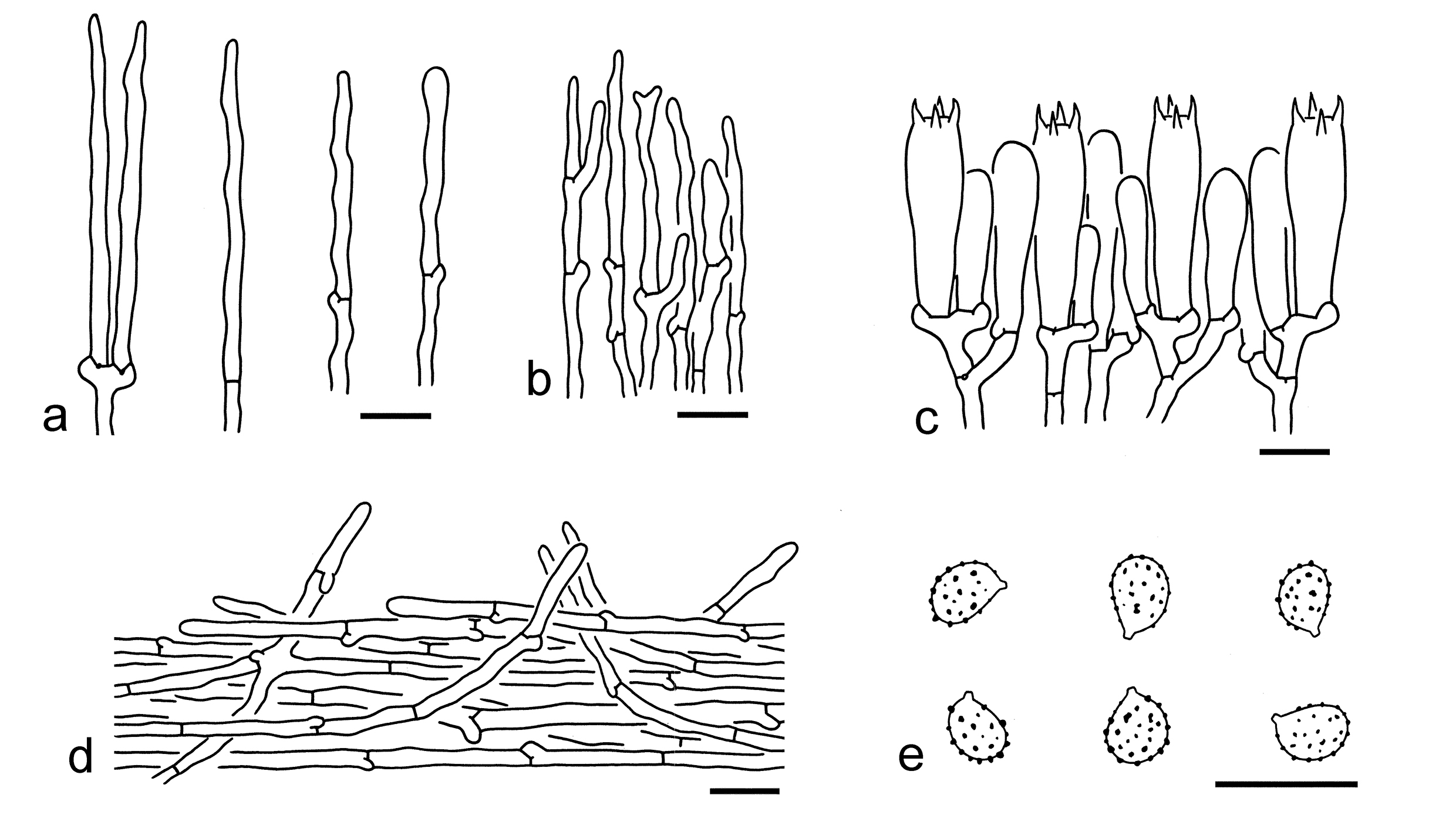
Ecology: On soil in temperate forests.
Known distribution: Our molecular data indicated this species occurs in North America and East Asia (Northwestern China).
Specimens examined: CHINA. Xinjiang, altitude 1865 m, 3 Aug. 2021, Jin Li 112 (KUN-HKAS124334); Changji Hui Autonomous Prefecture, Jimusaer, altitude 1987 m, 18 Jul. 2021, Jin Li 73 (KUN-HKAS124332); Urumqi, altitude 1885 m, 18 Jul. 2021, Jin Li 59 (KUN-HKAS124331).
Notes: Leucopaxillus laterarius was described from New York (Singer & Smith 1943) and is well separated by its small, broadly ellipsoid to ellipsoid basidiospores (4–5.5 3–4.5 µm, mean length = 4.8 µm, mean width = 3.8 µm) (this study). Leucopaxillus eucalyptorum (Cleland) Grgur. is related to L. laterarius (Fig. 1), but the former species grows in Eucalyptus forests of New Zealand (Grgurinovic 1997; Hubregtse 2019). Leucopaxillus pulcherrimus (Peck) Singer & A.H. Sm. is similar to L. laterarius, but differs by its subglobose basidiospores (4–5.8 3.5–4.5 µm) (Singer & Smith 1943, 1947).
5. Leucopaxillus verrucosus Y.Y. Cui & Zhu L. Yang, sp. nov. Figs. 15e, 22, 23
Fungal Names: FN 572331
Etymology: verrucosus = verrucose, referring to its verrucose basidiospores.
Diagnosis: Similar to L. laterarius, but differs by its larger basidiospores.
Type: CHINA. Xinjiang, Huocheng County, Guozigou, altitude ca. 2000 m, 5 Jul. 2017, Xiao-Xia Ding 167 (holotype, KUN-HKAS106509).
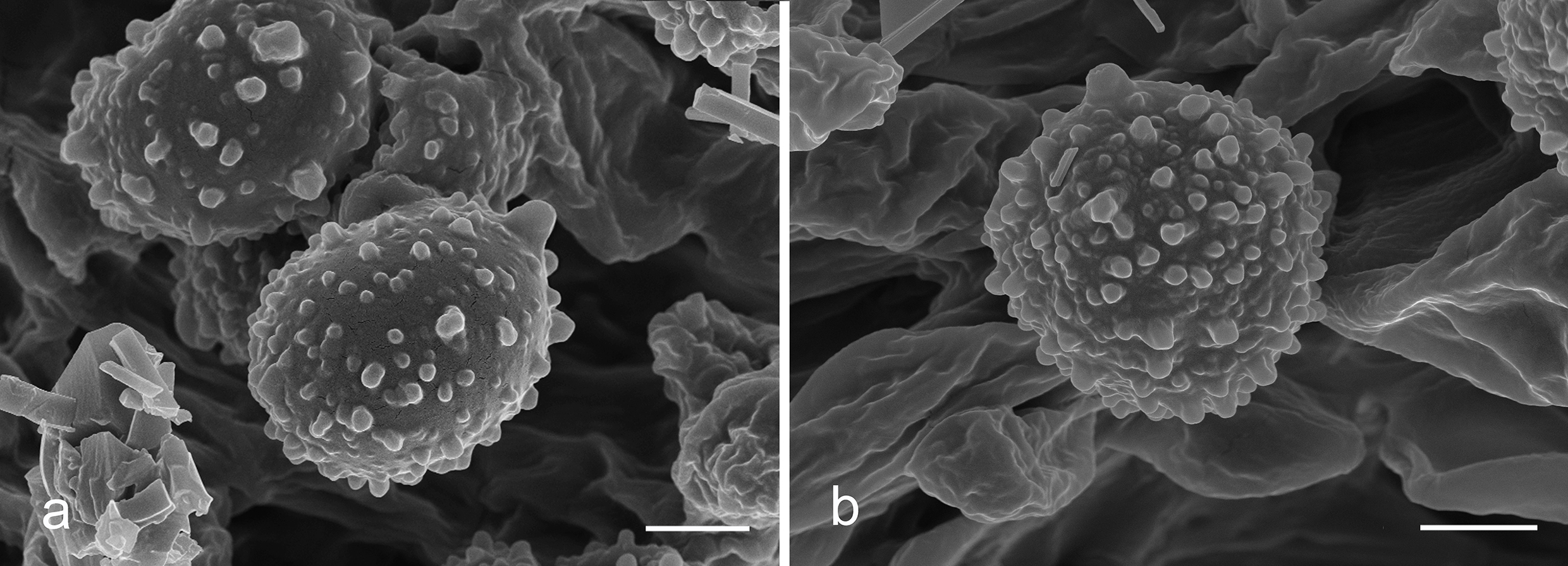
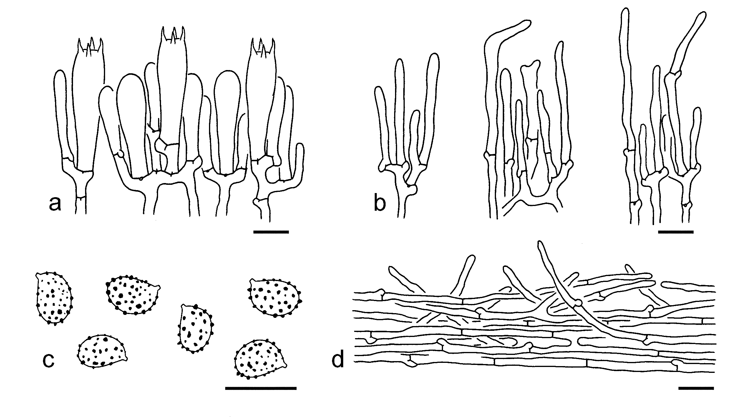
Description: Basidioma small to medium-sized, clustered. Pileus ca. 3.5 cm in diam., hemispherical, convex to plano-convex; surface dry, white (1A1), cream (1A2–4) to brownish (2B3–5), glabrous to fibrillose, in ages sometimes cracking into small pieces around the disc; margin incurved, often ribbed; context white (1A1). Lamellae subdecurrent, crowded, very narrow, sometimes forked, white (1A1) to pinkish (7A2); edges entire, concolor. Stipe ca. 4 1.5 cm, firm, solid, subclavate to clavate, white (1A1); basal mycelium white (1A1). Taste and odour not recorded.
Basidiospores [40/2/1] (5–) 5.5–7.5 4.5–6 µm (mean length = 6.7 µm, mean width = 5.1 µm), Q = (1.02–) 1.17–1.48 (–1.56), Qm = 1.32 ± 0.12, broadly ellipsoid to ellipsoid, amyloid, colorless, hyaline, thin-walled, verrucose. Basidia 30–45 7–9 µm, clavate, 4-spored, thin-walled, sometimes slightly thick-walled, hyaline; sterigmata 3–6 µm long; basal septa with clamps. Pleurocystidia absent. Cheilocystidia 20–50 2–5 µm, often clustered, subcylindrical to filiform, thin-walled, colorless, hyaline. Subhymenium composed of 2–4 µm wide filamentous hyphae, clamps present. Lamellar trama regular, composed of colorless; thin-walled hyphae 2–7 µm wide. Pileipellis a cutis, composed of radially arranged to interwoven, 3–6 µm wide, thin-walled, colorless hyphae, often mixed with more or less erected hyphae. Clamps present in all parts of basidioma.
Ecology: On soil in temperate forests with trees of Picea.
Known distribution: Currently known from Northwestern China.
Additional specimen examined: CHINA. Xinjiang, Tacheng, Shawan County, forest type not recorded, altitude ca. 1600 m, 2 Jul. 2017, Qi Zhao 2767 (KUN-HKAS134957).
Notes: Leucopaxillus verrucosus is delimitated by its brownish pileus, amyloid, verrucose, broadly ellipsoid to ellipsoid basidiospores (5.5–7.5 4.5–6 µm) and abundant cheilocystidia. No closely related species were detected based on our currently data (Fig. 1). Leucopaxillus laterarius is similar to this species due to their brown pileus but can easily be separated by its smaller basidiospores (4–5.5 3–4.5 µm) (this study).
Neodermoloma Sánchez-García, Matheny & Adamčík, IMA Fungus 16(e157337): 120 (2025)
Type: Neodermoloma campestre Sánchez-García, Matheny & Adamčík, IMA Fungus 16(e157337): 120 (2025).
Description: Basidiospores smooth, amyloid. Pileipellis an epithelium composed of inflated cells. Clamps present.
Known distribution: North America (Adamčíková et al. 2025).
Notes: Sánchez-García et al. (2021) identified a "Dermoloma-like" clade within the Tricholomataceae (Supplementary Table S4). Adamčíková et al. (2025) described this clade as a new genus named Neodermoloma (Supplementary Table S4). This study follows the study of Adamčíková et al. (2015). There have been no Neodermoloma species discovered in China thus far.
Porpoloma Singer, Sydowia 6(1-4): 198 (1952)
Type: Porpoloma sejunctum Singer, Sydowia 6(1-4): 198 (1952).
Description: Basidioma tricholomatoid. Basidiospores smooth, thin-walled, amyloid. Clamps present.
Ecology: Ectomycorrhizal (Garrido 1988; Tedersoo et al. 2010; Sánchez-García & Matheny 2017). On soil in forests with Myrtaceae and Nothofagus (Singer 1952; Sánchez-García & Matheny 2017).
Distribution: Mainly distributed in the Southern Hemisphere (Singer 1952; Sánchez-García & Matheny 2017), but also recorded in Europe (Hausknecht & Zuccherelli 1999; Arauzo 2012).
Notes: Porpoloma was described and divided into three subgenera (Singer 1952). Sánchez-García et al. (2014) promoted the subgen. Pogonoloma Singer and subgen. Pseudotricholoma (Singer) Singer to independent genera, as Pogonoloma (Singer) Sánchez-García and Pseudotricholoma (Singer) Sánchez-García & Matheny with molecular evidence respectively. Species of Porpoloma are mainly distributed in the Southern Hemisphere, but a few are recorded in Europe (Singer 1952; Hausknecht & Zuccherelli 1999; Arauzo 2012). To date, 12 species were described under this genus (https://nmdc.cn/fungalnames/currentfungal?searchval=porpoloma&radiosearch=2), but only three of them (Po. portentosum Singer, Po. sejunctum Singer and Po. terreum Singer) are supported by both molecular and morphological evidence (Sánchez-García et al. 2014). Currently no species of Porpoloma are found in China and the aforementioned descriptions are a summary based on the three species.
Pseudobaeospora Singer, Lloydia 5: 129 (1942)
Type: Pseudobaeospora oligophylla (Singer) Singer, Lilloa 22: 438 (1951) [1949].
Description: Basidioma mycenoid to collybioid, very small to small. Pileus mostly violaceous, purple to purple-brown, glabrous to slightly squamulose. Lamellae concolorous with pileus or sometimes paler. Stipe subcylindrical, often concolorous with pileus. Basidiospores very small to small, smooth, at first thin-walled, then becoming thick-walled and more or less dextrinoid when mature. Cheilocystidia present or absent. Pileipellis with color change or not in 5% KOH, composed of radical or erected chains of inflated cells, or sometimes separated into two layers, with a suprapellis made of radically arranged filamentous hyphae and a subpellis composed of chains of inflated cells. Clamps mostly present, but absent in some species.
Ecology: Probably non-ectomycorrhizal (Bas 2003). Solitary to scattered on soil in various environments (Bas 2002, 2003; Arnolds et al. 2004; Desjardin 2004; Vellinga 2009; Desjardin et al. 2014; Wu et al. 2017; Craig et al. 2023).
Known distribution: Probably cosmopolitan (Singer 1986; Bas 2002, 2003; Arnolds et al. 2004; Desjardin 2004; Adamčík et al. 2007; Vellinga 2009; Adamčík & Jančovičová 2011; Schwarz 2012; Desjardin et al. 2014; Wu et al. 2017; Gisotti et al. 2021; Craig et al. 2023).
Notes: Vizzini et al. (2025) described a new subfamily named Pseudobaeosporoideae for the genus Pseudobaeospora within Tricholomataceae, with the remaining members placed in another suborder Tricholomatoideae. However, this work has not followed thus far because the Tricholomatoideae clade did not receive strong support in our phylogenetic analysis (Fig. 1).
Pseudobaeospora, typified by P. oligophylla (Singer) Singer (originally as Baeospora oligophylla Singer), was proposed by Singer (1942), and species in this genus have thick-walled and dextrinoid basidiospores when mature (Bas 2002, 2003). Pseudobaeospora was placed in Tricholomataceae or different tribus of Agaricaeae based on morphological characters (Singer 1942, 1951, 1986; Kühner 1980; Bas 2003), and molecular evidence supported this genus in Tricholomataceae (Sánchez-García & Matheny 2017; Sánchez-García et al. 2021; this study).
In regard to the subdivision within this genus, Bas (2003) divided it into five groups (Albidula group, Celluloderma group, Pyrifera group, Frieslandica group and Pillodii group) according to the color of basidioma, the presence or absence of clamps and cheilocystidia, and the structure of pileipellis, while Voto (2009) proposed an infrageneric systematic organization of Pseudobaeospora based on the structure of pileipellis. Both studies were relied on morphological data only. To date, Fungal Names database (https://nmdc.cn/fungalnames/currentfungal?searchval=Pseudobaeospora&radiosearch=2) listed 35 species names under the genus. Only one species, P. lilacina X.D. Yu et al., was found in China (see below).
6. Pseudobaeospora lilacina X.D. Yu et al., Mycotaxon 132(2): 332 (2017) [2016]. Figs. 15f, g, 24
Basidiospores [84/4/4] 3.5–5 (–6) 3–4 µm (mean length = 4.5 µm, mean width = 3.4 µm), Q = 1.16–1.53, Qm = 1.34 ± 0.08, broadly ellipsoid to ellipsoid, non-amyloid, mostly dextrinoid and thick-walled. Basidia 25–30 5–7 µm, clavate, 4-spored, hyaline; sterigmata 2–4 µm long; basal septa without clamps. Pleurocystidia absent. Cheilocystidia 25–50 3–15 µm, conspicuous, scattered to clustered, filamentous, subcylindrical, cylindrical to clavate, thin-walled, sometimes slightly thick-walled (ca. 1 µm), colorless, hyaline. Subhymenium composed of 2–4 µm wide filamentous hyphae, clamps present. Lamellar trama regular, composed of colorless, filamentous, cylindrical, ellipsoid to clavate hyphae 5–35 µm wide. Pileipellis composed of two layers; suprapellis thin, greyish, grey-brown, grey-green to green in KOH, consisting of thin-walled, 2–10 µm wide, repent hyphae with scattered, interwoven to somewhat erected, filamentous, subcylindrical to clavate terminal cells, which sometimes gathered as clusters at regular intervals with the cracking of pileus in appearances; subpellis made of chains of inflated cells, 30–50 15–40 µm, colorless, thin- to slightly thick-walled, clavate, ellipsoid to subglobose. Clamps present in all parts of basidioma.
Ecology: On naked soil or on soil with mosses or grasses in subtropical forests.
Known distribution: Known from Southern and Southwestern China.
Specimens examined: CHINA. Guangdong Province, Guangzhou, Baiyun District, 28 May 2013, Xiao-Dan Yu 3450 (SYAU FUNGI-010); same province, Zhaoqing, Fengkai County, 3 Jun. 2013, Xiao-Dan Yu 5546 (SYAU FUNGI-011). Yunnan Province, Baoshan, Longling County, on naked soil, altitude 1500 m, 26 Jul. 2022, GLG-WGS 223 (KUN-HKAS134959); Dehong Dai and Jingpo Autonomous Prefecture, Ruili, on soil with mosses, altitude ca. 1800 m, 17 Jul. 2022, Xue-Ping Fan 208 (KUN-HKAS134960); same prefecture, Longchuan County, on soil with mosses, altitude 1900 m, 18 Jul. 2022, Xue-Ping Fan 239 (KUN-HKAS134961).
Notes: Pseudobaeospora lilacina is characterized by its reddish violet, rosy brown to purple-brown basidioma, broadly ellipsoid to ellipsoid basidiospores (3.5–5 3–4 µm), conspicuous cheilocystidia, a cutis becoming grey-brown to green in KOH with a well differentiated subpellis, and the presence of clamps (Wu et al. 2017; this study).
One of the generic characters of Pseudobaeospora is that the basidiospores become thick-walled and dextrinoid when mature, but this phenomenon only can be found from the basidiospores on the surface of the pileus and the apical part of the stipe (Bas 2002, 2003). In our study, both non-dextrinoid and thick-walled, dextrinoid basidiospores was observed in preparations of fragments of lamellae; and thick-walled and dextrinoid basidiospores are dominant on the pileipellis and on the apex of stipe. The size of basidiospores of this species provided in this paper mostly refer to these thick-walled ones.
The color change of pileipellis in KOH, the structure of pileipellis, and the presence or absence of cheilocystidia are key characters for species delimitation in this genus (Bas 2002, 2003). The pileipellis of P. lilacina becomes grey-brown to green in KOH, and is made up of a suprapellis with repent, filamentous hyphae and a subpellis with chains of inflated cells (Fig. 24). The cheilocystidia of P. lilacina are present and conspicuous, but variable from filamentous, cylindrical to clavate, mostly thin-walled, sometimes thick-walled (Fig. 24).
Pseudoporpoloma Vizzini & Consiglio, Phytotaxa 243(3): 274 (2016)
Type: Pseudoporpoloma pes-caprae (Fr.) Vizzini & Consiglio, Phytotaxa 243(3): 274 (2016).
Description: Basidioma tricholomatoid. Pileus conical, dry, radially fibrillose. Basidiospores smooth, thin-walled, amyloid. Pleurocystidia absent. Cheilocystidia rare or absent. Pileipellis a cutis. Clamps present.
Ecology: Probably saprotrophic and on soil in grasslands (Vizzini et al. 2016).
Known distribution: Widespread in Europe (Vizzini et al. 2016).
Notes: Pseudoporpoloma was described by Vizzini et al. (2016) and contains only a single species, namely Ps. pes-caprae. To date, no species are found in China and the aforementioned descriptions are based on the study of Vizzini et al. (2016).
Pseudotricholoma (Singer) Sánchez-García & Matheny, Taxon 63(5): 1000 (2014)
Basionym: Cantharellula subgen. Pseudotricholoma Singer, Sydowia 2(1-6): 30 (1948).
Type: Pseudotricholoma umbrosum (A.H. Sm. & M.B. Walters) Sánchez-García & Matheny, Taxon 63(5): 1000 (2014).
Description: Basidioma brown to grey, covered with grey to dark grey, felted to tomentose squamules. Lamellae dirty white, brownish to greyish, becoming red then black when bruised. Basidiospores thin-walled, smooth, amyloid. Pleurocystidia absent. Cheilocystidia present or absent. Clamps present.
Ecology: Recorded as possibly ectomycorrhizal in Sánchez-García et al. (2014), and then listed as non-ectomycorrhizal taxa in Sánchez-García & Matheny (2017). On soil in woods and grasslands (Sánchez-García et al. 2014).
Known distribution: Distributed in Northern Hemisphere (Sánchez-García et al. 2014).
Notes: Pseudotricholoma, typified by Pse. umbrosum, was proposed by Sánchez-García et al. (2014). To date, only two species are found in China (see below).
Key to the species of Pseudotricholoma from China
1. Basidiospores longer, mostly elongate, 6–8 3.5–4.5 µm (mean length = 7.2 µm, mean width = 3.9 µm), Q = 1.62–2, Qm = 1.83 ± 0.15; cheilocystidia poorly differentiated………. ……….... Pse. aff. azoricum
1. Basidiospores mostly ellipsoid, 5.5–7 4–5 µm (mean length = 6.5 µm, mean width = 4.4 µm), Q =1.35–1.65, Qm = 1.49 ± 0.08; cheilocystidia present Pse. griseum
7. Pseudotricholoma aff. azoricum P. Iglesias et al., Errotari 18: 279 (2021). Fig. 15h, i
Description: Basidioma small. Pileus 3–5 cm in diam., hemispherical to convex; surface dry, brown-grey (3D2–4), densely covered with brown-grey (3D2–5) to dark grey (2E1–3), felted to tomentose squamules; margin incurved. Lamellae brownish (2B2–4), brownish grey (2C2–4) to greyish (2D1–3), close. Stipe 5–9 1–2 cm, subcylindrical to cylindrical, white (1A1) to dirty white (2B1) background with longitudinally arranged, grey (2C1–3) fibrils, exannulate. The color change when bruised or cut not recorded. Taste and odour not recorded.
Basidiospores [27/1/1] 6–8 3.5–4.5 µm (mean length = 7.2 µm, mean width = 3.9 µm), Q = (1.59–) 1.62–2 (–2.2), Qm = 1.83 ± 0.15, mostly elongate, amyloid, colorless, hyaline, thin-walled, smooth.
Ecology: Solitary to scattered on soil in a temperate forest.
Known distribution: Southwestern China.
Specimen examined: CHINA. Yunnan Province, Diqing Tibetan Autonomous Prefecture, Shangri-La, in a subalpine temperate forest, 9 Aug. 2020, MF399 (KUN-HKAS134962).
Notes: Through years of field collections, a specimen of Pseudotricholoma (KUN-HKAS134962) somewhat sister to Pse. azoricum P. Iglesias et al. was collected from Yunnan, China (Fig. 1). Unfortunately, the bad status of this specimen hindered us from detailed studying on it, we therefore treated it as Pse. aff. azoricum for the time being.
8. Pseudotricholoma griseum Y.Y. Cui & Zhu L. Yang, sp. nov. Figs. 15j, k, 25
Fungal Names: FN 572332
Etymology: griseum = grey, referring to the grey basidioma.
Diagnosis: Similar to Pse. umbrosum, but differs by its conspicuous cheilocystidia.
Type: CHINA. Yunnan Province, Nujiang Lisu Autonomous Prefecture, Lushui, in a bamboo forest, altitude 2590–2690 m, 6 Aug. 2022, GLG-LJ024 (holotype, KUN-HKAS134963).
Basidiospores [20/1/1] 5.5–7 4–5 µm (mean length = 6.5 µm, mean width = 4.4 µm), Q =1.35–1.65, Qm = 1.49 ± 0.08, mostly ellipsoid, occasionally elongate, amyloid, colorless, hyaline, thin-walled, smooth. Basidia 25–45 7–10 µm, clavate, 4-spored, colorless to brown, hyaline; sterigmata 4–7 µm long; basal septa with clamps. Pleurocystidia absent, but some narrowly cylindrical hyphae or clavate with apex mucronate cells present with a cystidioid appearance. Cheilocystidia 20–35 6–10 µm, clavate to fusiform or narrowly cylindrical to flexuose 2–4 µm wide, thin-walled, colorless to brown. Subhymenium composed of 2–4 µm wide filamentous hyphae, clamps present. Lamellar trama subregular to regular, composed of brown, cylindrical to filamentous hyphae 2–6 µm wide. Pileipellis with a suprapellis composed of brown, 5–20 µm wide, thin- to slightly thick-walled, cylindrical to clavate hyphae often more or less erected to irregularly arranged; a subpellis made of radially arranged, colorless, thin-walled, subcylindrical to filamentous hyphae 5–10 µm wide. Clamps present in all parts of basidioma.
Ecology: Solitary to scattered on soil in a subtropical bamboo forest.
Distribution: Currently only known from Southwestern China.
Specimen of Pse. metapodium examined: MB002938.
Notes: To accelerate the understanding of the species diversity of Pseudotricholoma, we describe Pse. griseum based on molecular and morphological evidence, although only a single specimen is available. Pseudotricholoma griseum is characterized by its dark grey, felted to tomentose pileus, grey lamellae, ellipsoid basidiospores (5.5–7 4–5 µm), presence of cheilocystidia, a suprapellis composed of more or less erected to irregularly arranged, cylindrical to clavate hyphae, and presence of clamps (this study).
This species is related to Pse. umbrosum, Pse. azoricum and Pse. aff. azoricum (Fig. 1). However, Pse. umbrosum has poorly differentiated cheilocystidia (Smith & Walters 1943). Pseudotricholoma azoricum differs from Ps. griseum by its dark brown to reddish brown pileus and slightly narrower basidiospores (6.1–7.2 3.4–3.8 µm) (Iglesias et al. 2021). Pseudotricholoma aff. azoricum can be separated from Pse. umbrosum by its mostly elongate basidiospores (6–8 3.5–4.5 µm) (this study).
Pseudotricholoma metapodium (Fr.) Sánchez-García & Matheny, originally described in Europe and listed as an endangered species in the IUCN Red List of Threatened Species (Jordal & Olariaga Ibarguren 2019), has not been found in China but is similar to Pse. griseum. We examined a specimen of Ps. metapodium (MB002938), and found that Ps. metapodium can be separated from Pse. griseum by its relatively longer basidiospores, which are often elongate to cylindrical with its size as [26/1/1] 7–9 3.5–4.5 µm (mean length = 8 µm, mean width = 3.9 µm), Q =1.82–2.28 (–2.34), Qm = 2.05 ± 0.15 (this study).
Tricholoma (Fr.) Staude, Schwämme Mitteldeutschl. 1: xxviii, 125 (1857)
Basionym: Agaricus trib. Tricholoma Fr., Syst. mycol. (Lundae) 1: 9, 36 (1821).
Type: Tricholoma equestre (L.) P. Kumm., Führ. Pilzk. (Zerbst): 130 (1871).
Description: Basidioma tricholomatoid. Pileus umbonate or not, white, yellow, brown, grey or olivaceous, glabrous, squarrulose, squamose or radially fibrillose. Lamellae sinuate, lamellar trama regular. Annulus present or absent. Basidiospores broadly ellipsoid, ellipsoid to elongate, thin-walled, inamyloid, smooth. Pleurocystidia absent. Cheilocystidia conspicuous or poorly differentiated. Pileipellis mostly a cutis composed of more or less radially arranged elements, sometimes gelatinized. Clamps present or absent.
Ecology: Ectomycorrhizal. On soil with various trees (e.g. Fagaceae, Pinaceae and Salicaceae et al.). Some species are specific ectomycorrhizal with a single tree genus (e.g. T. populinum with Populus and T. orienticolossum with Abies), while others are associated with a wide range of plant partners.
Known distribution: Widely distributed and nearly cosmopolitan.
Notes: Tricholoma is one of the most species-rich genus in the family Tricholomataceae. Phylogenomic study and morphological characters supported the division of Tricholoma into four subgenera and 11 sections (Figs. 2, 14), which was consistent with Ding et al. (2023). Our study also found four unnamed clades in China, and they may represent a new subgenus (T. melleum clade) and three new sections (Cade A, Clade B and Clade C) (Figs. 2, 14). However, in this study no taxonomical treatments are made to them due to limited taxa sampling. Here, 74 species of this genus found in China are described and reported. In addition, a comparison between the Chinese Tricholoma species reported in this work and Ding et al. (2023) are provided in Supplementary Table S5.
Key to the subgenera and the sections in Tricholoma
1. Pileus glabrous; lamellae close to distant; annulus absent; basidiospores often amygdaliform (subgen. Sericeicutis) 2
1. Pileus glabrous, squarrulose, squamose or radially fibrillose; lamellae mostly crowded to close; annulus present or absent; basidiospores not amygdaliform 3
2. Pileus yellow to brown; lamellae close to distant; basidiospores large, often amygdaliform; clamps present or absent sect. Sericella
2. Pileus white colored; lamellae close; basidiospores small, not as amygdaliform shape; clamps present sect. Lasciva
3. Pileus mostly squarrulose; pileipellis often a cutis with somewhat erected, thick-walled hyphae inflated or not, occasionally an ixo-cutis or with its subpellis composed of inflated cells; basidiospores mostly ellipsoid to elongate (subgen. Terrea) 4
3. Pileus glabrous, squarrulose, squamose or radially fibrillose; pileipellis not with erected, thick-walled hyphae; basidiospores mostly broadly ellipsoid to ellipsoid 5
4. Basidioma small to medium-sized; pileus brown to grey, tomentose to felty or squamulose; basidiospores small to medium-sized sect. Terrea
4. Basidioma medium-sized to large; pileus grey, covered with fibrillose scales; basidiospores small……………… ….sect. Atrosquamosa
5. Pileus brown to grey colored, squamose; annulus absent; pileipellis a cutis (subgen. Pardinicutis) sect. Pardinicutis
5. Pileus white, yellow, brown, grey or olivaceous, glabrous, squarrulose, squamose or radially fibrillose; annulus absent or present; pileipellis various (subgen. Tricholoma) 6
6. Pileus mostly brown, occasionally white, squamose, densely covered with distinctly fibrillose squamules; annulus present and distinct, cottony sect. Matsutake
6. Pileus white, yellow, brown, grey or olivaceous, glabrous, squarrulose or radially fibrillose; annulus mostly absent, occasionally present and retain a ring zone at stipe, not obvious 7
7. Pileus umbonate at center, mostly radially fibrillose; cheilocystidia conspicuous; pileipellis a cutis to an ixo-cutis with its subpellis differentiated and composed of inflated cells 8
7. Pileus umbonate or not, mostly glabrous to squarrulose; cheilocystidia absent or conspicuous; pileipellis mostly with its subpellis poorly differentiated 9
8. Pileipellis not gelatinized sect. Muscaria
8. Pileipellis gelatinized sect. Fucata
9. Pileus mostly with olivaceous color; lamellae close to subdistant; basidiospores small; clamps present sect. Rigida
9. Pileus various in color, sometimes with olivaceous color; lamellae crowded to close; basidiospores small to medium-sized; clamps present or absent 10
10. Pileus various in color; lamellae white, yellow or grey, sometimes becoming yellow when old; annulus absent; basidiospores medium-sized; clamps absent or present …sect. Tricholoma
10. Pileus brown; lamellae white to yellowish, with brown spots when old; annulus absent or sometimes retain a ring zone; basidiospores small; clamps absent sect. Genuina
Tricholoma subgen. Tricholoma
Type: Tricholoma equestre (L.) P. Kumm., Führ. Pilzk. (Zerbst): 130 (1871).
Description: Basidioma tricholomatoid. Pileus various in color, white, yellow, brown, grey or olivaceous, glabrous, squarrulose, squamose or radially fibrillose. Lamellae crowded, close to distant. Annulus present or absent. Basidiospores broadly ellipsoid to ellipsoid, thin-walled, smooth, inamyloid. Pleurocystidia absent. Cheilocystidia present or absent. Clamps present or absent.
Ecology: Ectomycorrhizal. On soil with various trees.
Known distribution: Almost cosmopolitan.
Notes: Tricholoma subgen. Tricholoma forms a monophyletic group and comprises six sections, including sect. Tricholoma, sect. Rigida, sect. Muscaria, sect. Fucata, sect. Matasutake and sect. Genuina (Figs. 2, 14). This subgenus is sister to subgen. Terrea (Figs. 2, 14). However, members in subgen. Terrea have brown to grey, squarrulose to squamose pilei and ellipsoid to elongate basidiospores. To date, 53 species of subgen. Tricholoma are found in China (see below).
Tricholoma sect. Tricholoma
Type: Tricholoma equestre (L.) P. Kumm., Führ. Pilzk. (Zerbst): 130 (1871).
Description: Basidioma tricholomatoid. Pileus various in color, mostly yellow, olivaceous, brown or grey, occasionally white, squarrulose to radially fibrillose. Lamellae often white, yellow or grey, crowded to subclose. Annulus absent. Basidiospores medium sized, broadly ellipsoid to ellipsoid, smooth, thin-walled, inamyloid. Pleurocystidia absent. Cheilocystidia present or absent. Pileipellis a cutis or an ixo-cutis occasionally with a subpellis slightly differentiated. Clamps present or absent.
Ecology: Ectomycorrhizal. Solitary to scattered on soil with various trees.
Known distribution: To date, mainly distributed in Europe, North America, and Asia.
Notes: To date, 11 species, including two new and nine known species, were found in China (see below).
Key to the species of Tricholoma sect. Tricholoma from China
1. Pileus with yellow tinges, yellow, lemon yellow, sulphur-yellow, olivaceous yellow, yellowish brown to orange-brown, without innately radially fibrillose structure 2
1. Pileus with grey to brown tinges, greyish, grey, grey-brown, brown, dark brown to dark grey, with innately radially fibrillose structure 7
2. Pileus with broadly umbo at center; lamellae white to slightly cream; basidiospores subglobose to broadly ellipsoid with Q value 1.1–1.2 3
2. Pileus without umbo at center; lamellae yellow, lemon yellow, sulphur-yellow to greenish yellow; basidiospores broadly ellipsoid, ellipsoid to elongate with Q value 1.1–2 4
3. Pileus brown to orange-brown, with obvious olive color; pileipellis strongly gelatinized, composed of interwoven to somewhat erected filamentous hyphae, often with clavate terminal cells at lower layer T. olivaceoluteolum
3. Pileus brown to olivaceous yellow; pileipellis gelatinized, composed of radially arranged filamentous hyphae …………T. orienticum
4. Basidioma with dirty yellow tinges, sulphur-yellow to olivaceous yellow; basidiospores relatively smaller, 5–7 × 3.5–5 μm (mean length = 5.9 μm, mean width = 4 μm) T. citrinum
4. Basidioma chrome yellow, olivaceous yellow, yellowish brown to orange-brown; basidiospores relatively larger, with mean length > 6 μm and mean width > 4 μm 5
5. Basidiospores relatively shorter, ellipsoid shaped, with mean length < 7 μm; clamps observed and present ………….T. qiaomianjun
5. Basidiospores relatively longer, broadly ellipsoid, ellipsoid to elongate, with mean length > 7 μm; clamps absent 6
6. Pileus orange-brown to brown when mature; basidiospores broadly ellipsoid, ellipsoid to elongate, 6.5–9 4–5.5 μm, Q = 1.15–1.95, Qm = 1.54 ± 0.23; dominantly distributed on nutrient poor soil (often sandy soil) in pine forests, sometimes also in broad-leaved or mixed forests T. equestre
6. Pileus yellow to brownish colored when mature; basidiospores predominantly elongate, 7–8.5 4–5 μm, Q = 1.66–2, Qm = 1.83 ± 0.14; dominantly distributed on rich nutrient soil in broad-leaved forests T. frondosae
7. Pileus pale colored, yellow-brown to grey-brown at center, greyish to white towards margin; lamellae white, without distinct color change when old or damage T. mastoideum
7. Pileus dark colored, greyish, grey, dark brown, dark grey to mouse grey; lamellae ivory to green-brown, or white to greyish, becoming yellowish to yellow when old or damaged 8
8. Lamellae ivory to olivaceous brown; basidiospores relatively larger with mean length ≥ 8 μm, mean width ≥ 6 μm T. olivaceifolium
8. Lamellae white to greyish; basidiospores relatively smaller with mean length < 8 μm, mean width < 6 μm 9
9. Pileus with distinct yellow colors at least at the margin; basidiospores broadly ellipsoid, 6–8 5–6.5 μm, Q = 1.17–1.36, Qm = 1.27 ± 0.08; clamps present T. sinoportentosum
9. Pileus without distinct yellow colors; basidiospores broadly ellipsoid, ellipsoid to oblong; clamps absent 10
10. Pileus broadly umbonate, dark grey at center and greyish towards margin, sticky when wet and greasy when dry; taste mild; basidiospores ellipsoid to oblong, 6–7.5 4–5 μm, Q = 1.44–1.88, Qm = 1.66 ± 0.12 T. portentosum
10. Pileus acutely umbonate, grey, not sticky or greasy; taste acrid to bitter; basidiospores broadly ellipsoid to ellipsoid, 7–10 5–7 μm, Q = 1.25–1.6, Qm = 1.43 ± 0.1 T. virgatum
9. Tricholoma citrinum Y.Y. Cui & Zhu L. Yang, Mycol. Progr. 21(35): 6 (2022). Figs. 15l, m, 26
Description: Basidioma medium-sized. Pileus 4–7 cm in diam., convex, plano-convex to applanate, without an umbo or depression at center; surface dry, brownish yellow (4B4–6), sulphur-yellow (1B4–7, 2B3–6) to olivaceous yellow (2C5–8, 30A4–6, 30B5–7), often darker at center, with dirty yellow (3C3–5) tinges, often covered with brownish grey (3E6–8, 4D5–8) squamules; margin slightly uplifted and straight; context cream (1A2). Lamellae adnexed to sinuate, sulphur-yellow (2B6–8) to olivaceous yellow (1B5–8, 30A3–6), with dirty yellow (3C3–5) tinges, moderately crowded to subclose; edges smooth, undulate, sulphur-yellow (2B6–8) to olivaceous yellow (1B5–8, 30A3–6). Stipe 6–10 × 0.5–1 cm, subcylindrical, slightly enlarged downwards, yellowish (1A2–5, 3A4–6) to sulphur-yellow (2B2–7), but paler at basal part, smooth or covered with sulphur-yellow (2B4–7) to brownish grey (2C4–7) fibrillose squamules, exannulate. Taste and odour not recorded.
Basidiospores [57/5/1] 5–7 × 3.5–5 μm (mean length = 5.9 μm, mean width = 4 μm), Q = 1.25–1.7 (–1.75), Qm = 1.47 ± 0.13, predominately ellipsoid, sometimes broadly ellipsoid or elongate, non-amyloid, colorless, hyaline, thin-walled, smooth; apiculus relatively large. Basidia 25–40 × 7–9 μm, clavate, 4-spored, hyaline; sterigmata 4–6 μm long; basal septa without clamps. Pleurocystidia absent. Cheilocystidia 20–40 3–8 µm, poorly differentiated, clavate to subclavate, thin-walled, colorless, hyaline. Subhymenium composed of 2–5 μm wide filamentous hyphae, clamps absent. Lamellar trama regular, composed of colorless, thin-walled hyphae 2–20 μm wide. Pileipellis an ixocutis, gelatinized, composed of radially arranged to interwoven, 2–8 μm wide, thin-walled, brown to reddish brown filamentous hyphae; subpellis poorly differentiated, non-gelatinized. Clamps absent in all parts of basidioma.
Known distribution: Known from Southwestern China.
Specimen examined: CHINA. Yunnan Province, Kunming, Fangwang Forest Farm, in a subtropical forest with Pinus armandii, altitude 2000 m, 22 Oct. 2011, Zhu L. Yang 5601 (holotype, KUN-HKAS71086); Kunming, Qiongzhusi, in a forest of Pinus, altitude 2000 m, 4 Sep. 2000, Fu-Qiang Yu 407 (KUN-HKAS39072); Chuxiong, Zixishan, in a forest of Pinus, altitude 2200 m, 30 Aug. 2000, Fu-Qiang Yu 333 (KUN-HKAS39071).
Notes: Tricholoma citrinum is characterized by its sulphur-yellow basidioma, ellipsoid basidiospores (5–7 × 3.5–5 μm, Q = 1.25–1.7, Qm = 1.47 ± 0.13), the absence of clamps and associations with coniferous trees (Cui et al. 2022; this study). In the tree generated from the ITS sequences, T. citrinum is somewhat close to T. intermedium Peck (Fig. 4), but the latter differs from the former by its white to whitish lamellae and variously shaped cheilocystidia (Bessette et al. 2013; Landry et al. 2022).
In China, T. equestre, T. frondosae Kalamees & Shchukin and T. qiaomianjun Y.Y. Cui & Zhu L. Yang can be confused with T. citrinum due to their yellow pileus and lamellae. However, T. equestre differs by its relatively larger basidiospores (6.5–9 × 4–5.5 μm) (this study). Tricholoma frondosae has a warmer colored pileus, longer basidiospores (7–8.5 × 4–5 μm) and is distributed in broad-leaved forests (Christensen & Heilmann-Clausen 2013; Cui et al. 2022; this study). Tricholoma qiaomianjun is different from T. citrinum by its yellowish brown to orange-brown pileus and presence of clamps (Cui et al. 2022; this study).
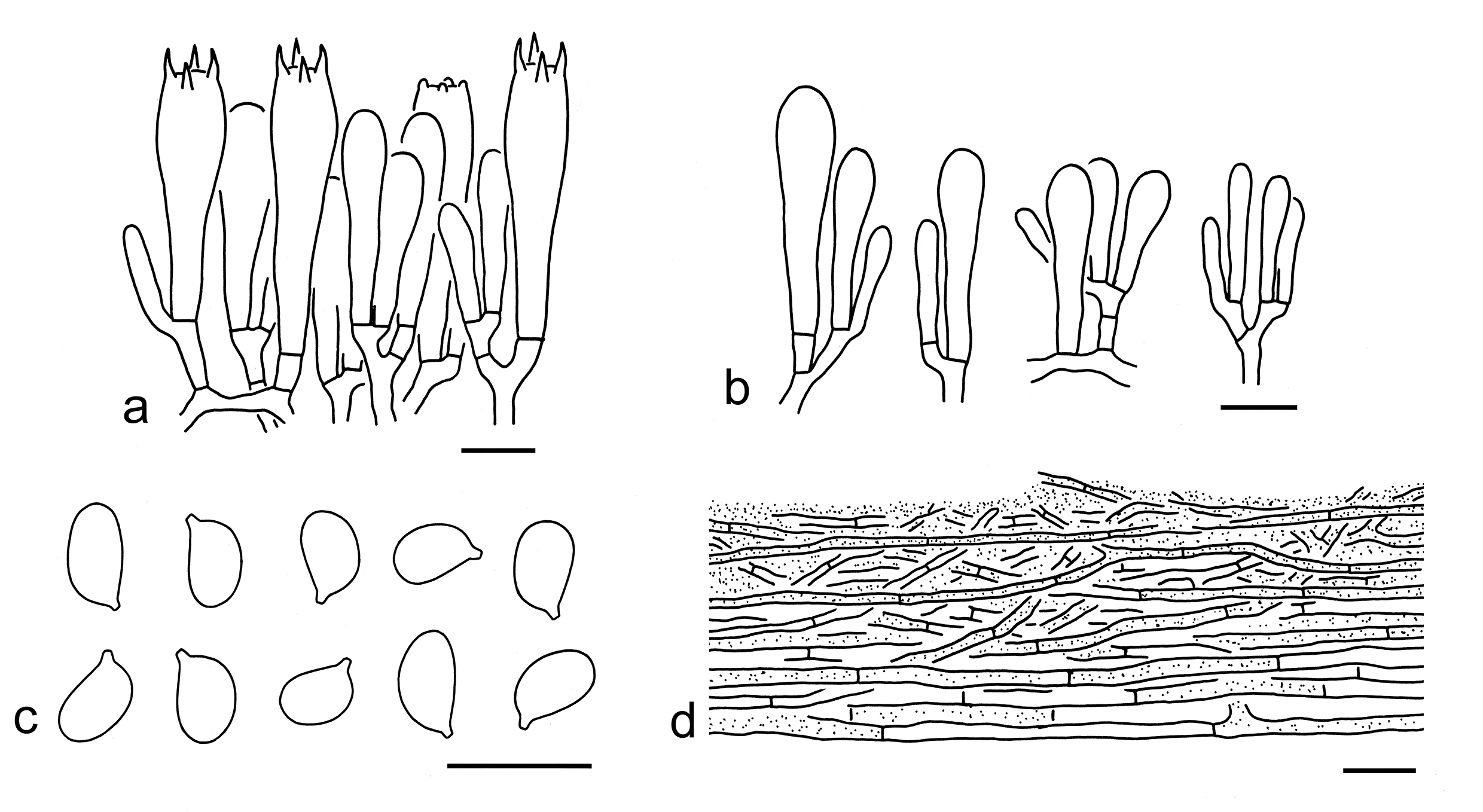
10. Tricholoma equestre (L.) P. Kumm., Führ. Pilzk. (Zerbst): 130 (1871). Figs. 15n, o, 27
Basionym: Agaricus equestris L., Sp. pl. 2: 1173 (1753).
Description: Basidioma small to medium-sized. Pileus 3.5–8 cm in diam., convex-applanate to applanate, without an obvious umbo or depression at center; surface dry, orangish brown (6C4–6, 5C5–7), often brownish (4B3–5) to yellow (3A2–5) towards margin, covered with orange-brown (5C5–7), fibrillose squamules; margin at first incurved, then straight; context cream (1A2). Lamellae sinuate, yellow (1A4–6), sulphur-yellow (2A6–8) to olivaceous yellow (30A4–6), crowed to subclose; edges concolor. Stipe 3–9 1–3 cm, cylindrical to subcylindrical, yellowish (1A2–4), olivaceous yellow (30A4–6) to brownish (5B3–5), glabrous to slightly fibrillose, solid to stuffed, exannulate. Taste and odour not recorded.
Basidiospores [239/9/6] (6–) 6.5–9 (–10) 4–5.5 (–6) µm (mean length = 7.3 µm, mean width = 4.8 µm), Q = (1.13–) 1.15–1.95 (–2.25), Qm = 1.54 ± 0.23, broadly ellipsoid, ellipsoid to elongate, non-amyloid, colorless, hyaline, thin-walled, smooth; apiculus relatively large. Basidia 30–40 7–12 µm, clavate, 4-spored, colorless or red brown, hyaline; sterigmata 3–6 µm long; basal septa without clamps. Pleurocystidia absent. Cheilocystidia 15–40 3–8 µm, poorly differentiated, clavate, subclavate to cylindrical, thin-walled, colorless or red-brown, hyaline. Subhymenium composed of 2–4 µm wide filamentous hyphae, clamps absent. Lamellar trama regular, composed of colorless, thin-walled hyphae 2–20 µm wide. Pileipellis a cutis, non- to very slightly gelatinized, composed of radially arranged to interwoven, 2–10 µm wide, thin-walled, yellow-brown to red-brown filamentous hyphae, often with transition to a trichoderm at regular intervals forming squamules; subpellis poorly differentiated, non-gelatinized. Clamps absent in all parts of basidioma.
Ecology: Solitary to scattered in Pinus forests and often on sandy soil, also collected in broad-leaved or mixed forests.
Known distribution: Known from Europe (Riva 1988, 2003; Christensen & Heilmann-Clausen 2013), North America (Bessette et al. 2013) and Asia (Reschke et al. 2018).
Notes: Tricholoma equestre is widely consumed as wild edible mushrooms in Southwestern China, but intoxications of rhabdomyolysis were reported in some countries (Bedry et al. 2001; Anand et al. 2009; Laubner & Mikulevičienė 2016; Yang et al. 2021). Christensen & Heilmann-Clausen (2013) reported its smell as weak but strongly farinaceous when cut, and its taste as mild and farinaceous. However, taste and odour were not recorded for Chinese collections. This species belongs to a species complex comprising several Tricholoma taxa with yellow pileus and lamellae. However, the delimitations within this complex remain unclear. To avoid unnecessary nomenclatural confusions, this study follows the treatment of T. equestre in Christensen & Heilmann-Clausen (2013), Reschke et al. (2018) and Cui et al. (2022).
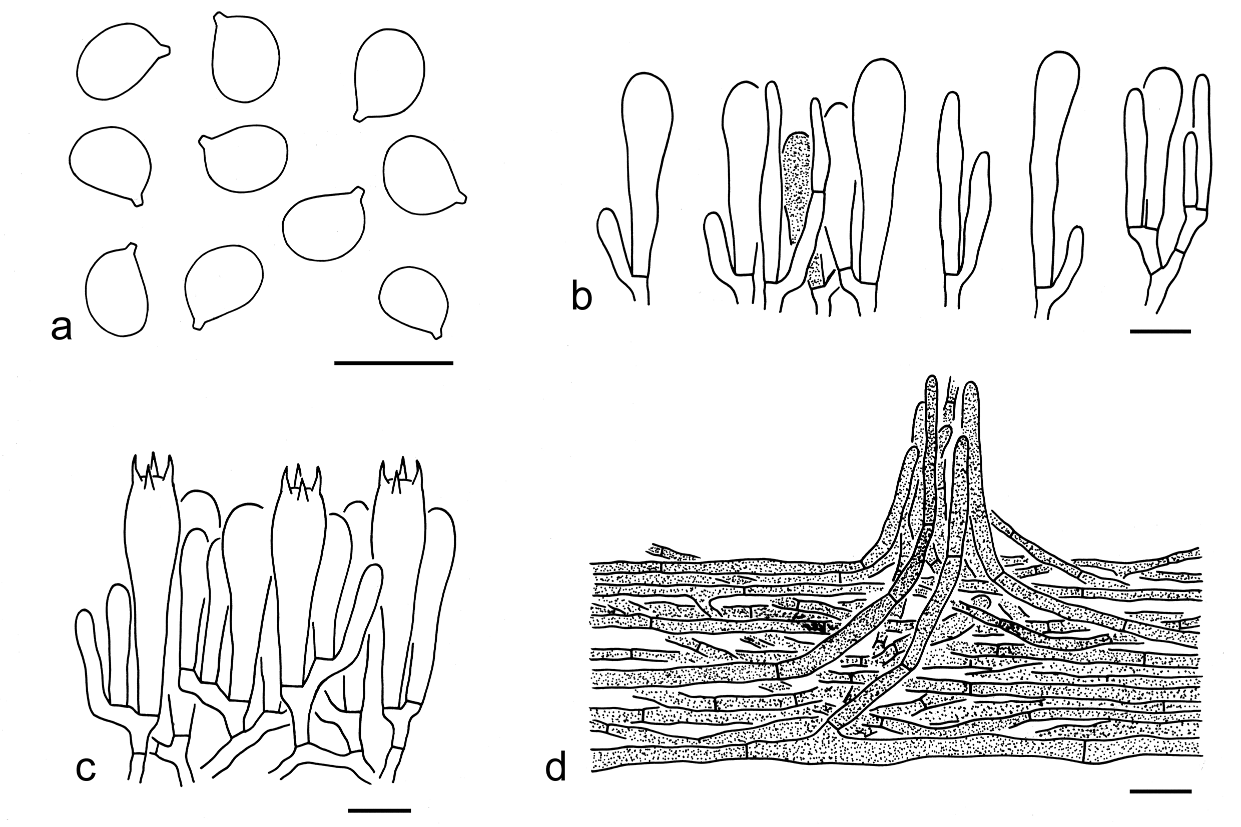
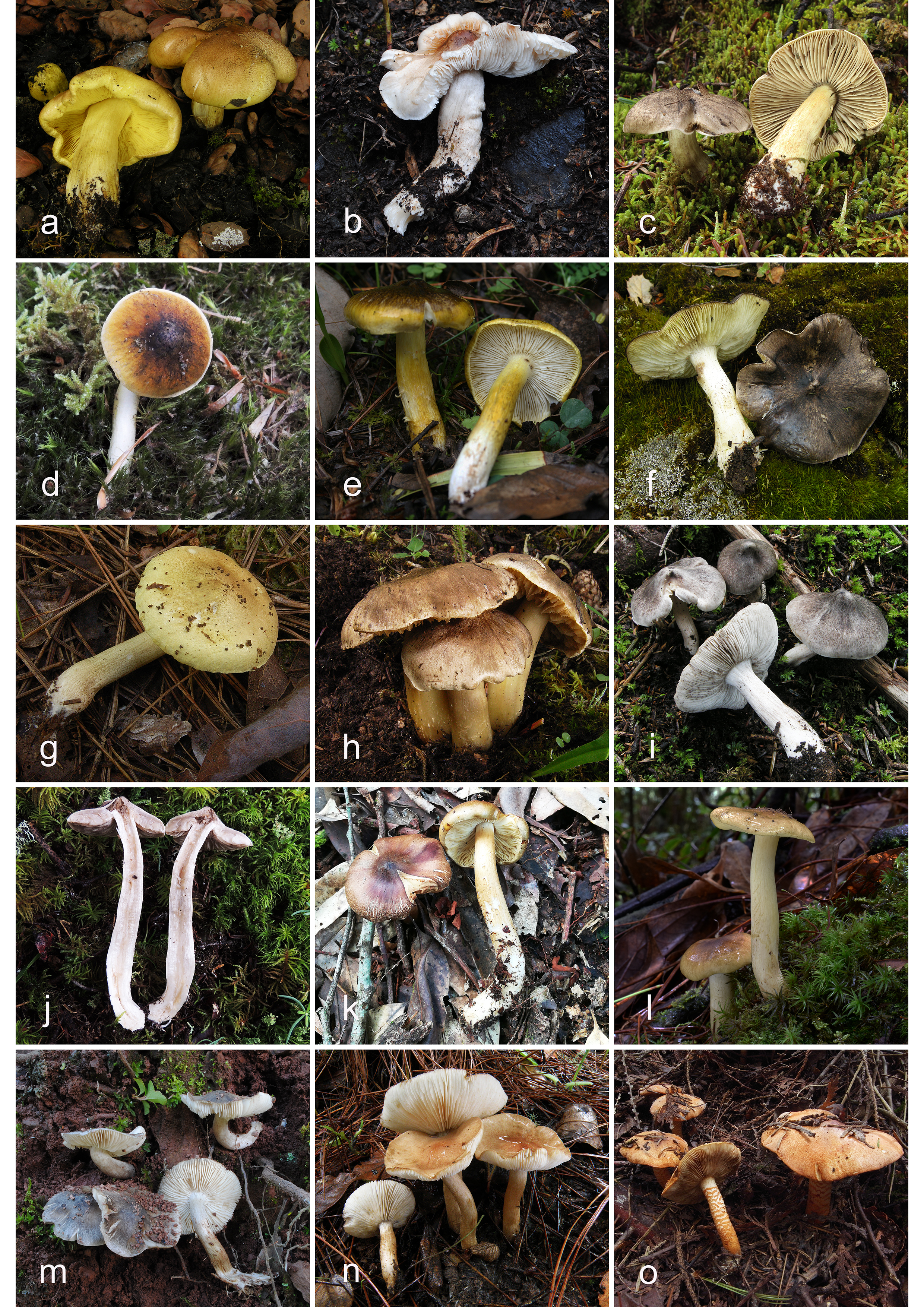
11. Tricholoma frondosae Kalamees and Shchukin, Folia Cryptogamica Estonica 38: 14 (2001). Figs. 28a, 29
Description: Basidioma medium-sized. Pileus 5–8 cm in diam., at first convex to convex-applanate, then applanate, often with a broadly low umbo at center; surface dry, reddish brown (6C5–7, 6B4–6), becoming brownish yellow (5A3–5, 4B3–5) to yellow (3A3–6) towards margin, with olivaceous (30A4–6) color, covered with red-brown (6C5–7) to brown (5C5–7) squamules; margin involute when young, then straight; context white (1A1) to yellowish (1A2–4). Lamellae sinuate, yellowish (1A2–4), yellow (2A3–5) to sulphur-yellow (3A5–7), often with olivaceous (30A4–6) color, crowded; edges concolor. Stipe 5–8 1–2 cm, cylindrical to subcylindrical, hollow, yellowish (2A2–4), olivaceous yellow (30A4–6, 1B4–6) to brownish (3B3–5), basal mycelium white (1A1), often with fine fibrils, solid to stuffed, exannulate. Taste and odour not recorded.
Ecology: Solitary to scattered in broad-leaved forests.
Known distribution: Known from Europe (Christensen & Heilmann-Clausen 2013; Heilmann-Clausen et al. 2017), North America (Heilmann-Clausen et al. 2017) and East Asia.
Specimens examined: CHINA. Sichuan Province, Ganzi Tibetan Autonomous Prefecture, Daocheng County, in a subalpine sclerophyllous evergreen forest dominated by Quercus semicarpifolia, altitude 3960 m, 7 Aug. 2016, Jian-Wei Liu 493 (KUN-HKAS98072). Yunnan Province, Lijiang, Yulong Naxi Autonomous County, in a subalpine sclerophyllous evergreen forest dominated by Quercus semicarpifolia, altitude 3500 m, 8 Aug. 2014, Xiao-Bin Liu 504 (KUN-HKAS87149).
Notes: Tricholoma frondosae was described by Kalamees (2001), and then separated into two types (T. frondosae type I and Ⅱ) according to Heilmann-Clausen et al. (2017). Tricholoma frondosae type I can be found in China and is characterized by its reddish brown pileus, sulphur-yellow lamellae, mostly elongate basidiospores (7–8.5 4–5 µm, Q = 1.66–2, Qm = 1.83 ± 0.14), absence of clamps, and associations with broad-leaved trees (this study). This species is mixed within T. equestre in our molecular analyses (Figs. 3, 4), but can be separated by its warm colored basidioma, mostly elongate basidiospores and being ectomycorrhizal with broad-leaved forests (Christensen & Heilmann-Clausen 2013). Here we follow the treatment of Christensen & Heilmann-Clausen (2013) and Heilmann-Clausen et al. (2017) for the time being.
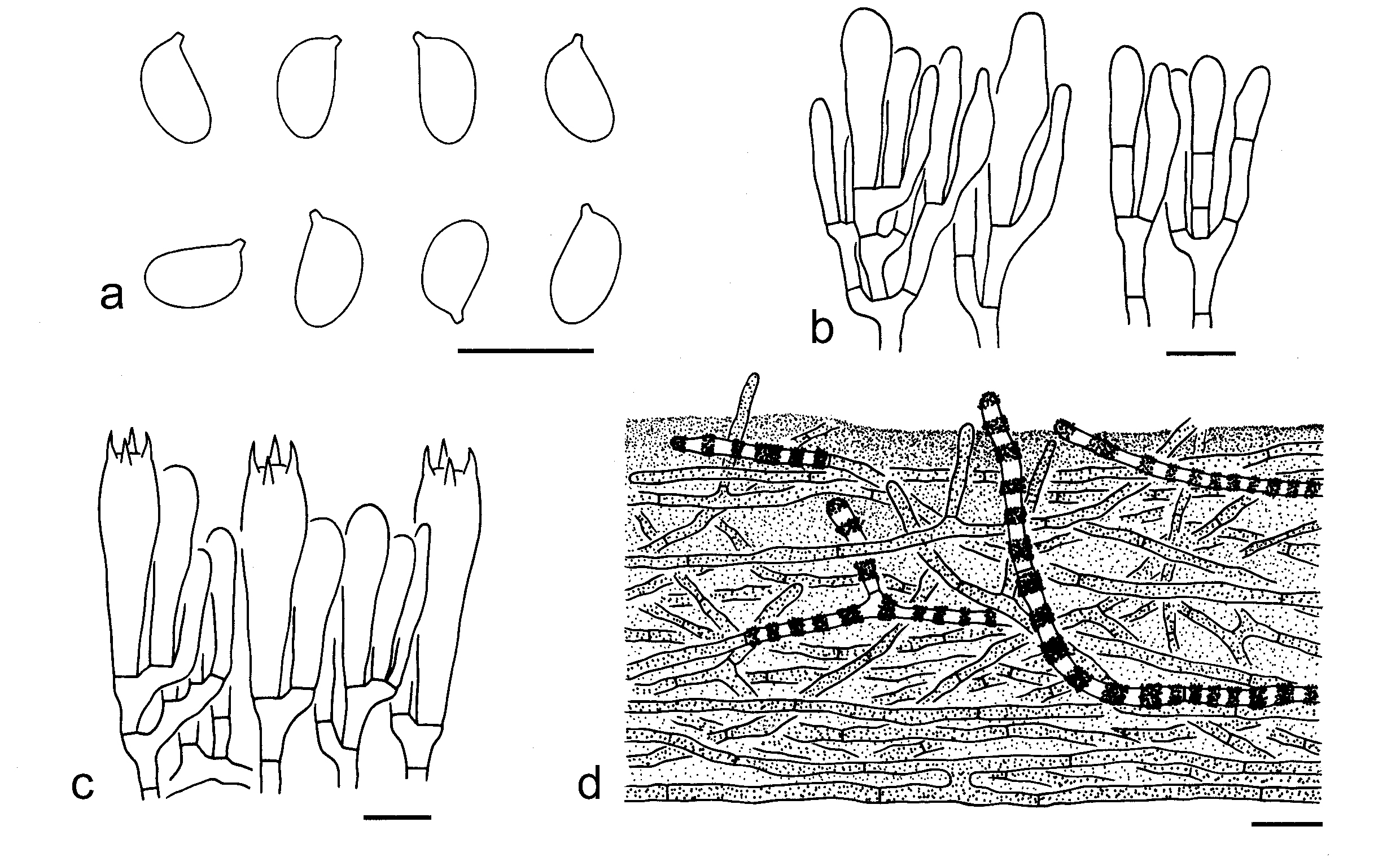
12. Tricholoma mastoideum Y.Y. Cui & Zhu L. Yang, Mycol. Progr. 21(35): 8 (2022). Figs. 28b, 30
Description: Basidioma small to medium-sized. Pileus 3–9 cm in diam., convex when young, applanate when mature, broadly umbonate at center; surface dry, often yellowish brown (5C5–7, 5D5–8), brown (5E5–7) to greyish brown (5D2–4, 5E3–5) at center, becoming greyish (1B1–3) to white (1A1) towards margin, sometimes greyish (1B1–3) to brownish grey (3B2–4, 4B3–5) over the entire surface, covered with concolor fibrillose squamules; margin undulating and slightly incurved; context white (1A1) to dirty white (1B1–3). Lamellae adnexed to sinuate, white (1A1) to dirty white (1B2), crowded; edges white (1A1) to dirty white (1B2). Stipe 6.5–9 1–1.5 cm wide, stuffed, subcylindrical to cylindrical, enlarged downwards, white (1A1) to dirty white (2B2–3), exannulate. Taste mild. Odour like trees of oaks.
Ecology: Solitary to scattered on soil in subalpine coniferous, broad-leaved or mixed forests with altitude 2500–3600 m.
Known distribution: Known from Southwestern China.
Specimens examined: CHINA. Sichuan Province, Ganzi Tibetan Autonomous Prefecture, Kangding, in a subalpine forest, altitude 3550 m, 5 Sep. 2016, KD-Ding 15 (holotype, KUN-HKAS97096); same location, in a subalpine sclerophyllous evergreen forest with Quercus semicarpifolia, altitude 3550 m, 5 Sep. 2016, KD-Ding 24 (KUN-HKAS97105); same city, in a subalpine forest, altitude 3550 m, 5 Sep. 2016, Bang Feng 32 (KUN-HKAS99385) same city, in a subalpine forest, altitude 3500 m, 6 Sep. 2016, Bang Feng 62 (KUN-HKAS99415). Yunnan Province, Diqing Tibetan Autonomous Prefecture, Shangri-La, in a subalpine mixed forest with trees of Pinus, Quercus and Betula, altitude 3000 m, 25 Aug. 2014, Qi Zhao 2284 (KUN-HKAS88051); Lijiang, Yulong Naxi Autonomous County, in a subalpine forest, altitude 2700 m, 3 Sep. 2019, Jian-Wei Liu 1876 (KUN-HKAS115907).
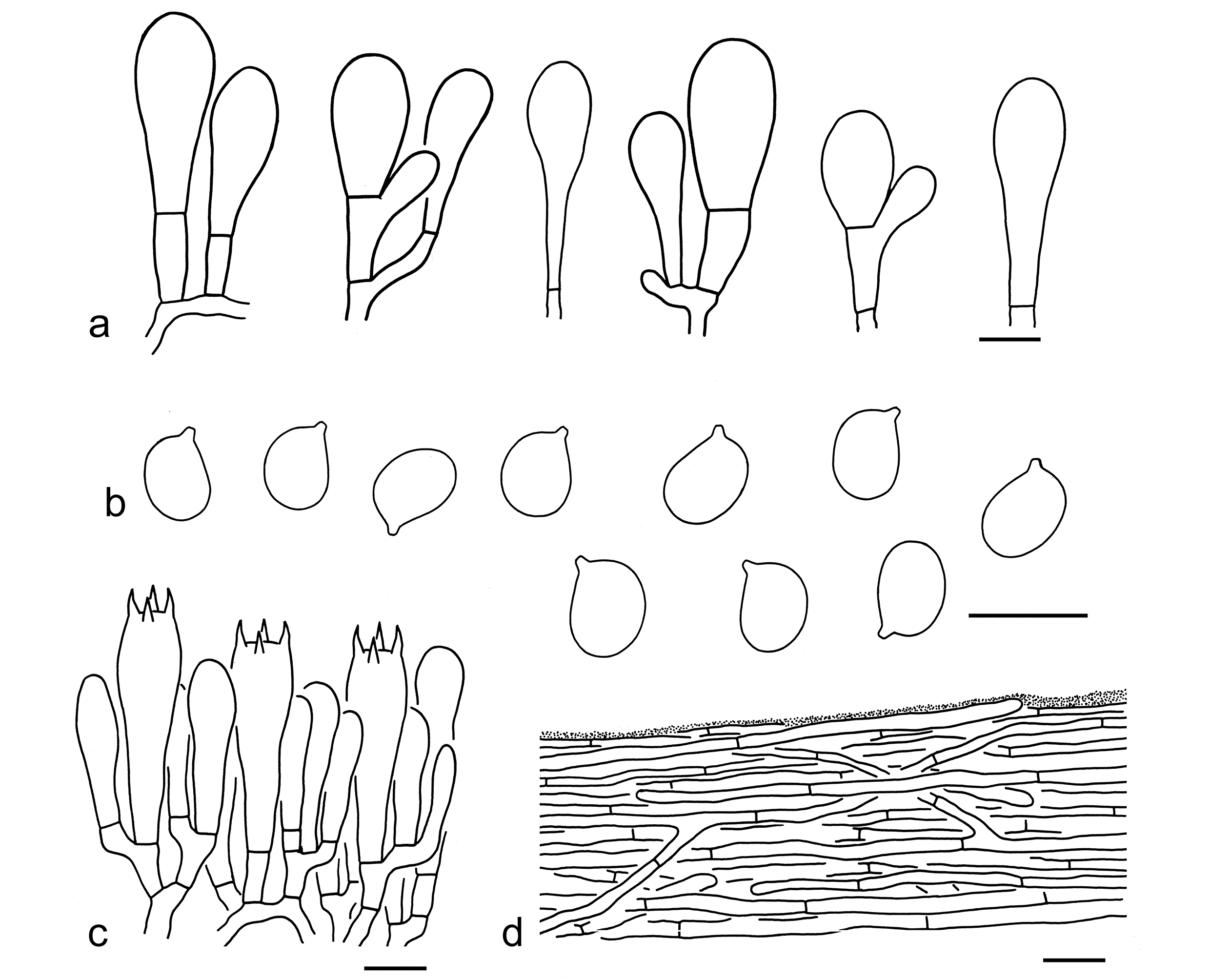
Notes: Tricholoma mastoideum is well delimitated by its umbonate pileus often yellowish brown to greyish brown at center and greyish to white towards margin, broadly ellipsoid to ellipsoid basidiospores (7–9 5–7 μm, Q = 1.15–1.38, Qm = 1.26 ± 0.08), and absence of clamps. Molecular data indicated that T. virgatum is closely related (Fig. 3), but differs by its grey, silvery shiny pileus and white to greyish lamellae becoming brownish when old (Riva 1988, 2003; Christensen & Heilmann-Clausen 2013; this study). Tricholoma guldeniae Mort. Chr. is somewhat similar to T. mastoideum, but has a greyish yellow pileus and heterogeneously sized basidiospores (Christensen & Heilmann-Clausen 2013).
13. Tricholoma olivaceifolium Y.Y. Cui & Zhu L. Yang, sp. nov. Figs. 28c, 31
Fungal Names: FN 572342
Etymology: olivaceifolium = olivaceous lamellae, referring its lamellae with green color.
Diagnosis: Similar to T. portentosum, but differs in its brownish grey pileus often with yellow tinge, larger basidiospores and a non-gelatinized pileipellis.
Type: CHINA. Xizang Autonomous Region, Linzhi, Bayi District, in a subalpine forest with Picea and Rhododendron, altitude 3995 m, 20 Jul. 2019, Gen-Shen Wang 454 (holotype, KUN-HKAS107574).
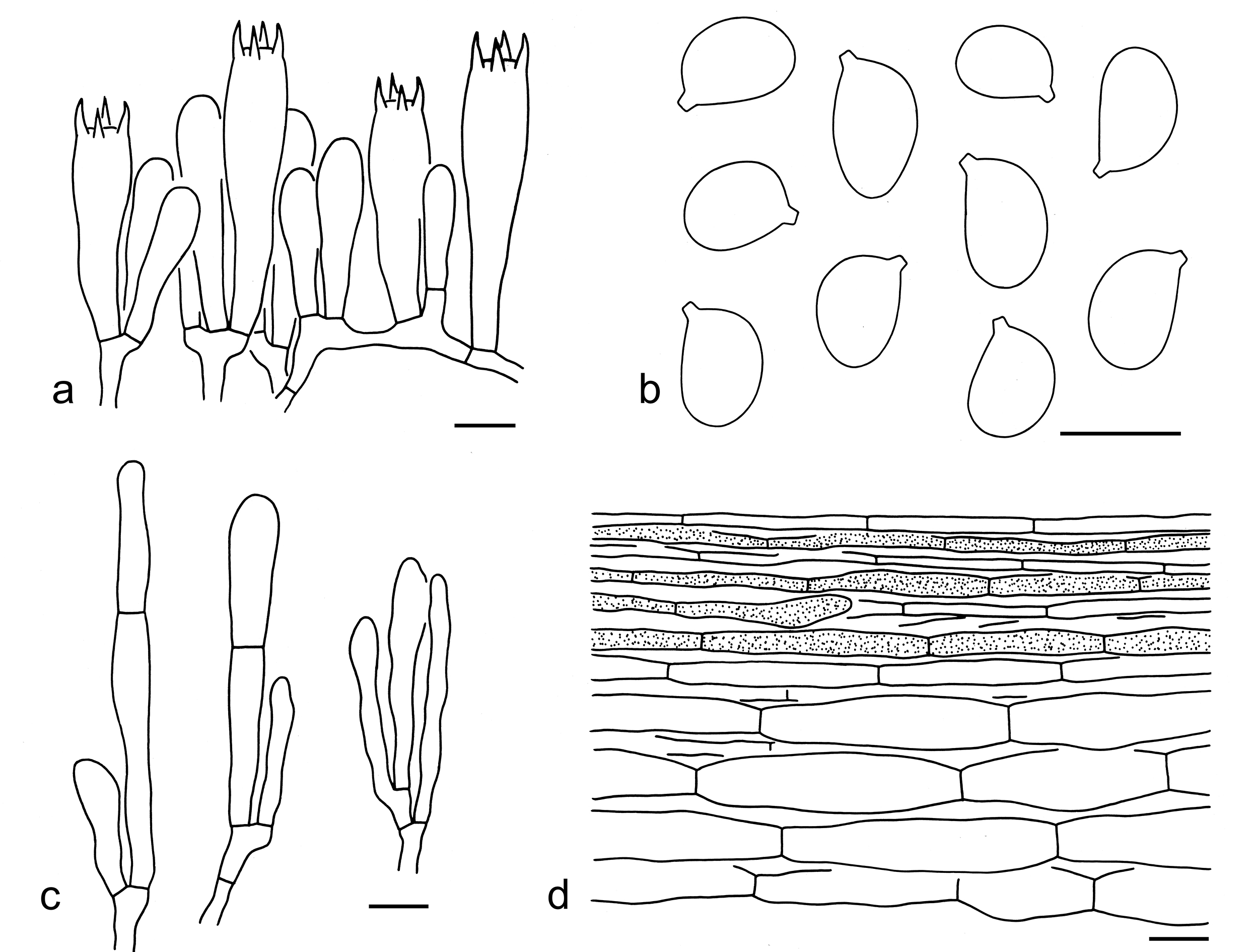
Description: Basidioma small to medium-sized. Pileus 4–7 cm in diam., conical, convex to plano-convex, papillate at center, often acute; surface dry, greyish (1B1, 2B1), grey (1D1) to brownish grey (2E1–4), sometimes with yellow (1A2–4) tinge, covered with grey (1D1) to brownish grey (2E1–4) fibrillose squamules; margin inflexed to straight; context grey (2E1–3), greyish (2D1–3) to dirty yellow (1B2–4). Lamellae adnexed, light ivory (1A2–3), ivory (2A2–4) to olivaceous brown (30A2–4, 30B2–4), subcrowded to subclose; edges ivory (2A2–4), sometimes brown (2E4–7). Stipe 5–8 0.5–1.5 cm, subcylindrical to subclavate, greenish yellow (30A2–5), glabrous or with brown (2E3–5) to greenish yellow (30A2–5) fibrils, stuffed, fistulose to hollow, exannulate. Taste and odour not recorded.
Basidiospores [20/1/1] 7.9–10 (–12) 6–8 µm (mean length = 9.4 µm, mean width = 7 µm), Q = 1.21–1.5 (–1.59), Qm = 1.35 ± 0.11, broadly ellipsoid to ellipsoid, non-amyloid, colorless, hyaline, thin-walled, smooth; apiculus relatively large. Basidia 30–50 9–12 µm, clavate, 4-spored, occasionally 2-spored, hyaline; sterigmata 5–7 µm long; basal septa without clamps. Pleurocystidia absent. Cheilocystidia 25–45 3–9 µm, clavate to filamentous, often septate, sometimes branching, thin-walled, colorless, hyaline. Subhymenium composed of 3–5 µm wide filamentous hyphae, clamps absent. Lamellar trama regular, composed of colorless, thin-walled filamentous hyphae 3–10 µm wide. Pileipellis a cutis, composed of radially arranged, 3–8 µm wide, thin-walled, colorless or brown filamentous hyphae; subpellis slightly differentiated, composed of cylindrical, clavate to ellipsoid inflated cells 10–25 µm wide. Clamps absent in all parts of basidioma.
Ecology: Solitary to scattered in subalpine forests.
Known distribution: Known from Southwestern China.
Additional specimens examined: CHINA. Yunnan Province, Diqing Tibetan Autonomous Prefecture, in a subalpine forest, altitude 3300 m, 22 Aug. 2008, Li-Ping Tang 690 (KUN-HKAS54922); Lijiang, Yulong Naxi Autonomous County, in a subalpine forest, altitude 3750 m, 31 Jul. 2005, Yan-Chun Li 255 (KUN-HKAS48488).
14. Tricholoma olivaceoluteolum Reschke et al., Mycologia 110(6): 1100 (2018). Figs. 28d, 32
Description: Basidioma small to medium-sized. Pileus 4–8 cm in diam., convex to plano-convex, often subumbonate at center; surface viscid, often dark brown (5F3–5) at center, becoming orangish brown (5D6–8) to yellow-brown (4D3–5) towards margin, with obviously olive (30B4–6) color, sometimes with a white (1A1) margin, fibrillose; margin undulating and slightly incurved; context white (1A1). Lamellae adnexed to sinuate, white (1A1), sometimes yellow (2A2–4) at the very margin, crowded; edges white (1A1), sometimes yellow (2A2–4). Stipe 5–12 0.5–1.5 cm wide, stuffed to hollow, cylindrical to clavate, white (1A1), often olivaceous yellow (30A2–4) in the middle part, exannulate. Taste slightly bitter. Odour not recorded.
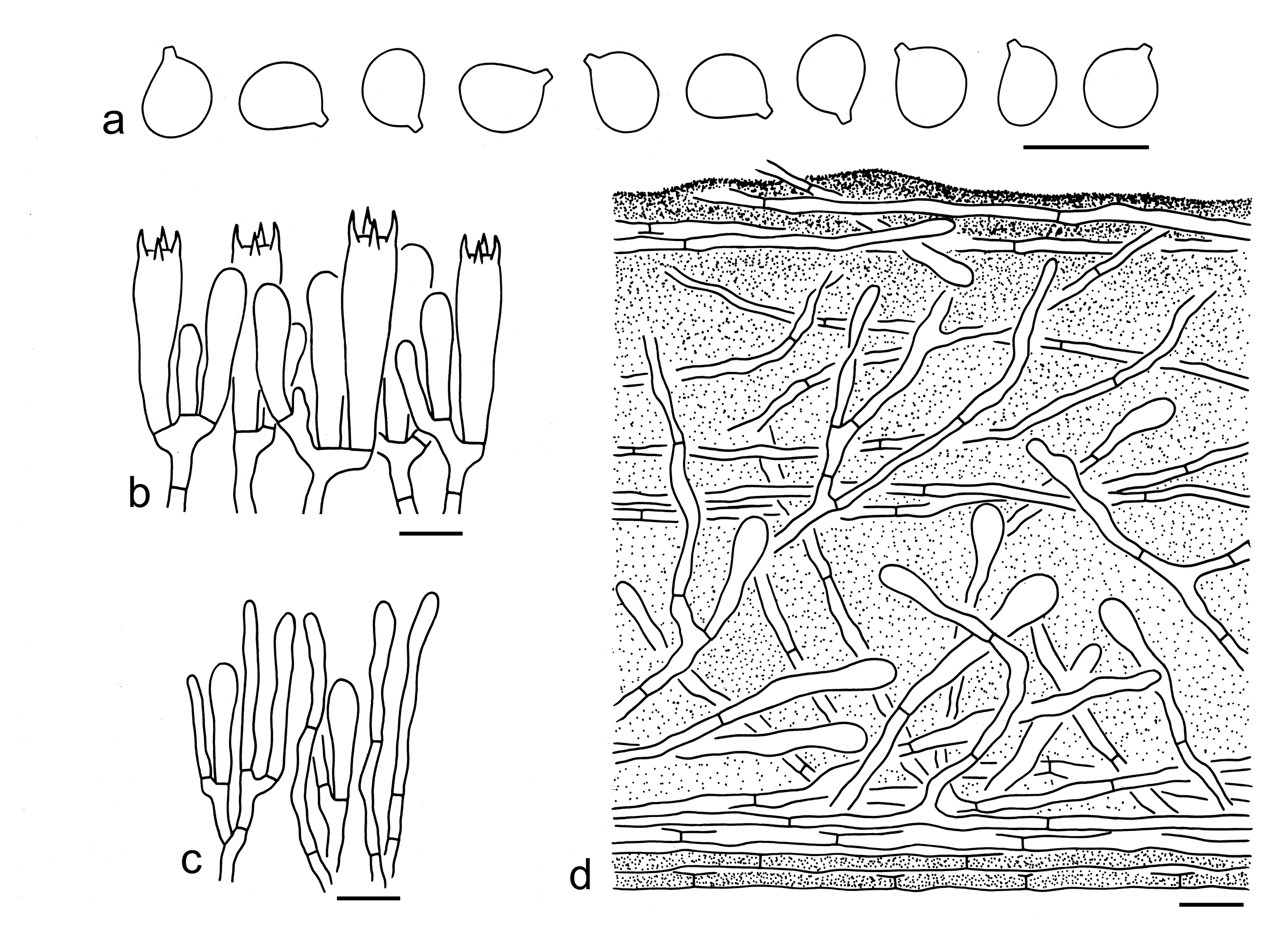
Basidiospores [60/3/3] 5.5–7.5 (–9) (4.5–) 5–7 (–8) μm, (mean length = 6.5 μm, mean width = 5.7 μm), Q = (1.01–) 1.05–1.25 (–1.4), Qm = 1.15 ± 0.08, subglobose to broadly ellipsoid, non-amyloid, colorless, hyaline, thin-walled, smooth, apiculus relatively large. Basidia 25–35 7–10 μm, clavate, 4-spored, hyaline; sterigmata 3–6 μm long; basal septa without clamps. Pleurocystidia absent. Cheilocystidia 20–30 2–5 μm, filamentous to clavate, thin thick-walled, colorless, and hyaline. Subhymenium composed of 2–4 μm wide filamentous hyphae, clamps absent. Lamellar trama regular, composed of colorless, thin-walled hyphae 3–20 μm wide. Pileipellis an ixocutis, strongly gelatinized, upper layer composed of more or less radially arranged to interwoven, 2–7 μm wide, thin-walled, colorless filamentous hyphae; lower layer composed of interwoven to somewhat erected, thin-walled, colorless filamentous hyphae, often with clavate terminal cells up to 15 μm wide. Clamps absent in all parts of basidioma.
Ecology: Solitary to scattered on soil in subtropical coniferous or broad-leaved forests.
Known distribution: Known from Southwestern China.
Specimens examined: CHINA. Yunnan Province, Baoshan, Longling County, in a subtropical coniferous forest, altitude 2100 m, 31 Aug. 2022, GLG-FXP858 (KUN-HKAS134964); same location and date, GLG-TJY610 (KUN-HKAS134965); Puer, Jingdong Yi Autonomous County, in a subtropical broad-leaved forest dominated by Quercus and Lithocarpus, altitude 2500 m, 20 Jul. 2006, MR 50 (holotype, KUN-HKAS93510).
Notes: Tricholoma olivaceoluteolum is characterized by its umbonate, olivaceous brown pileus, white lamellae sometimes yellow at the very margin, subglobose to broadly ellipsoid basidiospores (5.5–7.5 5–7 μm, Q = 1.05–1.25, Qm = 1.15 ± 0.08) and a strongly gelatinized pileipellis (this study). This species is related to T. orienticum Y.Y. Cui & Zhu L. Yang in the phylogenetic analysis based on five gene fragments (Fig. 3), but the latter species has poorly differentiated cheilocystidia and an ixo-cutis pileipellis with its subpellis composed of repent filamentous hyphae (this study).
Tricholoma sejunctum (Sowerby) Quél. looks like T. olivaceoluteolum, but the former differs from the latter by its more greenish pileus, medium spaced to distant lamellae and broadly ellipsoid to ellipsoid basidiospores (Riva 1988, 2003; Christensen & Heilmann-Clausen 2013). Tricholoma olivaceoluteolum may be confused as a member of sect. Muscaria, but species in sect. Muscaria have subclose to close lamellae, conspicuous cheilocystidia, a non-gelatinized pileipellis with its subpellis well differentiated and composed of inflated cells.
15. Tricholoma orienticum Y.Y. Cui & Zhu L. Yang, sp. nov. Figs. 28e, 33
Fungal Names: FN 572343
Etymology: orienticum = eastern, namely after its type locality from East Asia.
Diagnosis: Related to T. olivaceoluteolum, but differs by its poorly differentiated cheilocystidia and an ixo-cutis pileipellis with its subpellis composed of repent filamentous hyphae.
Type: CHINA. Yunnan Province, Lijiang, Yulong County, in a mixed forest with Fagaceae and Pinaceae, altitude 3200 m, 14 Aug. 2008, Qi Zhao 8198 (holotype, KUN-HKAS55189).
Description: Basidioma small to medium-sized. Pileus 4–7 cm in diam., plano-convex to applanate, broadly umbonate at center; surface dry, slightly viscid when wet, brown (1E3–5, 1F6–8) at center, becoming olivaceous yellow (30A6–8) towards margin, sometimes olivaceous yellow (30A6–8) overall, innately fibrillose or covered with brown (1F6–8), brownish (1E6–8), olivaceous brown (30E6–8) squamules, margin straight; context white (1A1) to yellowish (1A2–4). Lamellae adnexed to sinuate, white (1A1), sometimes yellow (30A3–5) at the very margin, crowded; edges white (1A1) to yellow (30A3–5). Stipe 5–8 1–1.5 cm, cylindrical, white (1A1) to olivaceous yellow (30A3–5, 2A5–7), glabrous, stuffed to hollow, exannulate. Taste not recorded. Odour pleasant.
Basidiospores [47/2/2] (5.5–) 6–7 (4–) 5–6 µm (mean length = 6.1 µm, mean width = 5.2 µm), Q = (1.05–) 1.1–1.28 (–1.4), Qm = 1.19 ± 0.07, subglobose to broadly ellipsoid, non-amyloid, colorless, hyaline, thin-walled, smooth; apiculus relatively large. Basidia 35–40 7–9 µm, clavate, 4-spored, hyaline; sterigmata 4–6 µm long; basal septa without clamps. Pleurocystidia absent. Cheilocystidia not observed. Subhymenium composed of 2–5 µm wide filamentous hyphae, clamps absent. Lamellar trama regular, composed of colorless, thin-walled, cylindrical hyphae 3–15 µm wide. Pileipellis an ixocutis, gelatinized, composed of radially arranged, 3–8 µm wide, thin-walled, colorless or brownish filamentous hyphae; subpellis poorly differentiated. Clamps absent in all parts of basidioma.
Ecology: Solitary to scattered in coniferous or mixed forests.
Known distribution: Known from Southwestern China.
Additional specimen examined: CHINA. Yunnan Province, Lijiang, Yulong County, in a coniferous forest with Pinus yunnanensis, altitude 3269 m, 17 Sep. 2012, Qi Zhao 1691 (KUN-HKAS78949).
Notes: Tricholoma orienticum is well delimitated by its basidioma with olivaceous yellow color, white lamellae with yellow color at the very margin, and subglobose to broadly ellipsoid basidiospores (6–7 5–6 µm, Q = 1.1–1.28, Qm = 1.19 ± 0.07). Our multi-gene phylogenetic analysis (Fig. 3) suggested that T. olivaceoluteolum is related to T. orienticum. For comparison, see notes under that name. Tricholoma subluteum Peck is closely related (Fig. 4) and similar to T. orienticum, but has a dull orange or golden-yellow to yellow pileus and is currently only recorded from North America (Peck 1904; Murrill 1914; Bessette et al. 2013).
Tricholoma subsejunctum Peck can be confused with T. orienticum, but the former species has relatively smaller basidiospores (5–6 4–5 µm in the protologue), and is a rare species currently only known from North America (Peck 1912; Ammirati & Ovrebo 1979; Landry et al. 2022; Trudell et al. 2022). Tricholoma atrofibrillosum S.A. Trudell et al. also looks like, but differs by its ixocutis pileipellis with its lower layer composed of cylindrical to moderately inflated elements and only found in North America (Trudell & Parker 2021; Trudell et al. 2022). Tricholoma sejunctum, T. viridilutescens M.M. Moser and T. viridifucatum Bon are similar to T. orienticum. However, T. sejunctum has broadly ellipsoid to ellipsoid basidiospores and an ixocutis pileipellis with its lower layer composed of cylindrical to slightly inflated elements (Riva 1988, 2003; Christensen & Heilmann-Clausen 2013). Tricholoma viridilutescens differs by its medium spaced to rather distant lamellae, slightly larger basidiospores (average 6.3–8 5.3–6.6 µm) and a subpellis with rather short inflated hyphae (Christensen & Heilmann-Clausen 2013). Tricholoma viridifucatum has medium-spaced lamellae and broadly ellipsoid to ellipsoid basidiospores (Riva 1988, 2003; Christensen & Heilmann-Clausen 2013).
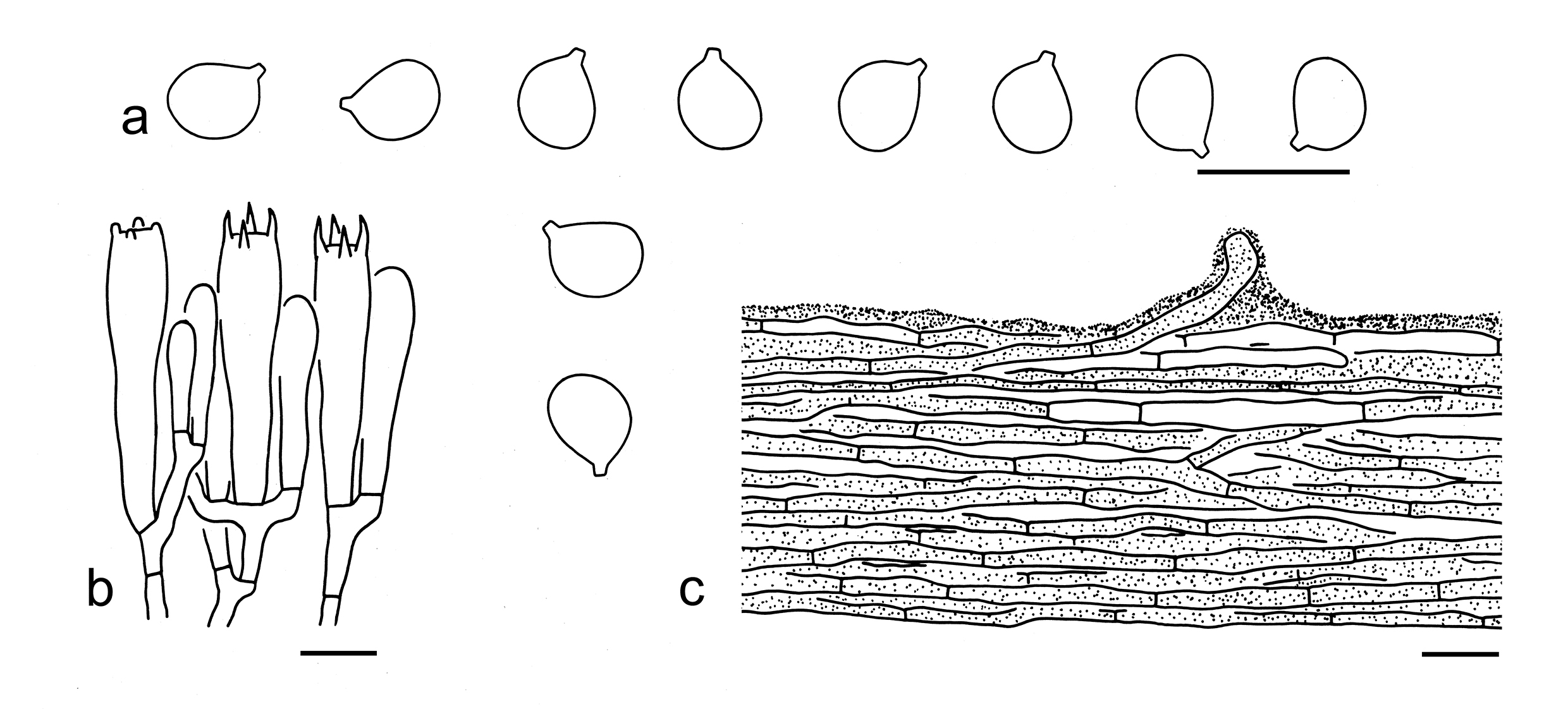
Basionym: Agaricus portentosus Fr., Syst. mycol. (Lundae) 1: 39 (1821).
Description: Basidioma medium-sized. Pileus 5–9 cm in diam., convex-applanate to applanate, sometimes with a broadly low umbo at center; surface viscid when wet, greasy when dry, dark grey (2F1–3), grey (1E1–3) to grey-brown (4E4–6), often darker (1F1–3) at center, glabrous or finely squamulose; margin straight; context white (1A1) to dirty white (1B1–2). Lamellae sinuate, white (1A1) to greyish (1B2), then olivaceous yellow (30A3–5) to yellow (1A4–6), crowded; edges concolor or brown (4E6–8). Stipe 4–11 1–2.5 cm, subcylindrical to cylindrical, white (1A1) to olivaceous yellow (30A2–4), fibrillose, solid, exannulate. Taste and odour indistinct.
Basidiospores [61/3/2] 6–7.5 (–8) 4–5 µm (mean length = 6.8 µm, mean width = 4.1 µm), Q = (1.33–) 1.44–1.88 (–1.95), Qm = 1.66 ± 0.12, ellipsoid to elongate, non-amyloid, colorless, hyaline, thin-walled, smooth; apiculus relatively large. Basidia 25–40 6–8 µm, clavate, 4-spored, hyaline; sterigmata 3–5 µm long; basal septa without clamps. Pleurocystidia absent. Cheilocystidia 20–35 3–5 µm and filiform or 30–50 10–15 µm and clavate, thin-walled, colorless, hyaline. Subhymenium composed of 2–5 µm wide filamentous hyphae, clamps absent. Lamellar trama regular, composed of colorless, thin-walled, more or less inflated to filamentous hyphae 2–20 µm wide. Pileipellis an ixo-cutis, gelatinized, composed of radially arranged to interwoven, 2–5 µm wide, thin-walled, colorless filamentous hyphae; subpellis poorly differentiated. Clamps absent in all parts of basidioma.
Ecology: Solitary to scattered dominated in coniferous or broad-leaved forests.
Known distribution: Known from Europe (Riva 1988, 2003; Christensen & Heilmann-Clausen 2013; Heilmann-Clausen et al. 2017), North America (Heilmann-Clausen et al. 2017; Bessette et al. 2013) and East Asia (Hongo 1988; Ushijima et al. 2021; this study).
Specimens examined: CHINA. Sichuan Province, Ganzi Tibetan Autonomous Prefecture, Daocheng County, in a forest with Quercus semicarpifolia, altitude 4000 m, 12 Sep. 2016, Xiao-Xia Ding 132 (KUN-HKAS97075); Ganzi Tibetan Autonomous Prefecture, Kanding, forest and altitude not recorded, 5 Sep. 2016, Bang Feng 28 (KUN-HKAS99381). Yunnan Province, Kunming, in a forest with Pinus armandii and Pinus yunnanensis, altitude 2200 m, 22 Oct. 2011, Yan-Jia Hao 619 (KUN-HKAS71728).
Notes: Tricholoma portentosum is characterized by its dark grey to grey pileus often viscid when wet and greasy when dry, yellow color on lamellae and stipe, and ellipsoid to elongate basidiospores (6–7.5 4–5 µm, Q = 1.44–1.88, Qm = 1.66 ± 0.12) (this study). This species was reported by Christensen & Heilmann-Clausen (2013) to have a weak but distinctly farinaceous smell when cut, and a rather mild, farinaceous taste. However, no distinct odour or taste have been observed in Chinese collections.
Phylogenetically, no closely related species were detected (Figs. 3, 4). Tricholoma sinoportentosum Zhu L. Yang et al. is similar to T. portentosum. However, T. sinoportentosum differs by its greenish yellow color on pileus, broader basidiospores (6–8 5–6.5 µm, Q = 1.17–1.36, Qm = 1.27 ± 0.08), and the clamped basidia (Reschke et al. 2018; this study).
17. Tricholoma qiaomianjun Y.Y. Cui & Zhu L. Yang, Mycological Progress 21(35): 10 (2022). Figs. 28g, 35
Description: Basidioma medium-sized. Pileus 4–8 cm diam., convex, plano-convex to applanate, often broadly umbonate; surface dry, slightly viscid when wet, greenish yellow (30A6–8), chrome yellow (2B6–8, 3B7–8), yellowish brown (3C6–8, 4C5–8) to orangish brown (5D8, 5E6–8, 6D5–8), covered with olivaceous brown (30D3–5, 30E4–6), orangish brown (5D8, 5E6–8, 6D5–8) to brown (6E6–8, 6F8) squamules; margin straight; context white (1A1). Lamellae adnexed to sinuate, pale yellow (1A1–3), yellow (2A4–6), citron yellow (3A6–8) to greenish yellow (30A4–7), crowded; edges pale yellow (1A1–3), citron yellow (3A6–8) to greenish yellow (30A3–6). Stipe 4–8 × 0.5–2 cm, subcylindrical to cylindrical, enlarged downwards, dry, cream (2A2–3) at apex, pale yellow (1A1–3), yellowish (3A2–4) to pale greenish yellow (30A2–6) below, covered with greenish yellow (30A6–8), brownish (3B5–8, 4C6–8) to orangish brown (5C6–8) fibrillose and more or less reflexed squamules, solid, exannulate. Taste and odour not recorded.
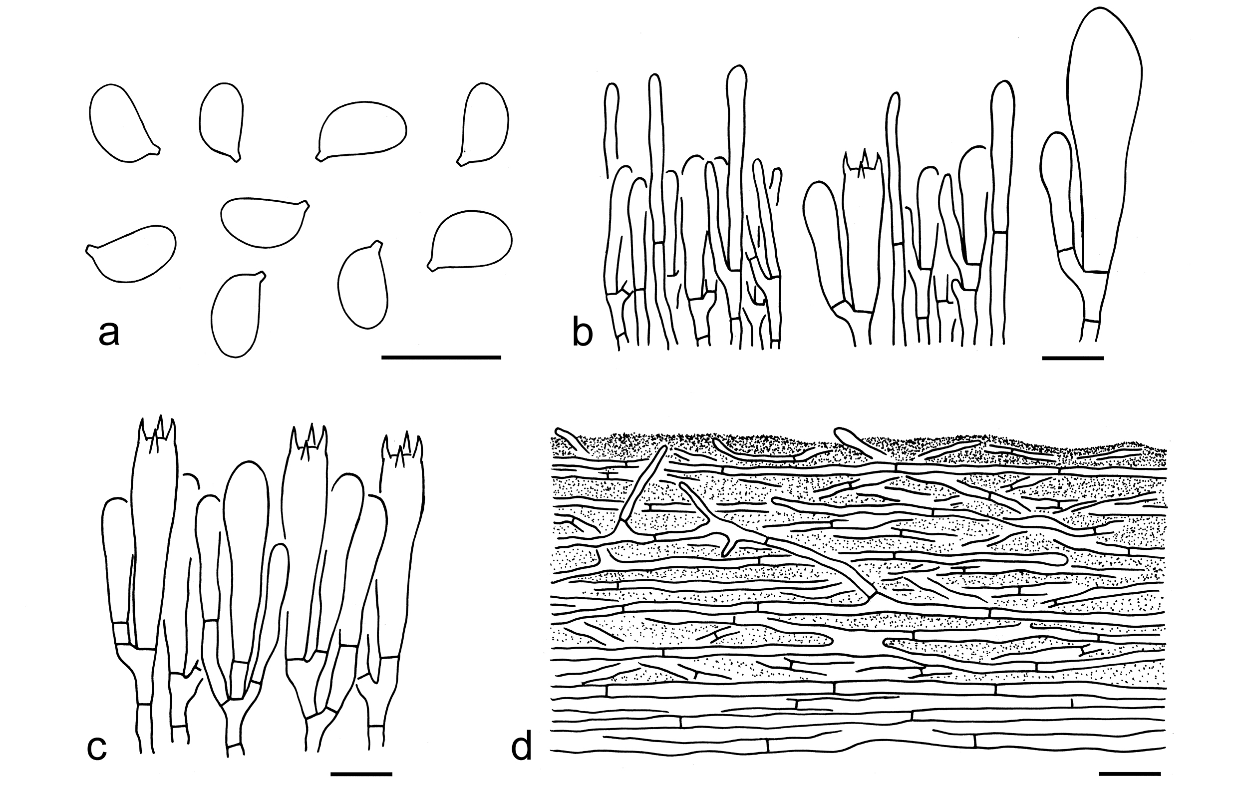
Ecology: Solitary to scattered on soil in coniferous or mixed forests with trees of Pinaceae and Fagaceae.
Known distribution: Known from early summer to late autumn in Southwestern China.
Specimens examined: CHINA. Yunnan Province, Baoshan, in a forest with Pinaceae, altitude 1900 m, 9 Aug. 2010, Yan-Jia Hao 211 (KUN-HKAS69195); the same city, Longyang, in a mixed forest with Pinaceae and Fagaceae, altitude 1700 m, 8 Aug. 2011, Qi Zhao 1203 (KUN-HKAS74156); the same city, Changning County, in a mixed forest of Pinus yunnanensis, Lithocarpus and Rhododendron, altitude 1800 m, 25 Jun. 2009, Yan-Chun Li 1801 (KUN-HKAS59548); Dali Bai Autonomous Prefecture, Yunlong County, in a forest with Pinus armandii, altitude 2370 m, 8 Aug. 2010, Li-Ping Tang 1201 (KUN-HKAS63020); Kunming, bought from Ciba Market, 23 Jul. 2017, Xiao-Xia Ding 220A (KUN-HKAS106561); same location, bought from Ciba Market, altitude unknown, 27 Jul. 2020, Yang-Yang Cui 378 (KUN-HKAS115902); same city, bought from Mushuihua Market, altitude unknown, 7 Oct. 2019, Yang-Yang Cui 346 (KUN-HKAS115901); Puer, Lancang Lahu Autonomous County, in a mixed forest with Pinaceae and Fagaceae, altitude 1090 m, 12 Nov. 2017, Zhu L. Yang 6114 (holotype, KUN-HKAS101303); same city, Jingdong Yi Autonomous County, Ailao Mountain, in a forest with Pinus yunnanensis, altitude 2500 m, 10 Aug. 2019, Zhu L. Yang 6243 (KUN-HKAS106855). Xizang Autonomous Region, Nyingchi, Bome County, in a subalpine sclerophyllous evergreen forest dominated by Quercus spp., altitude 2760 m, 17 Jul. 2019, Zhu L. Yang 6190 (KUN-HKAS106802).
Notes: Tricholoma qiaomianjun is widely consumed in Southwestern China and has been reported as “Tricholoma aff. flavovirens” in Yang et al. (2021). This species is well delimited by its greenish yellow to orange-brown pileus, yellow lamellae, ellipsoid basidiospores (5.5–7 × 4–5 μm, Q = 1.3–1.58, Qm = 1.44 ± 0.1) and presence of clamps (Cui et al. 2022; this study).
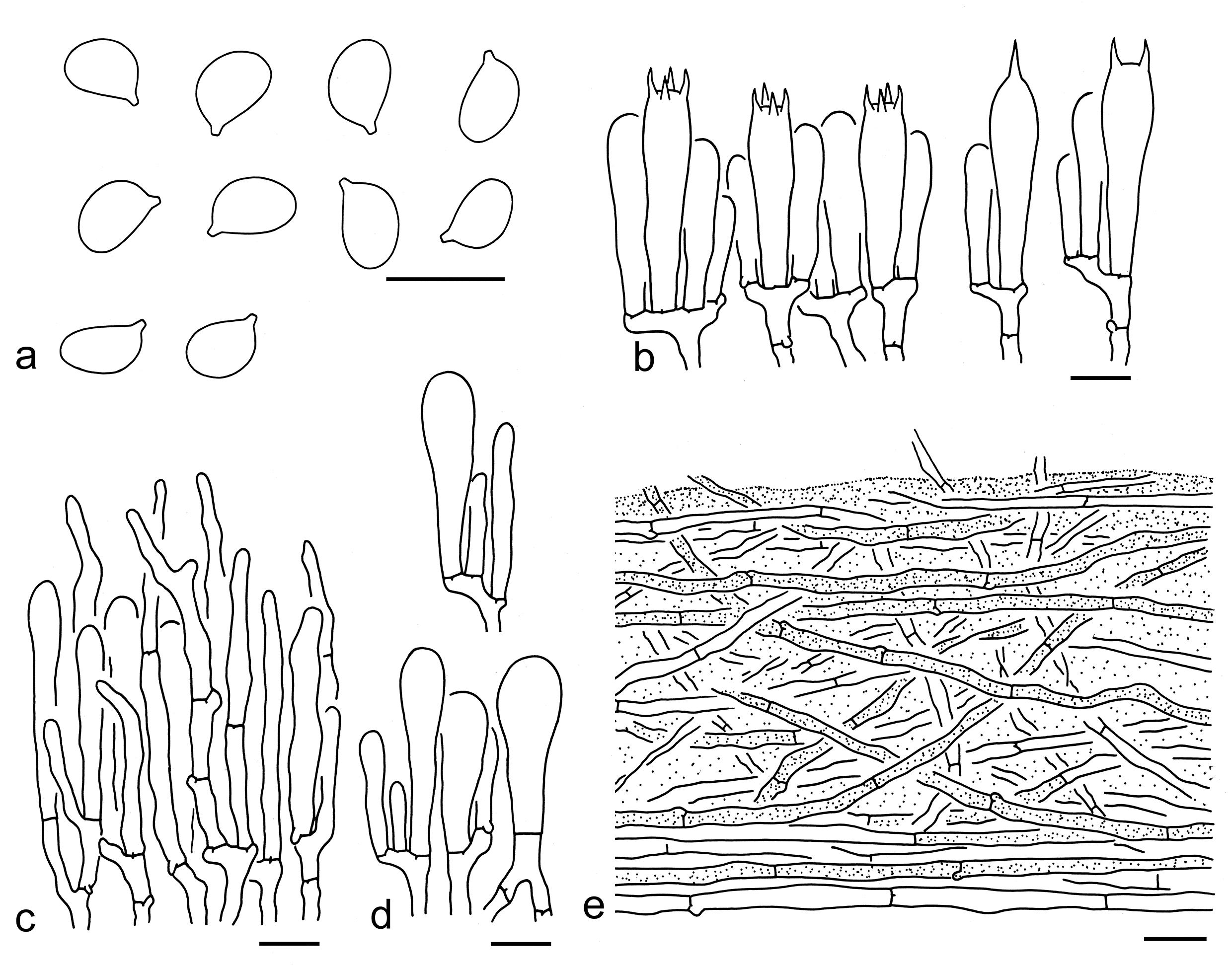
18. Tricholoma sinoportentosum Zhu L. Yang et al., Mycologia 110(6): 1098 (2018). Figs. 28h, 36
Description: Basidioma small, medium-sized to large. Pileus 3–12 cm in diam., convex to plano-convex, often irregularly convex, often acutely papillate or papillate at center; surface dry, often dark brown (1F1–3), grey-brown (2E4–5, 4E6–8) to greyish brown (2D3–5), with greenish yellow (30B5–7) color, sometimes grey-brown (2F6–8) at center, becoming olivaceous yellow (30B5–7) to olivaceous brown (30C5–7) towards margin, covered with blackish (1F1–3), dark grey (1F1–3) to brown (3E6–8) squamules; margin straight; context white (1A1). Lamellae sinuate, white (1A1), cream (1B2–4) to olivaceous yellow (30A4–6), subclose; edges concolor. Stipe 5–13 1–3 cm, subcylindrical to cylindrical, olivaceous yellow (30A4–6) to olivaceous brown (30C3–6), glabrous to fibrillose, exannulate. Taste and odour indistinct.
Basidiospores [40/2/1] 6–8 (–8.5) (4.5–) 5–6.5 µm (mean length = 7.1 µm, mean width = 5.6 µm), Q = (1.08–) 1.17–1.36 (–1.45), Qm = 1.27 ± 0.08, broadly ellipsoid to ellipsoid, non-amyloid, colorless, hyaline, thin-walled, smooth; apiculus relatively large. Basidia 25–40 6–8 µm, clavate, 4-spored, also 1-, 2- to 3-spored, hyaline; sterigmata 3–6 µm long; basal septa with clamps. Pleurocystidia absent. Cheilocystidia 20–35 5–9 µm, poorly differentiated, clavate to narrowly clavate, sometimes subfusiform, thin-walled, colorless or brownish, hyaline. Subhymenium composed of 2–5 µm wide filamentous hyphae, clamps present. Lamellar trama regular, composed of colorless, thin-walled, filamentous or inflated hyphae 3–25 µm wide. Pileipellis a cutis, slightly gelatinized or not, composed of radially arranged, 3–5 µm wide, thin-walled, yellow to brownish filamentous hyphae; subpellis poorly differentiated, non-gelatinized. Clamps present in all parts of basidioma.
Ecology: Solitary to scattered dominated with Picea and Pinus.
Known distribution: Known from East Asia (Reschke et al. 2018; this study).
Notes: Tricholoma sinoportentosum is well delimitated by its papillate, dark brown to brown pileus, presence of clamps and broadly ellipsoid to ellipsoid basidiospores (6–8 5–6.5 µm, Q = 1.17–1.36, Qm = 1.27 ± 0.08) (Yang et al. 2018; this study). In addition, this species grows in coniferous forests with Pinus (Yang et al. 2018; this study). Tricholoma portentosum is similar to T. sinoportentosum. For comparison of the two species, see notes under the name.
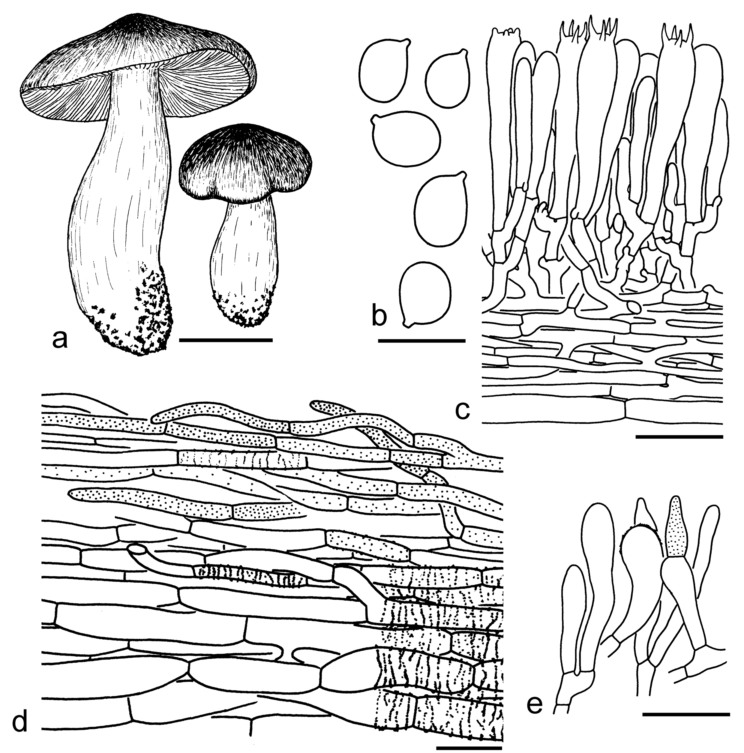
19. Tricholoma virgatum (Fr.) P. Kumm., Führ. Pilzk. (Zerbst): 134 (1871). Figs. 28i, j, 37
Basionym: Agaricus virgatus Fr., Observ. mycol. (Havniae) 2: 113 (1818).
Description: Basidioma small to medium-sized. Pileus 3–10 cm in diam., conical, convex to plano-convex, with an acute papilla or papilla at center; surface dry, often silvery shiny (1B1), sometimes dark grey (1F1–3) to grey (2D1–3), darker at center, glabrous or covered with dark grey (1F1–3) squamules; margin straight; context dirty white (1B1) to greyish (2B1–3). Lamellae sinuate, white (1A1) to greyish (2B1, 2C1), becoming greyish brown (1D2–4) to brownish (1C3–5) when old, crowded to subclose; edges white (1A1), grey (2E1–3) to brownish (1C3–5). Stipe 7–15 0.7–1.5 cm, cylindrical, often twisted, white (1A1), greyish (1B1–3), greyish brown (2B2–4) to brownish (4B2–4), glabrous to fibrillose, stuffed to hollow, exannulate. Taste not recorded. Odour unpleasant.
Ecology: Solitary to scattered in coniferous, broad-leaved, or mixed forests.
Known distribution: Known from Europe (Riva 1988, 2003; Christensen & Heilmann-Clausen 2013; Heilmann-Clausen et al. 2017), North America (Bessette et al. 2013) and East Asia.
Specimens examined: CHINA. Hubei Province, Yichang, Shennongjia Forest District, in a mixed forest, altitude 2300 m, 7 Aug. 2015, Xing He 3 (KUN-HKAS91176). Sichuan Province, Ganzi Tibetan Autonomous Prefecture, Kangding, in a subalpine forest, altitude 3450 m, 8 Sep. 2016, Bang Feng 98 (KUN-HKAS99452); the same county, in a mixed forest, altitude 3370 m, 9 Sep. 2016, KD-LJW 128 (KUN-HKAS97921); same county, in a mixed forest, altitude 3370 m, 9 Sep. 2016, KD-WPM 103 (KUN-HKAS98358). Yunnan Province, Diqing Tibetan Autonomous Prefecture, Shangri-La, in a mixed forest, altitude 4000 m, 13 Sep. 2016, Xiao-Xia Ding 136 (KUN-HKAS97079); the same location, in a mixed forest, altitude 4000 m, 13 Sep. 2016, Xiao-Xia Ding 135 (KUN-HKAS 97078); the same location, in a subalpine forest with Abies and Picea, altitude 3600 m, 30 Aug. 2021, Yang-Yang Cui 463 (KUN-HKAS135006); the same location, in a subalpine forest with Abies and Picea, altitude 3910 m, 8 Aug. 2016, Jian-Wei Liu 502 (KUN-HKAS98081); Kunming, Mushuihua Market, in a mixed forest, altitude unknown, 30 Aug. 2015, Pan-Meng Wang 468 (KUN-HKAS93474).
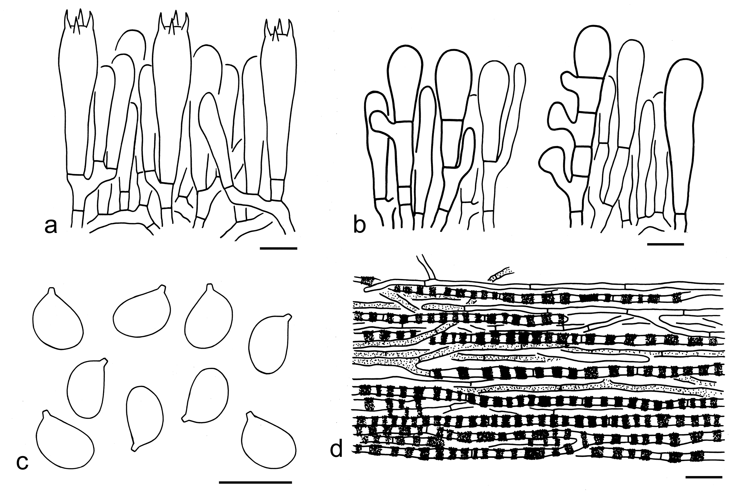
Notes: Tricholoma virgatum is characterized by its papillate, grey to silvery shiny pileus, a white to greyish stipe often twisted, broadly ellipsoid to ellipsoid basidiospores (7–10 5–7 µm, Q = 1.25–1.6, Qm = 1.43 ± 0.1) and absence of clamps (this study). Christensen & Heilmann-Clausen (2013) reported that this species has an indistinct or slightly spicy odour, and a taste that is initially mild but gradually becomes slightly to strongly acrid or bitterish. For the Chinese collections of this species, taste was not recorded, and odour was reported as unpleasant in the field.
In the phylogenetic tree based on five gene fragments (Fig. 3), T. mastoideum is sister to T. virgatum, and their differences have been discussed under its notes part. Morphologically, T. sciodes (Pers.) C. Martín can be confused. However, T. sciodes has darker colored basidioma, white to brownish lamellae without brown tinge, relatively longer basidiospores (5.6–8.3 4.5–7 µm) and is associated with broad-leaved trees (Riva 1988, 2003; Christensen & Heilmann-Clausen 2013).
Tricholoma sect. Fucata X.X. Ding et al.
Type: Tricholoma fucatum (Fr.) P. Kumm., Führ. Pilzk. (Zerbst): 130 (1871).
Description: Basidioma tricholomatoid. Pileus papillate at center, brown, grey or olivaceous, radially fibrillose. Lamellae white, dirty white, greyish or brownish. Annulus absent. Basidiospores medium-sized, broadly ellipsoid to ellipsoid, thin-walled, smooth, inamyloid. Pleurocystidia absent. Cheilocystidia present and conspicuous. Pileipellis an ixo-cutis with its lower layer composed with inflated cells. Clamps absent.
Ecology: Ectomycorrhizal. On soil with various trees.
Known distribution: Molecular evidence indicates that species from this section are found in Europe, North America, and Asia.
Notes: To date, two species, here newly describes as T. brunneiceps Y.Y. Cui & Zhu L. Yang and T. cheilocystidiatum Y.Y. Cui & Zhu L. Yang are known from China (see below).
Key to the species of Tricholoma sect. Fucata from China
1. Pileus dark grey to grey; lamellae white to dirty white, sometimes with grey edge; stipe dirty white, greyish to dirty brownish T. cheilocystidiatum
1. Pileus dark brown, brown to yellow-brown; lamellae white, cream to brownish, edges without grey color; stipe brownish to yellowish brown T. brunneiceps
20. Tricholoma brunneiceps Y.Y. Cui & Zhu L. Yang, sp. nov. Figs. 28k, l, 38
Fungal Names: FN 572334
Etymology: brunneiceps = brown-headed, referring to its brown pileus.
Diagnosis: Closely related to T. fucatum, but differs in relatively smaller basidiospores and being associated with trees of Fagaceae.
Type: CHINA. Yunnan Province, Honghe Hani and Yi Autonomous Prefecture, Lvchun County, Amoebin Forest Park, in a broad-leaved forest with Fagaceae, altitude 1600 m, 23 Sep. 2019, 532531MF0521 (holotype, KUN-HKAS134966).
Description: Basidioma small. Pileus 2.5–5 cm in diam., plano-convex to applanate, with an acute to blunt papilla; surface viscid, innately fibrillose, often blackish (1F1–3) to grey-brown (1E1–4) at center, becoming brown (4E4–8) to brownish (4D4–8, 3C4–8) towards margin, occasionally brown (2D4–8) overall; margin straight to inflexed; context white (1A1) to brownish (4D3–5). Lamellae adnexed to sinuate, white (1A1), cream (3A2–3) to yellow brownish (3C4–7), crowded to subclose; edges entire, white (1A1), cream (3A2–3) to brownish (3C4–7). Stipe 6–11 0.5–1 cm, cylindrical, stuffed to hollow, brownish (4B2–5) to yellowish brown (4C3–6), covered with brown (4D3–6) fibrillose squamules, exannulate. Taste and odour not recorded.
Basidiospores [100/5/4] 6–8.5 (–9) (4.5–) 5–6 µm (mean length = 7.2 µm, mean width = 5.3 µm), Q = 1.17–1.56 (–1.73), Qm = 1.37 ± 0.1, broadly ellipsoid to ellipsoid, non-amyloid, colorless, hyaline, thin-walled, smooth; apiculus relatively large. Basidia 30–40 7–10 µm, clavate, 4-spored, hyaline; sterigmata 3–5 µm long; basal septa without clamps. Pleurocystidia absent. Cheilocystidia 15–65 10–20 µm, conspicuous, fusiform, subfusiform to clavate, sometimes septate, thin- to thick-walled, brownish to brown or colorless, hyaline. Subhymenium composed of 3–5 µm wide filamentous hyphae, clamps absent. Lamellar trama regular, composed of colorless to thin-walled hyphae 3–15 µm wide. Pileipellis an ixo-cutis, strongly gelatinized, composed of radially arranged, 3–10 µm wide, thin-walled, colorless or brownish to brown filamentous hyphae; subpellis composed of radially arranged inflated cells, ellipsoid, clavate to cylindrical, 30–90 15–30 µm, thin-walled, yellow-brown to brownish. Clamps absent in all parts of basidioma.
Ecology: Solitary to scattered on soil in subtropical broad-leaved forests with trees of Fagaceae.
Known distribution: Known from Southwestern China.
Additional specimens examined: CHINA. Yunnan Province, Baoshan, Tengchong, in a subtropical broad-leaved forest with Fagaceae, altitude 1700 m, 10 Aug. 2011, Jiao Qin 291 (KUN-HKAS73277); same city, in a subtropical mixed forest with Fagaceae and Pinaceae, altitude 1960 m, 26 Jul. 2022, GLG-JLK2282 (KUN-HKAS134968); same city, in a subtropical forest dominated by Fagaceae, altitude 1900 m, 26 Jul. 2022, GLG-FXP236 (KUN-HKAS134971); same city, in a subtropical forest dominated by Fagaceae, altitude 1580 m, 24 Jul. 2022, GLG-FXP167 (KUN-HKAS134972), same city, in a subtropical forest dominated by Fagaceae, altitude 1900 m, 26 Jul. 2022, GLG-FXP250 (KUN-HKAS134973); same city, in a subtropical forest dominated by Fagaceae, altitude 1900 m, 26 Jul. 2022, GLG-FXP229 (KUN-HKAS134974); same city, in a subtropical broad-leaved forest with Fagaceae, altitude 1700 m, 10 Aug. 2011, Qi Zhao 1242 (KUN-HKAS74195); Baoshan, Longling County, in a subtropical mixed forest, altitude 1960 m, 30 Aug. 2022, GLG-FXP825 (KUN-HKAS134969); same county, in a subtropical mixed forest, altitude 1960 m, 30 Aug. 2022, GLG-FXP824 (KUN-HKAS134970); Honghe Hani and Yi Autonomous Prefecture, Pingbian Miao Autonomous County, Daweishan Nature Reserve, in a subtropical broad-leaved forest with Fagaceae, altitude 2300 m, 14 Sep. 2019, 532531MF0479 (KUN-HKAS108098); same prefecture, Lvchun County, Amoebin Forest Park, in a subtropical broad-leaved forest with Fagaceae, altitude 1600 m, 23 Sep. 2019, 532531MF0504 (KUN-HKAS134967); same province, Kunming, Qiongzhu Temple, in a subtropical broad-leaved forest, altitude 2200 m, 8 Aug. 2007, Bang Feng 32 (KUN-HKAS52753).
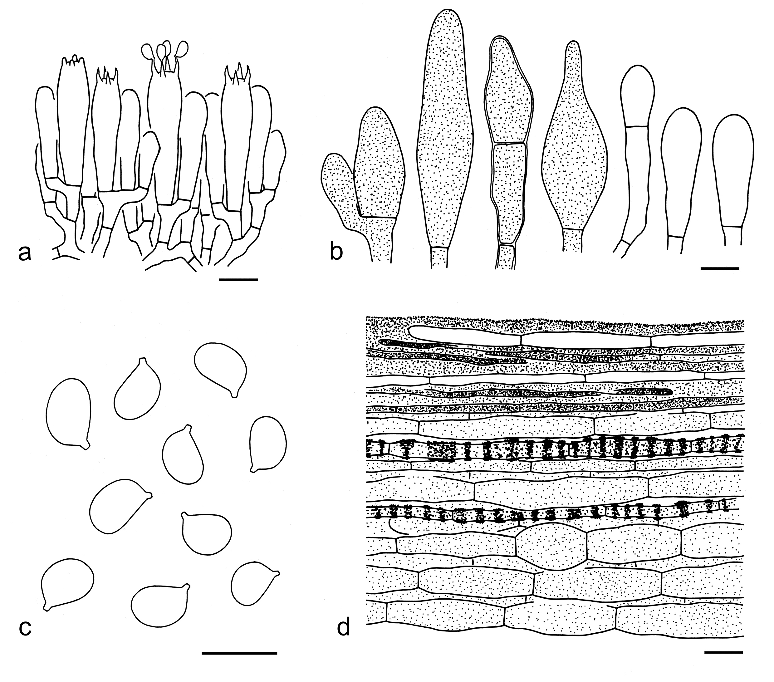
21. Tricholoma cheilocystidiatum Y.Y. Cui & Zhu L. Yang, sp. nov. Figs. 28m, 39
Fungal Names: FN 572336
Etymology: cheilocystidiatum = cheilocystidia, referring to its conspicuous cheilocystidia.
Diagnosis: Related to T. brunneiceps, but differs by its grey pileus, white to dirty white lamellae and a greyish stipe.
Type: CHINA. Yunnan Province, Chuxiong Yi Autonomous Prefecture, Nanhua County, in a subtropical broad-leaved forest dominated by plants of Fagaceae, altitude 1950 m, 10 Aug. 2016, Xiao-Xia Ding 108 (holotype, KUN-HKAS97051).
Description: Basidioma small to medium-sized. Pileus 3–7 cm in diam., plano-convex to applanate, often with a low umbo; surface dry to slightly viscid, radially rugose, innately fibrillose, dark grey (1E1–3) to blackish (1F1–4) at center, often grey (1E1–3, 2E1–3) to brownish grey (1E3–6), sometimes greyish brown (2E5–8) to olivaceous yellow (3D4–7) towards margin; margin straight or slightly reflexed, sometimes radially rimose; context white (1A1). Lamellae adnexed to sinuate, white (1A1), dirty white (1C1–2) to cream (1A2, 1B2–4), crowded to subclose; edges slightly serrulate, dirty white (1C1–2), cream (1A2, 1B2–4) to grey (2E1–2). Stipe 4–8 0.5–1.5 cm, cylindrical, hollow, dirty white (1B1–3), greyish (1C1–2) to dirty brownish (1B2–4), fibrillose, exannulate. Taste unknown. Odour like almond.
Basidiospores [140/7/2] 6–9 (–10) 4–6 (–6.8) µm (mean length = 7.2 µm, mean width = 5.0 µm), Q = 1.18–1.75 (–1.8), Qm = 1.46 ± 0.13, mostly ellipsoid, sometimes broadly ellipsoid or elongate, non-amyloid, colorless, hyaline, thin-walled, smooth; apiculus relatively large. Basidia 25–40 7–10 µm, clavate, 1-, 2- to 4-spored, hyaline; sterigmata 4–8 µm long; basal septa without clamps. Pleurocystidia absent. Cheilocystidia 15–40 8–15 µm, conspicuous, subclavate, clavate, fusiform to ellipsoid, sometimes irregular shaped, sometimes septate, thin- to slightly thick-walled, colorless, hyaline. Subhymenium composed of 2–5 µm wide filamentous hyphae, clamps absent. Pileipellis an ixo-cutis, gelatinized, composed of radially arranged, 4–8 µm wide, thin-walled, colorless to brown filamentous hyphae; subpellis composed of radially arranged inflated cells, fusiform, ellipsoid to cylindrical, 25–55 10–25 µm, thin-walled, colorless to brown. Clamps absent in all parts of basidioma.
Ecology: Solitary to scattered on soil in subtropical broad-leaved forests with trees of Fagaceae.
Known distribution: Known from Southwestern China.
Notes: Tricholoma cheilocystidiatum is recognized by its dark grey to grey pileus which is slightly viscid, white to dirty white lamellae sometimes with grey edges, and ellipsoid basidiospores (6–9 4–6 µm, mean length = 7.2 µm, mean width = 5.0 µm, Q = 1.18–1.75, Qm = 1.46 ± 0.13). In our -phylogenetic analysis based on five gene fragments (Fig. 3), this species is sister to T. brunneiceps. However, T. brunneiceps can be easily separated by its brown to yellow-brown pileus, white, cream to brownish lamellae and a brownish to yellowish brown stipe (this study).
Tricholoma felschii Ovrebo et al. is similar to T. cheilocystidiatum. However, T. felschii differs by having relatively smaller basidiospores (6–7.5 5–5.5 µm) and being found in the USA and Costa Rica (Ovrebo et al. 2019). Tricholoma mutabile Shanks and T. josserandii Bon can be confused with T. cheilocystidiatum. However, T. mutabile has a violet color on pileus, especially in old specimens, smaller basidiospores (5.8–7.7 4.3–5.8 µm) and is distributed in the USA (Shanks 1996; Bessette et al. 2013). Tricholoma josserandii is different from T. cheilocystidiatum in its shiny pileus and relatively narrower basidiospores (5.2–8 4.5–6.2 µm, Q = 1–1.4) (Riva 1988; Christensen & Heilmann-Clausen 2013).
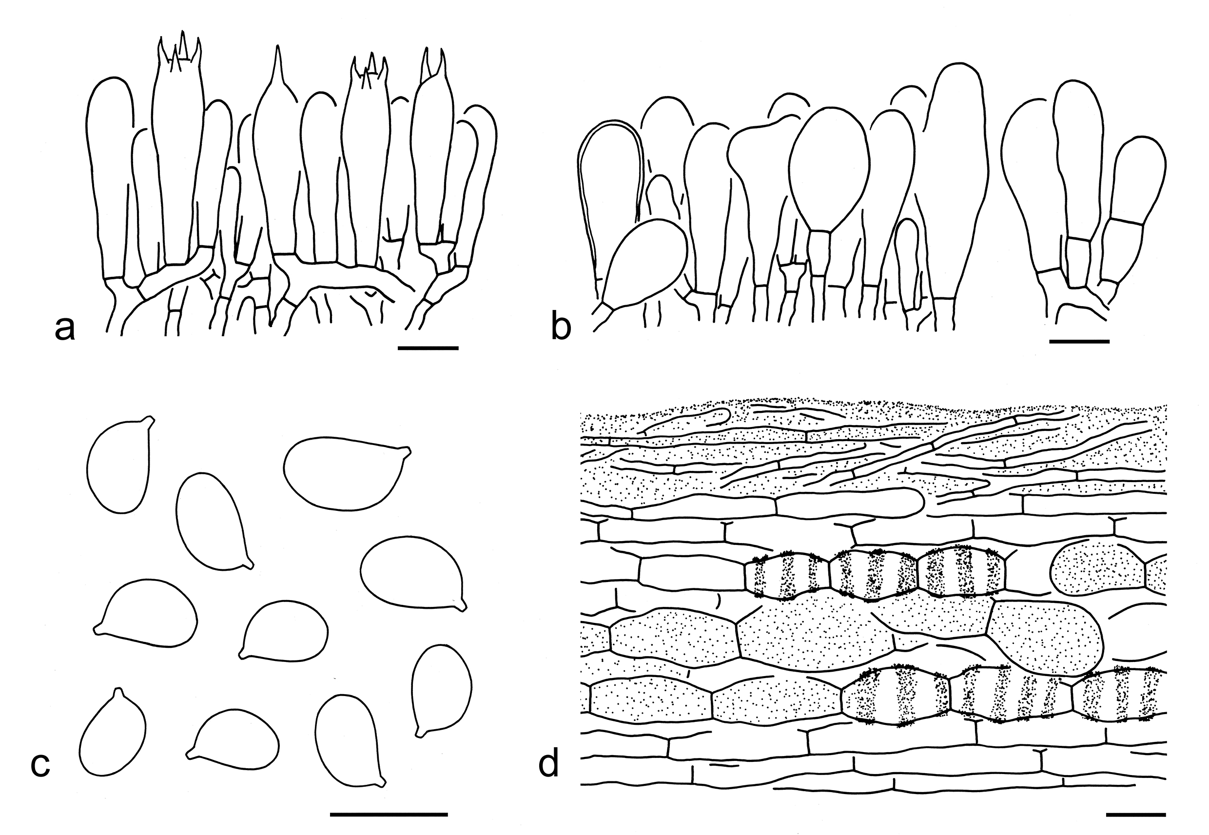
Tricholoma sect. Genuina (Fr.) Sacc.
Type: Tricholoma vaccinum (Schaeff.) P. Kumm., Führ. Pilzk. (Zerbst): 133 (1871).
Description: Basidioma tricholomatoid. Pileus umbonate or not, often with an involute margin especially in young stage, orange-brown to brown, glabrous, squarrulose to squamosa. Lamellae crowded, often with brown stains when damaged or old. Stipe cylindrical to subcylindrical, often without or occasionally with an evanescent ring. Basidiospores small, broadly ellipsoid, ellipsoid to elongate, thin-walled, smooth, inamyloid. Pleurocystidia absent. Cheilocystidia present or absent, elements in lamellar edge often gelatinized and pigmented. Pileipellis mostly a cutis or an ixo-cutis. Clamps absent.
Ecology: Ectomycorrhizal. Solitary to scattered on soil with various trees.
Distribution: Molecular data indicated members from this section are currently found in Europe, North America, Oceania, and Asia.
Notes: To date, 18 species, including three novel and 15 known ones, are found in China with both molecular and morphological evidence (see below).
Key to the species of Tricholoma sect. Genuina from China
1. Stipe with an obviously woolly to skin-like ring on the apical part of stipe T. focale
1. Stipe exannulate 2
2. Pileus yellowish to yellowish brown, with more yellow color; cheilocystidia poorly differentiated; distributions dominated in Larix or broad-leaved forests 3
2. Pileus pinkish brown, orange-brown to reddish brown, with more orange color; cheilocystidia conspicuous or poorly differentiated; distributions in coniferous, broad-leaved or mixed forests 4
3. Pileus fibrillose-felty to fibrillose, with obvious squamules; basidioma associations in forests with trees of Larix ………..T. psammopus
3. Pileus without distinct squamules; basidioma associations in forests with broad-leaved trees T. sinoacerbum
4. Pileus pinkish to pinkish brown at center, whitish towards margin; stipe shorter than pileus diameter, with pinkish brown color T. roseoacerbum
4. Pileus red-brown, orange-brown to brown; stipe often longer than pileus diameter, without pinkish brown color 5
5. Basidioma relatively robuster, large to very large; pileal margin strongly involute; basidiospores relatively larger, with mean length > 8 µm, mean width > 6 µm T. orienticolossum
5. Basidioma often medium-sized to large; pileal margin often involute when young, deflexed when mature; basidiospores relatively smaller, with mean length < 8 µm, mean width < 6 µm 6
6. Stipe with distinct apricot orange, ochraceous to brown squamules breaking into girdles; cheilocystidia conspicuous; pileipellis an ixo-trichoderm T. aurantium
6. Stipe glabrous or fibrillose, without distinct girdles; cheilocystidia conspicuous or poorly differentiated; pileipellis an ixo-cutis, cutis or ixo-trichoderm 7
7. Pileus dry, coarsely fibrillose to fibrillose-tomentose, with obvious squamules 8
7. Pileus dry or viscid when wet, glabrous or with distinct squamules 10
8. Basidiospores relatively narrower, with mean width < 5 µm; cheilocystidia poorly differentiated T. imbricatum
8. Basidiospores relatively broader, with mean width ≥ 5 µm; cheilocystidia conspicuous 9
9. Pileus tomentose, fibrillose-tomentose to fibrillose-hairy, often with a shaggy margin; lamellae white when young ……T. vaccinum
9. Pileus fibrillose-tomentose, without a shaggy margin; lamellae cream to yellowish colored when young …………….T. rufobrunneum
10. Lamellae yellowish to yellow when young 11
10. Lamellae white to whitish when young 13
11. Pileal margin somewhat ribbed when mature; basidiospores slightly shorter, with mean length < 6 µm; cheilocystidia not observed T. orientifulvum
11. Pileal margin not ribbed; basidiospores slightly longer, with mean length ≥ 6 µm; cheilocystidia present 12
12. Basidiospores slightly larger (6–7.5 4–5.5 µm, mean length = 6.6 µm, mean width = 4.8 µm); associations with Picea and Quercus T. fulvomaculatum
12. Basidiospores slightly smaller (5–7 4–5 µm, mean length = 6 µm, mean width = 4.3 µm); associations with Castanopsis and Pinus T. castaneiceps
13. Associations with Populus T. populinum
13. Associations in conifers, broad-leaved or mixed forests with Pinaceae and Fagaceae 14
14. Stipe without a white zone at top 15
14. Stipe with a white zone at top 17
15. Basidiospores relatively broader, subglobose to broadly ellipsoid; pileipellis not gelatinized; associations in broad-leaved forests T. rubiginosum
15. Basidiospores relatively narrower, broadly ellipsoid, ellipsoid to elongate; pileipellis gelatinized; associations in coniferous, broad-leaved or mixed forests 16
16. Pileus with dark brown spots; pileal margin not ribbed ….T. rubrocastaneum
16. Pileus without dark brown spots; pileal margin somewhat ribbed T. stans
17. Basidiospores relatively larger (6–7 4.5–5.5 µm, mean length = 6.3 µm, mean width = 5 µm); associations in coniferous or broad-leaved forests T. ustaloides
17. Basidiospores relatively smaller (5–7 3.5–5 µm, mean length = 5.7 µm, mean width = 4 µm); associations dominated with Pinus T. albobrunneum
22. Tricholoma albobrunneum (Pers.) P. Kumm., Führ. Pilzk. (Zerbst): 130 (1871). Figs. 28n, 40
Basionym: Agaricus albobrunneus Pers., Syn. meth. fung. (Göttingen) 2: 293 (1801).
Description: Basidioma small to medium-sized. Pileus 2–10 cm in diam., at first convex, then plano-convex to applanate, often slightly umbonate; surface dry, but viscid when wet, reddish brown (6C4–8, 6B4–6) to brown (4C4–6), darker at center, covered with reddish brown (6C4–8, 6B4–6) to brown (4C4–6), fine squamules; margin involute when young, deflexed when mature; context white (1A1) to cream (1A2). Lamellae sinuate, white (1A1) to cream (1A2), with brownish (5C3–5) spots then entirely brown (5D5–7), crowded; edges white (1A1) to brownish (5C3–5). Stipe 3–7 0.3–2 cm, cylindrical to subcylindrical, stuffed to hollow, whitish (1A1) to brownish (5C3–5), often white (1A1) at apical part, basal mycelium white (1A1), covered with brownish (5C3–5) fibrillose squamules, exannulate. Taste mild. Odour not recorded.
Basidiospores [113/5/4] (4–) 5–7 (–8) (3–) 3.5–5 µm (mean length = 5.7 µm, mean width = 4 µm), Q = 1.15–1.75 (–2), Qm = 1.44 ± 0.16, broadly ellipsoid, ellipsoid to elongate, non-amyloid, colorless, hyaline, thin-walled, smooth; apiculus relatively large. Basidia 20–30 6–8 µm, clavate, 4- or 2-spored, at first colorless, then yellow brown to brown, hyaline; sterigmata 3–7 µm long; basal septa without clamps. Pleurocystidia absent. Cheilocystidia 10–30 3–7 µm, poorly differentiated, clavate, sometimes septate, occasionally branching, thin-walled, hyaline, colorless when young, then brownish, at last gelatinized. Subhymenium composed of 2–5 µm wide filamentous hyphae, clamps absent. Lamellar trama regular, composed of colorless, thin-walled hyphae 2–8 µm wide. Pileipellis an ixo-cutis, gelatinized, composed of radially arranged, 3–9 µm wide, thin-walled, yellow-brown, brown to brownish filamentous hyphae; subpellis poorly differentiated. Clamps absent in all parts of basidioma.
Ecology: Solitary to scattered on soil in subtropical, sometimes temperate forests dominated with Pinus, sometimes mixed with Quercus.
Known distribution: Known from Europe and China.
Notes: Tricholoma albobrunneum is distinguished by its reddish brown to brown pileus and broadly ellipsoid, ellipsoid to elongate basidiospores (this study). In China, this species can be found not only in coniferous forest, but also in mixed forest with both Pinus and Quercus. According to Christensen & Heilmann-Clausen (2013), this species has a strongly farinaceous odour and a mild, farinaceous to slightly bitter taste, whereas taste and odour were not recorded in the Chinese specimens.
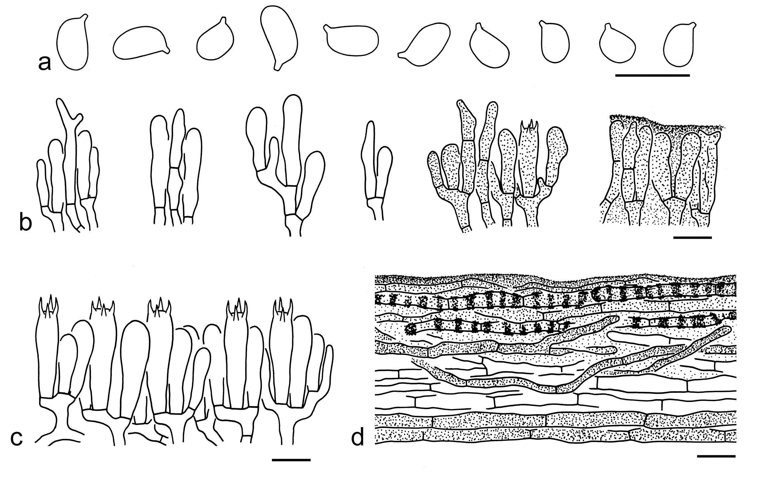
According to current data, no closely related species were discovered (Figs. 3, 4). Tricholoma stans (Fr.) Sacc. is similar to it, but has a more robust basidioma, a somewhat ribbed pileal margin and is distributed in coniferous and mixed forests (Riva 1988, 2003; Christensen & Heilmann-Clausen 2013; Landry et al. 2022; this study). Tricholoma rubrocastaneum Y.Y. Cui & Zhu L. Yang can be confused with it. For comparison, see notes under that name. Tricholoma fulvum (DC.) Bigeard & H. Guill. and T. ustaloides Romagn. are similar to T. albobrunneum. However, T. fulvum has distinct yellowish context (Christensen & Heilmann-Clausen 2013; Landry et al. 2022). Tricholoma ustaloides differs by its slight ring zone more obvious than T. albobrunneum, slightly larger basidiospores (6–7 4.5–5.5 µm, mean length = 6.3 µm, mean width = 5 µm) and association with only deciduous hosts (Riva 1988, 2003; Christensen & Heilmann-Clausen 2013; this study). Tricholoma batschii Gulden also similar to T. albobrunneum, but the former species has shorter and more broadly ellipsoid basidiospores (Christensen & Heilmann-Clausen 2013).
23. Tricholoma aurantium (Schaeff.) Ricken, Die Blätterpilze: 332 (1914). Figs. 28o, 41
Basionym: Agaricus aurantius Schaeff., Fung. bavar. palat. nasc. (Ratisbonae) 4: 18 (1774).
Description: Basidioma medium-sized to large. Pileus 4–12 cm in diam., convex to plano-convex, sometimes with a broadly low umbo; surface viscid when wet, often cracking into squamules when dry, apricot orange (1B4–6), ochraceous (5B5–7) to brown (5C5–7); margin involute, later inflexed; context white (1A1) to dirty white (1B2). Lamellae adnexed to sinuate, at first white (1A1) to cream (1A2), with brownish (5B3–5) to brown (5E6–8) spots when old, crowded; edges white (1A1) to cream (1A2), later with brown (5E6–8) spots or entirely brown (5E6–8). Stipe 3–10 0.5–3 cm, cylindrical to clavate, white (1A1) at apical part, a white (1A1) background covered with apricot orange (1B4–6), ochraceous (5B5–7) to brown (5C5–7) squamules often breaking into girdles, basal mycelium white (1A1), exannulate. Taste not recorded. Odour like cucumber.
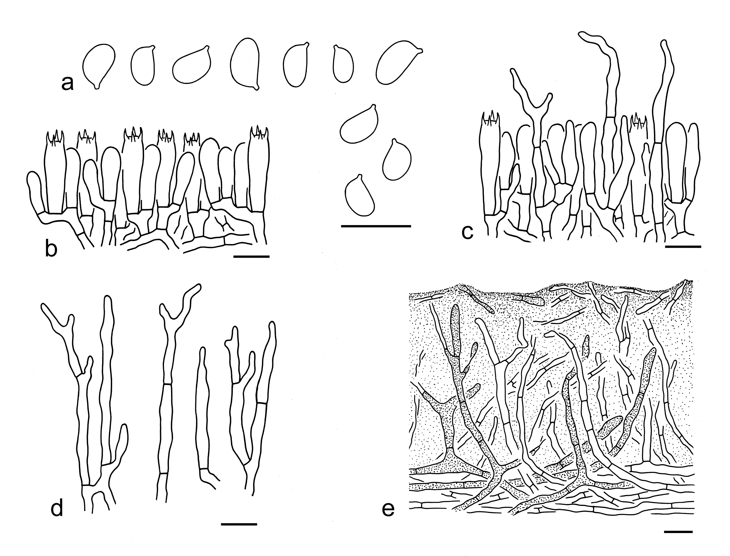
Basidiospores [90/5/2] 5–7 (–8) 3–4 (–4.5) µm (mean length = 5.9 µm, mean width = 3.7 µm), Q = 1.32–1.9 (–2.1), Qm = 1.6 ± 0.14, ellipsoid to elongate, non-amyloid, colorless, hyaline, thin-walled, smooth; apiculus relatively large. Basidia 15–35 5–7 µm, clavate, 4-spored, hyaline; sterigmata 2–4 µm long; basal septa without clamps. Pleurocystidia absent. Cheilocystidia 30–60 2–6 µm, conspicuous, cylindrical to filiform, often branching, sometimes septate, thin-walled, colorless, hyaline. Subhymenium composed of 2–4 µm wide filamentous hyphae, clamps absent. Lamellar trama regular, composed of colorless, thin-walled hyphae 2–8 µm wide. Pileipellis an ixo-trichoderm, strongly gelatinized, composed of more or less vertically arranged, 2–6 µm wide, colorless or yellowish filamentous hyphae; subpellis non-gelatinized, composed of radially arranged, 3–7 µm wide, thin-walled, colorless to yellowish hyphae. Clamps absent in all parts of basidioma.
Ecology: Solitary to scattered on soil in temperate coniferous forests with Abies and Picea.
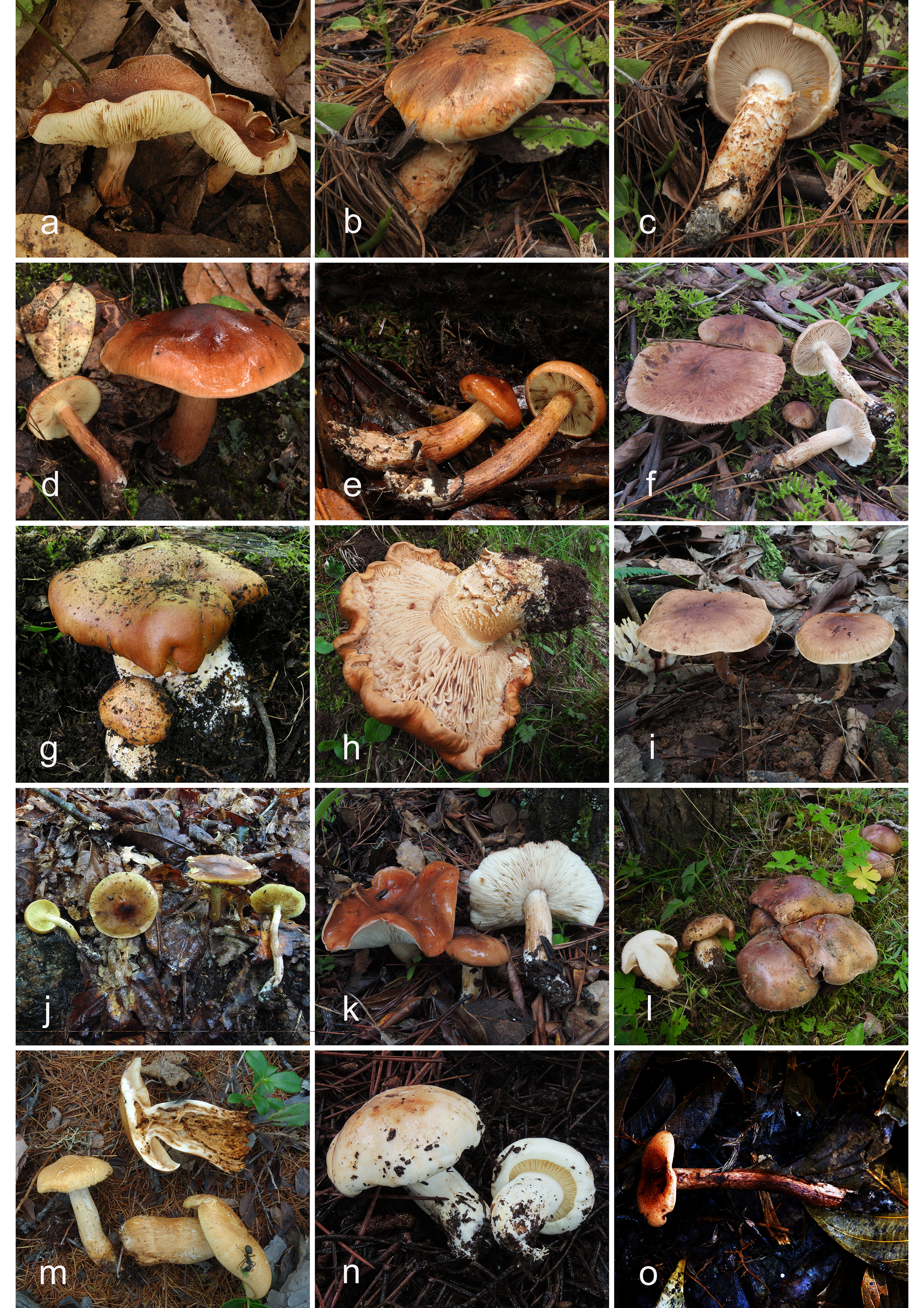
Distribution: Known from Europe (Christensen & Heilmann-Clausen 2013), North America (Bessette et al. 2013) and East Asia, distributed in Northern and Northwestern China.
Specimens examined: CANADA. Ontario, Bruce peninsula National Park, near the Emmett Lake, in a coniferous forest with Abies, altitude 200 m, 12 Sep. 2015, Bang Feng 1997 (KUN-HKAS94389); same location, in a coniferous forest with Abies, altitude 200 m, 12 Sep. 2015, Bang Feng 1998 (KUN-HKAS94390). CHINA. Inner Mongolia, Hulunbuir City, Genhe, altitude 800 m, 29 Aug. 2014, collector unknown (HMJAU28452). Xinjiang Uygur Autonomous Region, Yili Kazak Autonomous Prefecture, in a temperate coniferous forest with Picea, altitude 2300 m, 9 Jul. 2017, Xiao-Xia Ding 194 (KUN-HKAS106536).
Notes: Tricholoma aurantium can be easily delimitated by its apricot orange pileus, apricot orange girdles on stipe surface, ellipsoid to elongate basidiospores, conspicuous cheilocystidia and an ixo-trichoderm pileipellis (this study). Christensen & Heilmann-Clausen (2013) recorded this species as having a strongly farinaceous or linseed-oil-like odour and a strongly farinaceous to rancid-bitter taste. In the Chinese collections of this species, however, taste was not recorded, and the odour was described as cucumber-like in the field.
This species is sister to T. focale on the phylogenetic tree inferred from five gene fragments (Fig. 3), but the latter species has a coarsely radially fibrillose pileus, a woolly to skin-like ring and relatively shorter basidiospores (4–5.5 3–4 µm, mean length = 4.8 µm, mean width = 3.5 µm) (Riva 1988, 2003; Christensen & Heilmann-Clausen 2013; this study). Tricholoma batschii Gulden ex Mort. Chr. & Noordel. and T. stans (Fr.) Sacc. are somewhat similar to T. aurantium, but these two species have no girdles on stipe surface (Riva 1988; Christensen & Heilmann-Clausen 2013; Landry et al. 2022). Moreover, T. batschii has a thin, but distinct skin-like ring when young (Christensen & Heilmann-Clausen 2013). Tricholoma stans possesses poorly differentiated cheilocystidia and an ixo-cutis pileipellis (this study).
24. Tricholoma castaneiceps Y.Y. Cui & Zhu L. Yang, sp. nov. Figs. 42a, 43
Fungal Names: FN 572335
Etymology: castaneiceps = chestnut-headed, referring to its chestnut-colored pileus.
Diagnosis: Similar to T. fulvum, but differs by its non-ribbed pileal margin, presence of cheilocystidia and associations with subtropical trees of Castanopsis and Pinus.
Type: CHINA. Yunnan Province, Puer, Lancang Lahu Autonomous County, Qianliu Village, in a forest with Castanopsis and Pinus kesiya var. langbianensis, altitude 1300 m, 10 Nov. 2017, Zhu L. Yang 6092 (holotype, KUN-HKAS101281).
Description: Basidioma very small to small. Pileus 2–5 cm in diam., applanate to plano-concave, slightly umbonate at center; surface dry or viscid when wet, fulvous (5C4–7), often darker at center, covered with fulvous (5C4–7) fibrillose squamules; margin deflexed; context dirty white (1A1) to cream (1A2). Lamellae adnexed to sinuate, pale sulphur-yellow (3A2–4), crowded; edges often reddish (6A2–4) to brownish (5C4–6) when injured. Stipe 4–6 0.5–0.8 cm, cylindrical, attenuate upwards, brownish (5C4–6), covered with brown (5D6–8) squamules, basal mycelium dirty white (1A1) to pinkish (6A2), exannulate. Taste and odour not recorded.
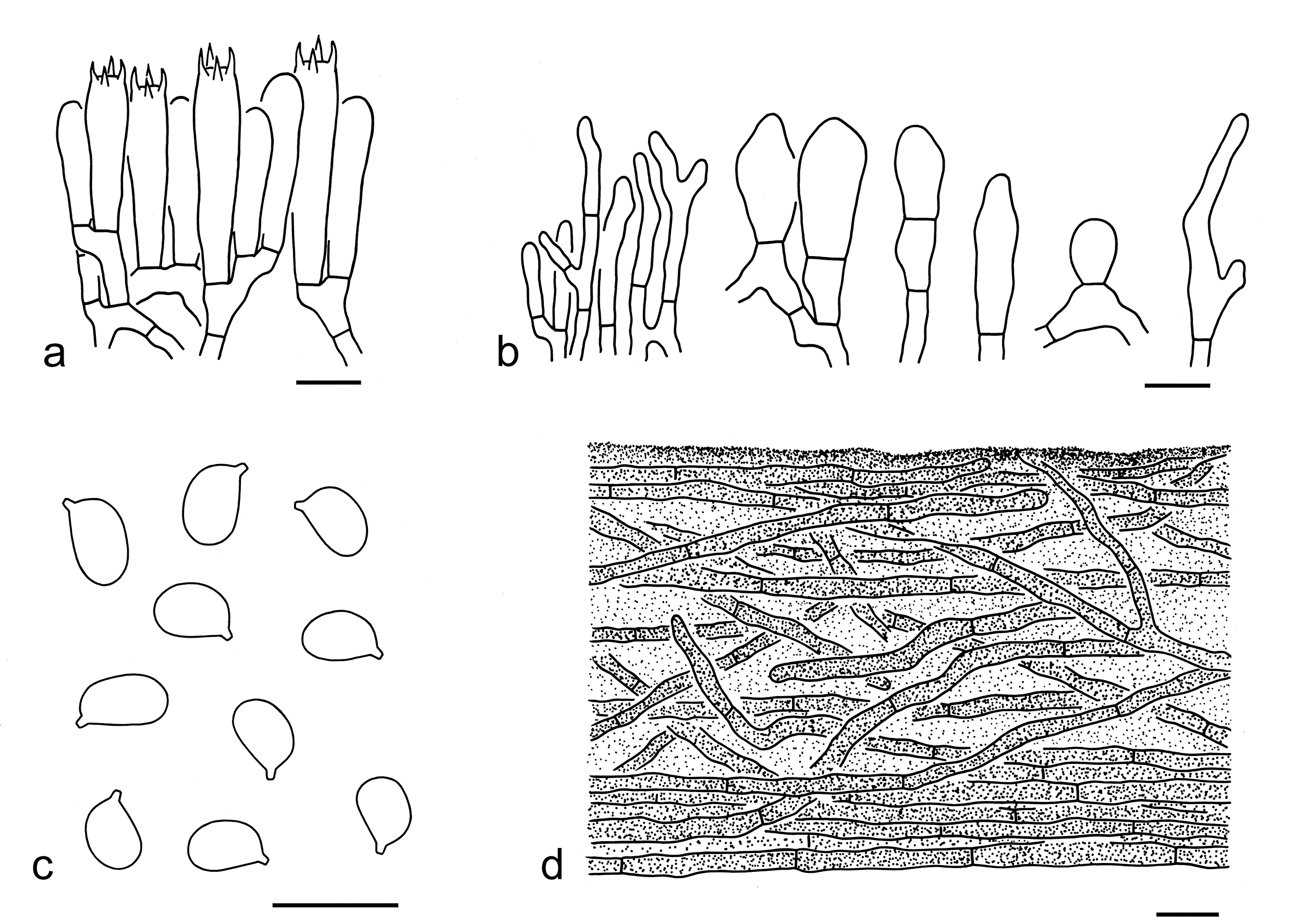
Basidiospores [80/4/1] 5–7 4–5 µm (mean length = 6 µm, mean width = 4.3 µm), Q = (1.2–) 1.3–1.5 (–1.75), Qm = 1.41 ± 0.1, ellipsoid, sometimes broadly ellipsoid or elongate, non-amyloid, colorless, hyaline, thin-walled, smooth; apiculus relatively large. Basidia 20–40 5–8 µm, clavate, 4-spored, sometimes 2- or 1-spored, hyaline; sterigmata 3–7 µm long; basal septa without clamps. Pleurocystidia absent. Cheilocystidia 10–45 3–10 µm, conspicuous, clavate, cylindrical to filamentous, often branching or septate, thin-walled, colorless, hyaline. Subhymenium composed of 2–5 µm wide filamentous hyphae, clamps absent. Lamellar trama regular, composed of colorless, thin-walled hyphae 2–15 µm wide. Pileipellis an ixo-cutis, gelatinized, composed of radially arranged to interwoven, 2–5 µm wide, thin-walled, yellow-brown filamentous hyphae; subpellis poorly differentiated, non-gelatinized, composed of radially arranged, 4–7 µm wide, yellow-brown filamentous hyphae. Clamps absent in all parts of basidioma.
Ecology: Solitary to scattered on soil in subtropical mixed forests with Castanopsis and Pinus kesiya var. langbianensis.
Known distribution: Known from Southwestern China.
Additional specimen examined: CHINA. Yunnan Province, Qin Cai 11 (KUN-HKAS138822).
Notes: Tricholoma castaneiceps is characterized by its fulvous pileus, ellipsoid basidiospores, conspicuous cheilocystidia and associations with subtropical trees of Castanopsis and Pinus. It is closely related to T. rubiginosum Y.Y. Cui & Zhu L. Yang and T. fulvomaculatum X.X. Ding et al. (Fig. 3). However, T. rubiginosum differs by its subglobose to broadly ellipsoid basidiospores with Q = 1.08–1.29, and a non-gelatinized pileipellis. Tricholoma fulvomaculatum has slightly larger basidiospores (6–7.5 4–5.5 µm, mean length = 6.6 µm, mean width = 4.8 µm) and is associated with Picea and Quercus (Ding et al. 2022; this study). This species is similar to T. fulvum, but the latter species differs by a somewhat ribbed pileal margin when mature, absence of cheilocystidia and associations with Betula, Abies and Picea (Christensen & Heilmann-Clausen 2013; Landry et al. 2022). Tricholoma orientifulvum X. Xu et al. Yang is comparable to T. castaneiceps, but possesses relatively smaller basidiospores (5–6 4–4.5 µm, mean length = 5.3 µm, mean width = 4.2 µm) and lacks cheilocystidia (Xu et al. 2020; this study).
25. Tricholoma focale (Fr.) Ricken, Die Blätterpilze: 332 (1914). Figs. 42b, c, 44
Basionym: Agaricus focalis Fr., Epicr. syst. mycol. (Upsaliae): 20 (1838) [1836-1838].
Description: Basidioma large. Pileus ca. 10 cm in diam., at first hemispherical, then plano-convex; surface dry, orange-brown (5B5–7), covered with appressed, orange-brown (5B5–7), fibrillose squamules, often with cottony remnants at margin; margin involute to inflexed; context white (1A1) to brownish (5A2–4). Lamellae adnate, adnexed to sinuate, white (1A1) to brownish (5B3–5), crowded; edges entire, white (1A1), sometimes with brownish (5B3–5) to brown (5C4–6) spots when mature. Stipe ca. 10 2.5 cm, cylindrical, sometimes slightly tapering downwards, solid to fistulose, with an orange-brown (5B5–7), cottony to fibrillose annulus on the apical part, white (1A1) above annulus, white (1A1) backgrounds densely covered with appressed, orange-brown (5B5–7), fibrillose squamules below annulus. Taste and odour not recorded.
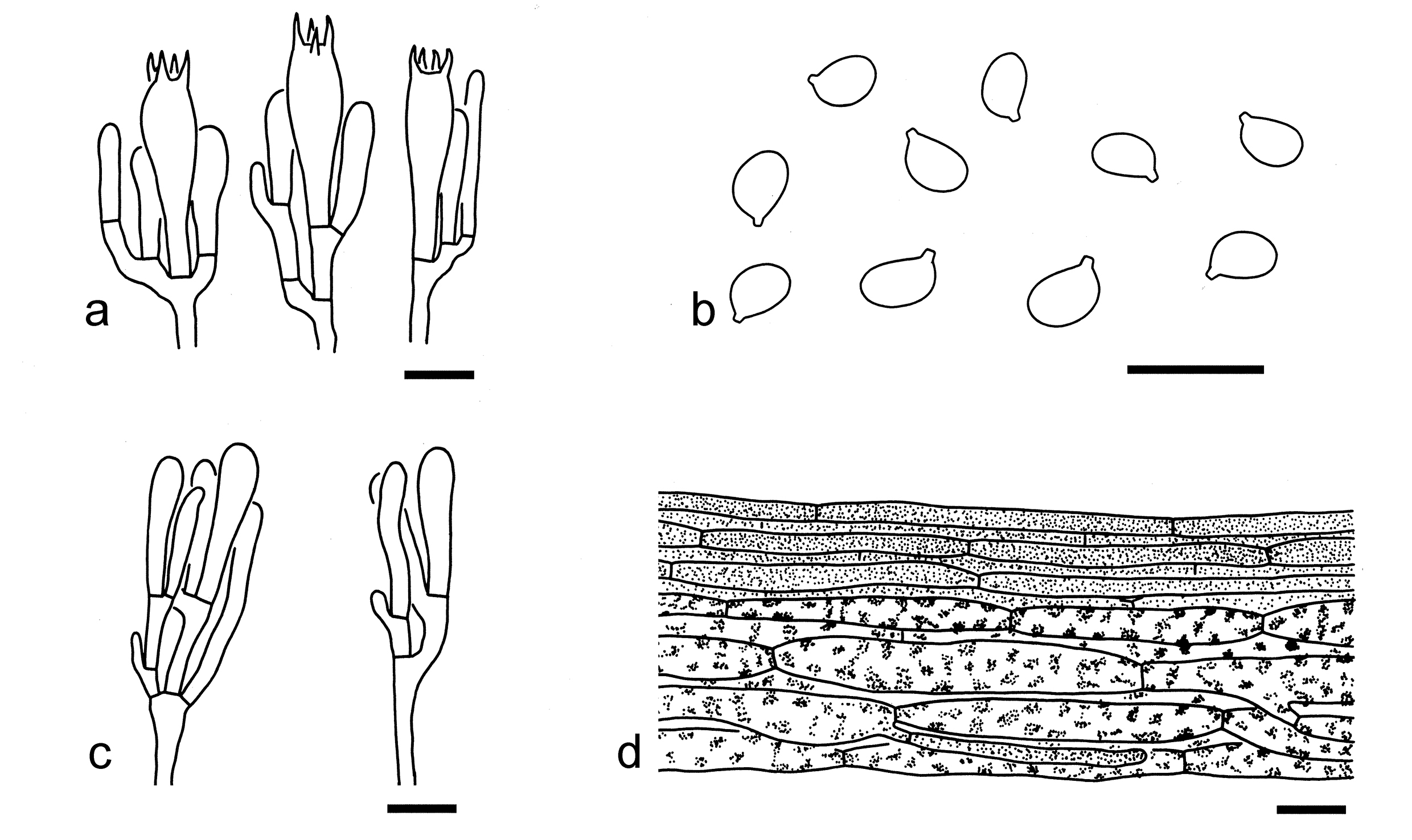
Basidiospores [20/1/1] 4–5.5 3–4 µm (mean length = 4.8 µm, mean width = 3.5 µm), Q = 1.26–1.47, Qm = 1.37 ± 0.08, broadly ellipsoid to ellipsoid, non-amyloid, colorless, hyaline, thin-walled, smooth; apiculus relatively large. Basidia 25–40 5–8 µm, clavate, 4-spored, hyaline; sterigmata 4–6 µm long; basal septa without clamps. Pleurocystidia absent. Cheilocystidia 15–30 3–5 µm, poorly differentiated, clavate to subclavate, thin-walled, colorless, hyaline. Subhymenium composed of 2–5 µm wide filamentous hyphae, clamps absent. Lamellar trama regular, composed of colorless, thin-walled hyphae 3–10 µm wide. Pileipellis a cutis, composed of radially arranged, 2–5 µm wide, thin-walled, brown filamentous hyphae; subpellis poorly differentiated, composed of radially arranged, 4–20 µm wide, brown cylindrical hyphae. Clamps absent in all parts of basidioma.
Ecology: Solitary to scattered on soil in coniferous forests.
Known distribution: Known from Europe (Riva 1988, 2003; Christensen & Heilmann-Clausen 2013), North America (Bessette et al. 2013) and Southwestern China.
Specimens examined: CHINA. Yunnan Province, Nujiang Lisu Autonomous Prefecture, Lanping Bai and Pumi Autonomous County, in a coniferous forest, altitude 2700 m, 31 Aug. 2018, Jian-Wei Liu 1428 (KUN-HKAS106309); Diqing Tibetan Autonomous Prefecture, Shangri-La, in a mixed forest with Pinus and Quercus, altitude 3540 m, 11 Aug. 2023, Zi-Rui Wang 198 (KUN-HKAS138827).
Notes: Tricholoma focale is characterized by an orange-brown pileus covered with appressed, concolorous fibrillose squamules, and an orange-brown, cottony to fibrillose annulus on the apical part of the stipe (this study). Christensen & Heilmann-Clausen (2013) reported a weak, sweetish-spicy odour becoming distinctly farinaceous to rancid after cutting, and a distinctly farinaceous taste, but taste and odour were not recorded in the field for the Chinese collections of this species.
In the phylogenetic analysis based on the ITS sequences (Fig. 4), this species is related to T. badicephalum (Zeller) N. Siegel, S.A. Trudell & M.J. Gordon, but T. badicephalum, originally described from Oregon, USA, is associated with spruce (Landry et al. 2022). In the tree generated from five gene fragments, it is closely related to T. aurantium (Fig. 3). However, T. aurantium can be easily separated by its more orange colors basidioma, absence of annulus, relatively longer basidiospores (5–7 3–4 µm, mean length = 5.9 µm, mean width = 3.7 µm), cylindrical to filiform cheilocystidia often branching, and an ixo-trichoderm pileipellis (this study).
26. Tricholoma fulvomaculatum X.X. Ding et al., Mycol. Progr. 21(37): 6 (2022). Figs. 42d, e, 45
Description: Basidioma small to medium-sized. Pileus 3–7 cm in diam., convex to plano-convex, slightly subumbonate or broadly umbonate at center; surface dry or viscid when wet, orange-brown (6B4–7, 6C6–8), often darker at center, covered with dark brown (6E6–8, 6F6–8) to orange-brown (6B4–7, 6C6–8) fibrillose squamules; margin involute to deflexed; context cream (2A2). Lamellae sinuate, yellowish (2A2–4), with orange-brown (6B4–7, 6C6–8) spots when injured or old, crowded; edges yellowish (2A2–4) to orange-brown (6B4–7, 6C6–8). Stipe 5–10 1–2 cm, subcylindrical, slightly inflated downwards, orange-brown (6B4–7, 6C6–8), often yellowish (2A2–4) at apical part, covered with orange-brown (6B4–7, 6C6–8) fibrillose squamules, basal mycelium white (1A1), solid, exannulate. Taste and odour not recorded.
Basidiospores [60/3/2] (5.5–) 6–7.5 (–8) 4–5.5 (–6) µm (mean length = 6.6 µm, mean width = 4.8 µm), Q = 1.2–1.55 (–1.67), Qm = 1.37 ± 0.11, broadly ellipsoid to ellipsoid, non-amyloid, colorless, hyaline, thin-walled, smooth; apiculus relatively large. Basidia 25–40 6–9 µm, clavate, 4-spored, hyaline; sterigmata 4–7 µm long; basal septa without clamps. Pleurocystidia absent. Cheilocystidia 15–45 2–7 µm, subcylindrical to clavate, sometimes septate or branching, thin-walled, colorless to yellowish, hyaline. Subhymenium composed of 2–5 µm wide filamentous hyphae, clamps absent. Lamellar trama regular, composed of colorless, thin-walled, filamentous, cylindrical, clavate to elongate hyphae 4–25 µm wide. Pileipellis an ixo-cutis, gelatinized, composed of radially arranged to interwoven, 3–10 µm wide, thin-walled, yellow-brown to brownish filamentous hyphae; subpellis poorly differentiated. Clamps absent in all parts of basidioma.
Ecology: Solitary to scattered on soil in subalpine temperate forests dominated by Picea and Quercus.
Known distribution: Known from Southwestern China.
Specimens examined: CHINA. Sichuan Province: Ganzi Tibetan Autonomous Prefecture, Danba County, altitude 2100 m, 23 Jul. 2007, Zai-Wei Ge 1493 (KUNHKAS53579). Xizang Autonomous Region, Nyingchi, in a forest dominated by Picea and Quercus, altitude 3000 m, 18 Jul. 2019, Zhu L. Yang 6203 (KUN-HKAS106815); same city, Bayi District, Lulang Town, in a forest dominated by Picea likiangensis var. rubescens and Quercus aquifolioides, altitude 2850 m, 21 Jul. 2019, Geng-Shen Wang 479 (holotype, KUN-HKAS107572); same district, Nyingchi Town, in a forest dominated by Picea and Quercus, altitude 3000 m, 22 Jul. 2019, Geng-Shen Wang 506 (KUN-HKAS107576).
Notes: Tricholoma fulvomaculatum is characterized by its orange-brown basidioma, a yellowish zone at the top of stipe, cream context, yellowish lamellae when young and broadly ellipsoid to ellipsoid basidiospores (Ding et al. 2022; this study). Tricholoma rubiginosum is related to this species (Fig. 3), but can be separated by its subglobose to broadly ellipsoid basidiospores with Q = 1.08–1.29, and a non-gelatinized pileipellis (this study).
Tricholoma ustaloides Romagn. and T. fulvum can be confused with T. fulvomaculatum. However, cheilocystidia from the former two species have not been observed (Riva 1988, 2003; Christensen & Heilmann-Clausen 2013; Landry et al. 2022). Moreover, T. ustaloides has white lamellae when young, white context and a white zone at top of stipe (Riva 1988, 2003; Christensen & Heilmann-Clausen 2013). Tricholoma fulvum possesses a ribbed pileal margin when mature (Christensen & Heilmann-Clausen 2013; Landry et al. 2022). Tricholoma rufobrunneum X.X. Ding et al. is similar to T. fulvomaculatum, but differs by its more fibrillose-tomentose pileus and an ixo-cutis pileipellis with cylindrical hyphae (Ding et al. 2022; this study). Tricholoma ligusticum M. Carbone, Boccardo & Calledda, T quercetorum Contu and T. ustale (Fr.) P. Kumm. are also comnparable to T. fulvomaculatum, but the former three species can be easily separated by their white context and more white colored lamellae with brown spot or becoming brown when old (Contu 2004; Christensen & Heilmann-Clausen 2013; Carbone et al. 2020; Overall 2021).
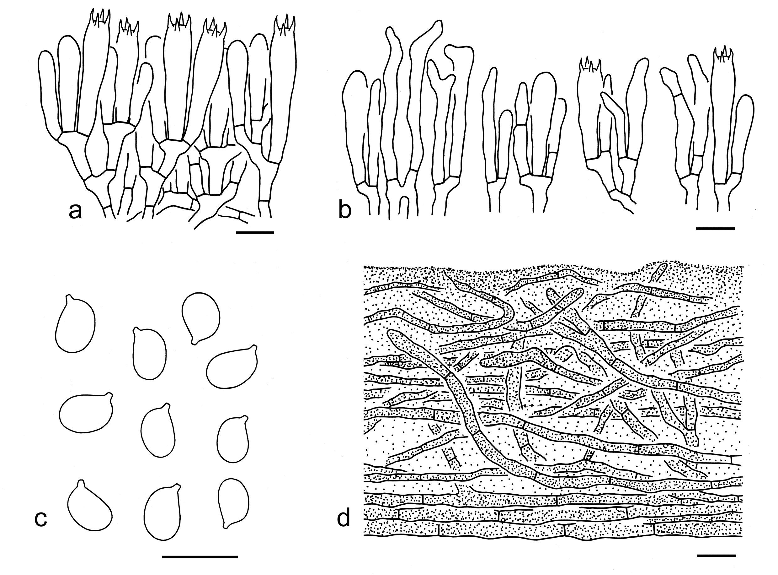
Basionym: Agaricus imbricatus Fr., Observ. mycol. (Havniae) 1: 27 (1815).
Description: Basidioma medium-sized to large. Pileus 4–12 cm in diam., at first convex, then plano-convex to applanate, sometimes with a broadly low umbo at center surface dry, dark brown (6F6–8), cinnamon (6E6–8) to orange-brown (6D5–7), covered with dark brown (6F6–8) to orange-brown (6D5–7) fibrillose squamules; margin involute to deflexed; context white (1A1). Lamellae adnexed to sinuate, white, with brownish (6C2–4) to brown (6D2–4) stains, crowded to subclose; edges brownish (6C2–4) to brown (6D2–4). Stipe 6–12 1–2 cm, cylindrical, dirty white (6B1), brownish (5B2–4) to orangish brown (6C3–5), basal mycelium white (1A1), fibrillose, exannulate. Taste and odour not recorded.
Basidiospores [70/3/2] (4.5–) 5.5–7 (–8) (3.5–) 4–5 µm (mean length = 6.2 µm, mean width = 4.2 µm), Q = (1.11–) 1.2–1.75 (–1.9), Qm = 1.48 ± 0.15, ellipsoid to elongate, sometimes broadly ellipsoid, non-amyloid, colorless, hyaline, thin-walled, smooth; apiculus relatively large. Basidia 30–40 5–7 µm, clavate, 4-spored, hyaline; sterigmata 4–6 µm long; basal septa without clamps. Pleurocystidia absent. Cheilocystidia not observed. Subhymenium composed of 2–4 µm wide filamentous hyphae, clamps absent. Lamellar trama regular, composed of colorless, thin-walled, ellipsoid, cylindrical to filamentous hyphae 5–20 µm wide. Pileipellis a cutis, made of radially arranged, 5–10 µm wide, thin-walled, yellow-brown filamentous hyphae; subpellis poorly differentiated, composed of colorless filamentous hyphae 10–20 µm wide. Clamps absent in all parts of basidioma.
Ecology: Solitary to scattered on soil dominated in coniferous, also found in broad-leaved or mixed forests with Pinus and Quercus.
Known distribution: Known from Europe, North America, Japan, and Southwestern China.
Specimens examined: CHINA. Xizang Autonomous Region, Nyingchi, Mainling County, Qiangna Village, in a forest with Pinus, altitude 2950 m, 29 Jul. 2014, Qi Zhao 2120 (KUN-HKAS87886). Yunnan Province, Lijiang, Yulong Naxi Autonomous County, Lijiang Alpine Botanic Garden, in a forest dominated by Pinus, mixed with Quercus, altitude 2700 m, 29 Sep. 2019, Jian-Wei Liu 2170 (KUN-HKAS134975); same location, in a forest with Quercus, altitude 2700 m, 9 Oct. 2019, Jian-Wei Liu 2226 (KUN-HKAS112559); same location, in a mixed forest with Pinus and Quercus, altitude 2700 m, 29 Sep. 2019, Jian-Wei Liu 2171 (KUN-HKAS134976).
Notes: Tricholoma imbricatum is recognized by its cinnamon, squamulose pileus, ellipsoid to elongate basidiospores and associations dominated with coniferous trees (this study). Christensen & Heilmann-Clausen (2013) reported that this species has an indistinct or fruity odour and a farinaceous, mild to slightly bitter taste. However, for the Chinese collections of this species, taste and odour were not recorded in the field.
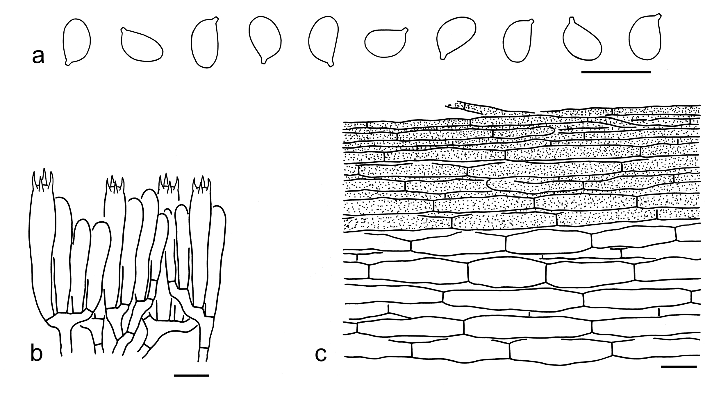
28. Tricholoma orienticolossum X. Xu et al., Phytotaxa 443 (2): 159 (2020). Figs. 42g, h, 47
Description: Basidioma large to very large. Pileus 13–18 cm in diam., convex to plano-convex, sometimes slightly depressed at center; surface dry, red-brown (6D6–8) to brown (6C5–7), covered with dark brown (6F6–8) to red-brown (6D6–8) squamules; margin strongly incurved; context white (1A1). Lamellae adnexed to sinuate, white (1A1) to cream (1A2), later becoming reddish brown (6C5–7), crowded; edges cream (1A2) to brown (6D6–8). Stipe 9–15 4.5–6.5 cm, robust, clavate, dirty white (1A1), covered with brownish (4A2–4) to red-brown (4C6–7), fibrillose squamules, basal mycelium white (1A1), exannulate. Taste and odour indistinct.
Basidiospores [40/2/2] (8–) 8.5–10 (–10.5) (6–) 6.5–7.5 (–8) µm (mean length = 9.1 µm, mean width = 6.8 µm), Q = (1.24–) 1.29–1.46 (–1.54), Qm = 1.35 ± 0.06, ellipsoid, sometimes broadly ellipsoid, non-amyloid, colorless, hyaline, thin-walled, smooth; apiculus relatively large. Basidia 60–70 9.5–11 µm, clavate, 4-spored, rarely 2-spored, hyaline; sterigmata up to 7 µm long; basal septa without clamps. Pleurocystidia absent. Cheilocystidia 50–120 4–8 µm, conspicuous, subcylindrical, often septate and slightly capitate, thin-walled, colorless or yellow to brownish, hyaline. Subhymenium composed of 3–5 µm wide filamentous hyphae, clamps absent. Lamellar trama regular, composed of colorless, thin-walled hyphae 3–10 µm wide. Pileipellis a cutis with transition to a trichoderm at regular intervals, composed of loosely and more or less radially arranged, 3–8 µm wide, thin-walled filamentous hyphae; subpellis poorly differentiated. Clamps absent in all parts of basidioma.
Ecology: Solitary to scattered on soil in subalpine temperate forests dominated by Abies.
Known distribution: Known from Southwestern China.
Specimens examined: CHINA. Sichuan Province, Daofu County, Bamei Town, in a forest dominated by Abies, altitude 3800 m, 4 Aug. 2016, Bang Feng 2051 (holotype, KUN-HKAS99341); same location, in a forest dominated by Abies, altitude 3800 m, 4 Aug. 2016, Jian-Wei Liu 466 (KUN-HKAS98645).
Notes: Tricholoma orienticolossum is well delimitated by large to very large, robust basidioma, a red-brown to brown pileus with an incurved margin, ellipsoid basidiospores and conspicuous cheilocystidia (this study). In the phylogenetic analysis based on the ITS sequences (Fig. 4), this species is sister to T. colossus (Fr.) Quél., but the latter species has a woolly-fibrillose ring that disappears as a zone with age, and relatively smaller and narrower basidiospores (6.8–9.4 5–6.4 µm, average 7.7–8.7 5.7–5.9 µm) (Riva 1988; Christensen & Heilmann-Clausen 2013). Tricholoma batschii is similar to T. orienticolossum, but has relatively smaller basidioma, a distinct ring when young and smaller basidiospores (3.8–6.4 3.1–5.4 µm, average 4.6–5.5 3.8–4.5 µm) (Riva 1988; Christensen & Heilmann-Clausen 2013).
29. Tricholoma orientifulvum X. Xu et al., Phytotaxa 443 (2): 161 (2020). Figs. 42i, j, 48
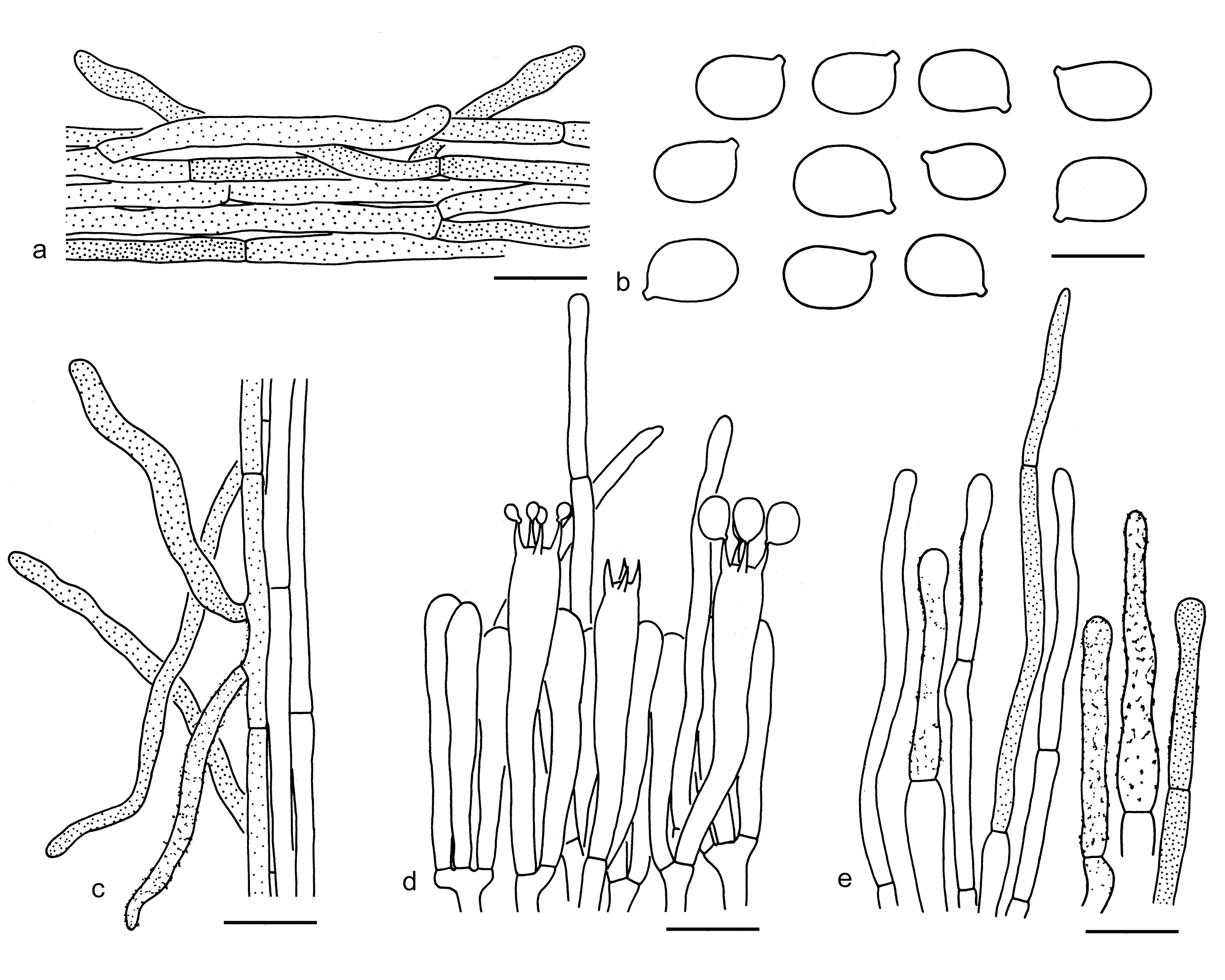
Basidiospores [40/2/2] (4.5–) 5–6 (–6.5) (3.5–) 4–4.5 (–5) µm (mean length = 5.3 µm, mean width = 4.2 µm), Q = 1.11–1.42 (–1.46), Qm = 1.26 ± 0.1, broadly ellipsoid to ellipsoid, non-amyloid, colorless, hyaline, thin-walled, smooth; apiculus relatively large. Basidia 18–30 5–7 µm, clavate, 4-spored, hyaline; sterigmata 3–5 µm long; basal septa without clamps. Pleurocystidia absent. Cheilocystidia not observed. Subhymenium composed of 2–5 µm wide filamentous hyphae, clamps absent. Pileipellis an ixo-cutis, composed of radially arranged to interwoven, 3–6 µm wide, thin-walled, brownish filamentous hyphae; subpellis poorly differentiated, non-gelatinized. Clamps absent in all parts of basidioma.
Ecology: Solitary to scattered on soil in subtropical mixed forests with Fagaceae and Pinaceae.
Known distribution: Known from Southwestern and Northeastern China.
Specimens examined: CHINA. Yunnan Province, Puer, Simao, in a subtropical mixed forest with Lithocarpus, Quercus and Pinus, altitude 1230 m, 31 Aug. 2019, Xiao-Xia Ding 632 (holotype, KUN-HKAS107157); same location, in a mixed forest with Lithocarpus, Quercus and Pinus, altitude 1230 m, 31 Aug. 2019, Xiao-Xia Ding 642 (KUN-HKAS107156). Liaoning Province, Dandong, Kuandian Manchu Autonomous County, in a temperate broad-leaved forest, 29 Jul. 2018, Xiao-Xia Ding 371 (KUN-HKAS105381).
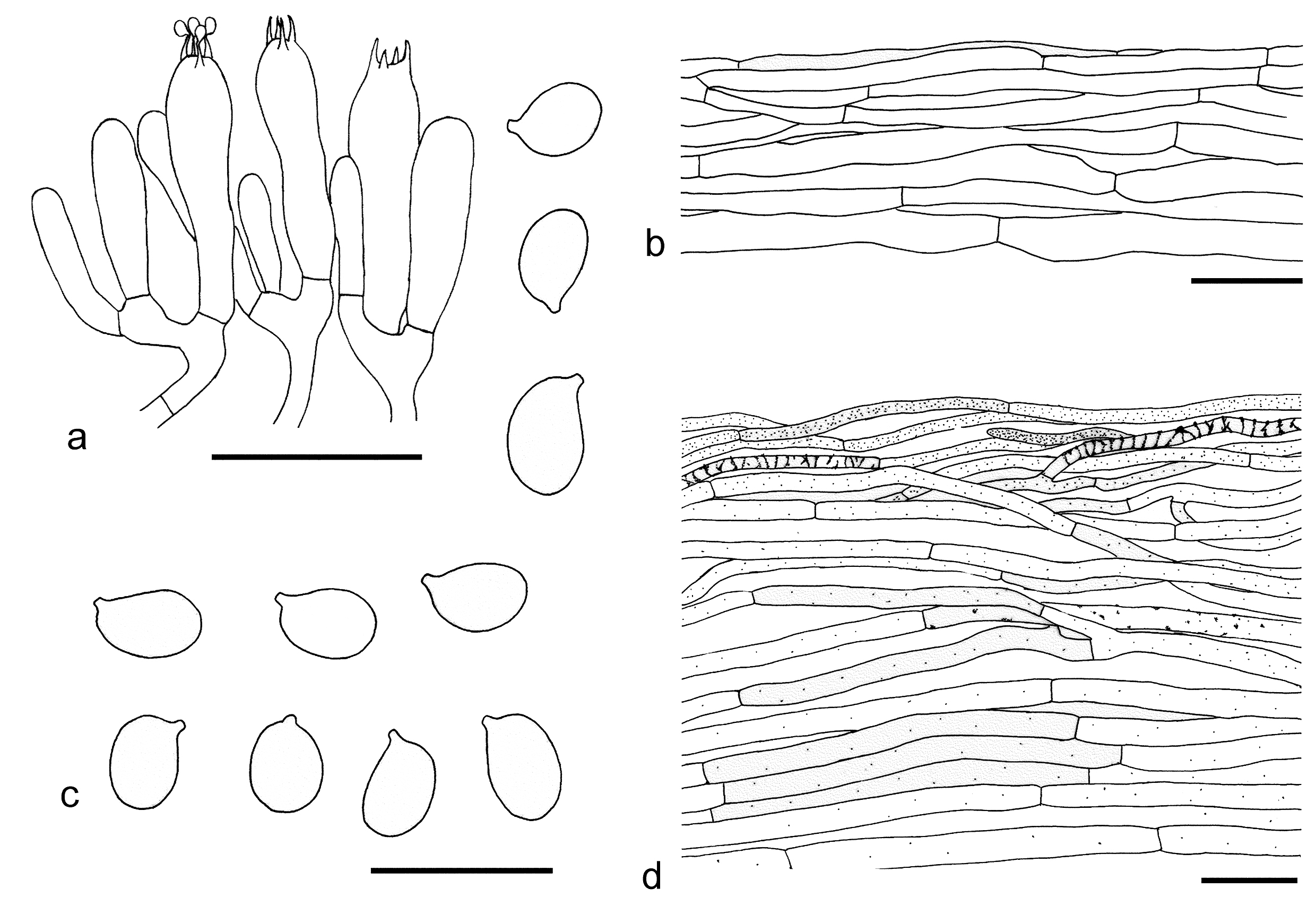
30. Tricholoma rubrocastaneum Y.Y. Cui & Zhu L. Yang, sp. nov. Figs. 42k, 49
Fungal Names: FN 572344
Etymology: rubrocastaneum referring to its red-brown pileus.
Diagnosis: Related to T. populinum, but differs by its red-brown to orange-brown pileus often with dark brown spots concentrated near margin and associations with Quercus, Pinus, Picea and Abies.
Type: CHINA. Yunnan Province, Lijiang, Yulong Naxi Autonomous County, Lijiang Alpine Botanic Garden, in a subalpine temperate forest dominated by Quercus semicarpifolia, mixed with Pinus, altitude 2700 m, 24 Aug. 2019, Jian-Wei Liu 1783 (holotype, KUN-HKAS134978).
Description: Basidioma medium-sized to large. Pileus 5–12 cm in diam., convex, plano-convex to applanate, center without umbo, often slightly depressed; surface dry or viscid when moist, red-brown (6B6–8) to orange-brown (5B6–8, 5C6–8), becoming paler towards margin, often with dark brown (5E6–8, 5F6–8) spots mainly concentrated near margin; margin involute, becoming deflexed when mature; context white (1A1) to dirty white (1A2, 1B2). Lamellae adnexed, white (1A1), dirty white (1B2) to cream (1A2), becoming brownish (5B3–5) to brown (5C5–7) when handed, damaged or old, very crowded; edges white (1A1), brownish (5B3–5) to brown (5C5–7). Stipe 4–7 1–2 cm, cylindrical to clavate, white (1A1), brownish (5B3–5) to brown (5C5–7), fibrillose, base white (1A1), solid, exannulate. Odour like cucumber. Taste indistinct.
Basidiospores [90/4/4] (4–) 4.5–6.5 (–7) 3–4.5 µm (mean length = 5.4 µm, mean width = 3.5 µm), Q = (1.2–) 1.32–1.81 (–2), Qm = 1.56 ± 0.15, ellipsoid to elongate, non-amyloid, colorless, hyaline, thin-walled, smooth; apiculus relatively large. Basidia 20–35 4–6 µm, clavate, 4-spored, hyaline; sterigmata 2–4 µm long; basal septa without clamps. Pleurocystidia absent. Cheilocystidia 15–30 3–5 µm, poorly differentiated, cylindrical to clavate, thin-walled, colorless, hyaline. Subhymenium composed of 2–4 µm wide filamentous hyphae, clamps absent. Lamellar trama regular, composed of colorless, thin-walled hyphae 2–7 µm wide. Pileipellis an ixocutis, strongly gelatinized, composed of irregularly to more or less radially arranged, 2–6 µm wide, thin-walled, yellow-brown filamentous hyphae; subpellis poorly differentiated, composed of radially arranged filamentous hyphae. Clamps absent in all parts of basidioma.
Ecology: Solitary to scattered on soil in subalpine temperate forests dominated by various trees, including Quercus, Pinus, Picea and Abies.
Known distribution: Known from Southwestern China.
Additional specimens examined: CHINA. Sichuan Province, Ganzi Tibetan Autonomous Prefecture, Daocheng County, Sangdui Village, in a forest with Quercus semicarpifolia, altitude 4000 m, 12 Sep. 2016, Xiao-Xia Ding 133 (KUN-HKAS97076); Ganzi Tibetan Autonomous Prefecture, Kangding, forest and altitude not recorded, 5 Sep. 2016, KD-LJW 34 (KUN-HKAS97827). Yunnan Province, Lijiang, Yulong Naxi Autonomous County, Lijiang Alpine Botanic Garden, in a forest dominated by Quercus semicarpifolia, mixed with Pinus, altitude 2700 m, 22 Sep. 2019, Jian-Wei Liu 2075 (KUN-HKAS134977); same location, in a forest dominated by Quercus semicarpifolia, altitude 2700 m, 30 Sep. 2019, Jian-Wei Liu 2186 (KUN-HKAS134979).
Tricholoma pessundatum can be confused, but differs by its slightly smaller basidiospores (4.2–4.8 2.8–3.1 µm) and distribution in Europe and North America (Christensen & Heilmann-Clausen 2013). Tricholoma stans, T. ustaloides and T. albobrunneum are similar to T. rubrocastaneum. However, the former three species have no dark brown spots near pileal margin (Riva 1988; Christensen & Heilmann-Clausen 2013; Landry et al. 2022). In addition, T. stans has a more or less ribbed pileal margin (Riva 1988; Christensen & Heilmann-Clausen 2013; Landry et al. 2022). Tricholoma ustaloides possess relatively larger basidiospores (6–7 4.5–5.5 µm, mean length = 6.3 µm, mean width = 5 µm) (this study). Tricholoma albobrunneum has slightly broader basidiospores with mean width ca. 4 µm and associated dominated with Pinus (Christensen & Heilmann-Clausen 2013; Landry et al. 2022; this study).
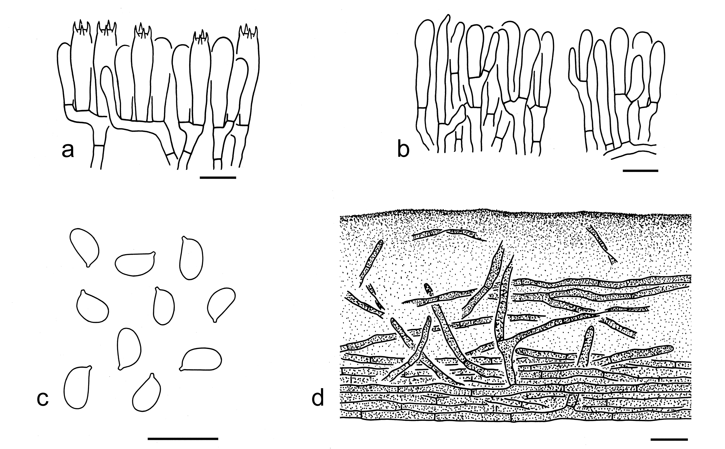
31. Tricholoma populinum J.E. Lange, Dansk bot. Ark. 8(no. 3): 14 (1933). Figs. 42l, 50
Description: Basidioma medium-sized to large. Pileus 4–12 cm in diam., convex to plano-convex, sometimes with a low, broad umbo; surface viscid when wet, shiny when dry, dark brown (6F6–8), brown (6D3–6, 5E6–8) to brownish (6C2–4, 5C3–5), often relatively paler towards margin, glabrous; margin involute when young, becoming inflexed to deflexed when mature; context white (1A1). Lamellae adnexed, dirty white (1B2), cream (2B2) to brownish (5B2–5), with brown (6C3–6) spots, crowded; edges dirty white (1B2) to brownish (5B2–5), with brown (6C3–6) areas. Stipe 3–10 1–4 cm, clavate, brownish (6C3–6), fibrillose, basal mycelium white (1A1), solid, exannulate. Taste not recorded, with a strong farinaceous odour.
Basidiospores [100/5/2] (4.5–) 5–7 (–7.5) (3–) 3.5–4 (–4.5) µm (mean length = 5.7 µm, mean width = 3.8 µm), Q = (1.11–) 1.3–1.67 (–1.88), Qm = 1.48 ± 0.14, ellipsoid to elongate, non-amyloid, colorless, hyaline, thin-walled, smooth; apiculus relatively large. Basidia 20–35 5–7 µm, clavate, 4-spored, hyaline; sterigmata 3–6 µm long; basal septa without clamps. Pleurocystidia absent. Cheilocystidia 10–30 3–7 µm, clavate, sometimes septate, thin-walled, yellow-brown, hyaline. Subhymenium composed of 2–4 µm wide filamentous hyphae, clamps absent. Lamellar trama regular, composed of colorless, thin-walled hyphae 2–5 µm wide. Pileipellis an ixo-cutis, strongly gelatinized, composed of radially arranged to interwoven, 2–6 µm wide, thin-walled, yellow-brown filamentous hyphae; subpellis poorly differentiated, composed of radially arranged, yellow-brown filamentous hyphae 3–8 µm wide.
Ecology: Solitary to scattered on soil in forests dominated with Populus.
Known distribution: Known from Europe (Grubisha et al. 2012; Christensen & Heilmann-Clausen 2013), North America (Grubisha et al. 2012; Bessette et al. 2013), Japan (Hongo 1988) and Southwestern China.
Specimens examined: CHINA. Sichuan Province, Ganzi Tibetan Autonomous Prefecture, Kangding, Waze Village, in a forest with Populus, altitude 3500 m, 18 Sep. 2017, Xiao-Xia Ding 315 (KUN-HKAS106656); same location, in a forest with Populus, altitude 3500 m, 18 Sep. 2017, Xiao-Xia Ding 316 (KUN-HKAS106657).
This species is closely related to T. fulvimarginatum Ovrebo & Halling in the tree generated from the ITS sequences (Fig. 4). However, T. fulvimarginatum, originally described from Massachusetts, USA, has a lighter reddish brown pileus and not found in China (Bessette et al. 2013; Landry et al. 2022). Tricholoma stans is similar to T. populinum, but has a somewhat ribbed pileal margin and is distributed in coniferous, broad-leaved or mixed forests (Riva 1988; Christensen & Heilmann-Clausen 2013; this study). Tricholoma albobrunneum is also similar to it, but differs by a white zone at the top of stipe and distributions in forests dominated by Pinus (Christensen & Heilmann-Clausen 2013; Landry et al. 2022). Tricholoma ustaloides is somewhat comparable, but can be separated by a white zone at the top of stipe, relatively larger basidiospores (6–7 4.5–5.5 µm, mean length = 6.3 µm, mean width = 5 µm) and associations with Fagaceae and Pinaceae (Riva 1988, 2003; Christensen & Heilmann-Clausen 2013; this study).
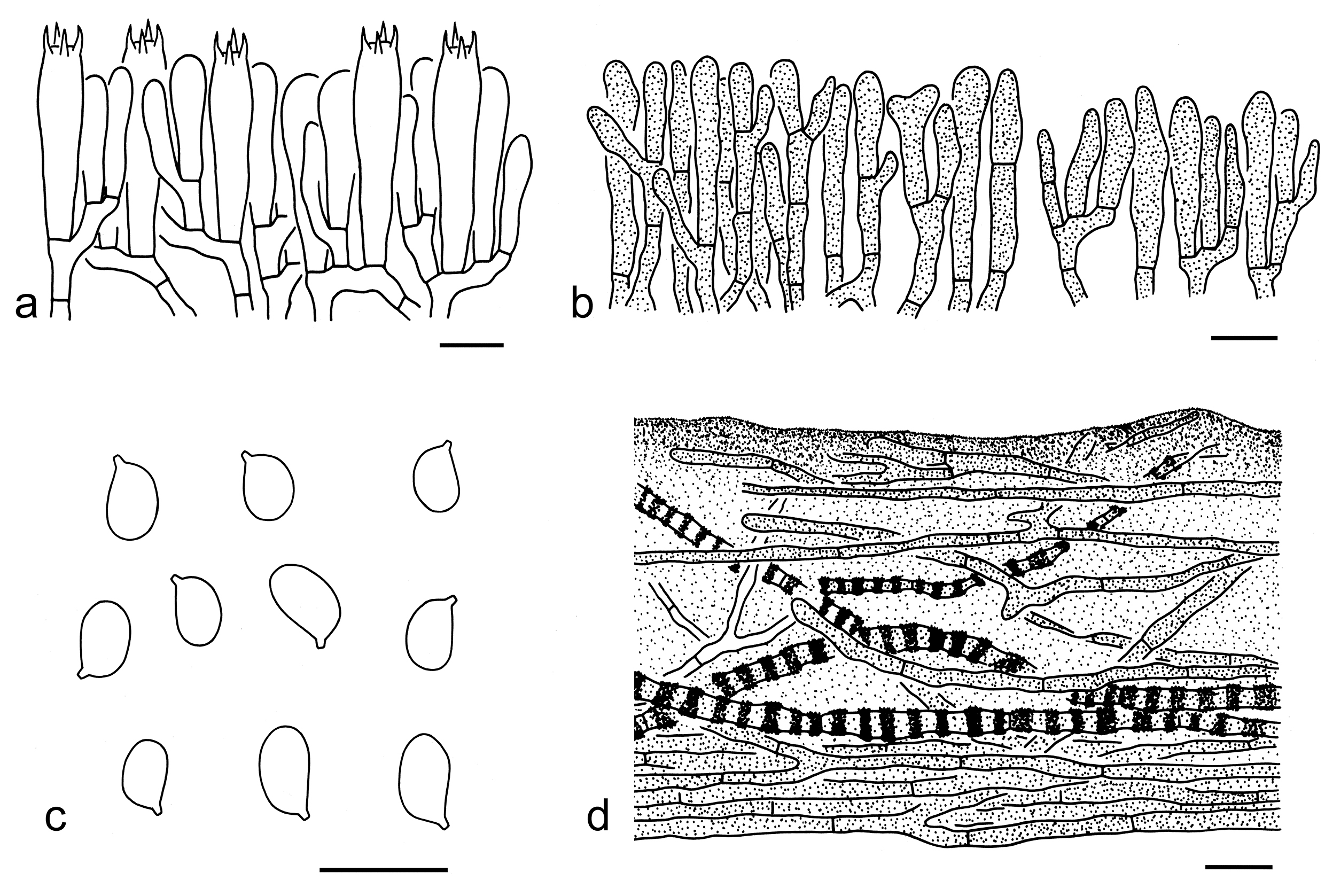
32. Tricholoma psammopus (Kalchbr.) Quél., Mém. Soc. Émul. Montbéliard, Sér. 2 5: 433 (1875). Figs. 42m, 51
Basionym: Agaricus psammopus Kalchbr., Icon. Sel. Hymenomyc. Hung. (Budapest) 1: tab. 3 (1873).
Description: Basidioma small to medium-sized. Pileus 2–7 cm in diam., convex to plano-convex, sometimes with a broad low umbo; surface dry, yellowish (3A2–4) to yellowish brown (3B3–5), covered with yellowish brown (3B3–5) to brown (4B4–6) fibrillose-felty to fibrillose squamules; margin involute to deflexed; context white (1A1) to cream (1A2). Lamellae adnate, cream (1A2–4) to brownish (5B3–5), often with brown (6A2–4) stains, subclose; edges brownish (5B3–5) to brown (6A2–4). Stipe 5–12 0.5–1 cm, subcylindrical to clavate, dirty white (1A1, 1B1) background covered with brownish (5B3–5) fibrillose squamules, often white (1A1) at apical part, stuffed to hollow, exannulate. Taste and odour not recorded.
Basidiospores [60/2/2] 5–6 (–6.5) 4–5 µm (mean length = 5.4 µm, mean width = 4.2 µm), Q = (1.04–) 1.16–1.37 (–1.5), Qm = 1.26 ± 0.09, broadly ellipsoid to ellipsoid, non-amyloid, colorless, hyaline, thin-walled, smooth; apiculus relatively large. Basidia 25–35 5–8 µm, clavate, 4-spored, hyaline; sterigmata 3–5 µm long; basal septa without clamps. Pleurocystidia absent. Cheilocystidia 20–30 3–6 µm, poorly differentiated, clavate to subclavate, thin-walled, colorless, hyaline. Subhymenium composed of 2–4 µm wide filamentous hyphae, clamps absent. Lamellar trama regular, composed of colorless, thin-walled, filamentous, cylindrical to subellipsoid hyphae 3–25 µm wide. Pileipellis a cutis, composed of radially to sometimes erected arranged, 3–8 µm wide, thin-walled, yellow-brown filamentous hyphae; subpellis made of radially arranged, colorless, filamentous to cylindrical hyphae 5–25 µm wide. Clamps absent in all parts of basidioma.
Distribution: Known from Europe, East Asia (Japan, and Northwestern and Northern China).
Specimens examined: CHINA. Gansu Province, Pingliang, in a coniferous forest, altitude 2160 m, 15 Jul. 2016, Xue-Tai Zhu 1797 (KUN-HKAS106314); same location, Xue-Tai Zhu 1536 (KUN-HKAS107579). Inner Mongolia, Hulunbuir, Genhe, altitude 840 m, 17 Aug. 2017, Gang Wu 2307 (KUN- HKAS106302).
Notes: Tricholoma psammopus is characterized by a yellowish to yellowish brown, fibrillose-felty pileus and broadly ellipsoid to ellipsoid basidiospores (this study). Christensen & Heilmann-Clausen (2013) reported that the taste of this species is initially mild, later becoming bitterish-astringent, but taste was not recorded in the Chinese collections.
Phylogenetically, it is closely related to the clade formed by T. imbricatum, T. vaccinum and T. rufobrunneum (Fig. 3). However, the latter three species can easily be separated by their dark brown to orange brown basidioma. In addition, T. imbricatum has longer basidiospores (5.5–7 4–5 µm, mean length = 6.2 µm, mean width = 4.2 µm) (this study). Tricholoma vaccinum possesses longer basidiospores (6–7.5 4.5–6 µm, mean length = 6.7 µm, mean width = 5 µm), conspicuous and gelatinized cheilocystidia, and a pileipellis with inflated cells (this study). Tricholoma rufobrunneum has larger basidiospores (6–7.5 5–6 µm, mean length = 6.7 µm, mean width = 5.2 µm) and a gelatinized pileipellis with cylindrical hyphae (this study).
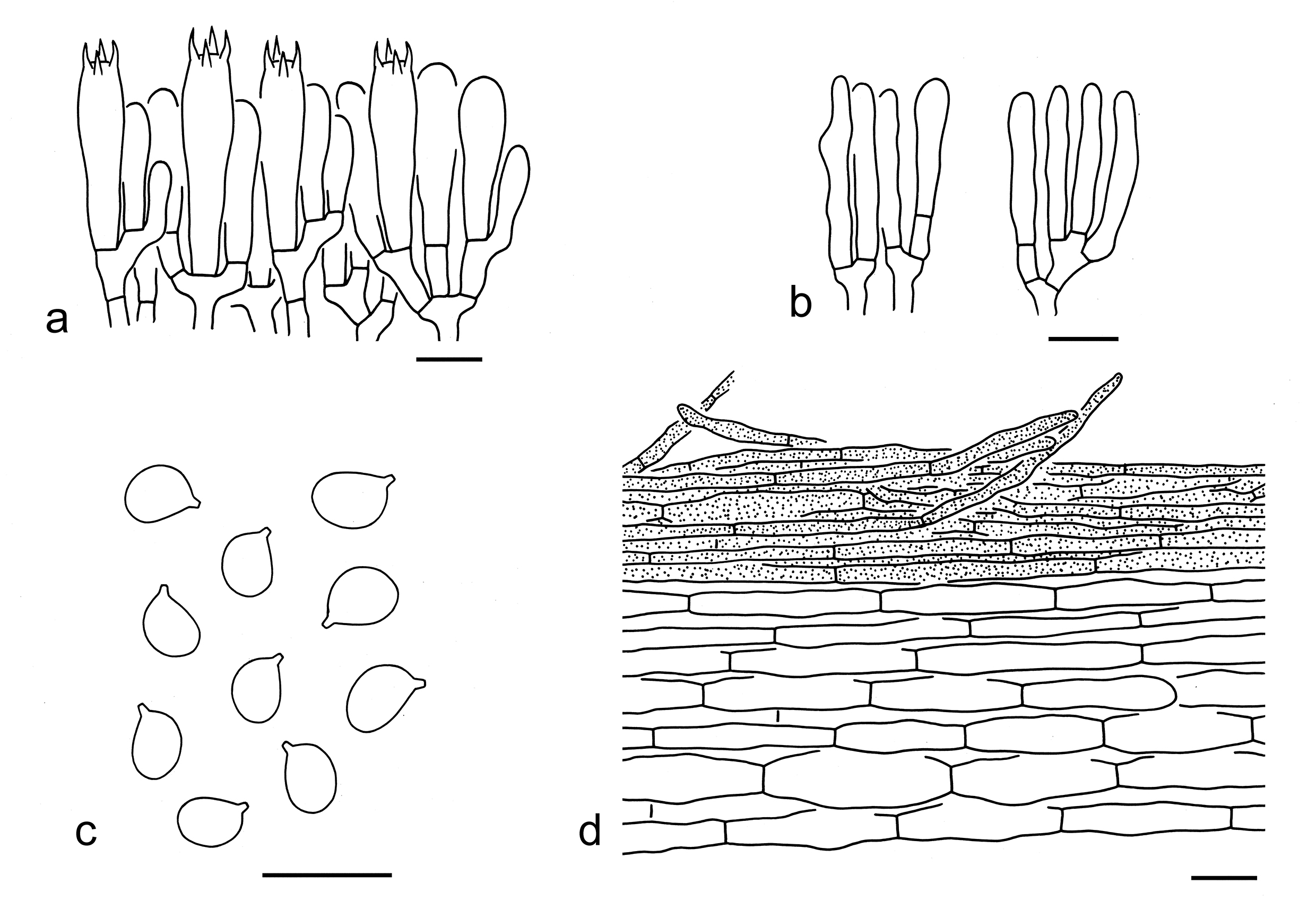
Morphologically, T. sinoacerbum, T. acerbum (Bull.) Quél. and T. apium Jul. Schäff. are similar to T. psammopus. However, T. sinoacerbum differs by its yellow-brown pileus without obvious squamules and associations dominated with broad-leaved trees (Hosen et al. 2016). Tricholoma acerbum has a much more robust and shorter stipe, more crowded lamellae and forms mycorrhizal relationships with Quercus, Castanea and other deciduous trees (Riva 1988; Christensen & Heilmann-Clausen 2013). Tricholoma apium possesses a much more robust and shorter stipe, relatively smaller basidiospores (3.4–5.6 2.9–4.6 µm, average 4.3–5 3.5–4 µm) and a strong smell of lovage (Riva 1988; Christensen & Heilmann-Clausen 2013).
33. Tricholoma roseoacerbum A. Riva, Mycol. helv. 1(3): 179 (1984). Figs. 42n, 52
Replaced synonym: Tricholoma pseudoimbricatum var. roseobrunneum A. Riva, Schweiz. Z. Pilzk. 57(1): 2 (1979).
Description: Basidioma small to medium-sized. Pileus 3–9 cm in diam., convex, without an umbo or a depression at center; surface viscid when wet, pinkish (7A2–4) to pinkish brown (6B3–6) at center, whitish (1A1, 5A2) towards margin, with white (1A1) fibrillose squamules; margin strongly involute; context white (1A1). Lamellae sinuate to emarginate, cream (2A2–4), crowded; edges cream (2A2–4). Stipe 4–6 1–2.5 cm, solid, clavate, white (1A1), with pinkish brown (6A2–4) hues, basal mycelium white (1A1), exannulate. Taste and odour not recorded.
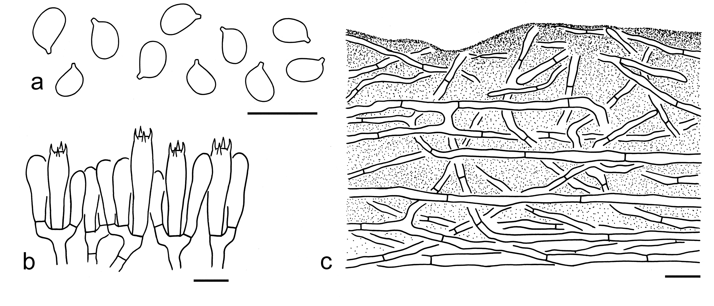
Basidiospores [20/1/1] (4–) 4.5–5.5 (–6) 3–4 µm (mean length = 5 µm, mean width = 3.6 µm), Q = 1.25–1.57, Qm = 1.4 ± 0.1, broadly ellipsoid to ellipsoid, non-amyloid, colorless, hyaline, thin-walled, smooth; apiculus relatively large. Basidia 20–30 5–7 µm, clavate, 4-spored, hyaline; sterigmata 3–5 µm long; basal septa without clamps. Pleurocystidia absent. Cheilocystidia not observed. Subhymenium composed of 2–4 µm wide filamentous hyphae, clamps absent. Lamellar trama regular, composed of colorless, thin-walled, cylindrical, clavate to filamentous hyphae 2–10 µm wide. Pileipellis gelatinized, composed of loosely arranged, interwoven to more or less erectly arranged, 2–5 µm wide, thin-walled, colorless filamentous hyphae; subpellis non-gelatinized, made of radially arranged, colorless filamentous hyphae 5–7 µm wide. Clamps absent in all parts of basidioma.
Ecology: Solitary to scattered on soil in mixed forests with Pinus, Quercus and Betula.
Known distribution: Known from Europe (Christensen & Heilman-Clausen 2013), North America (Bessette et al. 2013), East Asia (Japan and Southwestern China).
Specimen examined: CHINA. Yunnan Province, Diqing Tibetan Autonomous Prefecture, Shangri-La, Namucuo, in a forest with Pinus, Quercus and Betula, altitude 3400 m, 25 Aug. 2014, Qi Zhao 2279 (KUN-HKAS88046).
Notes: Tricholoma roseoacerbum is well recognized by a pinkish to pinkish brown pileus becoming whitish to strongly involute towards margin, broadly ellipsoid to ellipsoid basidiospores and associations with coniferous and broad-leaved trees (this study). Christensen & Heilmann-Clausen (2013) recorded this species as having a weak odour and a farinaceous to slightly bitterish taste, but these characters were not recorded in the field for the Chinese collections.
This species is sister to T. manzanitae T.J. Baroni & Ovrebo in the phylogenetic analysis based on the ITS sequences (Fig. 4). However, T. manzanitae has relatively larger basidiospores (5–7 4–5.5 µm) (Bessette et al. 2013). Tricholoma acerbum is similar to T. roseoacerbum, but differs by its relatively paler, often cream to buff pileus and associations with deciduous trees (Riva 1988; Christensen & Heilmann-Clausen 2013).
34. Tricholoma rubiginosum Y.Y. Cui & Zhu L. Yang, sp. nov. Figs. 42o, 53a, 54
Fungal Names: FN 572346
Etymology: rubiginosum = rust-colored, referring to its white lamellae with rust-colored stains when old.
Diagnosis: Related to T. fulvomaculatum, but can be separated by its white lamellae, subglobose to broadly ellipsoid basidiospores with Q = 1.08–1.29 and a non-gelatinized pileipellis.
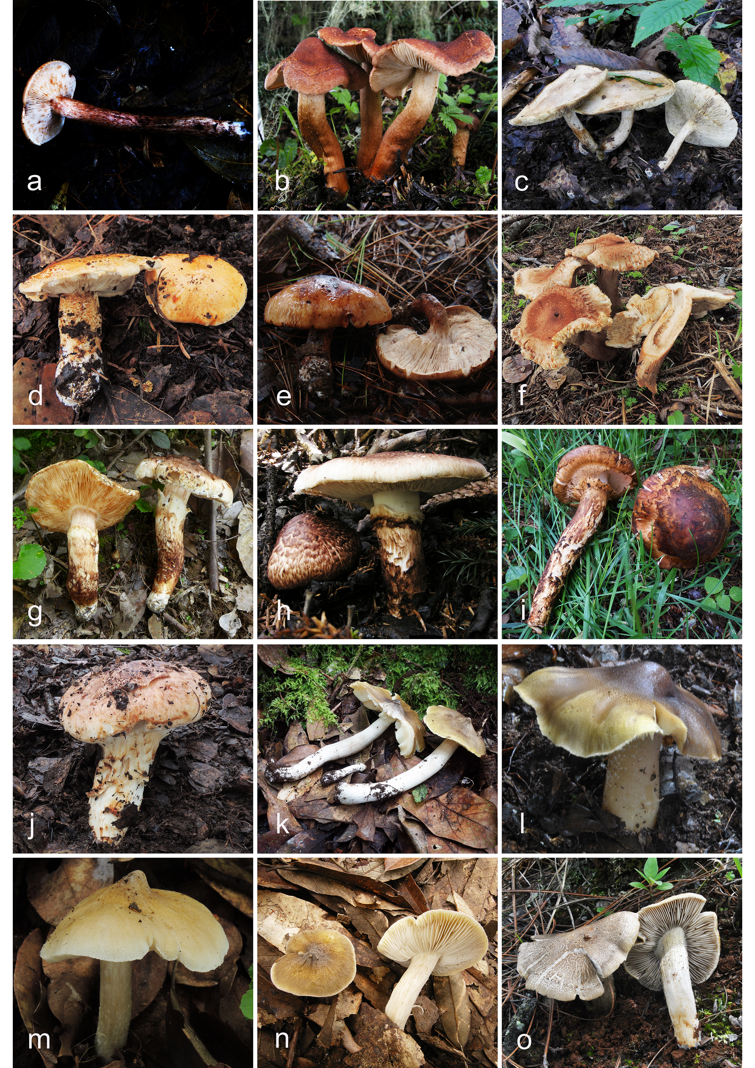
Type: CHINA. Yunnan Province, Baoshan, Tengchong, in a broad-leaved forest, altitude 2000 m, 25 Jul. 2022, GLG-FXP221 (holotype, KUN-HKAS134980).
Description: Basidioma medium-sized. Pileus 5–8 cm in diam., convex to plano-convex, without obviously umbo or depression at center; surface viscid when wet, orange-brown (6D6–8) to brown (6B3–5), sometimes dark brown (6E6–8) at center when old, glabrous or covered with orange-brown (6D6–8), fibrillose squamules; margin involute; context dirty white (1B1). Lamellae adnate, white (1A1), with brown (6C5–7) stains or spots when old, crowded; edges white (1A1), with brown (6C5–7) stains. Stipe 8–12 1–2 cm, slender, cylindrical, white (1A1) background with dark brown (6E6–8), orange-brown (6D6–8) to brown (6B3–5), fibrillose squamules, basal mycelium white (1A1), exannulate. Taste and odour not recorded.
Ecology: Solitary to scattered on soil in subtropical broad-leaved forests.
Known distribution: Known from Southwestern China.
Additional specimen examined: CHINA. Yunnan Province, Dali Bai Autonomous Prefecture, Dali, in a broad-leaved forest, altitude 2300 m, 1 Sep. 2017, Gang Wu 2467 (KUN-HKAS106303).
Notes: Tricholoma rubiginosum is characterized by its orange-brown pileus, white lamellae with brown spots when old, subglobose to broadly ellipsoid basidiospores (5.5–7 4.5–6.5 µm) and associations with broad-leaved trees. Tricholoma fulvomaculatum is related to this species in our four-gene phylogenetic analysis (Fig. 3), but has yellowish lamellae, broadly ellipsoid to ellipsoid basidiospores with Q = 1.2–1.55 and a gelatinized pileipellis (this study).
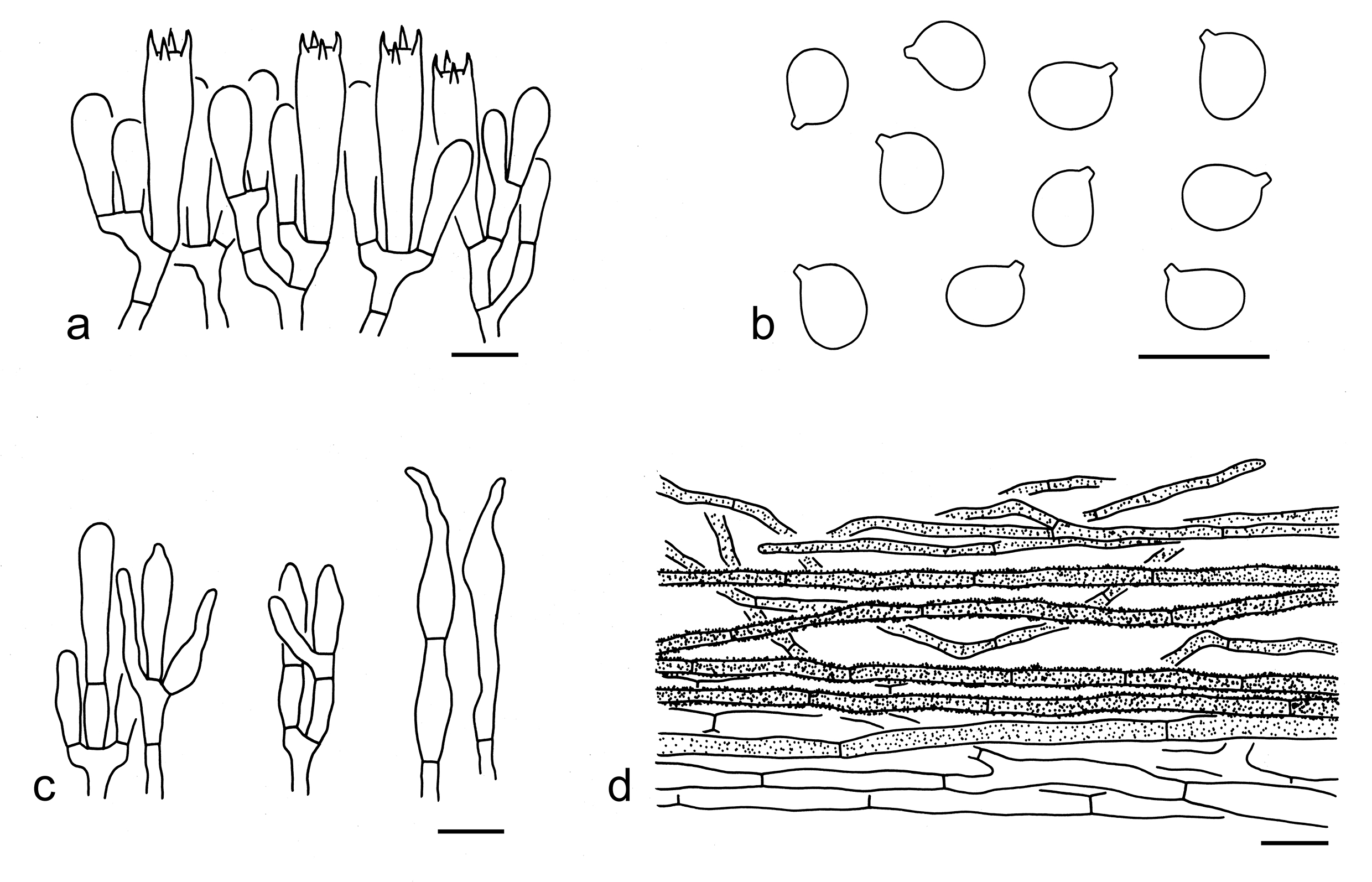
Tricholoma castaneiceps can be confused with T. rubiginosum, but differs by its pale sulphur-yellow lamellae, relatively narrower and ellipsoid basidiospores (5–7 4–5 µm), and a gelatinized pileipellis. Tricholoma orientifulvum and T. fulvum are similar to T. rubiginosum. However, T. orientifulvum can be separated by its olivaceous yellow to yellow-brown lamellae, narrower basidiospores with mean width ca. 4.2 µm and a gelatinized basidiospores. Tricholoma fulvum has pale yellow lamellae, broadly ellipsoid to ellipsoid basidiospores (4.7–7.4 3.6–6.1 µm), a gelatinized pileipellis, and associations with Betula, Abies and Picea (Christensen & Heilmann-Clausen 2013; Landry et al. 2022). Tricholoma ustale (Fr.) P. Kumm. and T. ustaloides are also similar to T. rubiginosum. However, T. ustale has cream to warm buff lamellae, broadly ellipsoid to ellipsoid basidiospores (5.3–7.7 4.4–6.4 µm), and a gelatinized pileipellis (Riva 1988, 2003; Christensen & Heilmann-Clausen 2013). Tricholoma ustaloides can be distinguished from T. rubiginosum by its broadly ellipsoid to ellipsoid basidiospores (6–7 4.5–5.5 µm) and a gelatinized pileipellis (Riva 1988, 2003; Christensen & Heilmann-Clausen 2013; this study).
35. Tricholoma rufobrunneum X.X. Ding et al., Mycol. Progr. 21(37): 8 (2022). Figs. 53b, 55
Description: Basidioma medium-sized to large. Pileus 5–12 cm in diam., convex to plano-convex, obtusely umbonate; surface dry or viscid when wet, dark brown (6E6–8) to orange-brown (6C5–8), densely covered with dark brown (6E6–8), fibrillose-tomentose squamules; margin involute; context dirty white (1B1). Lamellae sinuate, cream (1A2) to yellowish (2A2–4), with brown (7D5–7) stains when damaged or old, crowded to subclose; edges yellowish (2A2–4). Stipe 5–9 0.8–1.5 cm, cylindrical to subcylindrical, brownish (5B3–5) to brown (5C5–7), often yellowish (1A2) at apical part, covered with concolorous fibrillose squamules. Taste and odour not recorded.
Basidiospores [40/2/1] (5.5–) 6–7.5 (4.5–) 5–6 µm (mean length = 6.7 µm, mean width = 5.2 µm), Q = (1.09–) 1.17–1.4 (–1.5), Qm = 1.29 ± 0.1, broadly ellipsoid to ellipsoid, non-amyloid, colorless, hyaline, thin-walled, smooth; apiculus relatively large. Basidia 20–30 6–10 µm, clavate, 4-spored, sometimes 2-spored, colorless to brownish, hyaline; sterigmata 3–6 µm long; basal septa without clamps. Pleurocystidia absent. Cheilocystidia 10–90 3–10 µm, clavate, cylindrical to subcylindrical, sometimes septate, thin-walled, colorless or brownish, hyaline. Subhymenium composed of 2–5 µm wide filamentous hyphae, clamps absent. Lamellar trama regular, composed of colorless, thin-walled hyphae 2–10 µm wide. Pileipellis an ixo-cutis, slightly gelatinized, composed of radially arranged, interwoven to more or less erectly arranged, thin-walled, brownish to brown cylindrical hyphae (30–100 10–25 µm); subpellis poorly differentiated, non-gelatinized, composed of radially arranged, colorless or brownish hyphae 10–20 µm wide. Clamps absent in all parts of basidioma.
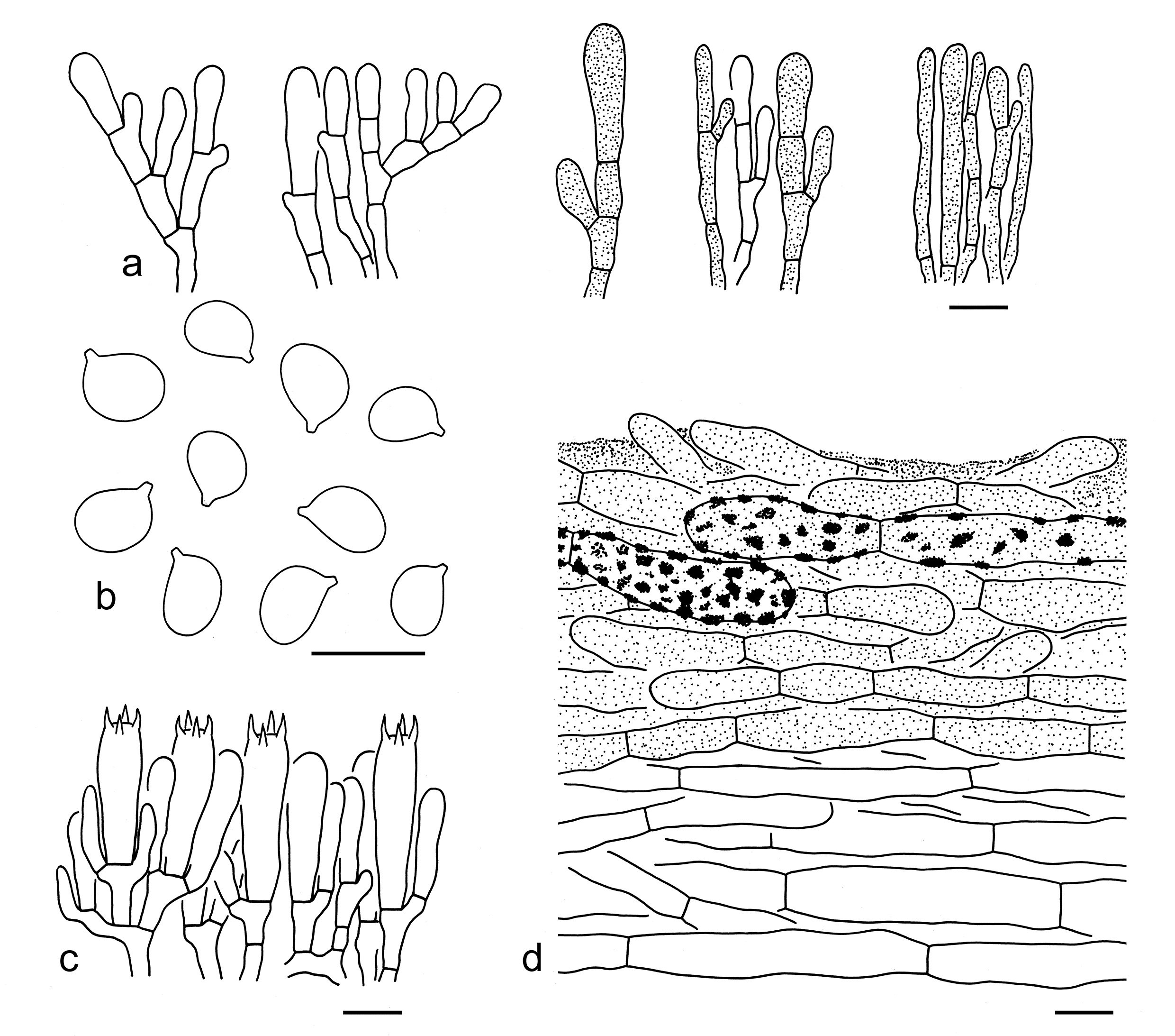
Ecology: Solitary to scattered on soil in subalpine temperate forests with Quercus, Abies, Picea and Salix.
Known distribution: Known from Southwestern China.
Specimens examined: CHINA. Sichuan Province, Ganzi Tibetan Autonomous Prefecture, Jiulong County, Tanggu Village, in a mixed forest with Quercus, Abies, Picea, Salix, adjacent meadow and boggy marsh, altitude 3600 m, 19 Jul. 2005, Zai-Wei Ge 574 (KUN-HKAS49069). Yunnan Province, Diqing Tibetan Autonomous Prefecture, Shangri-La, Tian Lake, in a forest dominated by Abies, altitude 3700 m, 9 Jul. 2014, Jian-Wei Liu 14 (holotype, KUN-HKAS90808).
Notes: Tricholoma rufobrunneum is characterized by an orange-brown, fibrillose-tomentose pileus, cream to yellowish lamellae when young, broadly ellipsoid to ellipsoid basidiospores and a gelatinized pileipellis with cylindrical hyphae (Ding et al. 2022; this study). It is closely related to T. imbricatum and T. vaccinum (Fig. 3). However, T. imbricatum differs by its ellipsoid to elongate basidiospores with Q = 1.2–1.75, and a non-gelatinized pileipellis composed of filamentous hyphae (this study). Tricholoma vaccinum has gelatinized cheilocystidia and a non-gelatinized pileipellis (this study). Tricholoma fulvum and T. fulvomaculatum share cream to yellowish lamellae and can be confused, but these two species have indistinct squamules on pileus and gelatinized pileipellis with filamentous hyphae (Christensen & Heilmann-Clausen 2013; Ding et al. 2022; Landry et al. 2022). Moreover, T. fulvum has a somewhat ribbed pileal margin when mature (Christensen & Heilmann-Clausen 2013; Landry et al. 2022). Tricholoma imbricatum is similar to T. rufobrunneum. For comparison, see notes under that name.
36. Tricholoma sinoacerbum T.H. Li et al., Mycoscience 57 (4): 234 (2016). Figs. 53c, 56
Description: Basidioma small, medium-sized to large. Pileus 3–15 cm in diam., at first convex to plano-convex, then applanate to plano-concave, slightly umbonate at center; surface dry or viscid when wet, yellow-brown (3A2–4), brown (5C4–6) to brownish (5B4–6); margin involute to deflexed when young, becoming straight to reflexed when mature; context white (1A1) to cream (1A2). Lamellae sinuate to subdecurrent, dirty white (1B1) to cream (1A2), with brownish (5C4–6) stains when damaged or old, crowded; edges cream (1A2) to brownish (5C4–6). Stipe 3–11 0.7–3 cm, clavate to cylindrical, dirty white (1A1, 2A2, 2B2), covered with brownish (3B3–5) to brown (3C3–5) fibrillose squamules, basal mycelium white (1A1), solid, exannulate. Odour fragrant. Taste bitter.
Basidiospores [80/3/3] (4–) 4.5–5.5 (–6) 3–4 µm (mean length = 5 µm, mean width = 3.7 µm), Q = 1.18–1.5 (–1.67), Qm = 1.35 ± 0.11, broadly ellipsoid to ellipsoid, non-amyloid, colorless, hyaline, thin-walled, smooth; apiculus relatively large. Basidia 20–30 5–7 µm, clavate, 4-spored, hyaline; sterigmata 3–5 µm long; basal septa without clamps. Pleurocystidia absent. Cheilocystidia not observed. Subhymenium composed of 2–4 µm wide filamentous hyphae, clamps absent. Lamellar trama regular, composed of colorless, thin-walled hyphae 4–10 µm wide. Pileipellis strongly gelatinized, composed of radially to more or less erectly arranged, 2–5 µm wide, colorless filamentous hyphae; subpellis not gelatinized, made of interwoven to radially arranged filamentous hyphae 3–7 µm wide. Clamps absent in all parts of basidioma.
Ecology: Solitary to scattered on soil, mainly in broad-leaved forests, occasionally in coniferous forests.
Known distribution: Known from Northeastern, Central, Southern and Southwestern China.
Specimens examined: CHINA. Anhui Province, Lu'an, Jinzhai County, in a mixed forest with Fagaceae and Pinaceae, altitude 940 m, Yan-Jia Hao 1686 (KUN-HKAS134981); Huangshan, forest type not recorded, altitude 1625 m, 12 Sep. 2018, Ting Guo 1401 (KUN-HKAS136950). Hubei Province, Yichang, Shennongjia Forest District, Muyu Town, in a forest with Quercus, Acer and Betula, altitude 1500 m, Qi Zhao 1454 (KUN-HKAS78713); same district, Hongping Town, in a forest with Quercus, altitude 1850 m, 15 Jul. 2012, Qi Zhao 1547 (KUN-HKAS78806). Liaoning Province, Dandong, Kuandian Man Autonomous County, Sipingjie Village, in a f broad-leaved forest, altitude 600 m, 29 Aug. 2018, Xiao-Xia Ding 378 (HKAS105388); same location, in a broad-leaved forest, altitude 600 m, 29 Aug. 2018, Xiao-Xia Ding 379 (KUN-HKAS105389). Yunnan Province, Baoshan, Longling County, Santai Village, in a broad-leaved forest, altitude 2000 m, 17 Jun. 2014, Xiao-Bin Liu 334 (KUN-HKAS86986); Kunming, Panlong District, Aziying Village, in a forest with Quercus, altitude 2000 m, 12 Jul. 2018, Xiao-Xia Ding 339 (KUN-HKAS105349); Lijiang, Yulong Naxi Autonomous County, Lijiang Alpine Botanic Garden, in a broad-leaved forest, altitude 2700 m, 22 Sep. 2019, Jian-Wei Liu 2089 (KUN-HKAS122161); Nujiang Lisu Autonomous Prefecture, Lushui, Luzhang Town, in a forest with Pinus yunnanensis and Keteleeria fortune, altitude 2000 m, 7 Aug. 2010, Qing Cai 307 (KUN-HKAS67869).
Notes: Tricholoma sinoacerbum is characterized by its yellow-brown pileus, dirty white and very crowed lamellae, bitter taste, broadly ellipsoid to ellipsoid basidiospores and associations with broad-leaved trees (Hosen et al. 2016; this study). In the five-gene phylogenetic analysis (Fig. 3), this species is sister to the clade formed by T. roseoacerbum and T. orienticolossum. However, T. roseoacerbum has a pinkish to pinkish brown pileus with a whitish, involute margin and is distributed in mixed forests (this study). Tricholoma orienticolossum can be easily separated from T. sinoacerbum by its large to very large basidioma with a red-brown pileus, larger basidiospores (8.5–10 6.5–7.5 µm) and a non-gelatinized pileipellis (this study).
Tricholoma psammopus is similar to T. sinoacerbum. For comparison, see notes under that name. The pileus color of T. sinoacerbum can be comparable to T. acerbum. However, T. acerbum has a relatively larger, robuster basidioma and an involute, ribbed pileal margin (Riva 1988; Christensen & Heilmann-Clausen 2013).
37. Tricholoma stans (Fr.) Sacc., Syll. fung. (Abellini) 5: 94 (1887). Figs. 53d, 57
Basionym: Agaricus pessundatus subsp. stans Fr., C. r. Congr. Soc. sav., Paris Dépts, Sec. Sci. 1(3): 25, tab. 28 (1869).
Description: Basidioma medium-sized to large. Pileus 4–14 cm in diam., at first convex, then plano-convex to applanate, without obvious umbo or depression at center; surface viscid, orange-brown (5B4–7), often relatively paler, sometimes ribbed towards margin, covered with orange-brown (5B4–7), fine squamules; margin involute to deflexed; context white (1A1). Lamellae adnate to sinuate, at first white (1A1) to cream (1A2), later brownish (5C5–7), crowded; edges
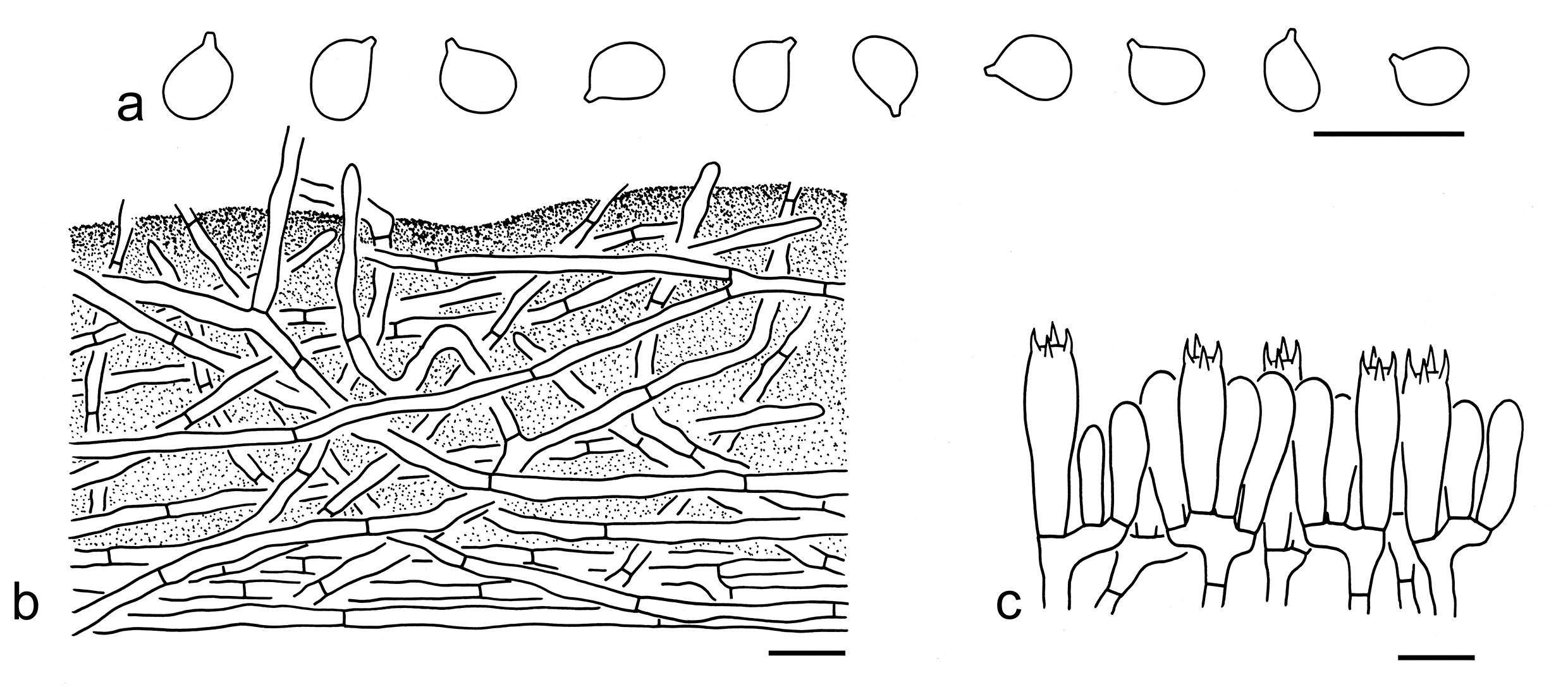
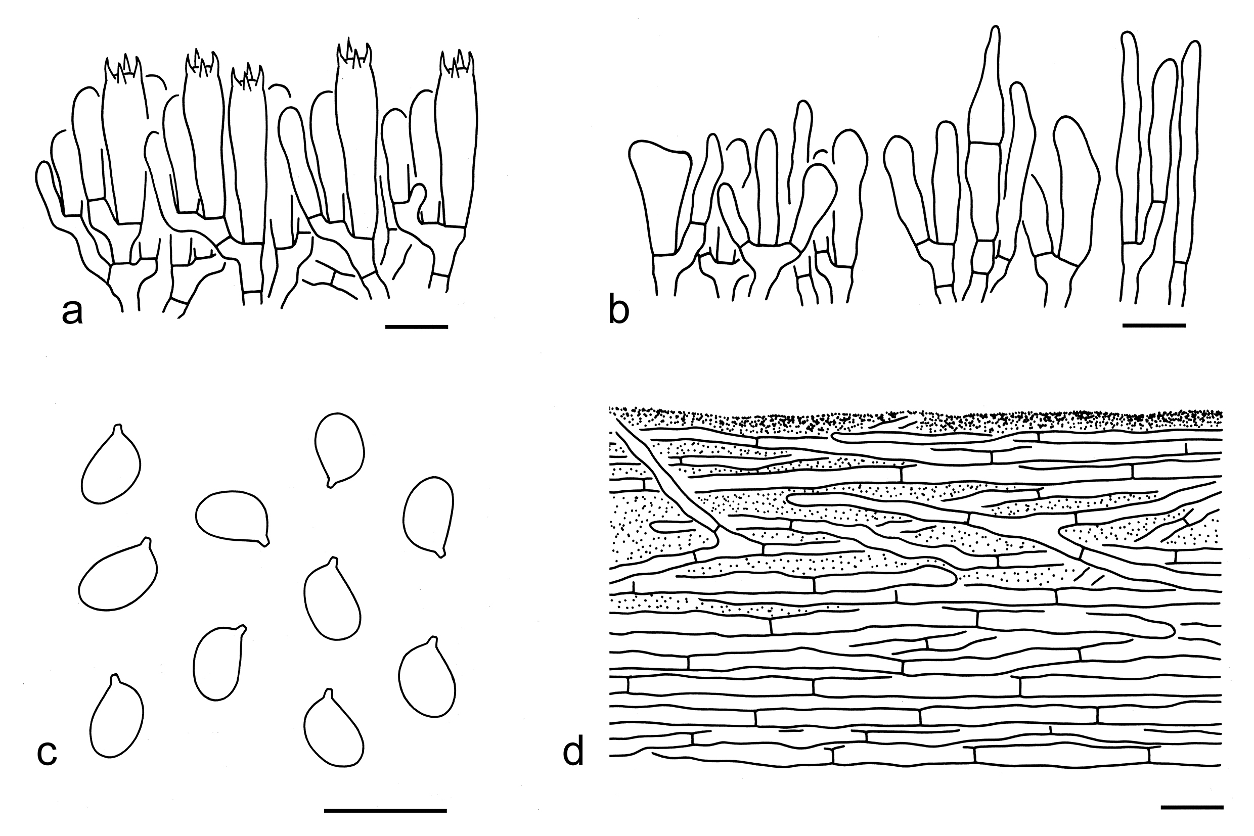
Basidiospores [92/5/5] 5–6.5 (–7.5) 3.5–4.5 µm (mean length = 5.7 µm, mean width = 4 µm), Q = 1.2–1.63 (–1.88), Qm = 1.42 ± 0.13, broadly ellipsoid to ellipsoid, non-amyloid, colorless, hyaline, thin-walled, smooth; apiculus relatively large. Basidia 20–35 5–8 µm, clavate, 4-spored, hyaline; sterigmata 3–5 µm long; basal septa without clamps. Pleurocystidia absent. Cheilocystidia 20–30 3–10 µm, poorly differentiated, clavate, cylindrical to filamentous or irregular-shaped, sometimes septate, thin-walled, colorless, hyaline. Subhymenium composed of 2–4 µm wide filamentous hyphae, clamps absent. Lamellar trama regular, composed of colorless, thin-walled hyphae 2–20 µm wide. Pileipellis an ixo-cutis with transition to an ixo-trichoderm at regular intervals, gelatinized, composed of 2–7 µm wide, thin-walled, colorless to brownish filamentous hyphae, often radially arranged, but becoming interwoven to more or less vertically arranged at regular intervals; subpellis poorly differentiated, composed of radially arranged, non-gelatinized, 4–10 µm wide filamentous hyphae. Clamps absent in all parts of basidioma.
Ecology: Solitary to scattered on soil in coniferous, broad-leaved or mixed forests.
Known distribution: Known from Europe and Southwestern China.
Specimens examined: CHINA. Sichuan Province, Ganzi Tibetan Autonomous Prefecture, Kangding, Gonggashan Village, in a subalpine temperate forest with Quercus, altitude 3550 m, 5 Sep. 2016, Bang Feng 29 (KUN-HKAS99382); same location, in a subalpine temperate forest with Quercus, altitude 3550 m, 5 Sep. 2016, KD-WPM 31 (KUN-HKAS98286). Yunnan Province, Diqing Tibetan Autonomous Prefecture, Shangri-La, Bitahai, in a subalpine temperate forest with Pinaceae and Fagaceae, altitude 3600 m, 20 Aug. 2014, Qi Zhao 2174 (KUN-HKAS87940).
Tricholoma mcneilii Lebeuf et al. is related to T. stans in the phylogenetic tree inferred from the ITS sequences (Fig. 4), but differs by its subglobose to broadly ellipsoid basidiospores (Trudell et al. 2022). Tricholoma populinum and T. rubrocastaneum may be confused with T. stans. For comparison, see notes under that name. Tricholoma ustaloides is similar to it, but has a non-ribbed pileus when mature and slightly larger basidiospores (6–7 4.5–5.5 µm, mean length = 6.3 µm, mean width = 5 µm) (Riva 1988, 2003; Christensen & Heilmann-Clausen 2013; this study).
38. Tricholoma ustaloides Romagn., Bull. Soc. nat. Oyonnax 8: 76 (1954). Figs. 53e, 58
Description: Basidioma medium-sized. Pileus 5–8 cm in diam., convex to plano-convex, without obvious umbo or depression at center; surface viscid when wet, orange-brown (6C4–8), darker at center; margin involute; context white (1A1). Lamellae adnexed to sinuate, dirty white (1A2), with brown (5F6–8) stains or spots when damaged or old, crowded; edges dirty white (1A1–2), with brown (5F6–8) stains. Stipe 4–8 1–2 cm, subcylindrical, orange-brown (6C4–8) to grey-brown (5F6–8), often white (1A1) to dirty white (1B1) at apical part, fibrillose, with a faint, poorly demarcated ring zone, basal mycelium white (1A1), stuffed. Taste and odour not recorded.
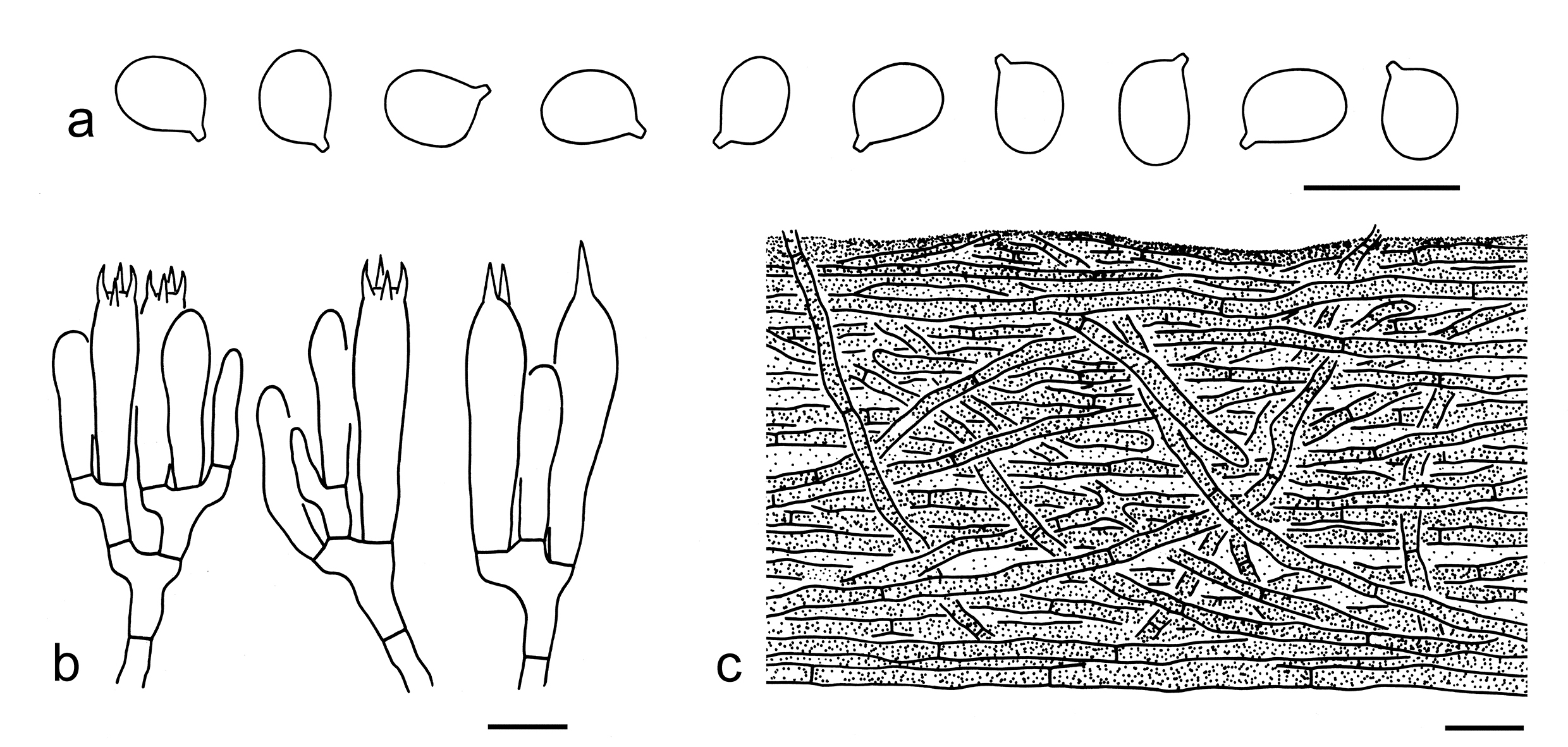
Basidiospores [40/2/1] (5.5–) 6–7 4.5–5.5 µm (mean length = 6.3 µm, mean width = 5 µm), Q = (1.09–) 1.15–1.4 (–1.46), Qm = 1.27 ± 0.09, broadly ellipsoid to ellipsoid, non-amyloid, colorless, hyaline, thin-walled, smooth; apiculus relatively large. Basidia 25–40 6–9 µm, clavate, 4-spored, sometimes 2- or 1-spored, hyaline; sterigmata 3–8 µm long; basal septa without clamps. Pleurocystidia absent. Cheilocystidia not observed. Subhymenium composed of 2–5 µm wide filamentous hyphae, clamps absent. Lamellar trama regular, composed of colorless, thin-walled, filamentous, cylindrical to clavate hyphae 3–30 µm wide. Pileipellis an ixo-cutis to an ixo-trichoderm, more or less gelatinized, composed of radially arranged, interwoven to more or less erected, 2–6 µm wide, thin-walled, brownish to brown filamentous hyphae; subpellis poorly differentiated, not gelatinized. Clamps absent in all parts of basidioma.
Ecology: Solitary to scattered on soil in coniferous or broad-leaved forests.
Known distribution: Known from Europe and China.
Notes: Tricholoma ustaloides is characterized by its orange-brown pileus, broadly ellipsoid to ellipsoid basidiospores and associations with Quercus in Europe and Pinus in southwestern China. According to Christensen & Heilmann-Clausen (2013), this species has a rather weak odour that becomes distinctly farinaceous after cutting, and a strongly farinaceous, bitter taste. For the Chinese collections of this species, however, these characters were not recorded in the field.
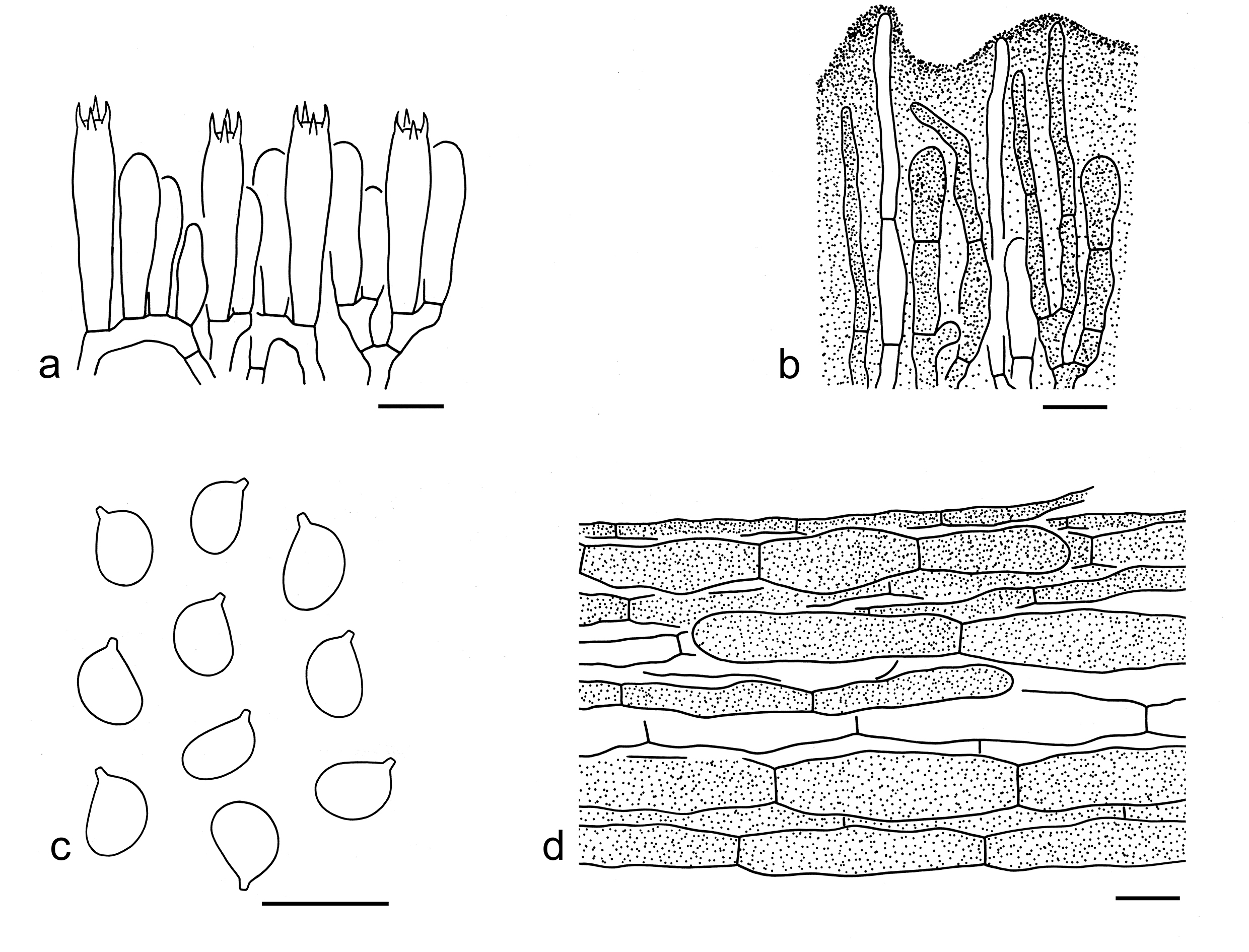
No closely related species were detected based on our current data (Figs. 3, 4). Tricholoma ustale could be confused with T. ustaloides, but differs by its stipe without a white zone at top and more viscid pileus (Riva 1988, 2003; Christensen & Heilmann-Clausen 2013). Tricholoma castaneiceps is similar to T. ustaloides, but has slightly smaller basidiospores (5–7 4–5 µm, mean length = 6 µm, mean width = 4.3 µm) and conspicuous cheilocystidia (this study). Tricholoma batschii is also similar to it, but can be separated by a distinct skin-like ring at stipe when young and slightly smaller basidiospores (3.8–6.4 3.1–5.4 µm, average 4.6–5.5 3.8–4.5 µm) (Riva 1988; Christensen & Heilmann-Clausen 2013). Tricholoma albobrunneum and T. populinum also can be comparable. For comparison, see notes under these names. Tricholoma ligusticum and T. quercetorum can be comparable to T. ustaloides, but the former two species have a stipe without any ring zone, and often in broad-leaved forests (Carbone 2020; Overall 2021).
39. Tricholoma vaccinum (Schaeff.) P. Kumm., Führ. Pilzk. (Zerbst): 133 (1871). Figs. 53f, 59
Basionym: Agaricus vaccinus Schaeff., Fung. bavar. palat. nasc. (Ratisbonae) 4: 13 (1774).
Description: Basidioma small to medium-sized. Pileus 3–10 cm in diam., convex to plano-convex, sometimes slightly umbonate at center; surface dry, dark brown (5E6–8) to orange-brown (6C5–7) at center, orangish brown (6B4–6) to brownish (4B3–5) towards margin, densely covered with concolorous squamules, often tomentose at center, fibrillose-tomentose to fibrillose-hairy at margin; margin involute, often deflexed when mature; context white (1A1), dirty white (1B1) to dirty cream (1B2). Lamellae sinuate to subdecurrent, white (1A1) at first, later cream (1A2) to brownish (5B3–6), with brown (5E6–8) stains or spots when injured or old, crowded; edges cream (1A2) to brownish (5B3–6). Stipe 3.5–12 0.7–2.5 cm, cylindrical to subcylindrical, white (1A1), dirty white (1B1), brownish (5B3–6) to orange-brown (6C5–7), often white (1A1) at apical part, covered with concolorous fibrillose squamules, basal mycelium white (1A1), hollow, exannulate. Taste not recorded. Odour fresh.
Basidiospores [80/4/4] (5.5–) 6–7.5 (–8) (4–) 4.5–6 µm (mean length = 6.7 µm, mean width = 5 µm), Q = 1.18–1.53 (–1.63), Qm = 1.35 ± 0.11, broadly ellipsoid to ellipsoid, non-amyloid, colorless, hyaline, thin-walled, smooth; apiculus relatively large. Basidia 30–40 6–8 µm, clavate, 4-spored, hyaline; sterigmata 3–5 µm long; basal septa without clamps. Pleurocystidia absent. Cheilocystidia 10–25 2–10 µm, conspicuous, often gelatinized, filamentous to clavate, often septate, thin-walled, colorless or brownish to brown, hyaline. Subhymenium composed of 2–5 µm wide filamentous hyphae, clamps absent. Lamellar trama regular, composed of colorless, thin-walled, ellipsoid, clavate, subglobose to cylindrical cells (25–90 10–35 µm) and filamentous hyphae (4–8 µm wide). Pileipellis a cutis, composed of radially arranged, slightly inflated hyphae (3–30 µm wide), which are often clavate to cylindrical, sometimes filamentous, thin-walled, yellow-brown, brownish to brown; subpellis poorly differentiated. Clamps absent in all parts of basidioma.
Ecology: Solitary to scattered on soil in forests dominated by Picea.
Known distribution: Known from Europe, North America, Japan and Southwestern China.
Specimens examined: CHINA. Sichuan Province, Ganzi Tibetan Autonomous Prefecture, Dege County, Yulonglacuo, in a forest with Picea, Salix, Lonicera and Sorbus, altitude 4000 m, 17 Aug. 2006, Zai-Wei Ge 1295 (KUN-HKAS50875); same location, in a forest with Picea, altitude 4000 m, 9 Aug. 2013, Kuan Zhao 339 (KUN-HKAS80764); same prefecture, Daofu County, Bamei Town, Kama Village, in a forest with Abies and Pinus, altitude 3812 m, 4 Aug. 2016, Jian-Wei Liu 458 (KUN-HKAS98037); same prefecture, Kangding, Tagong Town, in a forest with Picea, altitude 4021 m, Bang Feng 146 (KUN-HKAS99500). Yunnan Province, Diqing Tibetan Autonomous Prefecture, Shangri-La, Bitahai, in a forest dominated with Picea, altitude 3600 m, 20 Aug. 2014, Qi Zhao 2163 (KUN-HKAS87929).
Notes: Tricholoma vaccinum is well delimited by its orange brown, obviously squamulose pileus, broadly ellipsoid to ellipsoid basidiospores, conspicuous cheilocystidia and associations dominated with Picea (this study). This species is related to T. imbricatum (Fig. 3). For comparison, see notes under that name.
Tricholoma sect. Matsutake X.X. Ding et al.
Type: Tricholoma matsutake (S. Ito & S. Imai) Singer, Annls mycol. 41(1/3): 77 (1943).
Description: Basidioma tricholomatoid. Pileus umbonate or not, mostly brown, occasionally white, squamose. Lamellae crowded to close. Stipe covered with a cottony to fibrillose annulus on the apical part. Basidiospores medium-sized, broadly ellipsoid to ellipsoid, thin-walled, smooth, inamyloid. Pleurocystidia absent. Cheilocystidia present or absent. Pileipellis a cutis or an ixo-cutis. Clamps absent.
Ecology: Ectomycorrhizal. On soil with various trees.
Distribution: Molecular data indicated species from this section are distributed in Europe, North America and Asia.
Notes: Matsutakes and its allies are a group of the most widely known edible mycorrhizal mushrooms and highly prized in Japan and China (Yang et al. 2008; Vaario et al. 2017; Wang et al. 2017). Because high demands outstrip supplies in Japan, more than 1,000 tonnes of matsutake are imported from China annually (Yang et al. 2008; Vaario et al. 2017; Wang et al. 2017). Apart from the Japanese common name “Matsutake”, it is also called “songkoumo”, “songrong”, “songmaojun” and “qinggangjun” in China (Zang 1990; Wang et al. 2004; Wang et al. 2017). Traditionally, Matsutake growing under conifers are considered as the true, while these associated with broad-leaved trees are treated as false. According to the previous studies, Matsutake group harbours more than one species widely distributed in Asia, Europe, North America and North Africa and associated with both coniferous and broad-leaved trees (Zang 1990; Chapela & Garbelotto 2004; Ota et al. 2012; Bessette et al. 2013; Christensen & Heilmann-Clausen 2013; Trudell et al. 2017; Vaario et al. 2017). To date, seven species, including one new and six known species, are found in China (see below).
Key to the species of Tricholoma sect. Matsutake from China
1. Lamellae pale to creamy lavender T. lavendulophyllum
1. Lamellae white, cream to brownish 2
2. Pileus less coarse, squamules mainly concentrated in central part; basidioma currently only collected from northwestern China T. tianshanense
2. Pileus densely covered with appressed, brown, fibrillose squamules; basidioma is not restricted to northwestern China 3
3. Basidiospores relatively larger, 6.5–10 4.5–6.5 µm, with mean length > 7.2 µm 4
3. Basidiospores relatively smaller, 6–8 4–6.5 µm, with mean length ≤ 7.2 µm 5
4. Basidioma distributed in subalpine coniferous forest; basidiospores relatively smaller, 7–9 4.5–6 µm …………….T. xizangense
4. Basidioma only distributed in alpine broad-leaved forests; basidiospores relatively larger, 6.5–10 5–6.5 µm T. zangii
5. Basidiospores relatively larger, ellipsoid, with mean length > 7 µm; basidioma distributed in Europe, North America and Asia associated with both coniferous and broad-leaved trees T. matsutake
5. Basidiospores relatively smaller, subglobose, broadly ellipsoid to ellipsoid, with mean length < 7 µm; basidioma distributed in Asia associated with broad-leaved trees 6
6. Basidiospores subglobose to broadly ellipsoid shaped, 6–8 4.5–6.5 µm, Q = 1.08–1.33, Qm = 1.21 ± 0.09; cheilocystidia cylindrical, fusiform, subclavate to irregularly shaped, sometimes septate T. bakamatsutake
6. Basidiospores broadly ellipsoid to ellipsoid, 6–8 4–6 µm, Q = 1.15–1.5, Qm = 1.32 ± 0.09; cheilocystidia poorly differentiated T. fulvocastaneum
40. Tricholoma bakamatsutake Hongo, J. Jap. Bot. 49(10): 294 (1974). Figs. 53g, 60
Description: Basidioma medium-sized. Pileus 4–9 cm in diam., at first hemispherical to convex, then plano-convex to applanate, sometimes with a broadly umbo; surface dry, dark brown (4F6–8), red-brown (6E6–8), yellow-brown (4E5–8) to brown (3E5–8), sometimes brownish (3D6–8) to white (1A1) at margin, densely covered with appressed, dark brown (4F6–8) to brown (4D5–8, 4E5–8), fibrillose squamules; margin involute to inflexed; context white (1A1). Lamellae adnexed, at first white (1A1) to cream (1A2), then red-brownish (5A2–4) to red-brown (5C3–6), crowded; edges entire, fragile, white (1A1) when young, becoming brown (5E6–8) when mature. Stipe 8–14 1–2 cm, cylindrical, often tapering or sometimes slightly inflated downwards, solid, with a cream (1A2), brownish (4B2–5) to brown (4E6–8, 4F6–8), cottony to fibrillose annulus on the apical part, white (1A1) to brownish (4B2–4) above annulus, white (1A1) backgrounds densely covered with appressed, dark brown (4F6–8), yellow-brown (4E6–8) to reddish brown (5E6–8), fibrillose squamules below annulus. Taste slightly spicy and tongue-numbing. Odour strong, sweetish.
Ecology: Solitary to scattered on soil in broad-leaved forests with trees of Fagaceae.
Known distribution: Known from Japan (Hongo 1974) and China.
Specimens examined: CHINA. Gansu Province, Longnan, Wudu, the type of forest and altitude unknown, the year of 1993, Mao-Lin Tian (HMAS70231). Yunnan Province, Kunming, Guandu District, Mushuihua Market, the type of forest and altitude unknown, 12 Jul. 2019, Xiao-Xia Ding 482 (KUN-HKAS134983); same location and date, Xiao-Xia Ding 474 (KUN-HKAS134984); same location and date, Xiao-Xia Ding 477 (KUN-HKAS134985); same location and date, Xiao-Xia Ding 478 (KUN-HKAS134986); same location and date, Xiao-Xia Ding 481 (KUN-HKAS107573); same location and date, Xiao-Xia Ding 480 (KUN-HKAS134987); Wenshan Zhuang and Miao Autonomous Prefecture, Malipo County, in a broad-leaved forest, altitude 1100 m, 28 Jul. 2017, Gang Wu 2261 (KUN-HKAS106301).
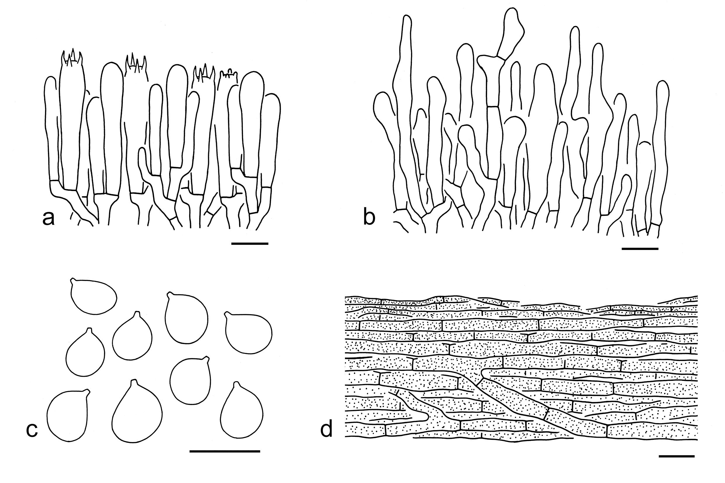
Notes: Tricholoma bakamatsutake was originally described from Japan in forests with Quercus (Hongo 1974), it also occurs in China and commonly called “jiasongrong”, “jiasongkoumo” and “lirong” in markets. This species is characterized by its dark brown, red-brown to brown pileus densely covered with concolor, appressed, fibrillose squamules, subglobose to broadly ellipsoid basidiospores (6–8 4.5–6.5 µm, Q = 1.08–1.33, Qm = 1.21 ± 0.09), cylindrical, subclavate to irregularly shaped, sometimes septate cheilocystidia, numbing and spicy tastes and strong odours.
In the phylogenetic analyses based on the ITS sequences (Fig. 4), T. bakamatsutake is closely related to the clade formed by T. dulciolens Kytöv., T. ilkkae Mort. Chr. et al. and T. tianshanense Zhao et al. However, these latter three relative species all grow with conifers (Christensen & Heilmann-Clausen 2013; this study). Tricholoma dulciolens can be easily distinguished from T. bakamatsutake by its pale brown basidioma, and predominantly broadly ellipsoid basidiospores (4.4–7.9 3.7–5.8 µm, Q = 1–1.6) (Kytövuori 1988; Christensen & Heilmann-Clausen 2013). Tricholoma ilkkae has a distinct ring at the margin of pileus and relatively smaller basidiospores (4.5–6.7 3.9–5.5 µm, average 5.1–6 4.4–4.9 µm) (Christensen & Heilmann-Clausen 2013; Heilmann-Clausen et al. 2017). Tricholoma tianshanense differs by its broadly ellipsoid to ellipsoid basidiospores and poorly differentiated cheilocystidia (this study).
Tricholoma fulvocastaneum Hongo occurs in broad-leaved forests and can be confused with T. bakamatsutake, but the former one has broadly ellipsoid to ellipsoid basidiospores (6–8 4–6 µm, Q = 1.15–1.5, Qm = 1.32 ± 0.09) and poorly differentiated cheilocystidia (Hongo 1960; this study). Tricholoma matsutake is similar to T. bakamatsutake, but differs by its strong odour, propotionally robuster stipe, ellipsoid basidiospores (6.5–8 5–6 µm, Q = 1.3–1.5, Qm = 1.39 ± 0.09) and associations with both coniferous and broad-leaved trees (this study).
41. Tricholoma xizangense Y.Y. Cui & Zhu L. Yang, sp. nov. Figs. 53h, 61
Fungal Names: FN 572338
Etymology: xizangense = Xizang Autonomous Region, named due to its type locality.
Diagnosis: Similar to T. matsutake, but differs in its relatively larger basidiospores and clavate cheilocystidia.
Type: CHINA. Xizang Autonomous Region, Qamdo, Baxoi County, Ranwu Town, in a subalpine temperate forest, altitude 3900 m, 4 Aug. 2014, Jian-Wei Liu 220 (holotype, KUN-HKAS91028).
Description: Basidioma medium-sized to large. Pileus ca. 10 cm in diam., at first hemispherical to convex, then plano-convex to applanate; surface dry, a white (1A1) background densely covered with appressed, dark brown (5F5–8) to brown (5E5–8), tomentose, floccose to fibrillose squamules, often becoming brownish (5D5–8) to cream (3B2) at margin; margin involute; context white (1A1). Lamellae adnexed to sinuate, white (1A1), crowded; edges entire, white (1A1). Stipe ca. 12 2 cm, cylindrical, solid, with a white (1A1) to brownish (5C3–6), cottony to fibrillose annulus on the apical part. white (1A1) above annulus, white (1A1) backgrounds densely covered with appressed, blackish (5F6–8), dark brown (5E5–8) to brown (4F6–8), fibrillose squamules below annulus. Taste bitter. Odour not recorded.
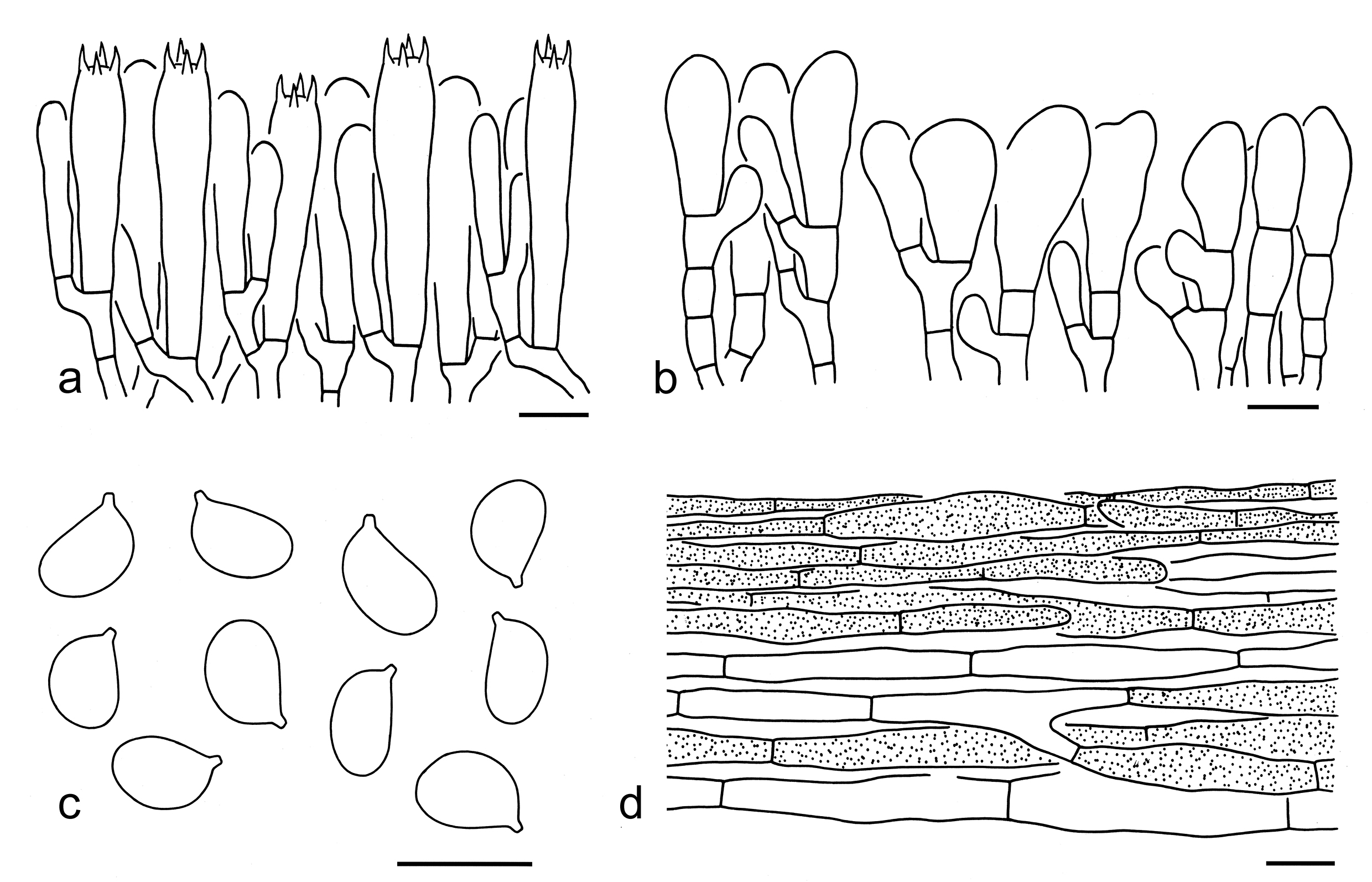
Basidiospores [30/1/1] (6–) 7–9 4.5–6 µm (mean length = 7.6 µm, mean width = 5.3 µm), Q = 1.17–1.69 (–1.78), Qm = 1.43 ± 0.15, broadly ellipsoid to ellipsoid, sometimes elongate, non-amyloid, colorless, hyaline, thin-walled, smooth; apiculus relatively large. Basidia 35–50 7–9 µm, clavate, 4-spored, hyaline; sterigmata 3–5 µm long; basal septa without clamps. Pleurocystidia absent. Cheilocystidia 15–35 8–15 µm, conspicuous, subclavate, clavate to irregularly shaped, sometimes septate, thin-walled, colorless, hyaline. Subhymenium composed of 2–5 µm wide filamentous hyphae, clamps absent. Pileipellis a cutis, composed of 3–20 µm wide, thin-walled, yellow-brown to brownish, filamentous to cylindrical hyphae; subpellis poorly differentiated. Clamps absent in all parts of basidioma.
Ecology: Solitary to scattered on soil in subalpine forests.
Known distribution: Southwestern China.
Additional specimen examined: CHINA. Xizang Autonomous Region, Qamdo, Baxoi County, in a subalpine temperate forest, altitude 3800 m, Jian-Wei Liu (KUN-HKAS152698).
Notes: Tricholoma xizangense is characterized by its dark brown to brown basidioma, broadly ellipsoid to ellipsoid basidiospores (7–9 4.5–6 µm, Q = 1.17–1.69, Qm = 1.43 ± 0.15) and distributed in subalpine forests (this study). In the tree based on the ITS sequences, it is related to two samples which labbled as T. caligatum in GenBank (Fig. 4).
Morphologically, T. matsutake is similar to T. xizangense, but differs by its relatively smaller basidiospores (6.5–8 5–6 µm) and filiform, cylindrical to narrowly cylindrical cheilocystidia (this study). Tricholoma caligatum also can be confused with it, but has relatively darker colored basidioma, relatively smaller basidiospores (average 6.5–7.1 5.3–5.6 µm) and is not distributed in China (Riva 1988, 2003; Christensen & Heilmann-Clausen 2013).
42. Tricholoma fulvocastaneum Hongo, J. Jap. Bot. 35: 86 (1960). Figs. 53i, 62
Basidiospores [80/4/4] (5.5–) 6–8 4–6 (–6.5) µm (mean length = 6.8 µm, mean width = 5.1 µm), Q = 1.15–1.5 (–1.56), Qm = 1.32 ± 0.09, broadly ellipsoid to ellipsoid, non-amyloid, colorless, hyaline, thin-walled, smooth; apiculus relatively large. Basidia 30–40 6–8 µm, clavate, 4-spored, hyaline; sterigmata 4–6 µm long; basal septa without clamps. Pleurocystidia absent. Cheilocystidia 15–40 3–7 µm, poorly differentiated, filiform, cylindrical, subclavate to clavate, sometimes septate, thin-walled, colorless, hyaline. Subhymenium composed of 2–4 µm wide filamentous hyphae, clamps absent. Pileipellis a cutis, often more or less lightly gelatinized, composed of 3–15 µm wide, thin-walled, brown, filamentous to cylindrical hyphae; subpellis poorly differentiated. Clamps absent in all parts of basidioma.
Ecology: Solitary to scattered on soil in broad-leaved forests with trees of Fagaceae.
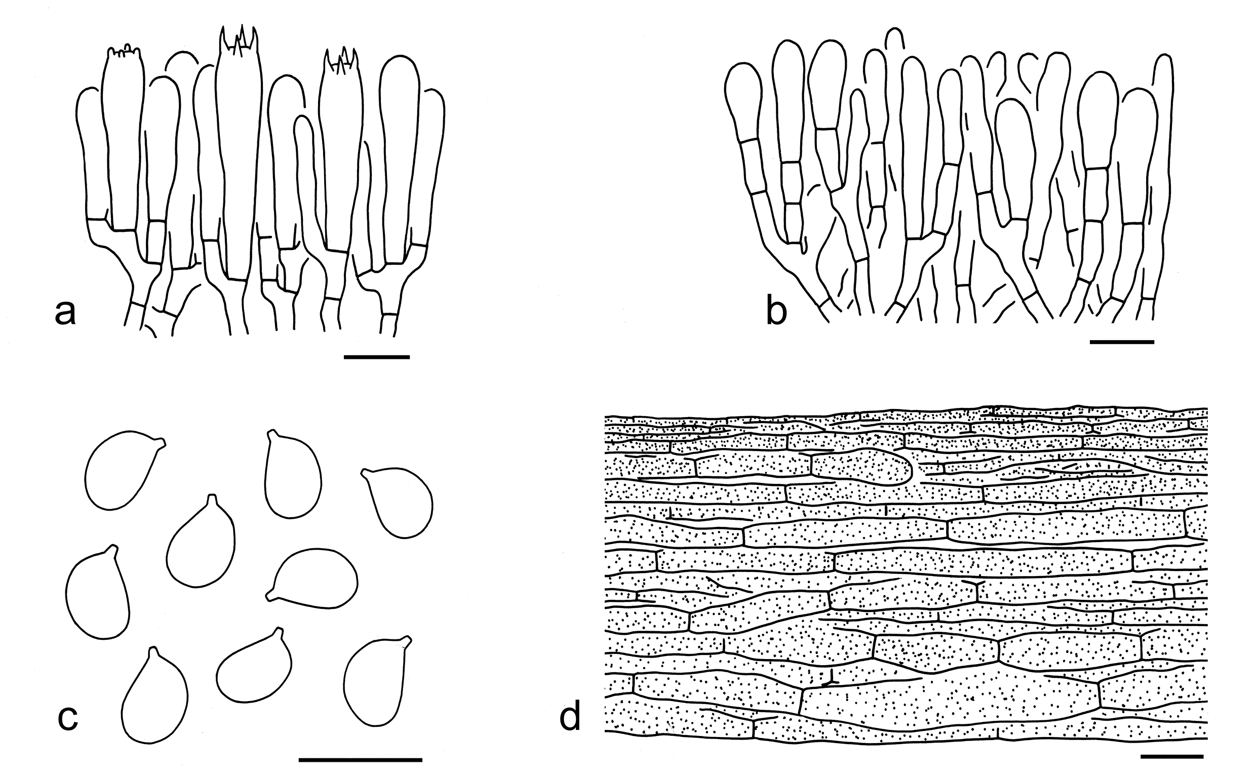
Known distribution: Known from Japan, Northern Thailand (Sanmee et al. 2007) and Southwestern China.
Specimens examined: CHINA. Yunnan Province, Kunming, Guandu District, bought from Mushuihua Market, 12 Jul. 2019, Xiao-Xia Ding 462 (KUN-HKAS134988); Xiao-Xia Ding 463 (KUN-HKAS134989); Xiao-Xia Ding 465 (KUN-HKAS107568); Xiao-Xia Ding 472 (KUN-HKAS134990); Xiao-Xia Ding 476 (KUN-HKAS107571).
Notes: Tricholoma fulvocastaneum was originally described from Japan (Hongo 1960) and also found in northern Thailand (Sanmee et al. 2007) and southwestern China (Zang 1990; Wang et al. 2004; Yu 2007). This species is characterized by its red-brown to brown pileus densely covered with appressed, concolor, fibrils squamules, broadly ellipsoid to ellipsoid basidiospores (6–8 4–6 µm, Q = 1.15–1.5, Qm = 1.32 ± 0.09), poorly differentiated cheilocystidia and broad-leaved associations.
Phylogenetically, T. fulvocastaneum is closely related and morphologically similar to the clade formed by T. matsutake, T. bakamatsutake and T. tianshanense (Fig. 3). However, T. matsutake has strong odour, propotionally robuster stipe, conspicuous cheilocystidia and is associated with both coniferous and broad-leaved trees. The comparisons between T. bakamatsutake and T. fulvocastaneum are provided under the notes of the former species. Tricholoma tianshanens is distributed in coniferous forests.
43. Tricholoma lavendulophyllum F.Q. Yu, Mycotaxon 95: 306 (2006)
Description: Pileus 4.5–7.5 cm in diam., hemispherical to convex when young, becoming subumbonate to plano-convex when mature; surface slightly viscid when wet, chestnut brown to fuscous in the center, with appressed dark brown zoned scales, often becoming broken up into rather indistinct pieces; margin brown to whitish brown, inrolled when young; context white, taste sweetish. Lamellae adnate, sinuate or arcuate, then separating from stem, pale to creamy lavender, with some lamellulae. Stipe 8–9.5 × 1.2–1.5 cm, equal or enlarged at the base, with a persistent but inconspicuous annulus on the upper part, 1.5–2.2 cm downward from the lamellae; concolorous with the pileus below the ring, with dark brown, appressed scales, whitish above the ring.
Pileipellis a layer of interwoven and thin-walled hyphae, 5–13 μm in diam., light yellowish brown, terminal elements subclavate to clavate, 7.5–9 × 22.5–67.5 μm. Lamellar trama subparallel, hyphae mostly 3–12 μm in diam., thin–walled, hyaline. Basidia 40–50 × 7.5–15 μm, clavate, hyaline, 4-spored, rarely 2- and 1-spored, sterigmata 2–5 μm long. Cystidia absent, but some clavate free hyphal ends present at the gill edge. Basidiospores (5.5–) 6–7.5 × (4.5–) 5–5.5 (–6) μm, Q= (1.18–) 1.25–1.33, broadly ellipsoid, thin-walled, hyaline, smooth, inamyloid. Stipitipellis with longitudinally arranged, appressed, parallel hyphae, 4–12 μm in diam., thin-walled, whitish brown to yellowish brown, terminal elements scattered, cylindrical, 6.8–9 × 30–54 μm, periclinal arranged.
Ecology: On soil in a broad-leaved forest (Yu et al. 2006).
Known distribution: Currently only known from Southwestern China (Yu et al. 2006).
Specimen examined: CHINA. Yunnan Province, Kunming City, Wujing Road, wild edible mushroom market, 23 Jul. 2005, F. Q. Yu 1273 (holotype, HKAS49804).
Notes: The description above and the citation of the holotype are from Yu et al. (2006). Tricholoma lavendulophyllum was described from Kunming, Yunnan, China, and is characterized by its lavender-colored lamellae (Yu et al. 2006). Although Yu et al. (2006) did not provide molecular evidence, Yu (2007) mentioned that this species possessed an ITS sequence and was labelled as ‘T19, T. lavendulophyllum, Yunnan’ in the MP tree analysis. Additionally, ‘T19, T. lavendulophyllum, Yunnan’ and ‘T. bakamatsutake’ form a monophyletic group with significant support, suggesting that T. lavendulophyllum may be a member of T. bakamatsutake group (Yu 2007). Unfortunately, the sequence was not submitted to a public database for additional confirmatory investigation.
Tricholoma lavendulophyllum was described based on two collections purchased from the wild edible mushroom markets in Kunming, namely F.Q. Yu1273 (HKAS49804, holotype) and F.Q. Yu1310B (HKAS49796). However, HKAS49796 was F.Q. Yu1310A, and was cited under the name T. bakamatsutake by Yu (2007). ITS phylogenetic analysis indicated that F.Q. Yu1310A (HKAS49796) is T. bakamatsutake (Yu 2007). Unfortunately, the type specimen for this species (F.Q. Yu1273, HKAS49804) and F.Q. Yu1310B were not traced.
44. Tricholoma matsutake (S. Ito & S. Imai) Singer, Annls mycol. 41(1/3): 77 (1943). Figs. 53j, 63
Basionym: Armillaria matsutake S. Ito & S. Imai, Bot. Mag. Tokyo: 327 (1925).
Description: Basidioma medium-sized, large to very large. Pileus 5–17 cm in diam., at first hemispherical to convex, becoming convex, plano-convex to applanate when mature, sometimes with a broadly low umbo; surface dry, clay buff (3D3–5), grey-brown (3F4–6, 4E3–6) to brown (4C5–8, 5D4–6), sometimes greyish brown (4E3–6) to brown (4C5–8, 5D4–6) at center, becoming paler towards margin, densely covered with appressed, dark greyish brown (3F4–6, 4E3–6), clay buff (3D3–5) to brown (4C5–8, 5D4–6), fibrillose squamules; margin involute when young, later inflexed to deflexed, often splitting to expose white flesh; context white (1A1). Lamellae adnexed to emarginate, dirty white (1B1), white (1A1) to cream (1A2), with brown (4D3–6) spots when old, crowded; edges entire, white (1A1) to cream (1A2). Stipe 6–13 1.5–3.5 cm, cylindrical, sometimes tapering downwards, robust and solid, covered with a large, white (1A1), brownish (3C2–4) to brown (4C5–8, 5D4–6), cottony annulus, white (1A1) with snake-shaped squamules above annulus, white (1A1) background with grey-brown (3F4–6, 4E3–6), clay buff (3D3–5) to brown (4C5–8, 5D4–6), fibrillose squamules below annulus. Taste mild. Odour strong, sweetish.
Basidiospores [40/1/1] (6–) 6.5–8 (4.5–) 5–6 µm (mean length = 7.2 µm, mean width = 5.2 µm), Q = (1.17–) 1.3–1.5 (–1.63), Qm = 1.39 ± 0.09, ellipsoid, sometimes broadly ellipsoid or elongate, non-amyloid, colorless, hyaline, thin-walled, smooth; apiculus relatively large. Basidia 35–50 7–10 µm, clavate, 4-spored, hyaline; sterigmata 4–6 µm long; basal septa without clamps. Pleurocystidia absent. Cheilocystidia 40–60 3–6 µm, conspicuous, filiform, cylindrical, narrowly cylindrical to subclavate, sometimes forked, thin-walled, colorless, hyaline. Subhymenium composed of 2–4 µm wide filamentous hyphae, clamps absent. Pileipellis a cutis to a more or less trichoderm, composed of 7–15 µm wide, thin-walled, yellow-brown to brown, cylindrical to filamentous hyphae; subpellis poorly differentiated. Clamps absent in all parts of basidioma.
Ecology: Solitary to scattered on soil in coniferous, broad-leaved or mixed forests.
Known distribution: Widely distributed throughout Northern Hemisphere, known from Europe, North America, and Asia.
Specimens examined: CHINA. Sichuan Province, Ganzi Tibetan Autonomous Prefecture, Kangding, Pusharong Village, in a broad-leaved forest, altitude 3500 m, 6 Sep. 2016, Pan-Meng Wang 68 (KUN-HKAS98323). Xizang Autonomous Region, Nyingchi, Bayi District, Lulang Town, Zhaxigang Village, in a broad-leaved forest, altitude 2850 m, 1 Aug. 2014, Bang Feng 1683 (KUN- HKAS94067). Yunnan Province, Chuxiong Yi Autonomous Prefecture, Nanhua County, Wujie Town, in a broad-leaved forest, altitude 2500 m, 13 Aug. 2017, Jian-Wei Liu 912 (KUN-HKAS106299); Dali Bai Autonomous Prefecture, Jianchuan County, Shanglan Village, altitude 2500 m, 31 Aug. 2009, Bang Feng 741 (KUN-HKAS57470); Kunming, Guandu District, Mushuihua Market, the type of forest and altitude unknown, 12 Jul. 2019, Xiao-Xia Ding 468 (KUN-HKAS134991); Lijiang, Ninglang Yi Autonomous County, the type of forest unknown, altitude 2200 m, 6 Aug. 2011, Li-Ping Tang 1457 (KUN-HKAS69918); same city, Yulong Naxi Autonomous County, Lijiang Alpine Botanic Garden, in a broad-leaved forest, altitude 2700 m, 13 Sep. 2019, Jian-Wei Liu 1979 (KUN-HKAS134992); Diqing Tibetan Autonomous Prefecture: Shangri-La, Potatso National Park, in a coniferous forest, altitude 3500 m, 20 Aug. 2014, Qi Zhao 2164 (KUN-HKAS87930).
In Japan, T. matsutake is mainly associated with the Japanese red pine (Pinus densiflora) and also called Matsutake or pine mushroom (Murata & Minamide 1989; Murata et al. 2001; Vaario et al. 2017), whereas in China can be collected from northeastern and eastern regions with conifers and in southwestern areas under both conifers and broad-leaved trees (Yu 2007; Vaario et al. 2017; Wang et al. 2017). In regarding to this kind of mushrooms from Taiwai, Sawada (1931) circumstanced it as Armillaria matsutake var. formosana Sawada, which is treated as a synonym of T. matsutake (Yu 2007).
In the phylogenetic analysis based on the ITS sequences, T. magnivelare (Peck) Redhead is clustered within T. matsutake (Fig. 4). However, T. magnivelare has a paler colored basidioma (beige to tan), slightly smaller basidiospores (5–7.5 3.5–5.5 µm, mean 6.1 4.5 µm) and currently only known from North America (Bessette et al. 2013; Trudell et al. 2017). Tricholoma caligatum, T. bakamatsutake and T. fulvocastaneum can be confused with T. matsutake. However, T. caligatum has a relatively darker (dark greyish brown to dark reddish brown or dark brick) pileus (Riva 1988, 2003; Christensen & Heilmann-Clausen 2013). For comparisons with T. bakamatsutake and T. fulvocastaneum, see notes under that these names.
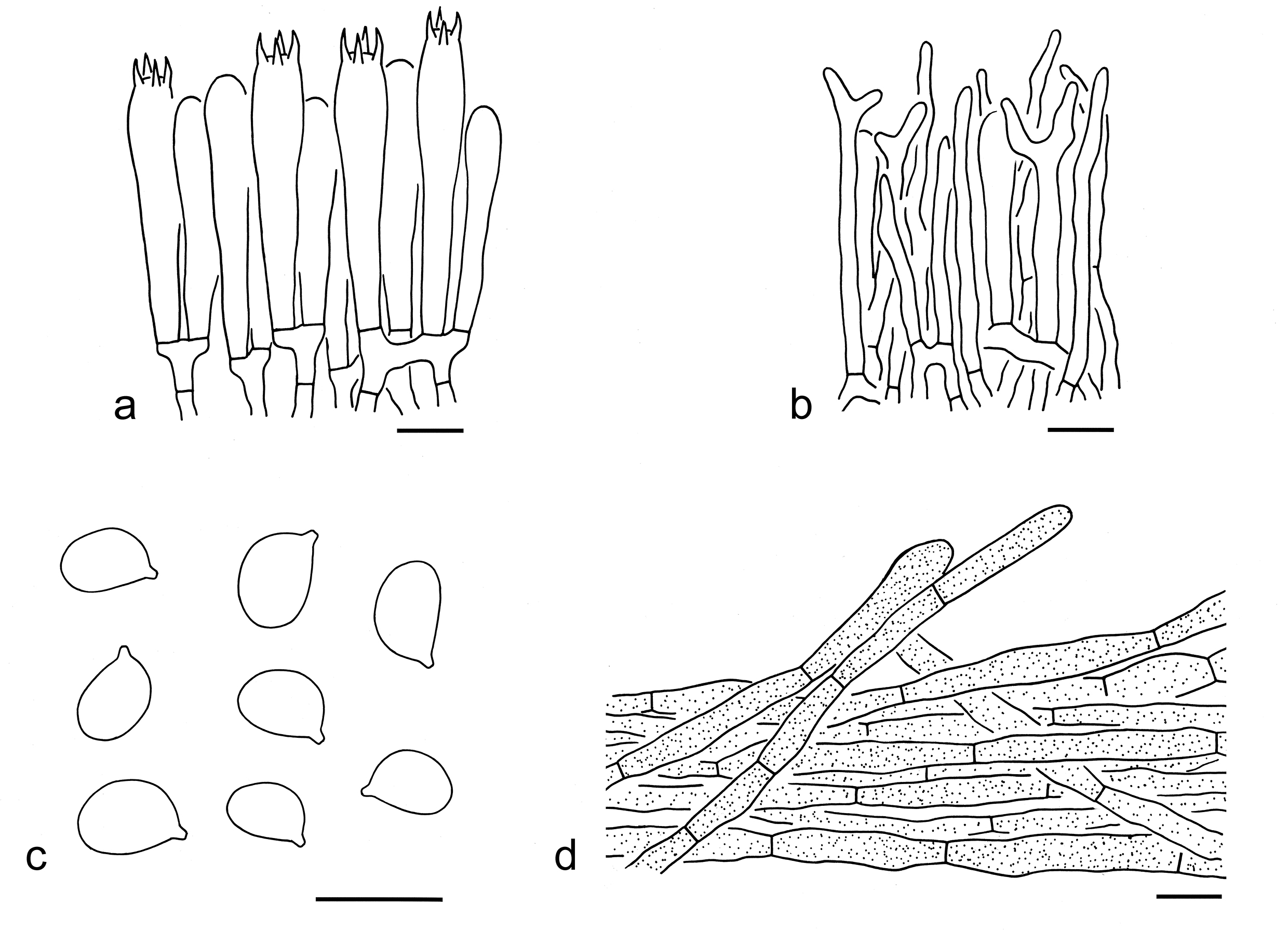
45. Tricholoma tianshanense Z.X. Zhao et al., Phytotaxa 549(1): 26 (2022). Fig. 64
Description: Basidioma medium-sized. Pileus 4–9 cm in diam., at first hemispherical, then plano-convex to applanate; surface dry, white (1A1) background covered with appressed, dark brown (4F6–8), brown (4E3–5) to brownish (4D4–6), fibrillose squamules; margin involute to inflexed; context white (1A1). Lamellae adnexed, white (1A1), crowded; edges entire, white (1A1). Stipe 5–10 2–2.5 cm, cylindrical, solid, with a brownish (4D4–6) to brown (4E3–5), cottony to fibrillose annulus on the apical part, white (1A1) above annulus, white (1A1) backgrounds covered with appressed, dark brown (4F6–8), brown (4E3–5) to brownish (4D4–6), fibrillose squamules below annulus. Taste not recorded. Odour very strong, like perfume.
Ecology: Solitary to scattered on soil in coniferous forests.
Known distribution: Currently only known from Northwestern China.
Specimen examined: CHINA. Xinjiang Province, Hejing County, in a coniferous forest dominated by Picea schrenkiana, altitude 2650 m, 11 September 2020, Z.X. Zhao & B. Xu (holotype, HMUT1112).
Notes: Tricholoma tianshanense is characterized by its white pileus densely covered with appressed, brown, fibrillose squamules, a white stipe with a brown cottony annulus on the apical part and covered with brown squamules below annulus, broadly ellipsoid to ellipsoid basidiospores (6–7.5 4.5–6 µm), and absence of clamps (this study). Zhao et al. (2022) reported that clamps are present in the species but rare. Our re-examination on the holotype cited above indicated that no clamps are present in the species. To our knowledge, no species of sect. Matsutake possesses clamps.
This species is related to T. ilkkae in phylogenetic tree based on the ITS sequences (Fig. 4), but the latter species has a more squamulose pileus and relatively smaller basidiospores (4.5–6.7 3.9–5.5 µm, average 5.1–6 4.4–4.9 µm) (Christensen & Heilmann-Clausen 2013; Heilmann-Clausen et al. 2017). Tricholoma dulciolens looks like T. tianshanense, but differs in its pale colored (brownish) basidioma, relatively smaller basidiospores (4.4–7.9 3.7–5.8 µm, average 5.7–6.3 4.7–4.9 µm) and has not been found in China (Christensen & Heilmann-Clausen 2013; Landry et al. 2022).
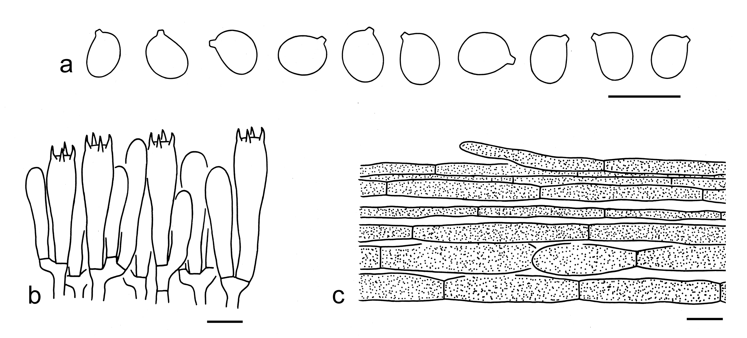
46. Tricholoma zangii Z.M. Cao et al., Mycotaxon 85: 161 (2003)
Replaced synonym: Tricholoma quercicola M. Zang, Acta Mycol. Sin. 9(2): 123 (1990).
Competing homonym: non Tricholoma quercicola (Murrill) Murrill, Lloydia 12: 67 (1949).
Description: Pileus 6–12 cm in diam., convex to plano-convex; surface light brown to pale grey, sometimes darker at center, dry, with light brown and appressed squamules; margin inrolled, entire, appendiculate. Lamellae sinuate-adnexed, adnexed to emarginate, crowded, whitish, 2.5–4 cm broad, with lamellulae of two lengths. Stipe 6–10 1.5–2.5 cm, robust, cylindrical, equal or slightly tapering towards the base; surface whitish and with small granules above the annulus, concolorous with pileus and scabrous below. Annulus apical, membranous. Context thick, whitish, comprising of thin-walled hyphae, 5–10 µm diam., lacking clamps-connections. Odour strong, sweetish. Taste mild, pleasant.
Basidiospores 6.5–10 5–6.5 µm, Q = 1.27, ellipsoid, hyaline, inamyloid, thin-walled, with a prominent hilar appendix. Basidia 16–26 7–12 µm, clavate, bearing four sterigmata. Lamellar edge heteromorphous, often with scattered cheilocystidia. Cheilocystidia 25–30 13–19 µm, clavate or ventricose-rostrate. Hymenophoral trama regular, hyaline, comprising parallel hyphae, 4–11 µm diam. Pileipellis a repent epicutis of tightly woven hyphae, 4–7 µm diam.
Ecology: Solitary to scattered on soil in broad-leaved forests.
Known distribution: Known from Southwestern China (Zang 1990; Cao et al. 2003).
Specimen examined: CHINA. Sichuan Province, Miyi, under Quercus pannosa, altitude 3000 m, 27 Jul. 1984, Ming-sheng Yuan 381 (holotype, KUN-HKAS19947).
Notes: The description above and specimen citation are from Zang (1990) and Cao et al. (2003). Zang (1990) described this matsutake from alpine Quercus forests in southwestern China as a new species, namely, T. quercicola M. Zang. Before that, Tominaga et al. (1988) proposed a variety, viz. T. matsutake var. qinggang Tominaga, based on matsutake mushrooms growing under alpine Quercus forests in Barkam, Sichuan Province, China. However, there is no Latin diagnosis or description, therefore this name is invalid (Turland et al. 2018). Cao et al. (2003) proposed a new name T. zangii to replace T. quercicola M. Zang 1990, which is a later homonym of T. quercicola (Murrill) Murrill 1949.
Seven specimens of this species were cited from Sichuan and Yunnan in Zang (1990) and Cao et al. (2003) (holotype, KUN-HKAS19947; KUN-HKAS16977; KUN-HKAS18210; KUN-HKAS14644; KUN-HKAS17632; KUN-HKAS32450 and KUN-HKAS32449). We traced five of them in the herbarium (KUN-HKAS17632; KUN-HKAS16977; KUN-HKAS32450; KUN-HKAS14644 and KUN-HKAS32449) and generated ITS sequences from the collections. ITS phylogenetic data indicated that they should be treated as T. matsutake. The sample KUN-HKAS18210, which was collected from alpine broad-leaved forests in Aba (Ngawa) Tibetan and Qiang Autonomous Prefecture, Sichuan Province, China, was not found. Unfortunately, the holotype (KUN-HKAS19947) for this species has not been traced, and, thus, no sequence from the holotype is available. The collections of T. zangii are probably populations of T. matsutake instead of an independent species. For the time being, T. zangii is listed here.
Tricholoma sect. Muscaria Reschke
Type: Tricholoma muscarium Kawam. ex Hongo, Mem. Fac. lib. Arts Educ. Shiga Univ., Nat. Sci. 9: 62 (1959).
Description: Basidioma tricholomatoid. Pileus more or less umbonate at center, yellow, brown or olivaceous, glabrous, squarrulose or radially fibrillose. Lamellae subclose to close. Basidiospores medium-sized, broadly ellipsoid to ellipsoid, thin-walled, smooth, inamyloid. Pleurocystidia absent. Cheilocystidia present and conspicuous. Pileipellis with a differentiated subpellis made of inflated cells. Clamps absent.
Ecology: Ectomycorrhizal. On soil with various trees.
Known distribution: Molecular data indicated species from this section are distributed in North America and Asia.
Notes: To date, six species, including three species described as new in this paper, occur in China (see below).
Key to the species of Tricholoma sect. Muscaria from China
1. Stipe with obvious orange color T. aurantiipes
1. Stipe white, dirty white, greyish, brownish, without orange color 2
2. Pileal margin bright yellow, yellow, dirty yellow to yellowish 3
2. Pileal margin without obvious yellow color 4
3. Pileal center with obvious olivaceous color, olive-grey to olivaceous brown T. muscarioides
3. Pileal center without obvious olivaceous color, dark brown, grey-brown to brown T. muscarium
4. Pileus without obvious olivaceous color, yellow-brown, grey-brown to brown; lamellae at first dirty white, greyish to grey, later becoming yellowish brown to brownish; stipe greyish, yellowish brown to brownish, with brownish stains after bruised T. sinense
4. Pileus with olivaceous color; lamellae white to cream; stipe white to dirty white 5
5. Basidioma medium-sized; pileus dark brown, grey-brown to brown at center, olivaceous brown, brown to brownish towards margin, without obvious squamules; basidiospores relatively shorter, with mean length = 6.3 µm T. turgidum
5. Basidioma small to medium-sized; pileus olivaceous, densely covered with olivaceous brown squamules; basidiospores relatively longer, with mean length = 6.9 µm …T olivaceiceps
47. Tricholoma aurantiipes Hongo, Trans. Mycol. Soc. Japan 32(4): 464 (1991)
Ecology: On soil under deciduous trees (Hongo 1991).
Known distribution: Currently known from East Asia (Hongo 1991, Ko et al. 2012, Reschke et al. 2018).
Notes: Tricholoma aurantiipes was originally described from Japan (Hongo 1991), and reported from Korea (Ko et al. 2012). Reschke et al. (2018) provided an ITS sequence (MF034227) from a specimen (MB-003000) collected from Yunnan, China and delimitated as T. aurantiipes. Because no additional material is available for this study, we follow the treatment of Reschke et al. (2018). The detailed morphological characters of T. aurantiipes was not provided here due to lack of specimens.
48. Tricholoma muscarioides Reschke et al., Mycologia 110 (6): 1096 (2018). Figs. 53k, l, 65
Description: Basidioma medium-sized. Pileus 5–9 cm in diam., convex, plano-convex to applanate, papillate; surface dry, olive-grey (1D2–4, 2D3–5) to olivaceous brown (3D3–5, 3E3–6) at center, becoming bright yellow (1A2–6) to yellow (2B3–7) towards margin, sometimes greyish brown (2D4–6, 3D4–7) to brown (4D3–6) overall, covered with olivaceous brown (3D3–5, 3E3–6) to greyish brown (2D4–6, 3D4–7) fibrillose squamules; margin irregularly undulating and slightly reflexed, sometimes radially splitting; context white (1A1). Lamellae sinuate, white (1A1) to cream (1B3), subclose to close; edges entire, white (1A1) to cream (1B3). Stipe 8–16 1–2.5 cm, cylindrical, sometimes tapering downwards, at first solid, then stuffed to hollow, white (1A1) to brownish (3B2–4), fibrillose, exannulate. Taste slightly bitter. Odour not recorded.
Ecology: Solitary to scattered on soil in subtropical forests dominated with trees of Fagaceae, or sometimes mixed with conifers.
Known distribution: Known from Southwestern China.
Specimens examined: CHINA. Yunnan Province, Honghe Hani and Yi Autonomous Prefecture, Lvchun County, Amoebin Forest Park, in a broad-leaved forest, altitude 1600 m, 22 Sep. 2019, 532531MF0354 (KUN-HKAS134993); same location, 23 Sep. 2019, 532531MF0484 (KUN-HKAS134994); same location; Lijiang, Yulong Naxi Autonomous County, Lijiang Alpine Botanic Garden, in a mixed forest with Quercus and Pinus, altitude 2700 m, 10 Sep. 2019, Jian-Wei Liu 1954 (KUN-HKAS134995); same city, Gucheng, Longshan Village, in a broad-leaved forest, altitude 2700 m, 21 Aug. 2010, Qi Zhao 1016 (KUN-HKAS69737); Puer, Jingdong Yi Autonomous County, Ailao Mountain, in a Quercus dominated broad-leaved forest, altitude 2500 m, 15 Jul. 2007, K. Donges, G. Kost and K.-H. Rexer (holotype, KUN-HKAS93512).
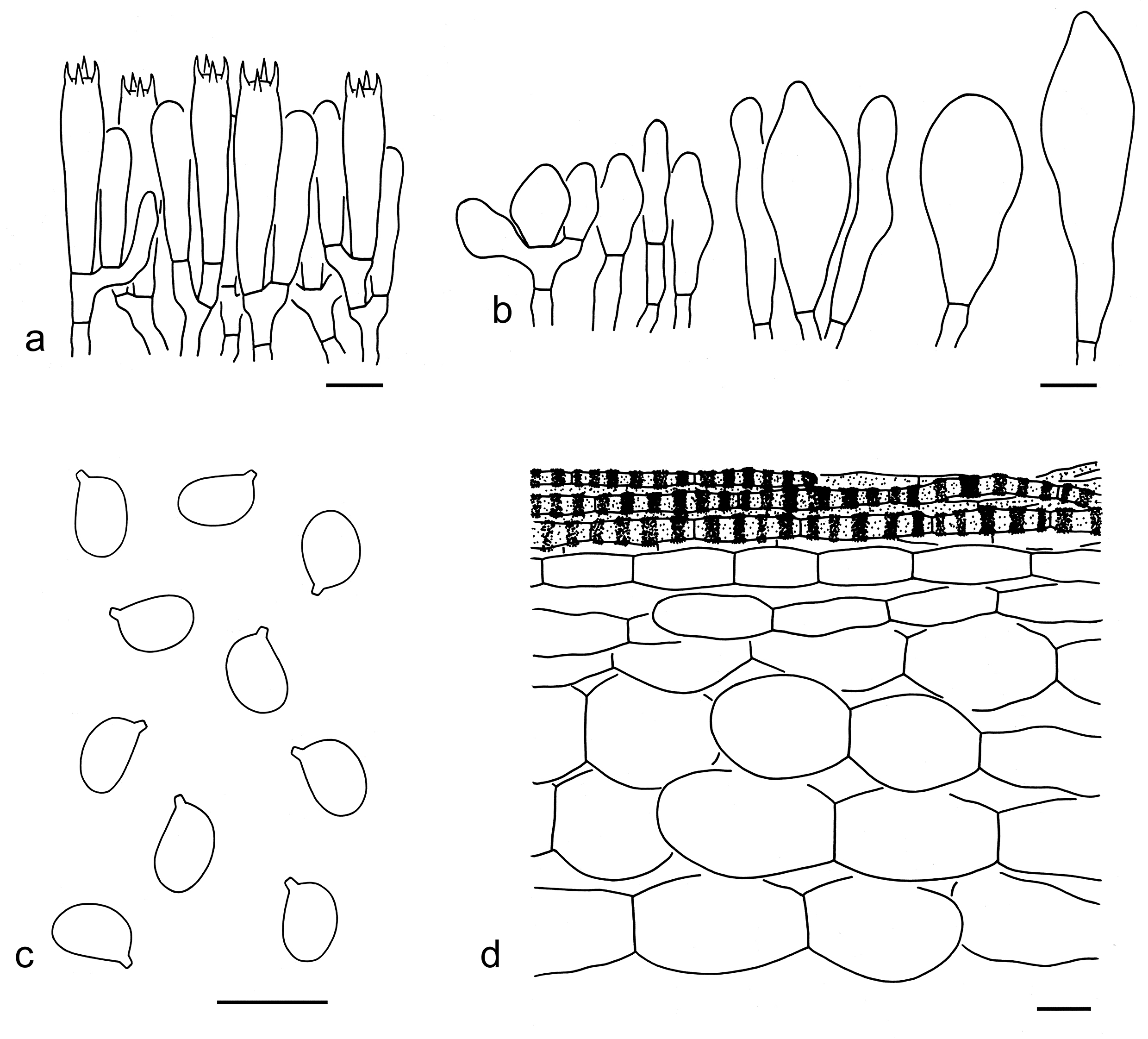
Notes: Tricholoma muscarioides was described by Reschke et al. (2018). This species is recognized by an papillate pileus with an olive-grey to olivaceous brown center and a bright yellow to yellow margin, and the main occurrence in broad-leaved forests (this study). Based on the tree inferred from the ITS sequences, T. muscarioides is closely related to T. turgidum Y.Y. Cui & Zhu L. Yang (Fig. 4), but the latter has a pileus without bright yellow color, which is grey-brown at center and brown to brownish at margin. In addition, T. turgidum is mainly distributed in conifers-dominated forests.
Tricholoma muscarioides can be confused with T. muscarium Kawam. ex Hongo due to its yellow pileal margin. However, the latter species has a dark brown, grey-brown to brown pileal center without obviously olivaceous color. Tricholoma davisiae Peck is somewhat similar to T. muscarioides, but has a relatively larger basidioma (3–16 cm), develops pink orange stains on the stipe and lamellar edges, and grows with conifers (Bessette et al. 2013).
49. Tricholoma muscarium Kawam. ex Hongo, Mem. Fac. lib. Arts Educ. Shiga Univ., Nat. Sci. 9: 62 (1959). Figs. 53m, 66
Basidiospores [40/2/2] 6–8 4–5.5 µm (mean length = 7 µm, mean width = 4.8 µm), Q = (1.27–) 1.3–1.6 (–1.63), Qm = 1.45 ± 0.08, ellipsoid, occasionally broadly ellipsoid or elongate, non-amyloid, colorless, hyaline, thin-walled, smooth; apiculus relatively large. Basidia 35–50 7–9 µm, clavate, 4-spored, hyaline; sterigmata 4–6 µm long; basal septa without clamps. Pleurocystidia absent. Cheilocystidia 10–60 5–12 µm, conspicuous, clavate to subclavate, sometimes irregularly shaped, often septate, thin-walled, colorless, hyaline. Subhymenium composed of 2–4 µm wide filamentous hyphae; clamps absent. Pileipellis a cutis, composed of radially arranged, 5–15 µm wide, thin-walled, brownish to brown filamentous hyphae, terminal elements subcylindrical, with round apex; subpellis composed of radially arranged inflated cells, ellipsoid, fusiform to clavate, 30–100 15–50 µm, thin-walled, yellowish to colorless. Clamps absent in all parts of basidioma.
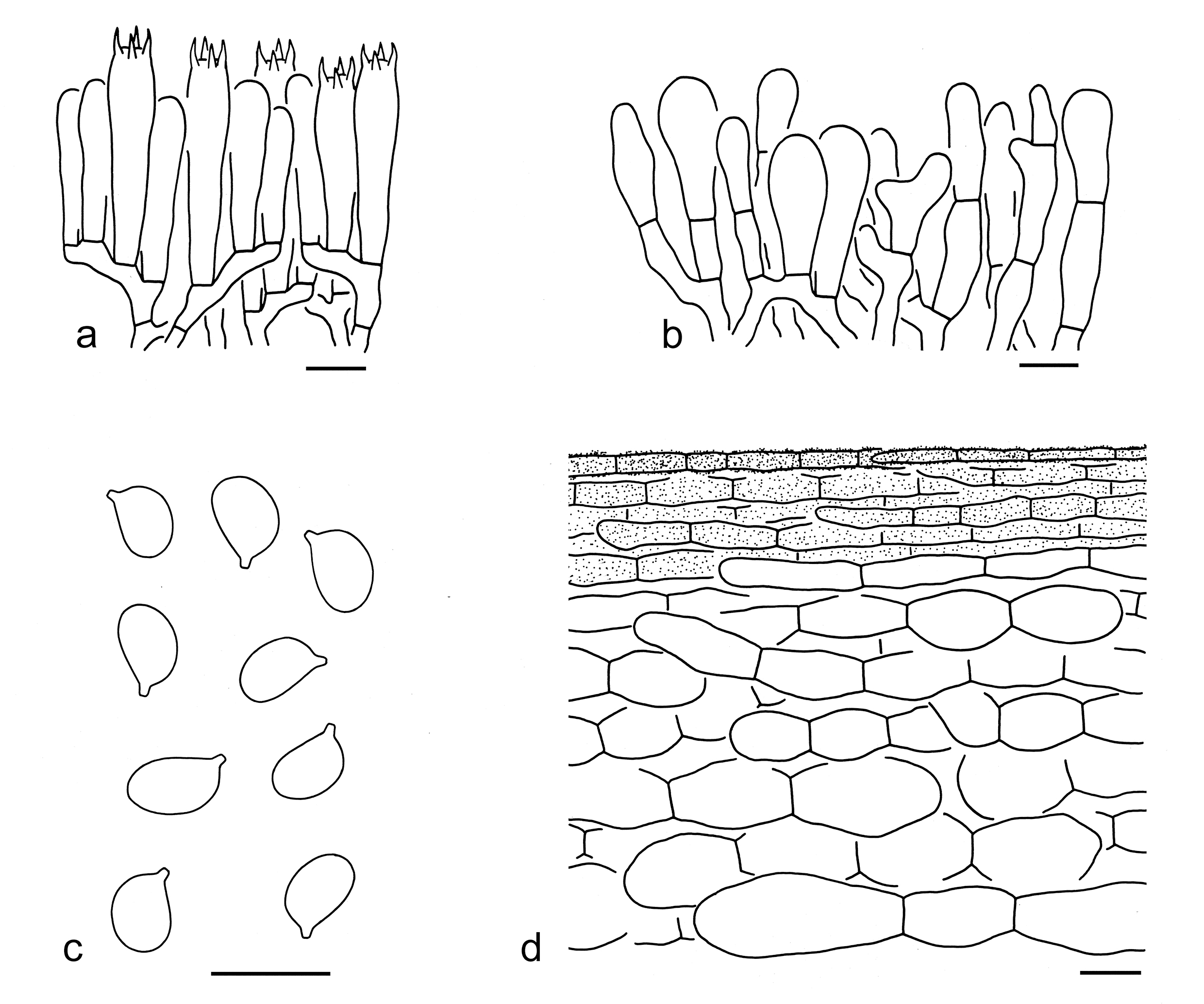
Ecology: Solitary to scattered on soil in subtropical broad-leaved forests.
Known distribution: Reported from Japan (Hongo 1959) and China (Deng et al. 2004).
Specimens examined: CHINA. Yunnan Province, Chuxiong Yi Autonomous Prefecture, Nanhua County, in a broad-leaved forest with trees of Fagaceae, altitude 2300 m, 12 Aug. 2017, Jian-Wei Liu 899 (KUN-HKAS106307); Kunming, Shuanglong Village, Yeyahu Lake, in a broad-leaved dominated forest, altitude 2100 m, 18 Aug. 2012, Hui Chen 40 (KUN-HKAS134996); same location, in a broad-leaved forest, altitude 2100 m, 18 Aug. 2012, Ting Guo 582 (KUN-HKAS76274).
50. Tricholoma olivaceiceps Y.Y. Cui & Zhu L. Yang, sp. nov. Figs. 53n, 67
Fungal Names: FN 572341
Etymology: olivaceiceps = olive-headed, referring to its olivaceous pileus.
Diagnosis: Similar to T. turgidum, but differs by its relatively smaller basidioma (3–7 cm), densely olivaceous brown squamules on pileal surface and association dominated with broad-leaved trees.
Type: CHINA. Yunnan Province, Puer, Lancang Lahu Autonomous County, Qianliu Village, in a forest with trees of Castanopsis and Pinus, altitude 1300 m, 10 Nov. 2017, Zhu L. Yang 6091 (holotype, KUN-HKAS101280).
Description: Basidioma small to medium-sized. Pileus 3–7 cm diam, plano-convex to applanate, papillate at center; surface dry, olivaceous (1D4–8, 30C4–7), but darker at center, densely covered with minute, olivaceous brown (1D3–6, 30D4–7) squamules; margin straight; context whitish. Lamellae sinuate, at first with greyish tinge, becoming whitish to cream when mature, subclose; edges white (1B1–2), dirty white (1A1, 1B2) to cream (1B2–3). Stipe 5–7 0.5–1 cm, cylindrical to subcylindrical, dry, dirty white (2B1–3), with grey (1B1) to greyish (1B2–3) fibrils, exannulate. Taste not recorded, odour indistinct.
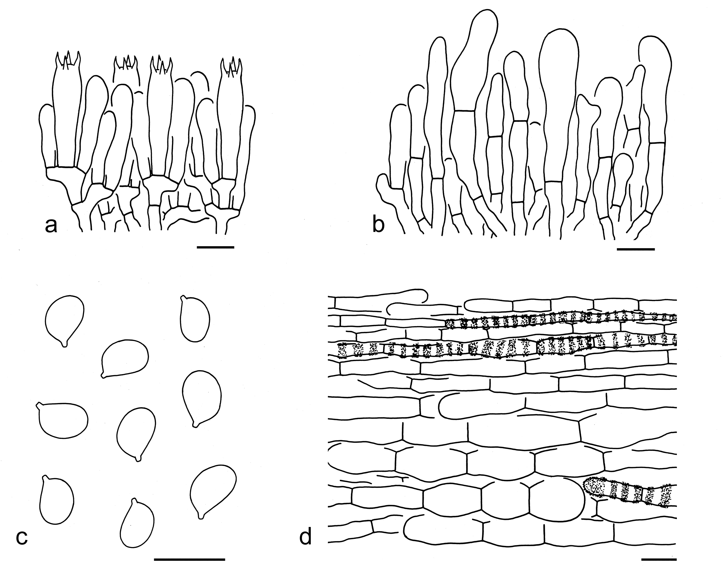
Basidiospores [60/3/2] 6–8 4–6 µm (mean length= 6.9 µm, mean width= 4.8 µm), Q = 1.2–1.67 (–1.71), Qm = 1.43 ± 0.11, broadly ellipsoid to ellipsoid, occasionally elongate, non-amyloid, colorless, hyaline, thin-walled, smooth; apiculus relatively large. Basidia 25–40 7–9 µm, clavate, 4-spored, rarely 2-spored or 1-spored, hyaline; sterigmata 4–7 µm long; basal septa without clamps. Pleurocystidia absent. Cheilocystidia 20–40 6–15 µm, conspicuous, clavate to subclavate, often septate, thin-or slightly thick-walled, brownish, hyaline. Subhymenium composed of 3–6 µm wide filamentous hyphae; clamps absent. Lamellar trama regular, composed of colorless, thin-walled, ellipsoid, fusiform, clavate to filamentous hyphae 3–30 µm wide. Pileipellis a cutis, composed of radially arranged, 5–10 µm wide, thin-walled, brownish to brown filamentous hyphae, terminal elements subcylindrical, with round apex; subpellis composed of radially arranged inflated cells, ellipsoid, subfusiform to clavate, 30–65 10–30 µm, thin-walled, brownish to brown or colorless. Clamps absent in all parts of basidioma.
Ecology: Solitary to scattered on soil in broad-leaved dominated forests.
Known distribution: Known from autumn in Southwestern China.
Notes: Tricholoma olivaceiceps can be recognized by its olivaceous pileus densely covered with olivaceous brown squamules, and white to dirty white lamellae. It is closely related to T. turgidum and T. muscarioides (Figs. 3, 4). Tricholoma turgidum is similar but can be separated. For comparisons, see notes under that that name. Tricholoma muscarioides differs from T. olivaceiceps by its bright yellow to yellow color on pileal margin.
Morphologically, T. luteomaculosum A.H. Sm. is similar to T. olivaceiceps. However, T. luteomaculosum has a mostly dark brownish grey or bluish grey pileus without olivaceous color, and its lamellar edges often stain dark brownish grey to blackish (Ovrebo 1986; Bessette et al. 2013).
51. Tricholoma sinense Y.Y. Cui & Zhu L. Yang, sp. nov. Figs. 53o, 68
Fungal Names: FN 572347
Etymology: sinense = chinese, referring its type locality from China.
Diagnosis: Similar to T. luteomaculosum, but differs by its relatively smaller basidioma and a greyish, yellowish brown to brownish stipe covered with grey, grey-brown to yellow-brown fibrils.
Type: CHINA. Yunnan Province, Lijiang, Dadong Village, in a subtropical mixed forest with Pinus armandii and Quercus semicarpifolia, altitude 2500 m, 18 Aug. 2010, Qi Zhao 948 (holotype, KUN-HKAS69669).
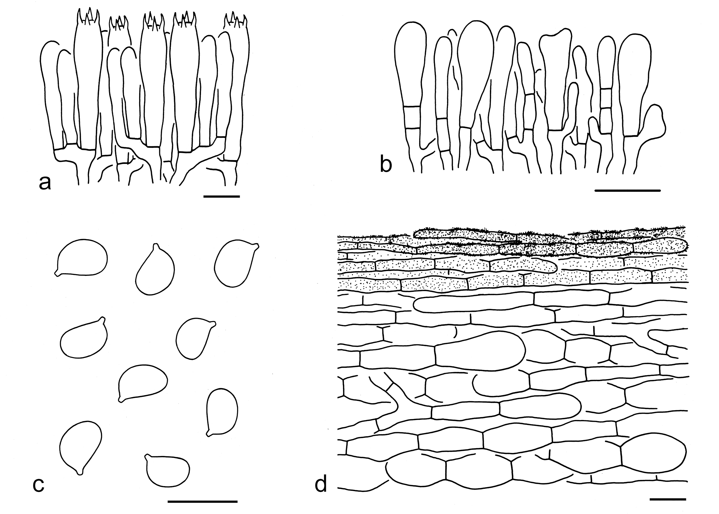
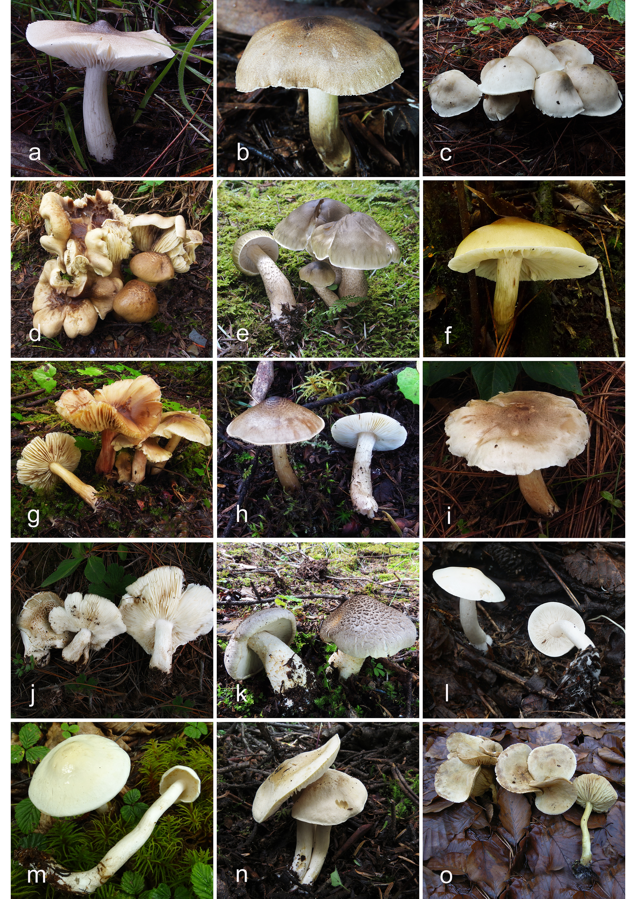
Description: Basidioma small to medium-sized. Pileus 3–8 cm diam, plano-convex to applanate, sometimes slightly broadly umbonate at center; surface dry, yellow-brown (4C4–7), grey-brown (4D3–5, 3D2–6) to brown (5D3–6), densely covered with minute, yellow-brown (4C4–7), grey-brown (4D3–5, 3D2–6) to brown (5D3–6) squamules; margin slightly incurved; context white (1A1). Lamellae sinuate, at first dirty white (1B1), greyish (1B1) to grey (2B1–3), becoming yellowish brown (4B2–4) to brownish (3C2–4) with age, subclose to close; edges smooth, entire to slightly undulate, greyish, yellow-brown (4B2–4) to brownish (3C2–4). Stipe 4–11 1–2 cm, cylindrical to subclavate, stuffed, dirty white (1B2–3), greyish (1B1), yellowish brown (4B2–4) to brownish (3C2–4), bruising brownish (4B2–4) after touch, covered with grey (1D1), grey-brown (3D2–4), yellow-brown (4D2–5) fibrils, exannulate. Taste not recorded. Odour similar to that of T. matsutake.
Basidiospores [120/6/4] (5.5–) 6–8 (–8.5) 4–6 µm (mean length = 6.7 µm, mean width= 5 µm), Q = 1.18-1.55 (–1.73), Qm = 1.36 ± 0.09, broadly ellipsoid to ellipsoid, occasionally elongate, non-amyloid, colorless, hyaline, thin-walled, smooth; apiculus relatively large. Basidia 30–45 7–9 µm, clavate, 4-spored, hyaline; sterigmata 3–5 µm long; basal septa without clamps. Pleurocystidia absent. Cheilocystidia 20–35 5–10 µm, clavate to subclavate, often septate, thin-walled, colorless, hyaline. Subhymenium composed of 3–5 µm wide filamentous hyphae; clamps absent. Lamellar trama regular, composed of colorless, thin-walled hyphae 2–20 µm wide. Pileipellis a cutis, composed of radially arranged, 5–12 µm wide, thin-walled, brownish filamentous hyphae, terminal elements subcylindrical, with round apex; subpellis composed of radially arranged inflated cells, ellipsoid, fusiform to clavate, 15–50 10–20µm, thin-walled, colorless. Clamps absent in all parts of basidioma.
Ecology: Solitary to scattered dominated in subtropical coniferous or mixed forests with trees of Pinus and Fagaceae.
Known distribution: Known from Southwestern China.
Additional specimens examined: CHINA. Yunnan Province, Kunming, Aziying, in a mixed forest, altitude 2050 m, 12 Jul. 2018, Xiao-Xia Ding 346 (KUN-HKAS105356); Lijiang, Qihe Village, in a pine forest with Pinus yunnanensis, altitude 2423 m, 27 Jul. 2011, Li-Ping Tang 1316 (KUN-HKAS69777); Nujiang Lisu Autonomous Prefecture, Lanping Bai and Pumi Autonomous County, in a pine forest with Pinus yunnanensis, altitude 2200 m, 15 Aug. 2011, Jiao Qin 375 (KUN-HKAS73361).
Notes: Tricholoma sinense is characterized by its yellow-brown, grey-brown to brown pileus densely covered with concolorous squamules, and dirty white, greyish to grey lamellae becoming yellowish brown to brownish with age. It is related to T. aurantiipes with moderate support (Fig. 4).
Tricholoma luteomaculosum has a similar pileal color and its stipe with brownish staining after touch. However, this species differs from T. sinense by its relatively larger basidioma (4–12 cm) and relatively light colored stipe (white to pale grey) without obvious squamules (Ovrebo 1986; Bessette et al. 2013). Tricholoma olivaceiceps also somewhat resembles, but has an olivaceous pileus with olivaceous brown squamules, white to cream lamellae and a white stipe without color change.
52. Tricholoma turgidum Y.Y. Cui & Zhu L. Yang, sp. nov. Figs. 69a, 70
Fungal Names: FN 572348
Etymology: turgidum = inflated, referring to its inflated cells in subpellis.
Diagnosis: Related and similar to T. muscarioides, but differs in its greyish brown to brown pileal margin without yellow color and the association with coniferous trees.
Type: CHINA. Yunnan Province, Lijiang, Yulong Naxi Autonomous County, Yulong Snow Mountain, in a forest with Pinus yunnanensis, altitude 2490 m, 16 Sep. 2012, Kuan Zhao 147 (holotype, KUN-HKAS77500).
Description: Basidioma medium-sized. Pileus 6–9 cm in diam., convex, plano-convex to applanate, sometimes with a pronounced or low papilla; surface dry, grey-brown (3D2–4, 3E5–8) to dark brown (3E2–4, 2E2–4) in the central part, greyish brown (2C2–5), brown (3C4–7) to brownish (3B3–5) towards margin, sometimes greyish brown (2C2–5), greyish yellow (2D2–5) to brown (3C4–7) overall, often with olivaceous (30B4–6) tinge over the entire part, covered with brown (3C4–7), minutely granular squamules; margin irregularly undulating; context white (1A1) to cream (1A2). Lamellae adnexed to sinuate, white (1A1) to cream (1A2, 1B3), subclose to close; edges entire, white to cream. Stipe 6–9 0.7–1.5 cm, cylindrical to subcylindrical, equal, stuffed to hollow, white (1A1), bruising brownish (3B2–4) after touch, smooth or fibrillose, exannulate. Taste not recorded. Odour like soap.
Basidiospores [160/8/4] 5–7 (–9) 4–5.5 (–6) µm (mean length= 6.3 µm, mean width = 4.5 µm), Q = (1.13–) 1.2–1.6 (–1.95), Qm = 1.4 ± 0.12, broadly ellipsoid to ellipsoid, occasionally elongate, non-amyloid, colorless, hyaline, thin-walled, smooth; apiculus relatively large. Basidia 28–40 7–10 µm, clavate, 4-spored, hyaline; sterigmata 4–6 µm long; basal septa without clamps. Pleurocystidia absent. Cheilocystidia 20–40 5–15 µm, conspicuous, cylindrical, subclavate to clavate, often septate, thin- to slightly thick-walled (≤ 1 µm), colorless, hyaline. Subhymenium composed of 2–4 µm wide filamentous hyphae, clamps absent. Lamellar trama regular, composed of colorless, thin- to slightly thick-walled, clavate, cylindrical to filamentous hyphae 5–30 µm wide. Pileipellis a cutis, composed of radially arranged, 5–18 µm wide, thin-walled, yellow-brown to brownish filamentous hyphae; subpellis composed of radially arranged inflated cells, subglobose, subfusiform, fusiform to ellipsoid, 25–80 15–35 µm, thin-walled, colorless. Clamps absent in all parts of basidioma.
Ecology: Solitary to scattered on soil dominated in forests with trees of Pinus, sometimes mixed with broad-leaved trees.
Known distribution: Known from Southwestern China.
Additional specimens examined: CHINA. Yunnan Province, Kunming, bought from Aziying Village, altitude 2050 m, 12 Jul. 2018, Xiao-Xia Ding 340 (KUN-HKAS 105350); Lijiang, Gucheng, Jinshan Village, in a forest with Pinus yunnanensis, altitude 2300 m, 16 Sep. 2012, Qi Zhao 1685 (KUN-HKAS78943); same city, Yulong Naxi Autonomous County, Lijiang Alpine Botanic Garden, in a broad-leaved forest, altitude 2700 m, 24 Aug. 2019, Jian-Wei Liu 1776 (KUN-HKAS134997).
Notes: Tricholoma turgidum is characterized by its papillate pileus, which is dark brown, grey-brown to brown in the central part and olivaceous brown, brown to brownish towards margin. It is predominantly associated with trees of conifers. Tricholoma turgidum is sister to T. muscarioides in the tree generated from the ITS sequences (Fig. 4), but differs by its non-yellow pileal margin and the association with conifers.
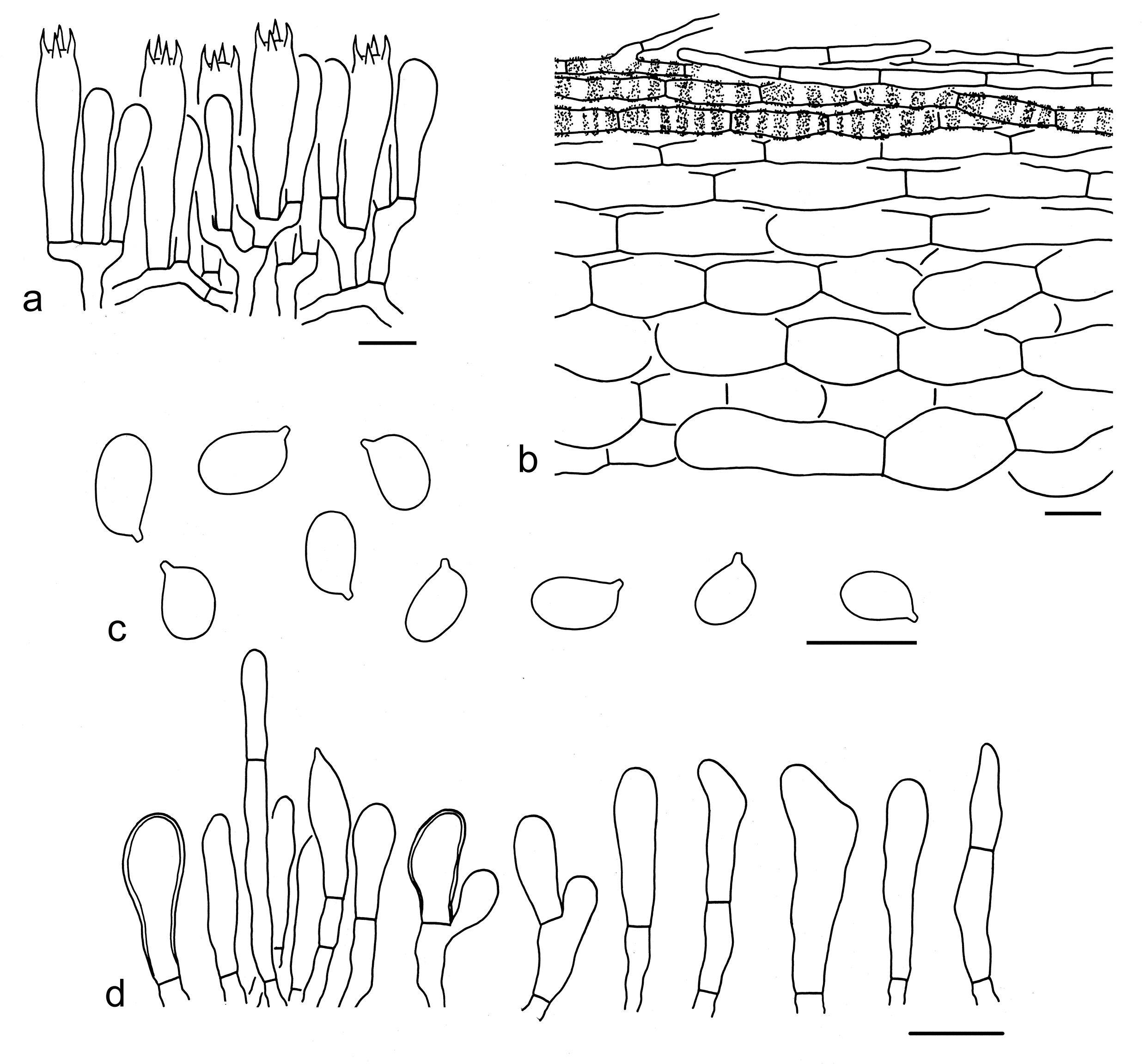
Tricholoma sect. Rigida (Fr.) Quél.
Type: Tricholoma saponaceum (Fr.) P. Kumm., Führ. Pilzk. (Zerbst): 133 (1871).
Description: Basidioma tricholomatoid. Pileus umbonate or not, grey-brown, yellow-brown to brown pileus, mostly glabrous and with greenish color. Lamellae close to subdistant. Stipe subcylindrical to cylindrical, without annulus. Basidiospores small, broadly ellipsoid to ellipsoid, thin-walled, smooth, inamyloid. Pleurocystidia absent. Cheilocystidia present or absent. Pileipellis a cutis or an ixo-cutis. Clamps present.
Ecology: Ectomycorrhizal. On soil with various trees.
Known distribution: Molecular data indicated species from this section are distributed in Europe, North America, and Asia.
Notes: Species in sect. Rigida are challenging to differentiate due to their similar morphologies. Compared with the phylogenetic analysis based on the ITS sequences, the analyses based on the five gene fragments are more accurate in recognizing the phylogenetic species of this section by using the GCPSR criteria. Based on the multi-gene phylogenetic analyses, morphological characters, and ecological data, nine species from this section, including four new and five known ones, are identified (see below).
Key to the species of Tricholoma sect. Rigida from China
1. Basidioma staining yellow when old, damaged or touched T. forteflavescens
1. Basidioma without yellow change when old, damaged or touched 2
2. Basidioma often with strong reddening reaction with age and when damaged T. boudieri
2. Basidioma without strong reddening 3
3. Subpellis composed of more or less slightly inflated hyphae, ellipsoid, cylindrical, cylindrical to filamentous; distributions in subalpine forests with Abies T. saponaceum
3. Subpellis composed of filamentous hyphae; distributions in coniferous, broad-leaved or mixed forests 4
4. Pileus blackish grey, grey-brown, brown to greyish white, without green tinge; basidiospores relatively smaller, mean length < 5.5 µm, mean width < 4 µm 5
4. Pileus grey-brown to yellow-brown, with green tinge; basidiospores relatively larger, mean length > 5.5 µm, mean width > 4 µm 7
5. Pileus dark blackish grey, greyish brown to brown, finely tomentose T. asiaticum
5. Pileus brown, grey-brown or greyish white, glabrous 6
6. Pileus paler, grey-brown to greyish white T. atrovirens
6. Pileus brown T. yunnanense
7. Basidiospores relatively smaller, with mean length less than 7 µm, mean width less than 5 µm 8
8. Cheilocystidia conspicuous T. sichuanense
8. Cheilocystidia poorly differentiated T. olivaceum
53. Tricholoma asiaticum Y.Y. Cui & Zhu L. Yang, sp. nov. Figs. 69b, 71
Fungal Names: FN 572333
Etymology: asiaticum = Asiatic, referring to its type locality from Asia.
Diagnosis: Somewhat related to T. yunnanense Y.Y. Cui & Zhu L. Yang, but differs by its greyish brown to brown pileus often finely tomentose.
Type: CHINA. Yunnan Province, Wenshan Zhuang and Miao Autonomous Prefecture, Guangnan County, Nanping Town, in a mixed forest with Pinaceae and Fagaceae, altitude 1300 m, 7 Aug. 2015, Kuan Zhao 813 (holotype, KUN-HKAS92443).
Description: Basidioma small to medium-sized. Pileus 3–10 cm in diam., convex to convex-applanate, slightly umbonate or not; surface dry, dark blackish grey (3E1–3), greyish brown (4D3–5) to brown (5C4–6), finely tomentose; margin straight; context white (1A1). Lamellae sinuate, white (1A1) to brownish (4B3–5), subclose to close; edges concolor. Stipe 3–8 1–2 cm, cylindrical to subclavate, greyish (1B1–3), basal part brownish (5B3–6) to pinkish (6A2), solid, exannulate. Taste not recorded. Odour slightly gas-like.
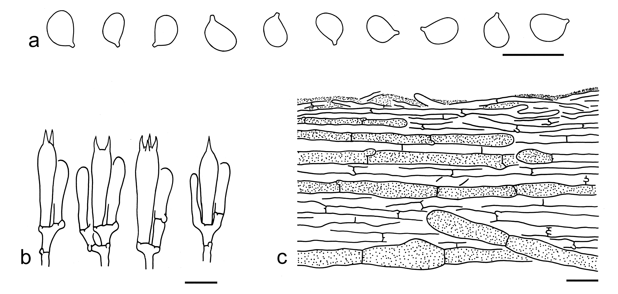
Basidiospores [60/3/1] 4–6 3–4.5 µm (mean length = 5.2 µm, mean width = 3.6 µm), Q = (1.15–) 1.3–1.57 (–1.67), Qm = 1.43 ± 0.13, broadly ellipsoid to ellipsoid, non-amyloid, colorless, hyaline, thin-walled, smooth; apiculus relatively large. Basidia 25–35 5–7 µm, clavate, 1-, 2- to 4-spored, hyaline; sterigmata 4–7 µm long; basal septa with clamps. Pleurocystidia and cheilocystidia absent. Subhymenium composed of 2–5 µm wide filamentous hyphae, clamps present. Lamellar trama regular, composed of colorless, thin-walled, clavate, cylindrical to filamentous hyphae 3–25 µm wide. Pileipellis a cutis, slightly gelatinized, composed of radially arranged, 3–8 µm wide, thin-walled, yellowish to brownish, filamentous hyphae; subpellis poorly differentiated, composed of radially arranged, colorless, yellowish to brownish, clavate to filamentous hyphae 5–20 µm wide. Clamps present in all parts of basidioma.
Ecology: Solitary to scattered on soil in mixed forests with Pinaceae and Fagaceae.
Known distribution: Southwestern China.
Additional specimen examined: CHINA. Yunnan Province, Kunming, Panlong District, Jindian, in a mixed forest with Pinus and Quercus, altitude 1900 m, 1 Sep. 2007, Zhu L. Yang 4943 (KUN-HKAS52260); Kunming, Panlong District, Heilongtan, forest and altitude not recorded, 4 Sep. 2011, Yan-Jia Hao 585 (KUN-HKAS71694); Kunming, Xishan District, Xishan, forest and altitude not recorded, 22 Sep. 2012, Ting Guo 624 (KUN-HKAS81826).
Notes: Tricholoma asiaticum can be well separated from other species of sect. Rigida by its dark blackish grey to greyish brown and tomentose pileus, and broadly ellipsoid to ellipsoid basidiospores (4–6 3–4.5 µm). This species is somewhat related to T. yunnanense in the five-gene analysis (Fig. 3), and has been compared under the latter species.
54. Tricholoma atrovirens (Pers.) Sacc., Syll. fung. (Abellini) 5: 106 (1887). Figs. 69c, 72
Basidiospores [20/1/1] 4.5–6 3–4 µm (mean length = 5.1 µm, mean width = 3.7 µm), Q = 1.24–1.51 (–1.87), Qm = 1.38 ± 0.14, broadly ellipsoid to ellipsoid, non-amyloid, colorless, hyaline, thin-walled, smooth; apiculus relatively large. Basidia 25–40 6–8 µm, clavate, 4-spored, hyaline; sterigmata 2–4 µm long; basal septa with clamps. Pleurocystidia absent. Cheilocystidia 25–50 3–5 µm, conspicuous, clavate, cylindrical to filiform, often branching, thin-walled, colorless, hyaline. Subhymenium composed of 2–4 µm wide filamentous hyphae, clamps present. Lamellar trama regular, composed of colorless, thin-walled, cylindrical to filamentous hyphae 4–15 µm wide. Pileipellis a cutis, non- to slightly gelatinized, composed of radially arranged, 2–10 µm wide, thin-walled, colorless filamentous hyphae; subpellis poorly differentiated. Clamps present in all parts of basidioma.
Ecology: Solitary to scattered on soil in broad-leaved or mixed forests with Fagaceae and Pinaceae.
Known distribution: Known from Europe and Southwestern China.
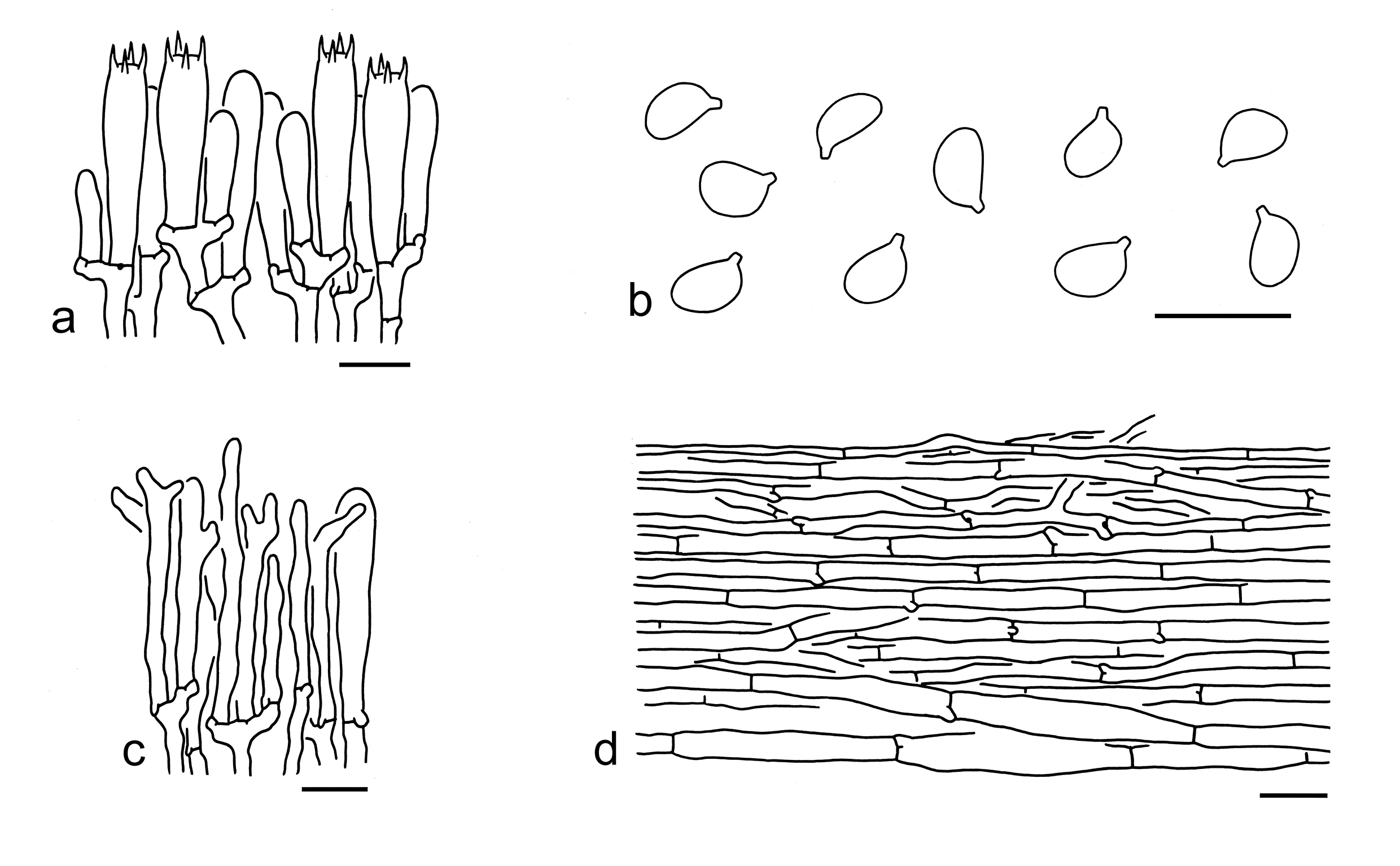
Specimens examined: CHINA. Sichuan Province, Ganzi Tibetan Autonomous Prefecture, Kangding, Pengta Village, in a broad-leaved forest, altitude 3100 m, 10 Sep. 2016, KD-LJW 156 (KUN-HKAS97949). Yunnan Province, Lijiang, Yulong Naxi Autonomous County, Lijiang Observatory, in a mixed forest with Lithocarpus, Picea and Pinus armandii, altitude 2400 m, 31 Aug. 2013, Kuan Zhao 371 (KUN-HKAS80796).
Notes: Tricholoma atrovirens collected from China has a pileus often grey-brown at center and dirty white towards margin, broadly ellipsoid to ellipsoid basidiospores (4.5–6 3–4 µm), conspicuous cheilocystidia and the presence of clamps (this study). This species was delimitated as T. saponaceum in Heilmann-Clausen et al. (2017), but it is morphological similar and appears to be T. atrovirens (https://svampe.databasen.org/taxon/72030). While the ITS sequence DQ494700 seem to be the best candidate for the true T. saponaceum and has been recorded below.
In the phylogenetic analysis based on the ITS sequences (Fig. 4), T. rapipes (Krombh.) Heilm.-Claus. & Mort. Chr. was clustered together with T. atrovirens, but can be separated by its more yellow colored pileus and associations with Abies and Picea (Christensen & Heilmann-Clausen 2013; Heilmann-Clausen et al. 2017). Because no more gene fragments of T. rapipes are available for multi-locus phylogenetic analysis, this study treats them as two independent species for the time being. Tricholoma yunnanense is similar to T. atrovirens, but differs by its more brown pileus. Tricholoma sudum (Fr.) Quél. may be confused with it, but has slightly larger basidiospores (5.1–7.9 3.3–5.1 µm) (Riva 1988; Christensen & Heilmann-Clausen 2013).
55. Tricholoma boudieri Barla, Bull. Soc. mycol. Fr. 3: 205 (1887). Figs. 69d, 73
Description: Basidioma medium-sized. Pileus 4–11 cm in diam., at first convex, then plano-convex to applanate, often concave when mature; surface dry, blackish (2E1–3, 2F1–3) to grey-brown (3E6–8) at center, becoming brown (4D2–4), brownish (4C2–4) to dirty white (1B1–2) towards margin, sometimes with greenish tinge (30A2–4), glabrous or covered with dark brown (2E1–3, 2F1–3) fibrillose scales often concentrated at center; margin at first involute, often straight to reflexed when mature; context white (1A1) to dirty white (1B1–2), sometimes with strong reddening reaction with age and when damaged. Lamellae sinuate, white (1A1), dirty white (2B1), cream (1A2) to greenish yellow (30A2–4), subclose; edges concolor. Stipe 7–12 1–2.5 cm, cylindrical to subcylindrical, greenish yellow (30A2–4), brownish (30B2–4) to brown (5B3–5), stuffed to hollow, exannulate. Taste not recorded. Odour strong, unpleasant, like cucumber.
Basidiospores [100/5/3] 5–7 (–8) 3.5–5 µm (mean length = 6.1 µm, mean width = 4.1 µm), Q = (1.2–) 1.33–1.6 (–1.78), Qm = 1.47 ± 0.12, ellipsoid, sometimes broadly ellipsoid or elongate, non-amyloid, colorless, hyaline, thin-walled, smooth; apiculus relatively large. Basidia 30–40 5–8 µm, clavate, 4-spored, colorless to yellowish, hyaline; sterigmata 3–5 µm long; basal septa with clamps. Pleurocystidia absent. Cheilocystidia poorly differentiated. Subhymenium composed of 2–4 µm wide filamentous hyphae, clamps present. Lamellar trama regular, composed of colorless to yellowish, thin-walled, cylindrical to filamentous hyphae 4–20 µm wide. Pileipellis a cutis, composed of radially arranged, 2–10 µm wide, thin-walled, brownish filamentous hyphae; subpellis poorly differentiated. Clamps present in all parts of basidioma.
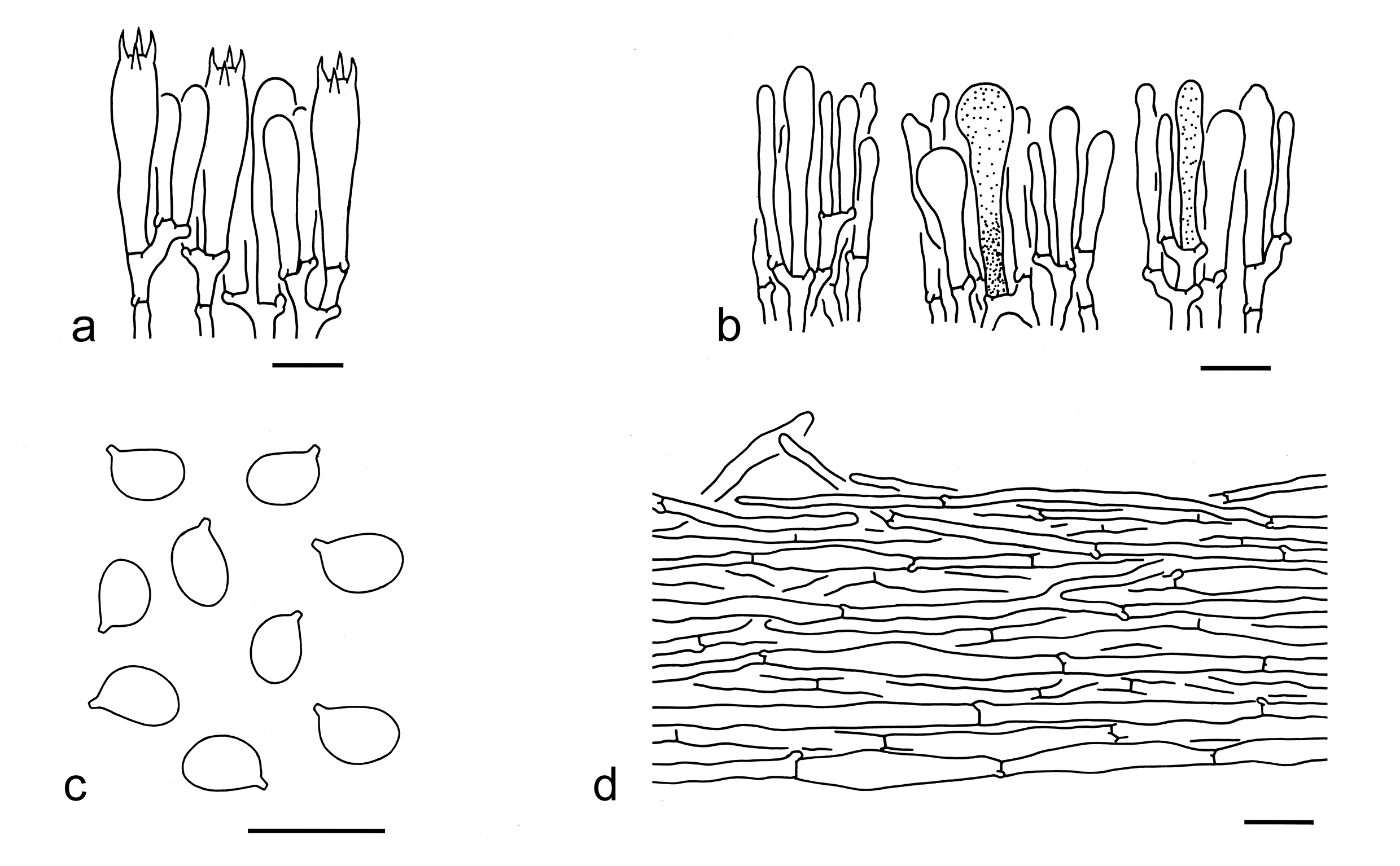
Ecology: Solitary to scattered on soil in forests with Fagaceae and Pinaceae.
Known distribution: Known from Europe, North America, and Southwestern China.
Specimens examined: CHINA. Sichuan Province, Ganzi Tibetan Autonomous Prefecture, Kangding, Waze Village, in a forest with Abies, altitude 3450 m, 8 Sep. 2016, KD-FB92 (KUN-HKAS99445); same location, in a mixed forest with Fagaceae and Pinaceae, altitude 3450 m, 8 Sep. 2016, KD-Ding 76 (KUN-HKAS97157); same location, in a mixed forest with Fagaceae and Pinaceae, altitude 3450 m, 8 Sep. 2016, Bang Feng 92 (KUN-HKAS99445); same city, Yala Village, in a mixed forest with Fagaceae and Pinaceae, altitude 3800 m, 9 Sep. 2016, KD-Ding 104 (KUN-HKAS97185), same city, forest and altitude not recorded, 10 Sep. 2016, Bang Feng 86 (KUN-HKAS99439). Yunnan Province: Lijiang, Ninglang, forest type not recorded, altitude 3610 m, 22 Sep. 2019, Xiang-Hua Wang 6872 (KUN-HKAS138819). NORTH MACEDONIA. The forest type and altitude not recorded, 2 Oct. 2018, Zhu L. Yang MK4 (KUN-HKAS104331).
Notes: Tricholoma boudieri is a morphological variable species, and has a grey, grey-brown, yellow-brown to olivaceous brown pileus, and white, dirty white, yellowish to olivaceous yellow lamellae (this study). Our field observations revealed that the reddening reaction is unstable; certain basidioma of this species exhibit a pronounced reddening reaction with age or injury, while others do not. No closely related species of T. boudieri were detected in our phylogenetic analyses (Figs. 3, 4). Tricholoma atrovirens is morphologically similar to it, but differs from it by its basidioma without reddish color change and relatively smaller basidiospores (4.5–6 3–4 µm, mean length = 5.1 µm, mean width = 3.7 µm) (this study).
56. Tricholoma ellipsosporum Y.Y. Cui & Zhu L. Yang, sp. nov. Figs. 69e, 74
Fungal Names: FN 572337
Etymology: ellipsosporum = ellipsoid, referring to its broadly ellipsoid to ellipsoid basidiospores.
Type: CHINA. Yunnan Province, Baoshan, Longling County, in a mixed forest, altitude 1900 m, 11 Aug. 2022, GLG-FXP 648 (holotype, KUN-HKAS134998).
Description: Basidioma very small, small to medium-sized. Pileus 1–8 cm in diam., conical to convex, often broadly umbonate; surface dry, olivaceous grey (30C5–8), often darker at center, glabrous; margin involute; context whitish. Lamellae white (1A1), crowded; edges white (1A1). Stipe 4–9 0.5–3 cm, subclavate, white (1A1) to greyish (1B1), covered with grey (2E2–4) squamules, exannulate. Taste and odour not recorded.
Basidiospores [24/1/1] 6–8 4–6 µm (mean length = 7 µm, mean width = 5 µm), Q = (1.19–) 1.24–1.56 (–1.63), Qm = 1.4 ± 0.12, broadly ellipsoid to ellipsoid, non-amyloid, colorless, hyaline, thin-walled, smooth; apiculus relatively large. Basidia 25–35 5–7 µm, clavate, 4-spored, sometimes 2-spored, hyaline; sterigmata 2–5 µm long; basal septa without clamps. Pleurocystidia absent. Cheilocystidia 25–45 3–6 µm, conspicuous, filiform, cylindrical, clavate to irregular shaped, sometimes branching, thin-walled, colorless, hyaline. Subhymenium composed of 2–4 µm wide filamentous hyphae, clamps present. Lamellar trama regular, composed of colorless, thin-walled, ellipsoid, clavate, cylindrical to filamentous hyphae 2–20 µm wide. Pileipellis a cutis, composed of radially arranged, 2–8 µm wide, colorless filamentous hyphae; subpellis poorly differentiated. Clamps present in all parts of basidioma.
Ecology: Solitary to scattered on soil in forests with Abies and Picea.
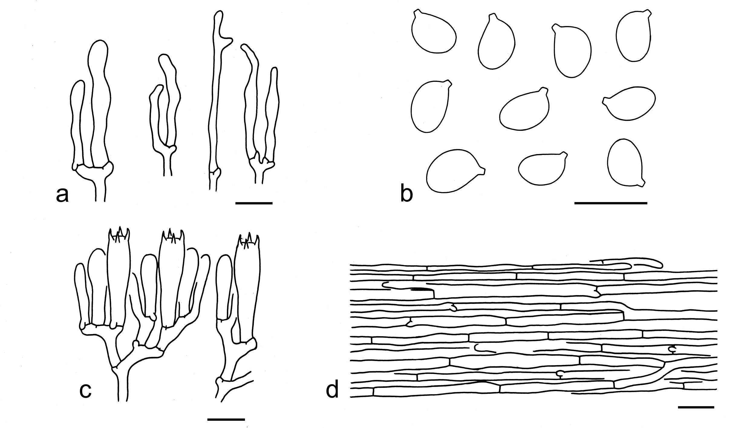
Known distribution: Known from Southwestern China.
Additional specimen examined: CHINA. Yunnan Province, Diqing Tibetan Autonomous Prefecture, Shangri-La, in a forest with Picea and Abies, altitude 3900 m, 4 Aug. 2018, Kui Wu 129 (KUN-HKAS134999).
Notes: Tricholoma ellipsosporum is well delimited by its olivaceous grey pileus, broadly ellipsoid to ellipsoid basidiospores (6–8 4–6 µm), conspicuous cheilocystidia and presence of clamps. Tricholoma olivaceum is sister to T. ellipsosporum in the phylogenetic analysis based on five gene fragments (Fig. 3). However, T. olivaceum has close lamellae, smaller basidiospores (5–6.5 3.5–5 µm) and poorly differentiated cheilocystidia (Reschke et al. 2018; this study).
57. Tricholoma forteflavescens Reschke et al, Mycologia 110(6): 1102 (2018). Fig. 75
Description: Basidioma medium-sized. Pileus 4–8 cm in diam., convex to plano-convex, without an umbo or depression at center; surface dry, light grey (1C1, 1D1) to silver grey (1B1), often darker at center and becoming paler towards margin, sometimes with reddish (7B4) tinge, staining yellow (3B4–6) when old, damaged or touched, glabrous or often centrically cracking; margin at first involute, then straight; context white (1A1) to greyish (1B1–2). Lamellae sinuate, greyish cream (3B2–4) to grey (1B1–2), staining yellow (3A3–5) when old or after damage, subclose; edges concolor. Stipe 5–10 1–2.5 cm, cylindrical to subclavate, greyish white (2B2–4), covered with dark grey (2E1–3) to grey (1D1–3) fibrils, staining yellow (3A3–5) after touch, solid, exannulate. Taste weak. Odour like unperfumed soap.
Ecology: Solitary to scattered on soil in mixed forests with Pinus and Quercus.
Known distribution: Known from Southwestern China.
Specimen examined: CHINA. Yunnan Province, Yulong Snow Mountain, in a forest with Quercus aquifolioides and Pinus sp., altitude 3500 m, 20 Aug. 2013, G. Kost & F. Popa FP1006 (holotype, KUN-HKAS93511).
Notes: Tricholoma forteflavescens is easily recognized in sect. Rigida by its yellow color change with age and when damaged and ellipsoid to elongate basidiospores (6–8 3.5–4 µm). Tricholoma atrovirens is related to this species with moderate support (Fig. 3), but differs by its basidioma without yellow discoloration and smaller basidiospores (4.5–6 3–4 µm) (this study).
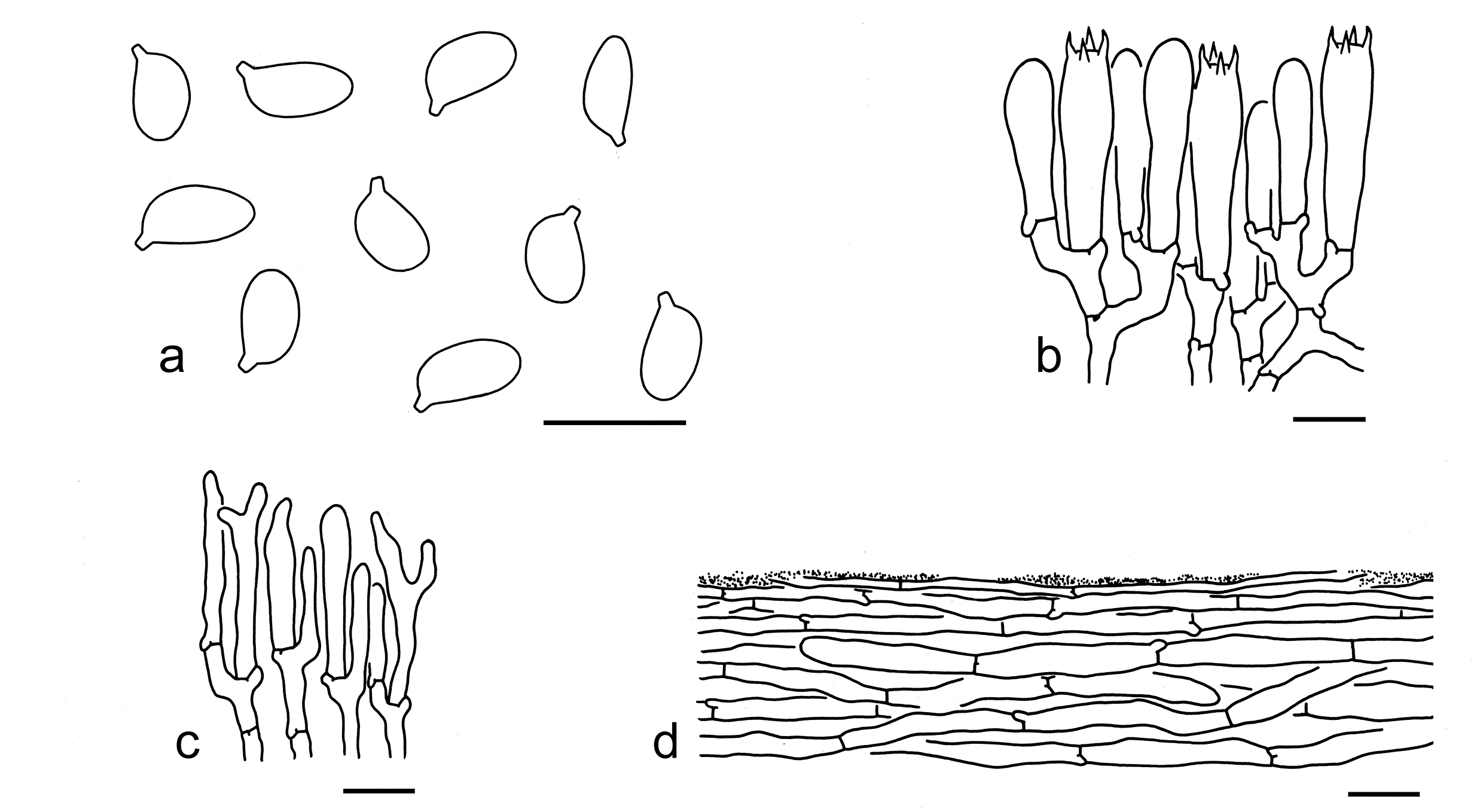
58. Tricholoma olivaceum Reschke et al., Mycologia 110(6): 1101 (2018). Figs. 69f, 76
Description: Basidioma small to medium-sized. Pileus 2–7 cm in diam., convex to plano-convex, sometimes umbonate at center; surface dry, grey (1E1–3), grey-brown (2E3–5) to yellow-brown (2C4–6), with distinct greenish (30B6–8) colors, glabrous; margin at first involute, then inflexed; context white (1A1). Lamellae adnexed to sinuate, white (1A1) to greenish yellow (30A4–6), close to subdistant; edges concolorous. Stipe 3–11 0.5–1.5 cm, cylindrical, often taper downwards, grey-brown (2E3–5) to brownish (1B2–4), with greenish (30B6–8) colors, exannulate. Taste not recorded. Odour like almond or cucumber.
Basidiospores [80/4/4] (4–) 5–6.5 (–8) (3–) 3.5–5 µm (mean length = 5.6 µm, mean width = 4.0 µm), Q = (1.2–) 1.25–1.55 (–1.83), Qm = 1.40 ± 0.12, broadly ellipsoid to ellipsoid, non-amyloid, colorless, hyaline, thin-walled, smooth; apiculus relatively large. Basidia 25–35 5–7 µm, clavate, 4-spored, hyaline; sterigmata 3–5 µm long; basal septa with clamps. Pleurocystidia absent. Cheilocystidia poorly differentiated. Subhymenium composed of 2–4 µm wide filamentous hyphae, clamps present. Lamellar trama regular, composed of colorless, thin-walled hyphae 3–20 µm wide. Pileipellis a cutis, non- to slightly gelatinized, composed of radially arranged, 2–8 µm wide, thin-walled, yellowish filamentous hyphae; subpellis poorly differentiated. Clamps present in all parts of basidioma.
Ecology: Solitary to scattered on soil in subtropical broad-leaved, coniferous or mixed forests with Fagaceae and Pinaceae.
Known distribution: Known from Southwestern China.
Notes: Tricholoma olivaceum is delimitated by its grey-brown to yellow-brown pileus with obviously greenish tinge, close and white to greenish yellow lamellae, broadly ellipsoid to ellipsoid basidiospores (5–6.5 3.5–5 µm) and presence of clamps (this study). Our phylogenetic analysis based on five gene fragments indicated that it is sister to T. ellipsosporum (Fig. 3). For comparison, see notes under that name.
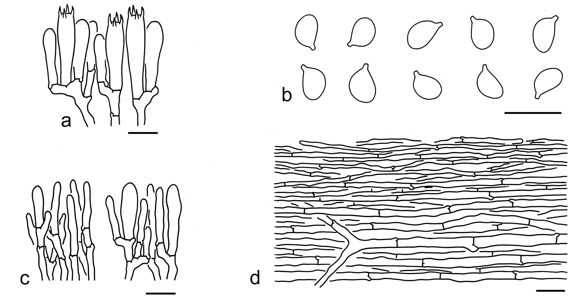
59. Tricholoma saponaceum (Fr.) P. Kumm., Führ. Pilzk. (Zerbst): 133 (1871). Figs. 69g, 77
Description: Basidioma medium-sized, with weaker reddish discoloration. Pileus 5–10 cm in diam., plano-convex to applanate, umbonate at center; surface dry, greyish brown (4E6–8) to brown (5E6–8, 5D6–8) at center, becoming yellowish brown (2B2–4), brownish (5C3–5) to yellowish grey (3B2–4) towards margin; margin straight; context dirty white (2B2) to white (1A1). Lamellae sinuate, cream (1A2) to yellowish (4A2–3), subdistant; edges cream (1A2) to yellowish (4A2–3). Stipe 8–12 1–2 cm, subcylindrical, yellowish (1A2–4) to brownish (5C5–7), longitudinally striate, stuffed, exannulate. Taste not recorded. Odour like rotten wood.
Basidiospores [31/2/2] (5–) 6–7.5 3.5–5 µm (mean length = 6.5 µm, mean width = 4.5 µm), Q = 1.32–1.61 (–1.75), Qm = 1.46 ± 0.1, ellipsoid, occasionally elongate, non-amyloid, colorless, hyaline, thin-walled, smooth; apiculus relatively large. Basidia 25–40 6–8 µm, clavate, 4-spored, hyaline; sterigmata 4–6 µm long; basal septa with clamps. Pleurocystidia absent. Cheilocystidia 25–50 2–5 µm, conspicuous, filiform, subclavate to clavate, often septate, thin-walled, colorless, hyaline. Subhymenium composed of 2–4 µm wide filamentous hyphae, clamps present. Lamellar trama regular, composed of colorless, thin-walled, cylindrical, subcylindrical to filamentous hyphae 5–35 (–60) µm wide. Pileipellis a cutis, composed of radially arranged, 5–10 µm wide, thin- to slightly thick-walled, colorless to brownish filamentous hyphae; subpellis composed of radially arranged, more or less slightly inflated hyphae, ellipsoid, cylindrical, cylindrical to filamentous, 5–25 µm wide, thin-walled, colorless to brownish. Clamps present in all parts of basidioma.
Ecology: Solitary to scattered on soil in forests with trees of Abies.
Distribution: Known from Southwestern China.
Specimens examined: CHINA. Sichuan Province, Ganzi Tibetan Autonomous Prefecture, Kangding, Gonggashan Village, in a forest with Abies, altitude 3650 m, 4 Sep. 2016, KD-FB 8 (KUN-HKAS99361); same location, in a forest with Abies, altitude 3650 m, 4 Sep. 2016, KD-FB 7 (KUN-HKAS99360).
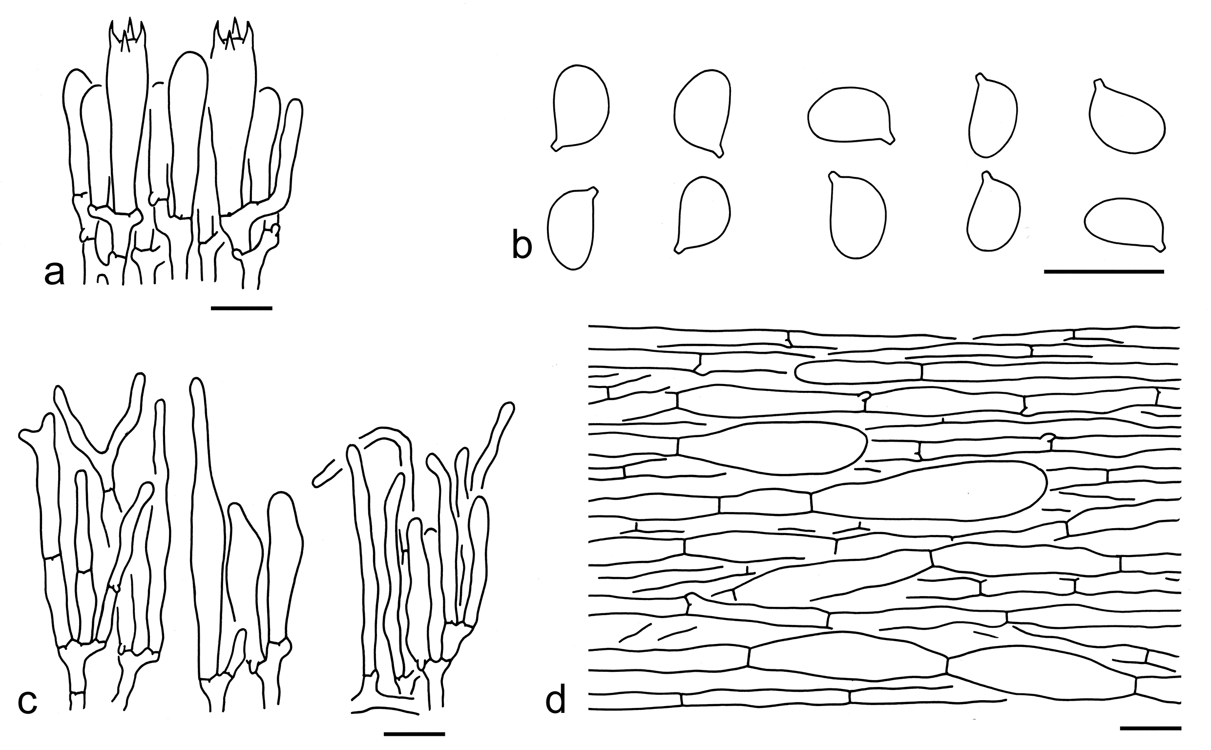
60. Tricholoma sichuanense Y.Y. Cui & Zhu L. Yang, sp. nov. Figs. 69h, 78
Fungal Names: FN 572349
Etymology: sichuanense = Sichuan Province, referring to its type locality.
Diagnosis: Similar to T. olivaceum, but differs in its pileus often with squamules at center and occurrence in subalpine forests dominated with Picea.
Type: CHINA. Sichuan Province, Ganzi Tibetan Autonomous Prefecture, Kangding, Gonggashan Village, in a forest with Picea and Rhododendron, altitude 4000 m, 21 Sep. 2017, Xiao-Xia Ding 331 (holotype, KUN-HKAS105344).
Description: Basidioma small to medium-sized. Pileus 3–7 cm in diam., convex to plano-convex, slightly umbonate or not; surface dry, often grey-brown (2F6–8) to brown (4E6–8) at center, becoming olivaceous yellow (1C6–8) towards margin, sometimes grey-brown (2F6–8) overall, covered with grey-brown (2F6–8) to brown (4E6–8) squamules; margin at first involute, then straight; context white (1A1) to cream (1A2). Lamellae sinuate, white (1A1) to pale olivaceous yellow (1B2–4), sometimes with pinkish (7A2–4) hues, subclose; edges concolorous. Stipe 7–10 0.5–1.5 cm, cylindrical to subcylindrical, dirty white (1B1), greyish olivaceous (1B2–4), brownish (4B3–5) to pinkish (7A2–4), exannulate. Taste and odour not recorded.
Basidiospores [50/3/2] 5–7 (–8) 4–5 µm (mean length = 6.4 µm, mean width = 4.5 µm), Q = 1.23–1.6 (–1.7), Qm = 1.42 ± 0.11, broadly ellipsoid to ellipsoid, non-amyloid, colorless, hyaline, thin-walled, smooth; apiculus relatively large. Basidia 25–35 6–8 µm, clavate, 4-spored, hyaline; sterigmata 4–6 µm long; basal septa with clamps. Pleurocystidia absent. Cheilocystidia 15–35 3–8 µm, filiform, subclavate to clavate or irregular shaped, thin-walled, colorless, hyaline. Subhymenium composed of 2–4 µm wide filamentous hyphae, clamps present. Lamellar trama regular, composed of colorless, thin-walled hyphae 5–25 µm wide. Pileipellis a cutis, sometimes slightly gelatinized, composed of radially arranged, 3–15 µm wide, thin-walled, yellowish to yellowish brown filamentous hyphae; subpellis poorly differentiated. Clamps present in all parts of basidioma.
Ecology: Solitary to scattered on soil in subalpine temperate forests dominated with Picea.
Known distribution: Known from Southwestern China.
Additional specimens examined: CHINA. Sichuan Province, Ganzi Tibetan Autonomous Prefecture, Kangding, Gonggashan Village, in a forest with Picea, altitude 3650 m, 4 Sep. 2016, KD-WPM15 (KUN-HKAS98270); same city, Yala Village, in a forest with Picea, altitude 3290 m, 21 Aug. 2017, Jian-Wei Liu 933 (KUN-HKAS106308).
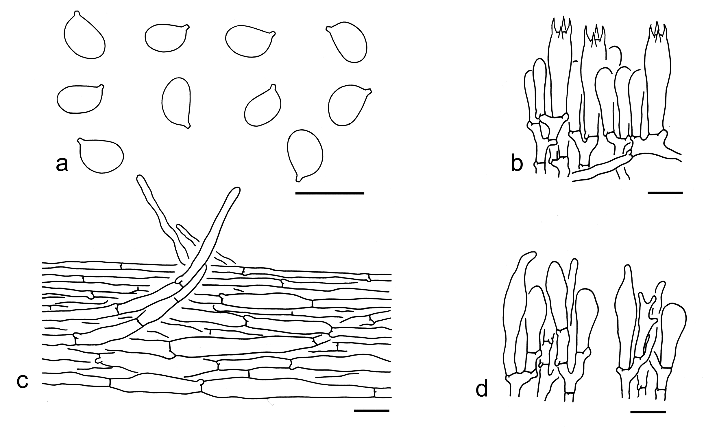
61. Tricholoma yunnanense Y.Y. Cui & Zhu L. Yang, sp. nov. Figs. 69i, 79
Fungal Names: FN 572351
Etymology: yunnanense = Yunnan, referring its type locality from Yunnan province.
Diagnosis: Related to T. asiaticum, but differs by its glabrous and brown, brownish to dirty white pileus.
Type: CHINA. Yunnan Province, Kunming, Panlong District, Shuanglong Village, Yeya Lake, in a mixed forest with Fagaceae and Pinaceae, altitude 2100 m, 1 Sep. 2013, Jiao Qin 847 (holotype, KUN-HKAS81285).
Description: Basidioma medium-sized. Pileus 4–10 cm in diam., at first convex, then convex-applanate, without an umbo or depression at center; surface dry, grey-brown (3E1–3) to brown (6E3–5) at center, becoming brownish (5D3–5) to dirty white (1B2) towards margin, sometimes brown (5D4–6) overall, glabrous; margin straight; context dirty white (1B2) to pinkish (5A2–3). Lamellae sinuate, white (1A1), staining brown (4B2–4) to pinkish (5A2–3) tinge when old, close; edges concolor. Stipe 6–9 0.7–2 cm, cylindrical to subcylindrical, dirty white (1A1, 1B1), greyish (1B2–3) to brownish (5B2–4), hollow, exannulate. Taste not recorded. Odour like sesame paste.
Basidiospores [20/1/1] (4–) 4.5–6 3–4 µm (mean length = 5 µm, mean width = 3.6 µm), Q = (1.13–) 1.25–1.57 (–1.83), Qm = 1.4 ± 0.18, broadly ellipsoid to ellipsoid, non-amyloid, colorless, hyaline, thin-walled, smooth; apiculus relatively large. Basidia 20–30 5–7 µm, clavate, 4-spored, hyaline; sterigmata 3–5 µm long; basal septa with clamps. Pleurocystidia absent. Cheilocystidia not observed. Subhymenium composed of 2–5 µm wide filamentous hyphae, clamps present. Lamellar trama regular, composed of colorless, thin-walled, cylindrical hyphae 5–15 µm wide. Pileipellis a cutis, non- to slightly gelatinized, composed of radially arranged, 3–7 µm wide, thin-walled, colorless filamentous hyphae; subpellis poorly differentiated. Clamps present in all parts of basidioma.
Ecology: Solitary to scattered on soil in subtropical conifers or mixed forests with Pinaceae and Fagaceae.
Known distribution: Known from Southwestern China.
Additional specimen examined: CHINA. Yunnan Province, Puer City, Lancang Lahu Autonomous County, Nuofu Village, in a coniferous forest with Pinus, altitude 2100 m, 1 Sep. 2013, Xiao-Xia Ding 268 (KUN-HKAS106610).
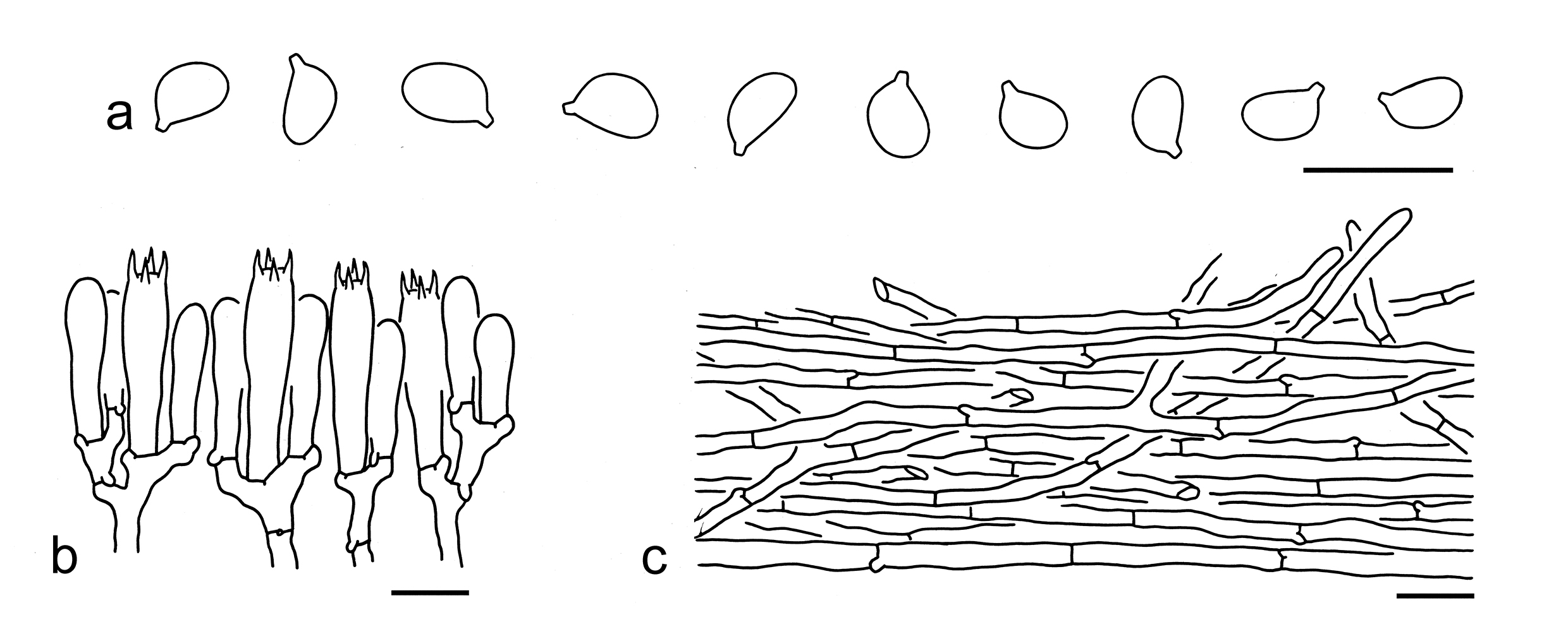
Tricholoma subgen. Pardinicutis Singer
Type: Tricholoma pardinum (Pers.) Quél., Mém. Soc. Émul. Montbéliard, Sér. 2 5: 339 (1873).
Description: Basidioma tricholomatoid. Pileus mostly brown or grey, squamose. Lamellae crowded to close. Annulus absent. Basidiospores broadly ellipsoid to ellipsoid, thin-walled, smooth, inamyloid. Pileipellis a cutis composed of repent filamentous hyphae.
Ecology: Ectomycorrhizal. On soil with various trees.
Known distribution: Molecular data indicated species from this subgenus are found in Europe, North America, and Asia.
Notes: Tricholoma subgen. Pardinicutis is supported to be a monophyletic group and comprises only one section named sect. Pardinicutis. Currently, two species of this subgenus are found in China (see below).
Tricholoma sect. Pardinicutis
Type: Tricholoma pardinum (Pers.) Quél., Mém. Soc. Émul. Montbéliard, Sér. 2 5: 339 (1873).
Description: Basidioma tricholomatoid and robust. Pileus umbonate or not, whitish, greyish to brownish, squamose. Lamellae crowded to close. Annulus absent. Basidiospores medium-sized to large, broadly ellipsoid to ellipsoid, thin-walled, smooth, inamyloid. Pleurocystidia absent. Cheilocystidia conspicuous or absent. Pileipellis a cutis composed of repent filamentous hyphae. Clamps present.
Ecology: Ectomycorrhizal. On soil with various trees.
Known distribution: Molecular data indicated that species from this section are collected from Europe, North America, and Asia.
Notes: To date, T. highlandense and T. nigrosquamosum are found in China (see below).
Key to the species of Tricholoma sect. Pardinicutis from China
1. Basidiospores relatively smaller (6.5–8 5–6 µm, mean length = 7.3 µm, mean width = 5.5 µm); cheilocystidia clavate or mucronate; associations mainly with Pinus …………………T. highlandense
1. Basidiospores relatively larger (8.5–10.5 6.5–7.5 µm, mean length = 9.3 µm, mean width = 6.9 µm); cheilocystidia clavate without mucronate shaped; associations mainly in forests with trees of Picea T. nigrosquamosum
62. Tricholoma highlandense Zhu L. Yang et al., Phytotaxa 305(1): 5 (2017). Figs. 69j, 80
Description: Basidioma medium-sized to large. Pileus 5–10 cm in diam., plano-convex to applanate, broadly umbonate or not; surface dry, white (1A1), dirty white (3B1) to brownish (1B2–4), covered with brown (2C2–4) to grey-brown (2E3–5), somewhat reflexed scales; margin involute to inflexed; context white (1A1). Lamellae adnexed to sinuate, white (1A1), dirty white (1B1) to cream (1B2), crowded; edges white (1A1) to cream (1B2). Stipe 3–9 1–3 cm, cylindrical, often slightly inflated at base, white (1A1) to cream (1B2), covered with dirty white (1B1) to brownish (3C2–5) fibrillose squamules, exannulate. Taste and odour indistinct or farinaceous.
Basidiospores [60/3/3] 6.5–8 (–9.5) 5–6 (–6.5) µm (mean length = 7.3 µm, mean width = 5.5 µm), Q = (1.12–) 1.21–1.46 (–1.50), Qm = 1.33 ± 0.08, broadly ellipsoid to ellipsoid, non-amyloid, colorless, hyaline, thin-walled, smooth; apiculus relatively large. Basidia 37–50 8–10 µm, clavate, 4-spored, rarely 2-spored, hyaline; sterigmata 4–5 µm long; basal septa with clamps. Pleurocystidia absent. Cheilocystidia 40–50 8–10 µm, conspicuous, clavate, often mucronate, thin-walled, colorless, hyaline. Subhymenium composed of 2.5–5 µm wide filamentous hyphae, clamps present. Lamellar trama regular, composed of colorless, thin-walled hyphae 3–15 µm wide. Pileipellis a cutis with transition to trichoderm at regular intervals, composed of radially arranged, 3–8 µm wide, thin-walled, yellowish to brownish filamentous hyphae; subpellis poorly differentiated. Clamps present in all parts of basidioma.
Ecology: Solitary to scattered on soil in subtropical evergreen mixed or coniferous forests.
Known distribution: Known from Southwestern China (Yang et al. 2007).
Notes: Tricholoma highlandense is characterized by its white pileus covered with coarsely brown to grey-brown scales, broadly ellipsoid to ellipsoid basidiospores, clavate to mucronate cheilocystidia, and the presence of clamps (this study). In the phylogenetic analyses (Figs. 3, 4), this species is sister to T. filamentosum (Alessio) Alessio, but the latter has a shiny pileus with little contrasting-coloured scales, which are pale grey or olivaceous buff or smoke grey, poorly differentiated cheilocystidia and are mainly with deciduous trees (Christensen & Heilmann-Clausen 2013).
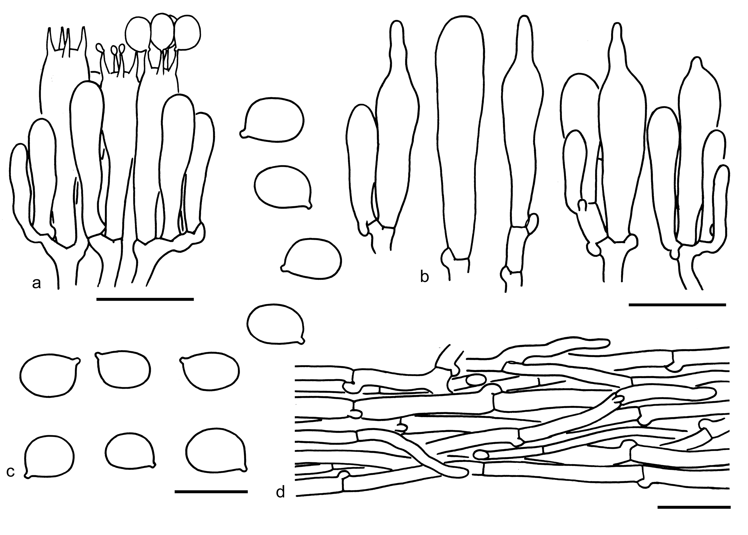
Tricholoma nigrosquamosum can be confused, but differs by its relatively larger basidiospores (8.5–10.5 6.5–7.5 µm, mean length = 9.3 µm, mean width = 6.9 µm), clavate cheilocystidia without mucronate shapes, and associations with subalpine temperate forests (Yang et al. 2017; Wang et al. 2024; this study). Tricholoma highlandense is also similar to T. pardinum. However, T. pardinum has relatively larger basidiospores (7.5–9.5 5.5–6.5 µm, mean length = 8.5 µm, mean width = 6.1 µm), poorly differentiated cheilocystidia and forms mycorrhizal relationships with Fagus and Abies on calcareous soil (Riva 1988; Bessette et al. 2013; Christensen & Heilmann-Clausen 2013; Yang et al. 2017).
63. Tricholoma nigrosquamosum (P.G. Liu) Zhu L. Yang & G.S. Wang, Mycology 15(2): 205 (2023) [2024]. Figs. 69k, 81
Basionym: Tricholomopsis nigrosquamosa P.G. Liu, Acta Mycol. Sin. 13(3): 183 (1994).
Synonymy: Tricholoma sinopardinum Zhu L. Yang et al., Phytotaxa 305(1): 6 (2017).
Description: Basidiomata medium-sized to large. Pileus 5–11 cm in diam., at first convex, then plano-convex to applanate, often broadly umbonate; surface dry, white (1A1), dirty white (1B1–3) to greyish (2B1–3), covered with brown (2B2–4, 3C2–4) to dark brown (3D2–4, 3E2–4), fibrillose scales; margin involute to inflexed; context white (1A1) to cream (1A2). Lamellae adnexed to sinuate, white (1A1), dirty white (1B1) to cream (1B2–3), crowded; edges brownish (4B3–5) to yellow-brown (3B3–5). Stipe 5–18 1–3 cm, cylindrical to clavate, often slightly inflated at base, white (1A1) to brownish (3B2–4), covered with brownish (3B2–4) to brown (3D2–4), fibrillose squamules, exannulate. Taste and odour indistinct or farinaceous or fishlike.
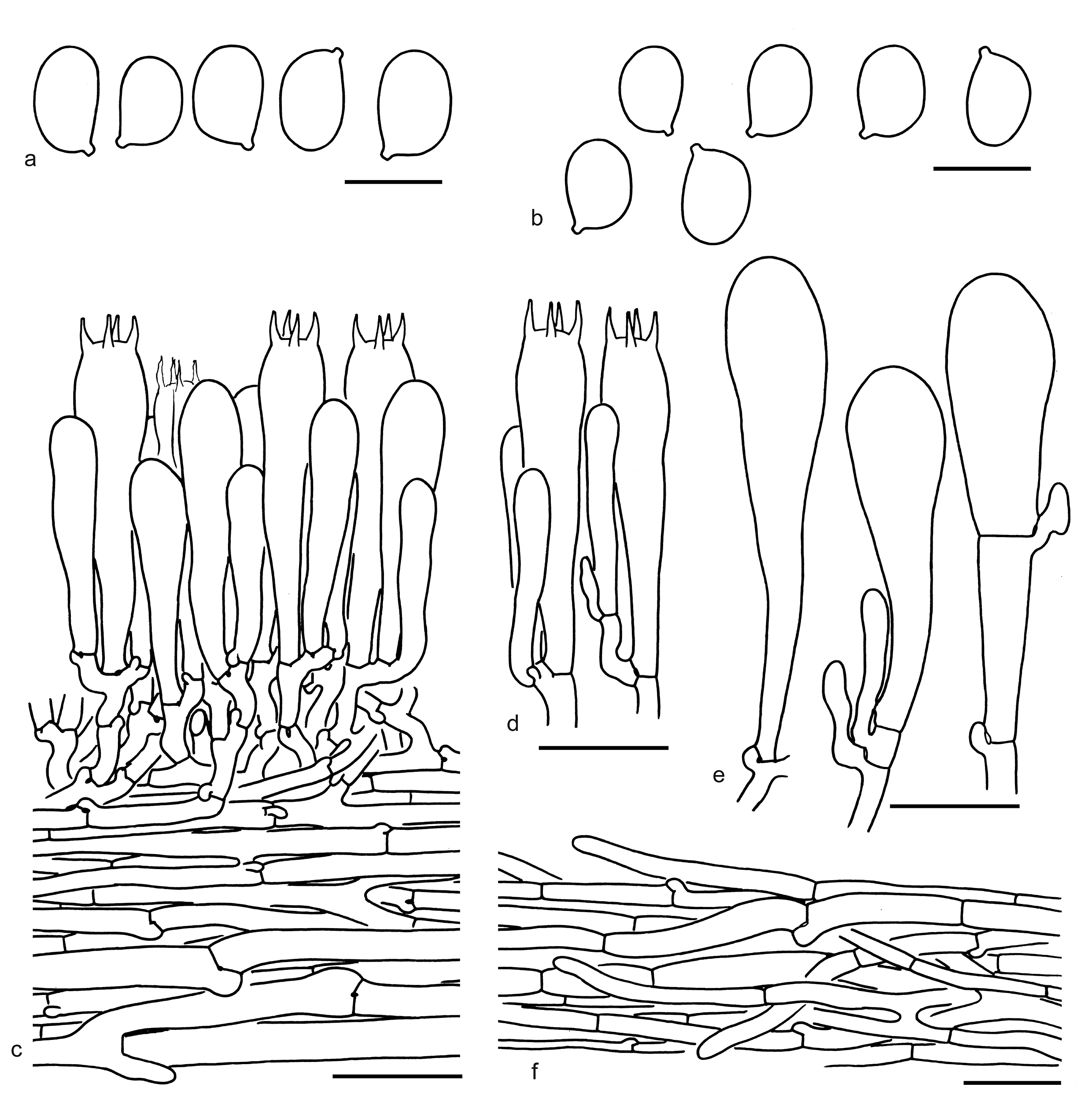
Basidiospores [115/6/5] (8–) 8.5–10.5 (–11.5) (5.5–) 6.5–7.5 (–8) µm (mean length = 9.3 µm, mean width = 6.9 µm), Q = (1.20–) 1.25–1.50 (–1.62), Qm = 1.36 ± 0.09, mostly ellipsoid, sometimes broadly ellipsoid, non-amyloid, colorless, hyaline, thin-walled, smooth; apiculus relatively large. Basidia 45–55 (–65) 9–11 (–15) µm, clavate, 4-spored, rarely 2-spored, hyaline; sterigmata 4–5 µm long; basal septa with clamps. Pleurocystidia absent. Cheilocystidia 40–75 15–25 µm, conspicuous, clavate, sometimes septate, thin- to slightly thick-walled (≤ 0.5 µm), colorless, sometimes brownish or yellowish, hyaline. Subhymenium composed of 2.5–4 µm wide filamentous hyphae, clamps present. Lamellar trama regular, composed of colorless, thin-walled hyphae 3–30 µm wide. Pileipellis a cutis with transition to a trichoderm at regular intervals, composed of radially arranged, 3–10 (–15) µm wide, thin-walled, yellowish to brownish filamentous hyphae; subpellis poorly differentiated. Clamps present in all parts of basidioma.
Ecology: Solitary to scattered on soil in subalpine forests dominated by Picea, sometimes mixed with Betula, Populus or Quercus.
Known distribution: known from Southwestern China and Nepal.
Specimens examined: CHINA. Xizang Autonomous Region, Jiangda County, Jiangda Town, in a forest dominated by Picea and Populus, altitude 3500 m, 8 Aug. 2013, Bang Feng 1427 (KUN-HKAS82533); same town, in a forest dominated by Picea and Betula, altitude 4100 m, 4 Aug. 2009, Zhu L. Yang 5459 (KUN-HKAS58001); same county, Tongpu Town, in a forest dominated by Picea, altitude 3300 m, 2 Aug. 2004, Zai-Wei Ge 261 (KUN-HKAS46041); Mangkang County, on the way to Sichuan, in a forest dominated by Picea likiangensis, altitude unknown, 16 Jul. 2009, Bang Feng 470 (KUN-HKAS57199); Xiangcheng County, Reda Town, in a forest dominated by Picea and Quercus, altitude 3600 m, 16 Jul. 2004, Zhu L. Yang 4143 (KUN-HKAS45529). Sichuan Province, Ganzi Tibetan Autonomous Prefecture, Luhuo County, in a forest dominated by Picea, altitude 3100 m, 16 Jul. 2014, Kuan Zhao 551 (KUN-HKAS89214); Litang County, in a forest dominated by Picea, altitude 4000 m, 8 Aug. 2014, Kuan Zhao 712 (KUN-HKAS89643); same county, forest type and altitude not recorded, 5 Aug. 2016, Jian-Wei Liu 474 (KUN-HKAS98053).
Notes: Tricholoma nigrosquamosum is delimitated by its white pileus covered with brown to dark brown scales, mostly ellipsoid basidiospores, clavate cheilocystidia, the presence of clamps and associated with subalpine temperate forests dominated by Picea (this study). Tricholoma nigrosquamosum is closely related to the clade formed by T. highlandense and T. filamentosum in the phylogenetic analyses (Figs. 3, 4). Indeed, T. nigrosquamosum can be confused with T. highlandense, and the differences between them has been discussed under T. highlandense. Tricholoma filamentosum has a shiny pileus with little contrasting colored scales, relatively smaller basidiospores (6.6–9.8 4.7–7.1 µm, average 7.6–8.5 5.8–6.2 µm), poorly differentiated cheilocystidia and is mainly with deciduous trees (Christensen & Heilmann-Clausen 2013). Tricholoma pardinum is similar to T. nigrosquamosum, but the former has relatively smaller basidiospores (7.5–9.5 5.5–6.5 µm, mean length = 8.5 µm, mean width = 6.1 µm) and poorly differentiated cheilocystidia (Riva 1988; Bessette et al. 2013; Christensen & Heilmann-Clausen 2013; Yang et al. 2017).
Tricholoma subgen. Sericeicutis Singer
Type: Tricholoma sulphureum (Bull.) P. Kumm., Führ. Pilzk. (Zerbst): 133 (1871).
Description: Basidioma tricholomatoid. Pileus umbonate or not, white, yellow or brown, glabrous. Lamellae close to distant. Annulus absent. Basidiospores variable in shape and size, broadly ellipsoid, ellipsoid to elongate, sometimes amygdaliform, thin-walled, smooth, inamyloid. Pileipellis a cutis, sometimes with its subpellis composed of more or less inflated hyphae. Clamps present or absent.
Ecology: Ectomycorrhizal. On soil with various trees.
Known distribution: Molecular data showed that species from this subgenus are collected from Europe, North America, and Asia.
Notes: Tricholoma subgen. Sericeicutis is proved to be a monophyletic group and includes two sections named sect. Sericella and sect. Lasciva. To date, six species of subgen. Sericeicutis are collected from China (see below).
Tricholoma sect. Lasciva Bon
Type: Tricholoma lascivum (Fr.) Gillet, Hyménomycètes (Alençon): 111 (1874) [1878].
Description: Basidioma tricholomatoid and pale colored. Pileus umbonate or not, white, glabrous. Lamellae close. Annulus absent. Basidiospores small, broadly ellipsoid, ellipsoid to elongate, smooth, inamyloid. Cheilocystidia absent. Pileipellis a cutis with repent filamentous hyphae. Clamps present.
Ecology: Ectomycorrhizal. On soil with various trees.
Known distribution: Molecular data indicated species from this section are found in Europe, North America, and Asia.
Notes: To date, Tricholoma lishanense L. Fan & J.J. Yang and T. stiparophyllum (N. Lund) P. Karst. have been reported from China (see below).
Key to the species of Tricholoma sect. Lasciva from China
1. Pileus relatively smaller, 3–5 cm in diam.; context with a honey-like smell; associations with Quercus T. lishanense
1. Pileus relatively larger, 6–12 cm in diam.; context with an unpleasant smell; associations with Betula…………………… ……………………………T. stiparophyllum
64. Tricholoma lishanense L. Fan & J.J. Yang, Phytotaxa 598(1): 92 (2023). Figs. 69l, 82
Description: Basidioma small. Pileus 3–5 cm in diam., convex to plano-convex, sometimes slightly umbonate at center; surface dry, smooth, glabrous, white (1A1) to cream (1A2); margin straight; context white (1A1). Lamellae sinuate, white (1A1), very crowded; edges white (1A1). Stipe 4–7 0.5–0.8 cm, cylindrical, slightly widen at base, white (1A1), glabrous, exannulate. Taste not recorded. Odour like honey.
Basidiospores [40/2/1] 5.5–6.5 (–7) (3–) 3.5–4.0 µm (mean length = 6.0 µm, mean width = 3.8 µm), Q = 1.37–1.75 (–1.83), Qm = 1.55 ± 0.1, ellipsoid to elongate, non-amyloid, colorless, hyaline, thin-walled, smooth; apiculus relatively large. Basidia 25–30 6–8 µm, clavate, 4-spored, hyaline; sterigmata 3–5 µm long; basal septa with clamps. Pleurocystidia absent. Cheilocystidia 20–35 4–8 µm, poorly differentiated, narrowly clavate to clavate, thin-walled, colorless, hyaline. Subhymenium composed of 3–5 µm wide filamentous hyphae, clamps present. Lamellar trama regular, composed of colorless, thin-walled hyphae 3–10 µm wide. Pileipellis a cutis, composed of radially arranged to interwoven, 2–8 µm wide, thin-walled, colorless filamentous hyphae; subpellis poorly differentiated. Clamps present in all parts of basidioma.
Ecology: Solitary to scattered on soil in broad-leaved forests with Quercus.
Known distribution: Known from northern (Yang et al. 2023) and Northeastern China.
Notes: Tricholoma lishanense is characterized by its small, white basidioma, ellipsoid to elongate basidiospores (5.5–6.5 3.5–4.0 µm, Q = 1.37–1.75, Qm = 1.55 ± 0.1), honey-like smells and associations with Quercus (this study). This species is closely related and very similar to T. album (Schaeff.) P. Kumm (Figs. 3, 4), but differs by its very crowded lamellae.
Tricholoma lascivum (Fr.) Gillet is similar to T. lishanense, but has a pale greyish yellow pileus and a rancid smell (Riva 1988, 2003; Christensen & Noordeloos 1999; Christensen & Heilmann-Clausen 2013). Tricholoma stiparophyllum has a whitish basidioma and can be confused with T. lishanense. However, T. stiparophyllum has relatively larger basidioma and an unpleasant smell (Christensen & Noordeloos 1999; Christensen & Heilmann-Clausen 2013).
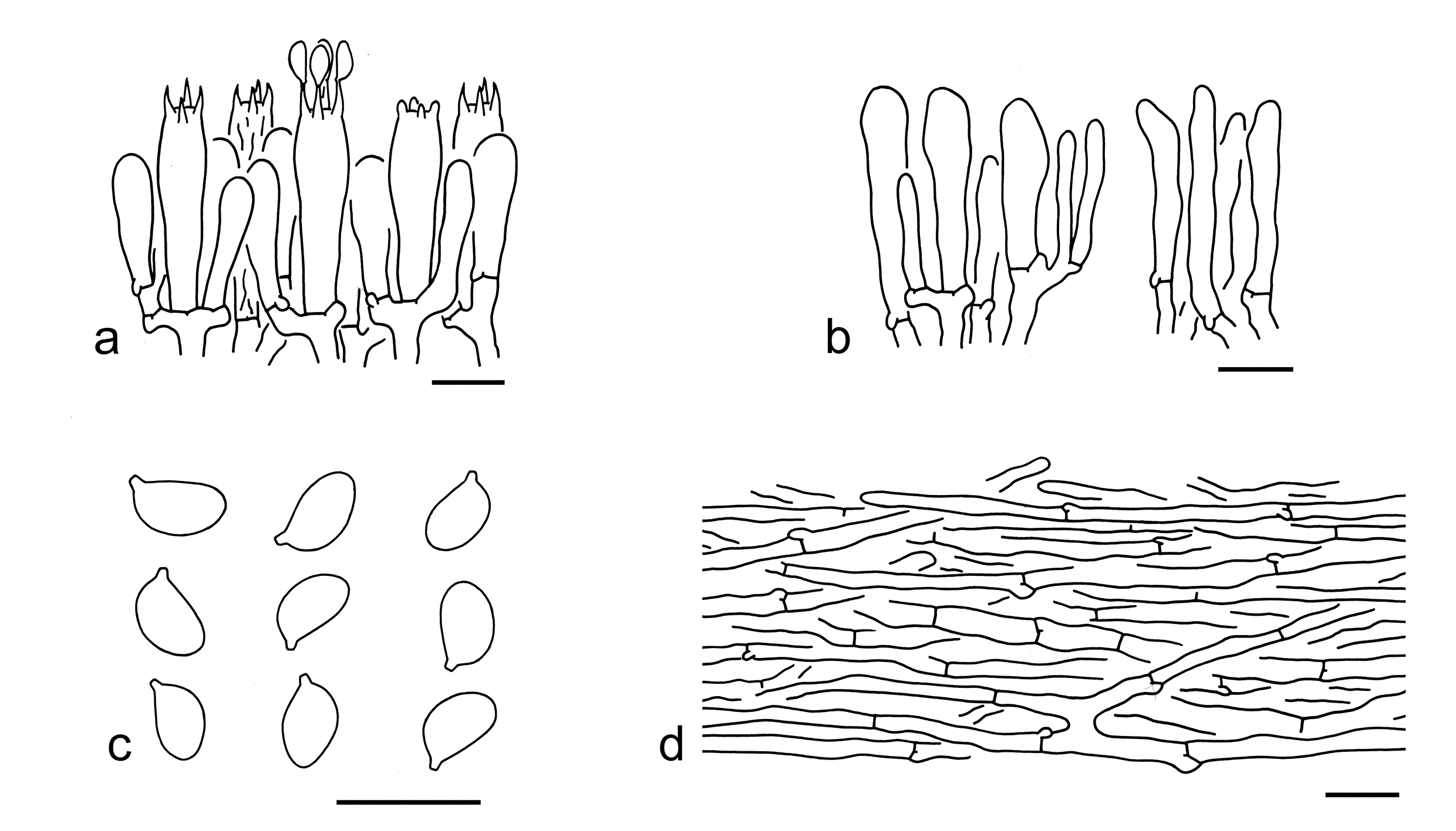
65. Tricholoma stiparophyllum (N. Lund) P. Karst., Meddn Soc. Fauna Flora fenn. 5: 42 (1879). Figs. 69m, 83
Basionym: Agaricus stiparophyllus N. Lund, Consp. Hymen. Holm (Christianiae): 10 (1845).
Description: Basidioma medium-sized to large. Pileus 6–12 cm in diam., convex to plano-convex, sometimes slightly umbonate at center; surface dry, smooth, glabrous, yellowish (2A2–4) to cream (1A2–4) at center, becoming pale cream (1A2) to white (1A1) towards margin; margin involute when young, becoming inflexed to straight when mature; context white (1A1). Lamellae adnate to sinuate, white (1A1) to cream (1A2–4), with brownish (4B4) spots when damaged or old, crowded; edges concolor. Stipe 8–11 1–2.5 cm, cylindrical, white (1A1) to cream (1A2–4), glabrous, exannulate. Taste not recorded. Odour unpleasant.
Basidiospores [20/1/1] 6–7 3.5–4.5 µm (mean length = 6.5 µm, mean width = 4 µm), Q = (1.47–) 1.5–1.8 (–1.91), Qm = 1.65 ± 0.12, ellipsoid to elongate, non-amyloid, colorless, hyaline, thin-walled, smooth; apiculus relatively large. Basidia 30–40 6–9 µm, clavate, 4-spored, hyaline; sterigmata 3–5 µm long; basal septa with clamps. Pleurocystidia absent. Cheilocystidia 35–50 3–5 µm, narrowly cylindrical to filamentous, thin-walled, colorless, hyaline. Subhymenium composed of 3–5 µm wide filamentous hyphae, clamps present. Lamellar trama regular, composed of colorless, thin-walled hyphae 3–15 µm wide. Pileipellis a cutis, composed of radially arranged, 3–10 µm wide, thin-walled, colorless filamentous hyphae; subpellis poorly differentiated. Clamps present.
Ecology: Solitary to scattered on soil in broad-leaved forests with Betula.
Known distribution: Known from Europe (Christensen & Noordeloos 1999; Christensen & Heilmann-Clausen 2013), North America (Trudell et al. 2022) and China.
Specimens examined: CHINA. Inner Mongolia, Hinggan League, Arxan, 30 Aug. 2006, HMGAU7829; same location and date, HMGAU7821. Xinjiang Uygur Autonomous Region, Altay Prefecture, forest unknown, altitude 1100 m, 6 Aug. 2003, Man-Shu Song 043 (HMAS96586). Xizang Autonomous Region, Zayü County, forest unknown, altitude 3585 m, 25 Aug. 2018, Xiang-Hua Wang 5825 (KUN-HKAS135000).
This species forms a strongly supported clade with T. album, T. lishanense, and T. lascivum (Figs. 3, 4). However, T. album has medium-spaced lamellae and association with Quercus (Riva 1988; Christensen & Heilmann-Clausen 2013). Tricholoma lishanense differs by its relatively smaller basidioma, honey-like smell and associations with Quercus (this study). Tricholoma lascivum has a pale greyish yellow basidioma and larger basidiospores (4.8–9.7 3–5.7 µm) (Riva 1988; Christensen & Heilmann-Clausen 2013). Tricholoma sulphurescens Bres. is morphologically comparable to T. stiparophyllum, but can be separated by its white basidioma strongly yellowing when touched and broadly ellipsoid basidiospores (4.5–7.6 3.9–6 µm) (Riva 1988; Christensen & Heilmann-Clausen 2013).
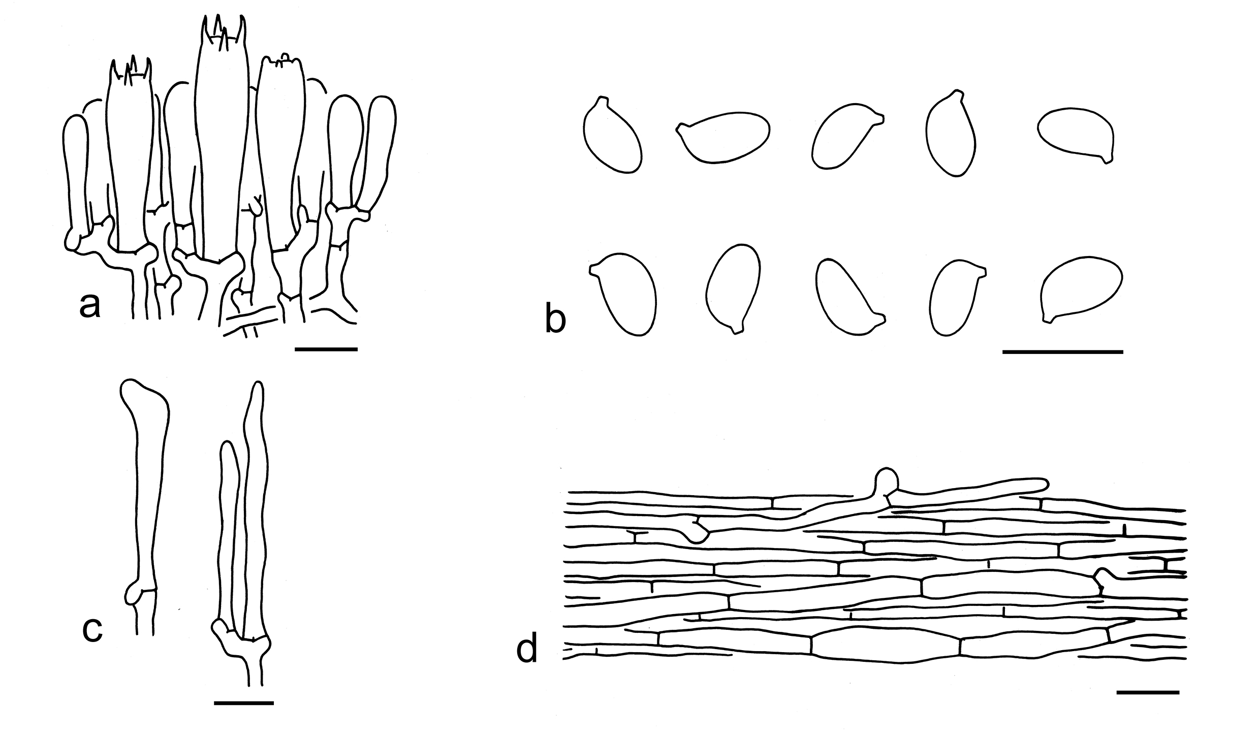
Tricholoma sect. Sericella (Fr.) Quél.
Type: Tricholoma sulphureum (Bull.) P. Kumm., Führ. Pilzk. (Zerbst): 133 (1871).
Description: Basidioma tricholomatoid. Pileus umbonate or not, whitish, cream, pinkish, sulphur-yellow, orangish brown to brown, glabrous. Lamellae close to distant, white, cream to sulphur-yellow. Annulus absent. Basidiospores large, ellipsoid to elongate, often somewhat amygdaliform, smooth, inamyloid. Cheilocystidia poorly differentiated. Pileipellis a cutis with its subpellis composed of radially arranged filamentous hyphae or inflated cells. Clamps present or absent.
Ecology: Ectomycorrhizal. On soil with various trees.
Known distribution: In Europe, North America, and Asia.
Notes: To date, four species have been reported from China (see below).
Key to the species of Tricholoma sect. Sericella from China
1. Basidioma paler colored, off-white, ivory to brownish colored; subpellis with obviously inflated cells………………. T. inamoenum
1. Basidioma sulphur-yellow, orangish brown to brown; subpellis poorly differentiated, mostly composed of radially arranged filamentous hyphae, occasionally with slightly inflated cells 2
2. Pileus orangish brown to brown at center and brownish to white at margin; lamellae white to cream T. tangerinum
2. Pileus sulphur-yellow; lamellae sulphur-yellow 3
3. Basidiospores relatively larger (10–12 5.5–7 µm, mean length = 10.6 µm, mean width = 6.2 µm); subpellis composed of radially arranged slightly inflated cells……… …………………T. sulphureum type Ⅰ
3. Basidiospores relatively smaller (9–11.5 5–6.5 µm, mean length = 9.9 µm, mean width = 5.4 µm); subpellis composed of radially arranged filamentous hyphae…………………………. …………………………T. sulphureum type Ⅱ
66. Tricholoma inamoenum (Fr.) Gillet, Hyménomycètes (Alençon): 112 (1874) [1878]. Figs. 69n, 84
Description: Basidioma small to medium-sized. Pileus 3–7 cm in diam., convex to plano-convex, with or without a broadly low umbo; surface dry, smooth to finely rugosus, off-white (1B1), ivory (1A2–3, 2A2–3), pale chrome to pale ochraceous (2B1, 3B1) to brownish (3B2–4); margin inflexed to straight, often waved; context off-white (1A1, 1B1). Lamellae sinuate, off-white (1A1, 1B1) to ivory (1A2–3, 2A2–3), distant; edges entire, off-white (1A1, 1B1) to ivory (1A2–3, 2A2–3). Stipe 6–12 0.7–1.2 cm, cylindrical, stuffed, off-white (1B1) to ivory (1A2–3, 2A2–3), with brownish (2B3–5) colors, fibrillose, exannulate. Taste mild. Odour strong of bitter almond.
Ecology: Solitary to scattered on soil in subalpine temperate forests with trees of Abies.
Known distribution: Known from southwestern China.
Specimens examined: CHINA. Xizang Autonomous Region, Qamdo, Markam County, Hongla Mountain, in a forest dominated by Abies, altitude 4200 m, 4 Aug. 2013, Xiao-Bin Liu 225 (KUN-HKAS79926). Yunnan Province, Diqing Tibetan Autonomous Prefecture, Daxueshan, in a forest dominated by species of Picea, Quercus and Rhododendron, altitude 4100 m, 8 Aug. 2009, Zhu L. Yang 5504 (KUN-HKAS58046).
Notes: Two Chinese specimens (KUN-HKAS79926 and KUN-HKAS58046) clustered with the neotype of T. inamoenum as a monophyletic group (Fig. 4). Because subtle morphological and ecological differences were observed among them, the Chinese collections are regarded as T. inamoenum for the time being. Tricholoma inamoenum is well recognized by its off-white to ivory basidioma, inflated cells in subpellis and associations with Abies and Picea trees (this study).
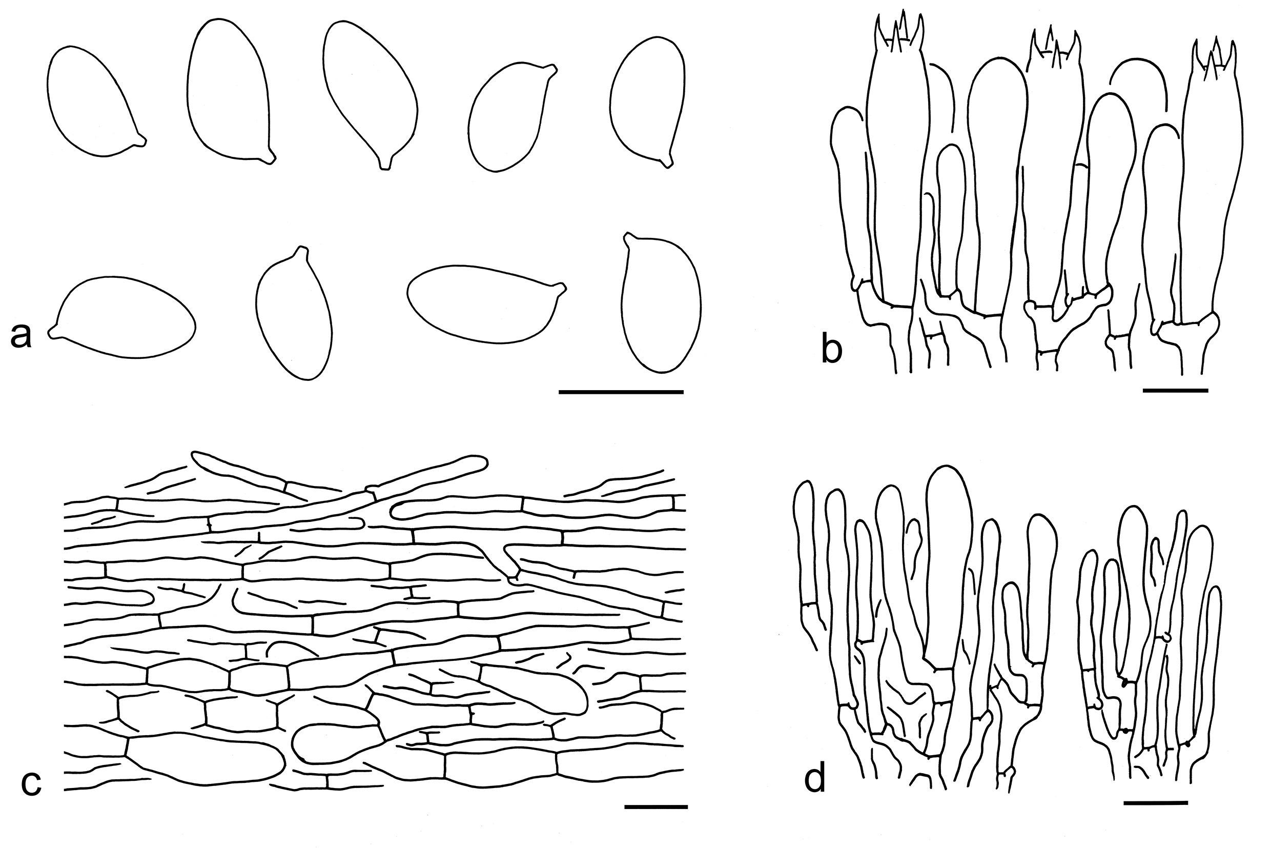
67. Tricholoma sulphureum (Bull.) P. Kumm. type Ⅰ, Führ. Pilzk. (Zerbst): 133 (1871). Figs. 69o, 85
Basionym: Agaricus sulphureus Bull., Herb. Fr. (Paris) 4: pl. 168 (1784) [1783-84].
Description: Basidioma small to medium-sized. Pileus 4–8 cm in diam., at first convex, then applanate, sometimes concave, sometimes slightly umbonate; surface dry, glabrous, sulphur-yellow (2A4–6, 2B2–5) with pale ochraceous (4C3–5) tinge; margin straight to slightly reflexed; context cream (1A2). Lamellae sinuate, yellow (1A2–4) to sulphur-yellow (1B4–7), often with grass greenish yellow (30A2–4) tinge, close; edges yellow (1A2–4) to sulphur-yellow (1B4–7). Stipe 7–10 0.8–1.5 cm, cylindrical, yellow (1A2–4) to sulphur-yellow (1B4–7), fibrillose, base with white-yellowish (1A2–3) hairs, exannulate. Taste unpleasant. Odour with strong gas or almond.
Ecology: Solitary to scattered on soil in broad-leaved forests.
Known distribution: Known from Europe, North America, and East Asia.
Specimens examined: CHINA. Yunnan Province, Kunming, Panlong District, Shuanglong Village, Yeya Lake, the type of forest unknown, altitude 2100 m, 8 Sept. 2008, Xi-Hui Du 57 (KUN-HKAS55509). GERMANY. Hessen, Marburg, in a broad-leaved forest with Fagus, altitude 300 m, 2 Dec. 2006, Zhu L. Yang M10 (KUN-HKAS53473).
Notes: Our phylogenetic inference based on the ITS sequences (Fig. 4) indicated that T. sulphureum s. l. is not monophyletic but clusters into three clades (T. sulphureum type Ⅰ, Ⅱ and Ⅲ) (Comandini et al. 2004; Christensen & Heilmann-Clausen 2013; Heilmann-Clausen et al. 2017). Because no epitype of T. sulphureum is selected, the phylogenetic position of this species remains unclear. Among the three clades, the samples collected in China were grouped in T. sulphureum type Ⅰ and Ⅱ, and the two types can be separated by their size of basidiospores and structure of subpellis. Tricholoma sulphureum type Ⅲ is currently only known in Europe. Tricholoma sulphureum type Ⅰ is recognized by its sulphur-yellow basidioma, elongate to amygdaliform basidiospores (10–12 5.5–7 µm, mean length = 10.6 µm, mean width = 6.2 µm, Q = 1.6–1.83, Qm = 1.71 ± 0.13), slightly inflated hyphae in subpellis and a strong odour. Tricholoma sulphureum type Ⅱ is characterized by its sulphur yellow basidioma, elongate to amygdaliform basidiospores (9–11.5 5–6.5 µm, mean length = 9.9 µm, mean width = 5.4 µm, Q = 1.64–2, Qm = 1.82 ± 0.12) and a strong odour.
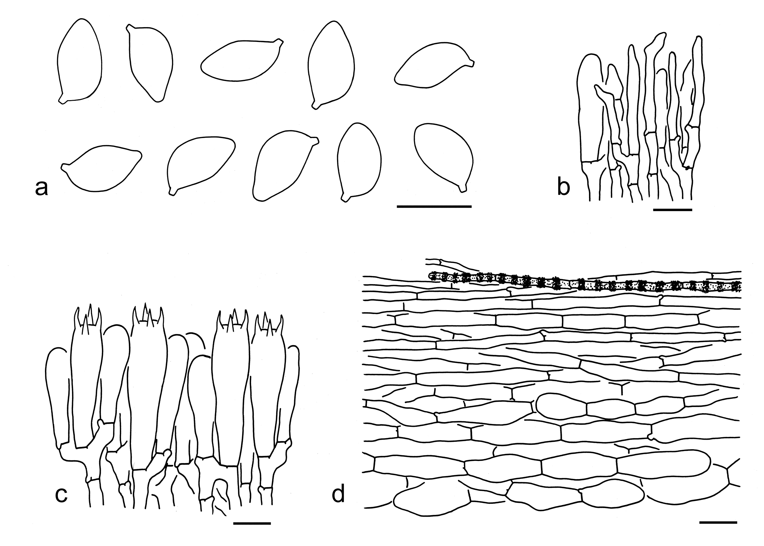
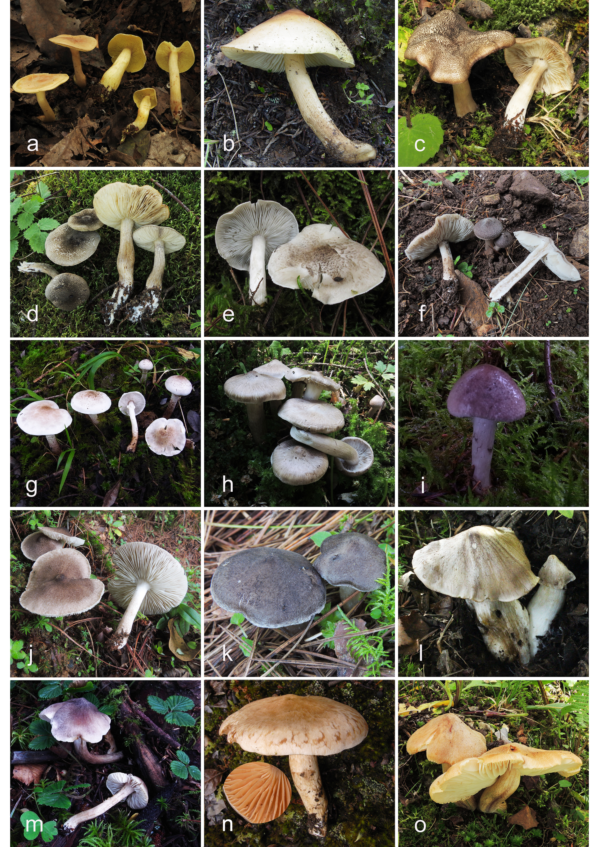
Tricholoma hemisulphureum (Kühner) A. Riva ex Boffelli is sister to T. sulphureum type Ⅰ with low support (Fig. 4), but the former has a pinkish pileus, pale pinkish lamellae and a poorly differentiated subpellis (Riva 2003; Christensen & Heilmann-Clausen 2013). Tricholoma bryogenum Mort. Chr., Heilm.-Claus. & Vauras is somewhat similar to this, but differs by its pileus with a pinkish buff to ochraceous orange center and a chrome margin, a poorly differentiated subpellis and absence of clamps (Christensen & Heilmann-Clausen 2013; Heilmann-Clausen et al. 2017).
68. Tricholoma sulphureum (Bull.) P. Kumm. type Ⅱ, Führ. Pilzk. (Zerbst): 133 (1871). Figs. 86a, 87
Basionym: Agaricus sulphureus Bull., Herb. Fr. (Paris) 4: pl. 168 (1784) [1783-84].
Description: Basidioma small to medium-sized. Pileus 3–12 cm in diam., at first bell-shaped to convex, soon plano-convex to applanate, occasionally with a low umbo; surface dry, smooth, pruinose to tomentose, orangish brown (4A6–8, 5A4–7), sulphur-yellow (3A4–7) to greenish yellow (30A3–6), often darker at center, context white (1A1), cream (1A2) to yellow (2A3–5); margin inflexed to involute. Lamellae sinuate to emarginate, sulphur-yellow (2A4–6), lemon yellow (1A5–7) to greenish yellow (30A4–6), crowded; edges entire, sulphur-yellow (2A4–6). Stipe 4–12 0.5–2 cm, cylindrical, orangish yellow (4A3–5), sulphur-yellow (1A4–6) to greenish yellow (30A2–5), fibrillose, exannulate. Taste and odour strong, unpleasant.
Ecology: Solitary to scattered on soil dominated in broad-leaved forests.
Known distribution: Known from Europe, North America, and East Asia.
Specimens examined: CHINA. Xizang Autonomous Region, Xigaze, Gyirong County, the type of forest and altitude unknown, 11 Aug. 2017, Ai-Guo Xu 2076 (AF0001459). Yunnan Province, Kunming, Kunming Botanical Garden, in a broad-leaved forest, altitude 1950 m, 25 Sep. 2018, Jian-Wei Liu 1640 (KUN-HKAS106305). NORTH MACEDONIA. The type of forest not recorded, altitude 1400 m, 12 Oct. 2019, Zhu L. Yang 6294 (KUN-HKAS106906); same location, 22 Oct. 2019, Pan-Meng Wang 2666 (KUN-HKAS131889).
Notes: As mentioned under T. sulphureum type Ⅰ, T. sulphureum s. l. is a polyphyletic group and separated into three clades (T. sulphureum type Ⅰ, Ⅱ and Ⅲ). Tricholoma odorum Peck is closely related to T. sulphureum type Ⅱ (Fig. 4), but differs by its relatively paler, yellowish buff to yellowish tan pileus and close lamellae (Bessette et al. 2013; Landry et al. 2022). Tricholoma hemisulphureum is somewhat similar to T. sulphureum type Ⅱ, but the former differs by its pinkish color in pileus and lamellae (Riva 2003; Christensen & Heilmann-Clausen 2013).
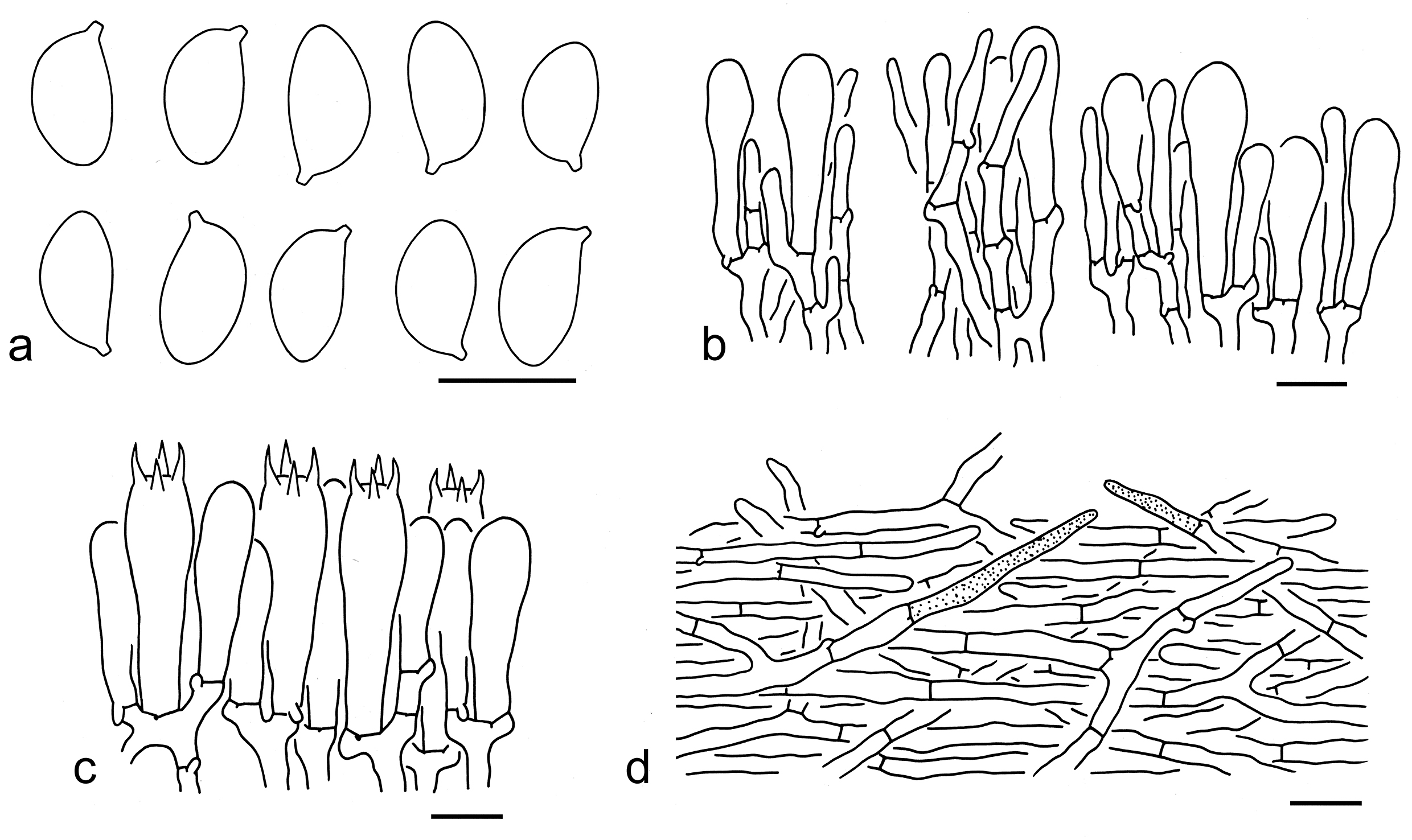
69. Tricholoma tangerinum Y.Y. Cui & Zhu L. Yang, sp. nov. Figs. 86b, 88
Fungal Names: FN 572350
Etymology: tangerinum = orange colored, referring to its orange colored pileal center.
Diagnosis: Related to T. sulphureum type Ⅰ, but differs by its orangish brown pileus and smaller basidiospores (9–10 5–6 µm, mean length = 9.7 µm, mean width = 5.8 µm).
Type: CHINA. Sichuan Province, Ganzi Tibetan Autonomous Prefecture, Seda County, Huoxi Village, in a subalpine forest, altitude 3650 m, 11 Aug. 2005, Zai-Wei Ge 860 (holotype, KUN-HKAS49355).
Description: Basidioma small to medium-sized. Pileus 5–7 cm in diam., convex, with a broadly umbo; surface dry, smooth, orangish brown (5A4–6, 4A6–8) to brown (4B6–8) at center, brownish (5B3–4, 3B3–5) to white (1A1) towards margin; margin straight. Lamellae sinuate to emarginate, white (1A1) to cream (1A2), close; edges entire, white (1A1) to cream (1A2). Stipe 6–9 0.5–1.5 cm, cylindrical, brownish (3B2–5), fibrillose, exannulate. Taste and odour not recorded.
Ecology: Solitary to scattered on soil in subalpine forests.
Known distribution: Known from Southwestern China.
Additional specimen examined: CHINA. Gansu Province, Gannan, Lintan County, in a forest with trees of Abies, Betula and Prunus, altitude 2670 m, 26 Oct. 2020, Xin Xu 419 (KUN-HKAS138838).
Notes: Tricholoma tangerinum is characterized by an orangish brown to brown pileus with a paler margin, and ellipsoid, elongate to amygdaliform basidiospores (9–10 5–6 µm, mean length = 9.7 µm, mean width = 5.8 µm, Q = 1.5–1.85, Qm = 1.68 ± 0.14). This species is related to T. sulphureum type Ⅰ based on the five-gene phylogenetic inference (Fig. 3). However, T. sulphureum type Ⅰ has a sulphur basidioma and larger basidiospores (10–12 5.5–7 µm, mean length = 10.6 µm, mean width = 6.2 µm). Tricholoma hemisulphureum is similar, but differs from T. tangerinum by its coarsely velutinous pileus and relatively larger basidiospores (8.1–13 5.5–8.2 µm) (Riva 2003; Christensen & Heilmann-Clausen 2013).
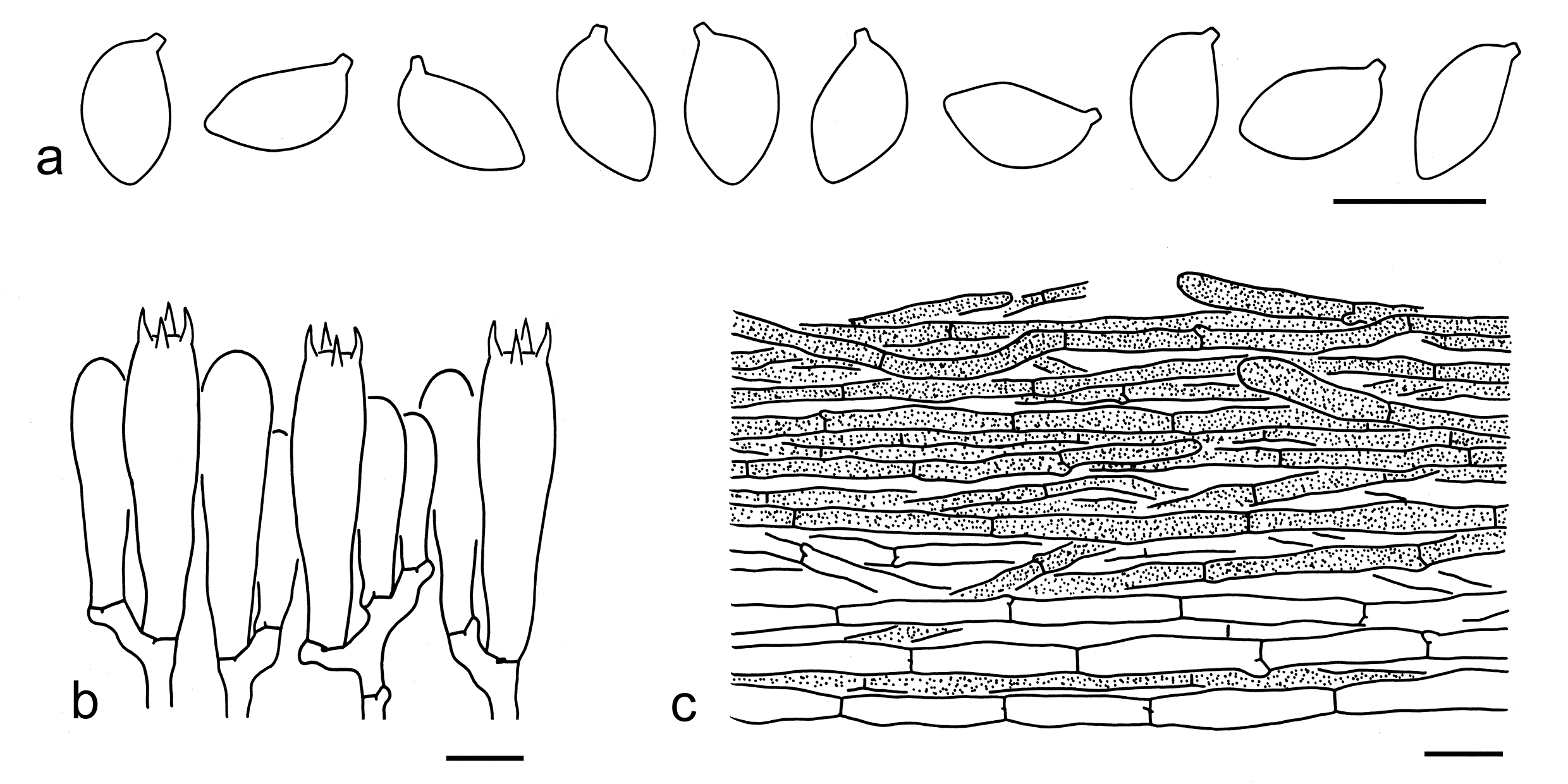
Tricholoma subgen. Terrea Ding et al.
Type: Tricholoma terreum (Schaeff.) P. Kumm., Führ. Pilzk. (Zerbst): 134 (1871).
Description: Basidioma tricholomatoid. Pileus umbonate or not, brown or grey, radially fibrillose or squarrulose. Lamellae crowded to close. Annulus present or absent. Basidiospores small, medium-sized to large, ellipsoid to elongate, smooth, inamyloid. Cheilocystidia present or absent. Pileipellis an ixo-cutis with filamentous hyphae, a cutis with its subpellis composed of inflated cells, or a cutis with somewhat erected, thick-walled hyphae. Clamps present or absent.
Ecology: Ectomycorrhizal. On soil with various trees.
Known distribution: Molecular data indicates species from this subgenus are found in Europe, North America, and Asia.
Notes: Tricholoma subgen. Terrea forms a clade sister to subgen. Sericeicutis (Figs. 2, 14). Members in this subgenus can be divided into two sections (sect. Atrosquamosa and sect. Terrea) and two unnamed clades (clades A and clade B). Currently, nine species of this subgenus are found in China (see below).
Tricholoma sect. Atrosquamosa Kühner ex Bon
Type: Tricholoma atrosquamosum Sacc., Syll. fung. (Abellini) 5: 104 (1887).
Description: Basidioma tricholomatoid. Pileus umbonate or not, dirty white to greyish, densely covered with dark grey to black squamules to scales. Lamellae crowded to close. Annulus absent. Basidiospores small, ellipsoid to elongate, smooth, inamyloid. Cheilocystidia conspicuous or absent. Pileipellis a cutis with transition to a trichoderm at regular intervals, composed of thick-walled hyphae, subpellis composed of radially arranged, thin-walled hyphae. Clamps present or absent.
Ecology: Ectomycorrhizal. On soil with various trees.
Known distribution: Molecular data shows species from this section are distributed in Europe, North America, and Asia.
Notes: Tricholoma atrosquamosum and T. olivaceotinctum Heilm.-Claus. & Mort. Chr. are found in China (see below).
1. Pileus white to dirty white background without olivaceous color; cheilocystidia poorly differentiated; clamps absent……. ……T. atrosquamosum
1. Pileus cream, yellow-grey to olivaceous grey background with obvious olivaceous color; cheilocystidia conspicuous; clamps present T. olivaceotinctum
70. Tricholoma atrosquamosum Sacc., Syll. fung. (Abellini) 5: 104 (1887). Figs. 86c, 89
Replaced synonym: Agaricus atrosquamosus Chevall., Strauch- und Laubflechten Mitteleur. 1: 45 (1837).
Competing homonym: non Agaricus atrosquamosus Batsch, Elench. fung. (Halle): 81, tab. 6, fig. 27 (1783).
Description: Basidioma medium-sized. Pileus 5–10 cm in diam., plano-convex to applanate, sometimes with a broadly low umbo; surface dry, a white (1A1) to dirty white (1B1–2) background densely covered with greyish (1B1), grey (1C1, 2C1) to dark grey (1E1–3, 1F1), more or less recurved, fibrillose scales; margin often radially splitting; context white (1A1). Lamellae sinuate, dirty white (1B2) to greyish (1B1), subclose; edges greyish (1B1) to cream (2B2) often with black (1F8) spots. Stipe 6–9 1–1.5 cm, cylindrical, stuffed, white (1A1) to dirty white (1B2), glabrous, exannulate. Taste not recorded. Odour sweet.
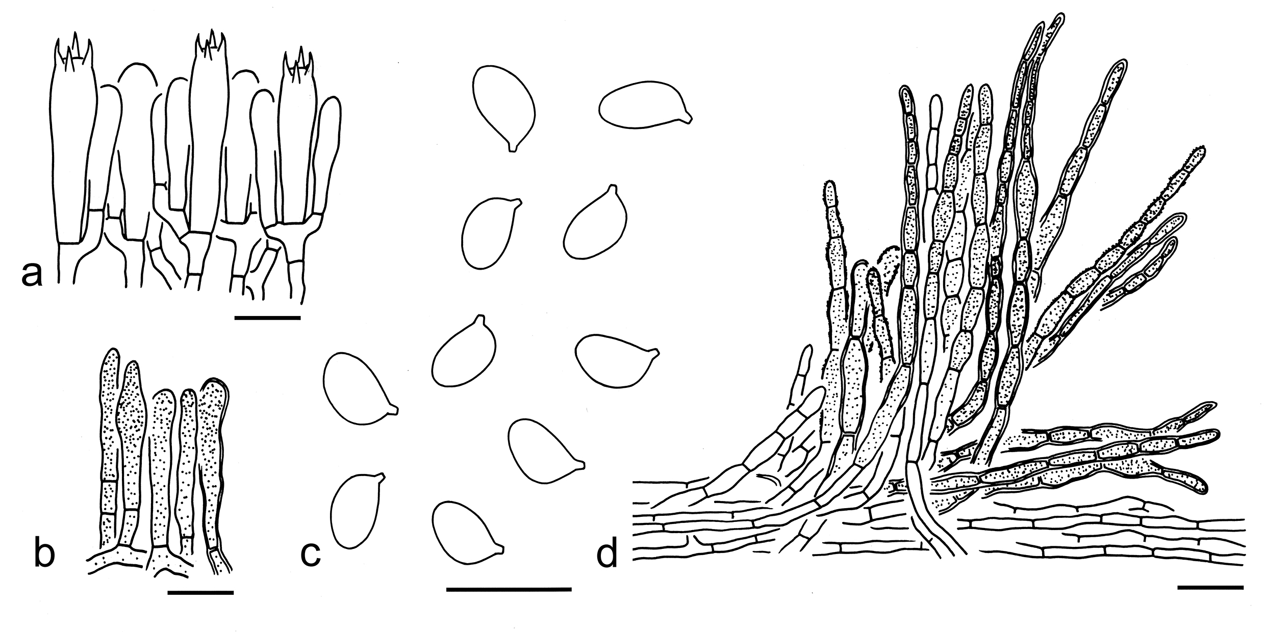
Basidiospores [60/3/2] (5–) 5.5–7 (–8) (3–) 3.5–4.5 µm (mean length = 6.4 µm, mean width = 4 µm), Q = (1.25–) 1.4–1.8 (–2), Qm = 1.6 ± 0.14, ellipsoid to elongate, non-amyloid, colorless, hyaline, thin-walled, smooth; apiculus relatively large. Basidia 25–35 5–8 µm, clavate, 4-spored, hyaline; sterigmata 3–6 µm long; basal septa without clamps. Pleurocystidia absent. Cheilocystidia poorly differentiated. Subhymenium composed of 2–4 µm wide filamentous hyphae, clamps absent. Lamellar trama regular, composed of colorless, thin-walled hyphae 2–6 µm wide. Pileipellis a cutis with transition to a trichoderm at regular intervals, composed of 5–25 µm wide, thick-walled (ca. 1 µm), yellow-brown, cylindrical to filamentous hyphae; subpellis poorly differentiated, composed of radially arranged, thin-walled hyphae. Clamps absent in all parts of basidioma.
Ecology: Solitary to scattered on soil in broad-leaved or coniferous forests.
Known distribution: Known from Europe (Riva 1988, Christensen & Heilmann-Clausen 2013), North America (Bessette et al. 2013, Trudell et al. 2022) and Southwestern China.
Specimens examined: CHINA. Sichuan Province, Ganzi Tibetan Autonomous Prefecture, Kangding, Waze Village, in a forest with Picea, altitude 386 m, 8 Sep. 2016, KD-LJW 117 (KUN-HKAS97910); same city, Xinduqiao Town, in a broad-leaved forest, altitude 3500 m, 11 Sep. 2016, KD-Ding 130 (KUN-HKAS97211). Yunnan Province, Diqing Tibetan Autonomous Prefecture, Shangri-La, the type of forest unknown, altitude 3500 m, 5 Sep. 2018, Jian-Wei Liu 1596 (KUN-HKAS106310).
71. Tricholoma olivaceotinctum Heilm.-Claus. & Mort. Chr., Mycotaxon 107: 436 (2009). Figs. 86d, 90
Description: Basidioma small, medium-sized to large. Pileus 3–10 cm in diam., at first hemispherical to convex, then plano-convex to applanate, slightly umbonate at center; surface dry, a cream (1A2), yellow-grey (1B3–5) to olivaceous grey (30B3–5, 30C4–6) background densely covered with dark grey (1E1–3) to black (1F1–3), recurved fibrillose scales; margin inflexed to straight; context white (1A1). Lamellae adnexed to sinuate, white (1A1), cream (1A2) to olivaceous (30B4–6), subclose; edges cream (1A2) to olivaceous grey (30D6–8). Stipe 7–13 0.7–2 cm, cylindrical, sometimes slightly inflated at base, white (1A1) to olivaceous grey (30B3–5, 30C4–6) often with a white (1A1) base, covered with brownish grey (1D4–6) to olivaceous grey (30B3–5, 30C4–6) fibrils, exannulate. Taste and odour not recorded.
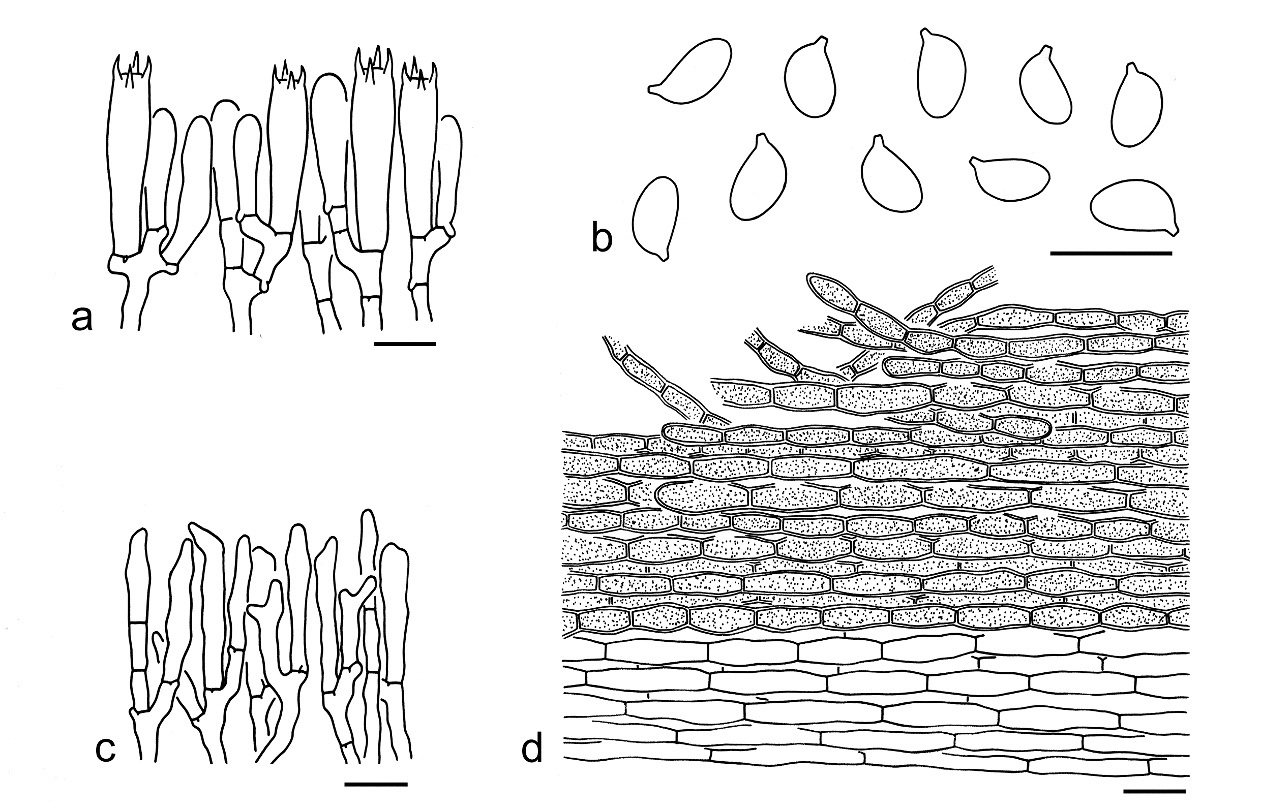
Basidiospores [40/2/2] 6–7.5 3.5–5 µm (mean length = 6.5 µm, mean width = 4.1 µm), Q = (1.2–) 1.33–1.88 (–2), Qm = 1.6 ± 0.17, ellipsoid to elongate, non-amyloid, colorless, hyaline, thin-walled, smooth; apiculus relatively large. Basidia 25–35 6–8 µm, clavate, 4-spored, hyaline; sterigmata 3–5 µm long; basal septa with clamps. Pleurocystidia absent. Cheilocystidia 15–30 3–5 µm, conspicuous, cylindrical to subclavate, thin-walled, colorless, hyaline. Subhymenium composed of 2–4 µm wide filamentous hyphae, clamps present. Lamellar trama regular, composed of colorless, thin-walled hyphae 2–5 µm wide. Pileipellis a cutis, composed of radically arranged, ellipsoid, clavate to cylindrical hyphae, often breaking up into curved trichoderm scales, 5–25 µm wide, thick-walled (ca. 1 µm), yellow-brown; subpellis poorly differentiated, composed of radially arranged, thin-walled hyphae. Clamps present in all parts of basidioma.
Ecology: Solitary to scattered on soil in temperate forests with trees of Picea.
Known distribution: Known from Europe (Christensen & Heilmann-Clausen 2009, 2013) and Southwestern China.
Specimens examined: CHINA. Sichuan Province, Ganzi Tibetan Autonomous Prefecture, Kangding, Xiaba Village, in a subalpine forest, altitude 3750 m, 23 Aug. 2017, Jian-Wei Liu 959 (KUN-HKAS107586); same city, Tagong Town, in a subalpine forest, altitude 3700 m, 22 Aug. 2017, Jian-Wei Liu 948 (KUN-HKAS107583); Ngawa Tibetan and Qiang Autonomous Prefecture, Hongyuan County, Shuajingsi Town, in a subalpine forest with Picea, altitude 3300 m, 12 Aug. 2013, Kuan Zhao 357 (KUN-HKAS80782).
Notes: Tricholoma olivaceotinctum is well delimitated by its olivaceous grey pileus covered with dark grey to black scales, ellipsoid to elongate basidiospores (6–7.5 3.5–5 µm, Q = 1.33–1.88, Qm = 1.6 ± 0.17), the presence of clamps and associations with trees of Picea (this study). In the protologue of T. olivaceotinctum, the absence of clamps was described (Christensen & Heilmann-Clausen 2009). However, clamps were found in the Chinese specimens (Fig. 90), which clustered well with the holotype of T. olivaceotinctum (Fig. 4). Although the taste and odour of the Chinese collections of this species were not recorded in the field, European collections were reported to have a weak but distinctly spicy and sweetish odour, reminiscent of honey and often intermixed with components similar to ground black pepper, becoming faintly to strongly farinaceous after cutting; the taste was mild to slightly bitter, and distinctly farinaceous (Christensen & Heilmann-Clausen 2013).
In the phylogenetic analyses based on the ITS sequences (Fig. 4), T. olivaceotinctum is related to T. squarrulosum, T. michiganense A.H. Sm. and T. basirubens (Bon) A. Riva & Bon. However, T. squarrulosum differs by its tomentose to squarrulose, dark greyish brown to black pileus often paler towards margin, relatively larger basidiospores (4.8–10.5 2.8–6.4 µm), the absence of clamps, and associations with deciduous trees (Hongo 1988; Riva 1988; Bessette et al. 2013; Christensen & Heilmann-Clausen 2013). Tricholoma michiganense possesses a dark grey pileus without olivaceous color and is associated with oak (Trudell et al. 2022). Tricholoma basirubens has a pale cream to buff pileus background without olivaceous color, the absence of clamps and associations with deciduous trees (Riva 1988; Christensen & Heilmann-Clausen 2013).
Tricholoma sect. Terrea
Type: Tricholoma terreum (Schaeff.) P. Kumm., Führ. Pilzk. (Zerbst): 134 (1871).
Description: Basidioma tricholomatoid. Pileus umbonate or not, white, grey, brown to black, densely covered with felty, tomentose to somewhat fibrillose squamules. Lamellae crowded to close. Veil absents or somewhat present especially in young basidioma. Basidiospores small to medium-sized, ellipsoid to elongate. Pleurocystidia absent. Cheilocystidia present or absent. Pileipellis a cutis with transition to a trichoderm at regular intervals composed of thick-walled, filamentous to cylindrical hyphae with poorly or well differentiated subpellis. Clamps absent.
Ecology: Ectomycorrhizal. On soil with various trees.
Known distribution: Molecular data indicates species from this section are distributed in Europe, North America, and Asia.
Notes: To date, seven species, T. argyraceum, T. bonii asso & Candusso, T. cingulatum, T. inocybeoides A. Pearson, T. terreum, T. triste and T. gansuense Y.Y. Cui & Zhu L. Yang, occur in China. Among them, T. cingulatum and T. terreum are widely distributed in Europe, North America and Asia, T. gansuense is currently only collected in China, while the remaining four species can be found in Europe and Asia.
Key to the species of Tricholoma sect. Terrea from China
1. A white stipe with a white, felty to cottony annulus on the upper part T. cingulatum
1. A stipe with or without a cobweb-like zone in the upper part 2
2. Veil is more or less present, often form a cobweb-like zone in the upper part of stipe; subpellis poorly differentiated with radially arranged filamentous hyphae 3
2. Veil absent, without a cobweb-like zone in the upper part of stipe; subpellis composed of inflated cells 5
3. Basidiospores relatively longer, elongate, 6–8 3.5–4 µm, mean length = 7.1 µm, mean width = 3.9 µm Q = 1.63–2, Qm = 1.82 ± 0.15 T. inocybeoides
3. Basidiospores relatively shorter, ellipsoid to elongate, with mean length less than 5.5 µm 4
4. Veil weakly developed, only distinct in very young basidioma; cheilocystidia poorly differentiated T. gansuense
4. Veil developed, often form a cobweb-like zone in the upper part of stipe; cheilocystidia conspicuous T. argyraceum
5. Pileus dark colored, black, dark grey to grey, with a distinct, white to dirty white zone at margin T. triste
5. Pileus relatively paler, greyish, grey, brownish, brown, brownish grey to dark grey, without a distinct, white to dirty white zone at margin 6
6. Basidiospores relatively longer, 6–9 4–6 μm, mean length = 7.2 µm; cheilocystidia conspicuous T. bonii
6. Basidiospores relatively shorter, 6–7 3.5–5 μm, mean length = 6.2 µm; cheilocystidia poorly differentiated………… …………T. terreum
72. Tricholoma argyraceum (Bull.) Gillet, Hyménomycètes (Alençon): 103 (1874) [1878]. Figs. 86e, 91
Basionym: Agaricus argyraceus Bull., Herb. Fr. (Paris) 9: tab. 423 (1789).
Description: Basidioma small to medium-sized. Pileus 2–6 cm in diam., at first hemispherical to convex, then plano-convex to applanate, often slightly umbonate at center; surface dry, pale grey (1C1–3), grey-brown (2E3–6), brown (4D3–6, 4E5–7, 5D3–6) to dark brown (4E2–4, 4F6–8), covered with grey-brown (2E3–6) to dark brown (4E2–4, 4F6–8), felty-tomentose, tomentose to fibrillose squamules; margin straight to inflexed; context white (1A1). Lamellae sinuate to emarginate, white (1A1), grey-white (1B1) to pale grey (1B1–3), becoming brown (4E6–8) to yellow-brown (4D6–8) when old or damaged, crowded to subclose; edges entire, white (1A1) to grey-white (2E3–6). Stipe 3–8 cm long 0.5–1 cm wide, cylindrical, white (1A1), grey-white (2E3–6) to brownish (4D2–5), glabrous, sometimes with a white (1A1) to dark brown (4E2–4, 4F6–8) cobweb-like zone on the upper part. Taste and odour not recorded.
Basidiospores [60/3/3] (4.5–) 5–6.5 3–4 μm (mean length = 5.4 µm, mean width = 3.4 µm), Q = (1.25–) 1.3–2, Qm = 1.6 ± 0.15, ellipsoid to elongate, non-amyloid, colorless, hyaline, thin-walled, smooth, apiculus relatively large. Basidia 20–40 5–7 μm, clavate, 4-spored, hyaline; sterigmata 3–5 μm long; basal septa without clamps. Pleurocystidia absent. Cheilocystidia 15–40 3–6 μm, conspicuous, filiform, narrowly cylindrical, narrowly clavate to clavate, sometimes fusiform, often septate, thin-walled, colorless and hyaline. Subhymenium composed of 3–6 μm wide filamentous hyphae, clamps absent. Lamellar trama regular, composed of colorless, thin-walled hyphae 2–25 μm wide. Pileipellis a cutis with transition to a trichoderm at regular intervals, composed of 3–8 µm wide, thick- (ca. 1 μm) to thin-walled, colorless, brown to yellow-brown filamentous hyphae, terminal elements subcylindrical with round or abrupt apex; subpellis poorly differentiated. Clamps absent in all parts of basidioma.
Ecology: Solitary to scattered on soil in temperate or subtropical forests with a wide range of trees (Pinus, Populus and Picea et al.).
Known distribution: Known from Europe and China.
Notes: Tricholoma argyraceum is characterized by its pale grey, grey-brown, brown to dark brown pileus covered with grey brown to dark brown, felty-tomentose, tomentose to fibrillose squamules, a white to dark brown, cobweb-like zone often on the upper part of stipe, ellipsoid to elongate basidiospores (5–6.5 3–4 μm, Q = 1.3–2, Qm = 1.6 ± 0.15). Even though taste and odour were not recorded in the field for the Chinese collections, Christensen & Heilmann-Clausen (2013) noted a strongly farinaceous odour after cutting and a farinaceous to rancid taste for the European ones.
Tricholoma argyraceum is related to T. cingulatum and T. gansuense based on the five-gene phylogenetic analysis (Fig. 3). However, T. cingulatum has a relatively paler colored pileus, a white, felty to cottony annulus in the upper part of stipe and poorly differentiated cheilocystidia (Riva 1988, 2003; Bessette et al. 2013; Christensen & Heilmann-Clausen 2013; this study). Tricholoma gansuense differs by a poorly developed veil only present as cortina on the upper part of stipe in young stage, and poorly differentiated cheilocystidia.
A cobweb-like zone also can be found in young basidioma of T. scalpturatum (Fr.) Quél., but it differs from T. argyraceum by its indistinctly broad and low umbo at pileus center, and relatively broader basidiospores (Q = 1.2–8, average Q = 1.4–1.54) (Riva 1988; Christensen & Heilmann-Clausen 2013). Tricholoma inocybeoides A. Pearson also has a weakly developed veil only distinct in very young basidioma and somewhat similar to T. argyraceum, but has relatively longer basidiospores (6–8 3.5–4 µm, mean length = 7.1 µm) and poorly differentiated cheilocystidia (this study).
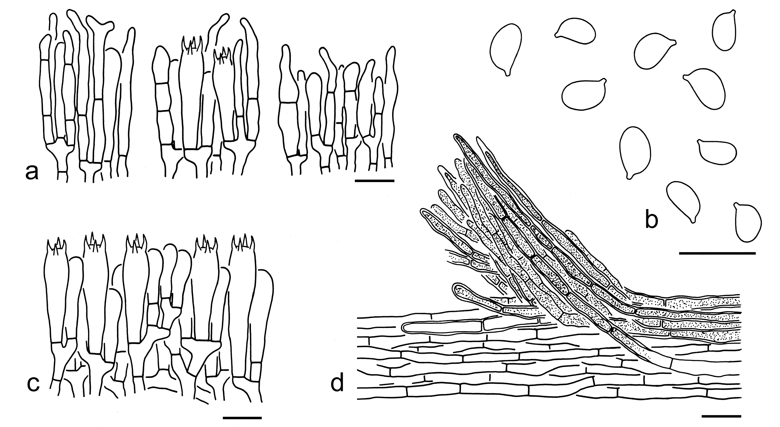
73. Tricholoma bonii Basso & Candusso, Docums Mycol. 27(no. 107): 64 (1997). Figs. 86f, 92
Description: Basidioma small to medium-sized. Pileus 2–7 cm in diam., at first conical to broadly conical, then plano-convex to applanate, often acutely umbonate at center; surface dry, grey-brown, grey to dark grey, covered with dark grey, felty-tomentose to tomentose squamules, often fibrillose at margin; margin straight; context greyish. Lamellae adnexed to sinuate, white, dirty white to greyish, subclose; edges entire to slightly undulate, white to greyish. Stipe 4–8 cm long 0.5–1 cm wide, cylindrical, stuffed to hollow, white, dirty white to brownish, glabrous or covered with greyish to brownish fibrillose squamules. Taste and odour not recorded.
Basidiospores [110/5/3] (5.5–) 6–9 (–10) 4–6 μm (mean length = 7.2 µm, mean width = 4.6 µm), Q = (1.15–) 1.33–1.8 (–2.14), Qm = 1.56 ± 0.15, ellipsoid to elongate, non-amyloid, colorless, hyaline, thin-walled, smooth, apiculus relatively large. Basidia 25–35 7–9 μm, clavate, 4-spored; hyaline; sterigmata 3–6 μm long; basal septa without clamps. Pleurocystidia absent. Cheilocystidia 25–40 7–18 μm, conspicuous, cylindrical, fusiform, clavate to broadly clavate, often septate, thin-walled, colorless and hyaline. Subhymenium composed of 3–5 μm wide filamentous hyphae, clamps absent. Lamellar trama regular, composed of colorless, thin-walled hyphae 3–40 μm wide. Pileipellis a cutis with transition to a trichoderm at regular intervals, composed of 4–10 μm wide, thick- (ca. 1 μm) to thin-walled, colorless to brownish filamentous hyphae, terminal elements subcylindrical with round apex; subpellis composed of radially arranged inflated cells, subglobose, ellipsoid, broadly ellipsoid, oblong to cylindrical, (20–) 30–60 (–120) 10–35 μm, thin-walled, colorless. Clamps absent in all parts of basidioma.
Ecology: Solitary to scattered on soil in temperate or subtropical coniferous or mixed forests.
Known distribution: Known from Europe and China.
Specimens examined: CHINA. Heilongjiang Province, Heihe, Wudalianchi, the type of forest unknown, altitude 300 m, 26 Sep. 2014, collecting no. unknown (HMJAU35946). Inner Mongolia, Chifeng, Ar Horqin Banner, the type of forest unknown, altitude 500 m, 14 Aug. 2014, collecting no. unknown (HMJAU34437). Liaoning Province, Dandong, Kuandian Man Autonomous County, Sipingjie Village, altitude 500 m, 29 Aug. 2018, Xiao-Xia Ding 385 (KUN-HKAS105395). Yunnan Province, Dali Bai Autonomous Prefecture, Binchuan County, Zhusheng Temple, in a forest with Pinus yunnanensis, altitude 2000 m, 20 Sep. 2012, Qi Zhao 1722 (KUN-HKAS78979); Kunming, Panlong District, bought from Ciba market, forests and altitude unknown, 23 Jul. 2017, Xiao-Xia Ding 221 (KUN-HKAS106563). Sichuan Province, Ngawa Tibetan and Qiang Autonomous Prefecture, Jiuzhaigou, in a mixed forest of Picea, Pinus and Fagaceae, altitude 2431 m, 20 Jun. 2014, Yan-Jia Hao 1085 (KUN-HKAS82876).
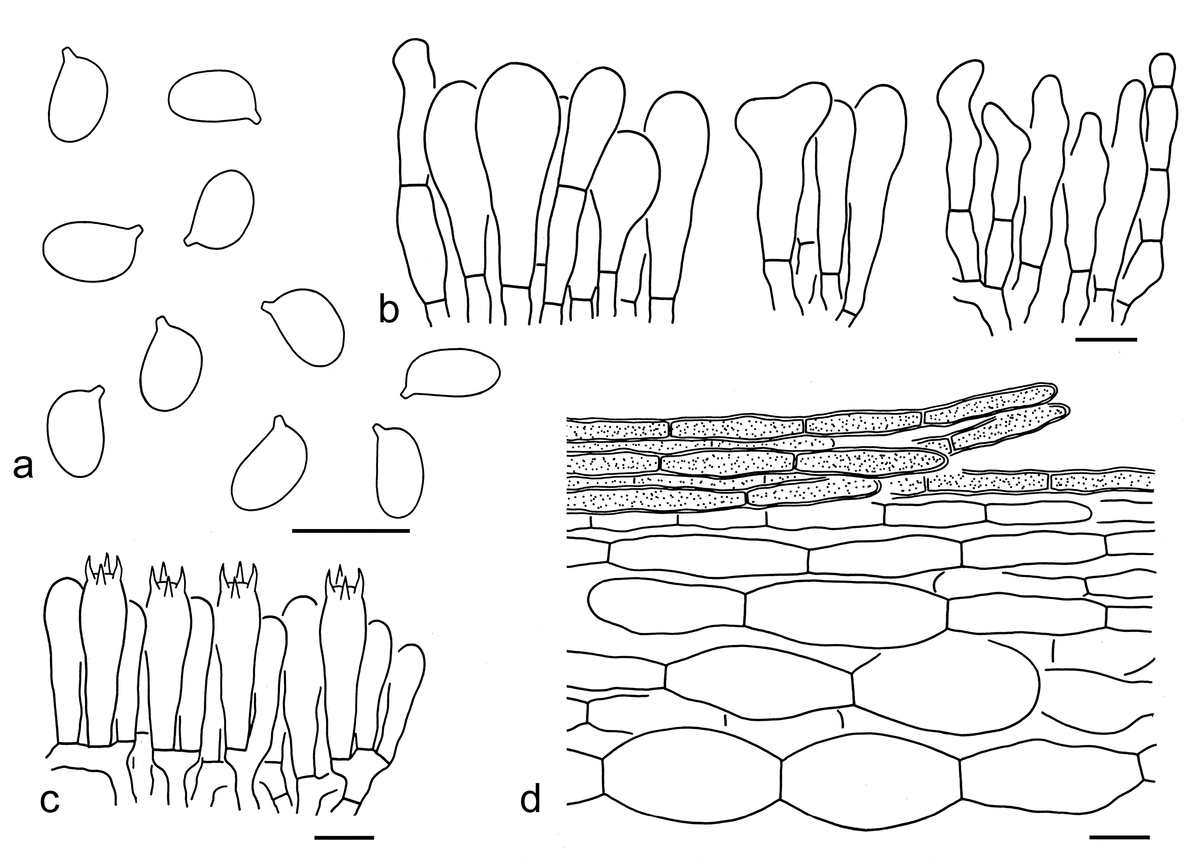
Notes: Tricholoma bonii is well defined by its grey-brown, grey to dark grey pileus covered with dark grey, felty-tomentose to tomentose squamules, a well differentiated subpellis with inflated cells, and ellipsoid to elongate basidiospores (6–9 4–6 μm, Q = 1.33–1.8, Qm = 1.56 ± 0.15). Tricholma terreum is closely related to T. bonii (Fig. 3), and these two species are indeed similar to each other in their color of basidioma and structure of pileipellis. However, T. terreum has relatively smaller basidiospores (6–7 3.5–5 μm, mean length = 6.2 µm, mean width = 4 µm) and poorly differentiated cheilocystidia (Riva 1988; Bessette et al. 2013; Christensen & Heilmann-Clausen 2013; this study).
Morphologically, T. scalpturatum and T. inocybeoides resemble T. bonii in their color of basidioma. However, T. scalpturatum differs by its cobweb-like ring in young basidioma, poorly differentiated subpellis with filamentous hyphae, and relatively smaller basidiospores (3.9–6.8 2.5–4.5 μm, average 4.9–5.6 3.2–3.8 μm) (Riva 1988; Christensen & Heilmann-Clausen 2013). Tricholoma inocybeoides has a weakly developed veil in young basidioma, pileipellis with filamentous hyphae, poorly differentiated cheilocystidia, and relatively narrower basidiospores (6–8 3.5–4 µm, mean length = 7.1 µm, mean width = 3.9 µm, Q = 1.63–2, Qm = 1.82 ± 0.15) (Riva 1988, 2003; Christensen & Heilmann-Clausen 2013; this study).
74. Tricholoma cingulatum (Almfelt ex Fr.) Jacobashch, Verh. bot. Ver. Prov. Brandenb. 33: 59 (1892). Figs. 86g, 93
Basionym: Agaricus cingulatus Almfelt ex Fr., Linnaea 5: 507 (1830).
Basidiospores [61/3/2] (4–) 4.5–6 (–6.5) (2.5–) 3–4 µm (mean length = 5.2 µm, mean width = 3.1 µm), Q = 1.4–2, Qm = 1.7 ± 0.15, ellipsoid to elongate, non-amyloid, colorless, hyaline, thin-walled, smooth; apiculus relatively large. Basidia 20–30 5–7 µm, clavate, 4-spored, colorless, hyaline; sterigmata 4–6 µm long; basal septa without clamps. Pleurocystidia absent. Cheilocystidia poorly differentiated. Subhymenium composed of 2–4 µm wide filamentous hyphae, clamps absent. Lamellar trama regular, composed of colorless, thin- to slightly thick-walled (ca. 1 µm), clavate to cylindrical cells, 35–150 15–25 µm, mixed with filamentous hyphae 4–7 µm wide. Pileipellis a cutis, composed of radially arranged to interwoven, 4–10 µm wide, slightly thick-walled (ca. 0.5–1 µm), colorless or yellow-brown to brownish filamentous hyphae; subpellis poorly differentiated. Clamps absent in all parts of basidioma.
Ecology: Solitary to scattered on soil in temperate forests with Fagaceae, Pinaceae and Salix.
Known distribution: Known from Europe (Riva 1988, 2003; Christensen & Heilmann-Clausen 2013; this study), North America (Bessette et al. 2013) and Asia (Kudo & Nagasawa 1998; Deng et al. 2004).
Specimens examined: CHINA. Sichuan Province, Ganzi Tibetan Autonomous Prefecture, Kangding, Gonggashan Village, in a mixed forest with Fagaceae and Pinaceae, altitude 2700 m, 16 Sep. 2017, Xiao-Xia Ding 292 (KUN-HKAS106634); same location, in a mixed forest with Fagaceae and Pinaceae, altitude 2700 m, 16 Sep. 2017, Xiao-Xia Ding 293 (KUN-HKAS106635); Pingwu County, along a roadside in a mixed forest, altitude 2500 m, 12 Oct. 1999, Zhu L. Yang 2848 (KUN-HKAS34313). Yunnan Province, Shangri-La, Wongshui Village, in a coniferous forest, altitude 3400 m, 21 Aug. 2000, Zhu L. Yang 2962 (KUN-HKAS36663).
Notes: Tricholoma cingulatum is well delimitated by its pale colored pileus covered with brown to greyish brown, tomentose to fibrillose squamules, a white stipe with a white, felty to cottony annulus on the upper part, and ellipsoid to elongate basidiospores (4.5–6 3–4 µm, Q = 1.4–2, Qm = 1.7 ± 0.15). In Europe this species is exclusively associated with Salix, while it can also be found in coniferous or mixed forests with Fagaceae and Pinaceae in China. In the molecular phylogenetic analyses (Figs. 3, 4), T. gansuense is closely related to T. cingulatum. For morphological comparison, see notes under that name.
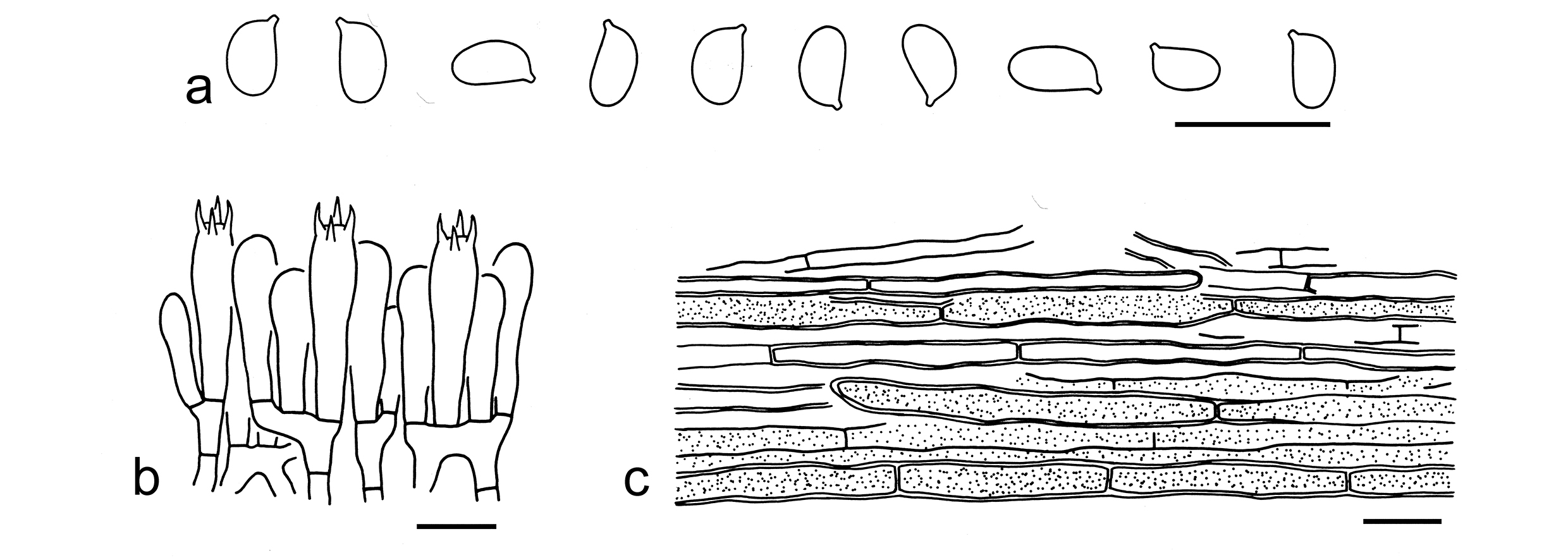
75. Tricholoma inocybeoides A. Pearson, Trans. Br. mycol. Soc. 22(1-2): 29 (1938). Figs. 86h, 94
Description: Basidioma very small to small. Pileus 2–5 cm in diam., at first broadly conical, then convex to plano-convex, umbonate at center; surface dry, greyish (1B1, 1C1), brownish (4B2–4), grey (1D1–4) to brownish grey (3D2–5), covered with dark grey (1E1–4), grey (1D1–4) to brown (3E6–8), fibrillose squamules; margin involute when young, straight when mature; context white (1A1). Lamellae adnexed, white (1A1) to dirty white (1B1–3), subclose; edges white (1A1), dirty white (1B1–3) to brownish (3B2–4). Stipe 4–6 0.5–1 cm, cylindrical, stuffed to hollow, white (1A1), greyish (1B1, 1C1) to brownish (3B2–4). Taste not recorded. Odour like soap.
Basidiospores [20/1/1] 6–8 (–9) (3–) 3.5–4 µm (mean length = 7.1 µm, mean width = 3.9 µm), Q = 1.63–2 (–2.25), Qm = 1.82 ± 0.15, elongate, occasionally cylindrical, non-amyloid, colorless, hyaline, thin-walled, smooth; apiculus relatively large. Basidia 20–30 5–7 µm, clavate, 4-spored, occasionally 1- or 2-spored, colorless, hyaline; sterigmata 3–5 µm long; basal septa without clamps. Pleurocystidia absent. Cheilocystidia 10–40 2–7 μm, poorly differentiated, filamentous, cylindrical to clavate, thin-walled, colorless and hyaline. Subhymenium composed of 2–5 µm wide filamentous hyphae, clamps absent. Lamellar trama regular, composed of colorless, thin-walled, filamentous to cylindrical hyphae 3–25 µm wide. Pileipellis a cutis to a trichoderm, composed of radially arranged, interwoven to more or less vertically arranged, 8–20 µm wide, thick-walled (ca. 1 µm), colorless to yellowish, clavate to cylindrical hyphae; subpellis poorly differentiated, composed of radially arranged, thin-walled, colorless, hyaline, filamentous hyphae 5–10 µm wide. Clamps absent in all parts of basidioma.
Known distribution: Known from Europe (Riva 1988, 2003; Christensen & Heilmann-Clausen 2013) and China.
Specimens examined: CHINA. Gansu Province, Longnan, Wen Couty, Jianshan Village, in a forest with Quercus, altitude 1000 m, 15 Aug. 2018, Xue-Tai Zhu 2495 (KUN-HKAS106315). Sichuan Province, Ganzi Tibetan Autonomous Prefecture, Luhuo County, Renda Village, in a subalpine forest, altitude 3100 m, 16 Jul. 2014, Kuan Zhao 552 (KUN-HKAS89215). Xizang Autonomous Region, Nyingchi City, Bome County, in a subalpine sclerophyllous evergreen forest dominated by Quercus spp., altitude 2760 m, 17 Jul. 2019, Zhu L. Yang 6188 (KUN-HKAS106800). Xinjiang Uygu Autonomous Region, Yili Kazak Autonomous Prefecture, in a forest with Picea, altitude 2300 m, 8 Jul. 2017, Xiao-Xia Ding 183 (KUN-HKAS106525); same location, in a forest with Picea, altitude 2300 m, 8 Jul. 2017, Xiao-Xia Ding 184 (KUN-HKAS106526); same location, in a forest with Picea, altitude 2300 m, 8 Jul. 2017, Xiao-Xia Ding 185 (KUN-HKAS106527).
Notes: Tricholoma inocybeoides is characterized by its greyish, brownish, grey to brownish grey pileus covered with dark grey, grey to brown, fibrillose squamules, a weakly developed veil only distinct in very young basidioma, and elongate basidiospores (6–8 3.5–4 µm, Q = 1.63–2, Qm = 1.82 ± 0.15) (this study). Tricholoma inocybeoides is closely related to T. scalpturatum in the five-gene phylogenetic analyses (Fig. 3), but the latter has conspicuous cheilocystidia, and relatively shorter basidiospores (average 4.9–5.6 3.2–3.8 μm) (Riva 1988; Christensen & Heilmann-Clausen 2013). Besides T. inocybeoides and T. scalpturatum, T. argyraceum also has a dark colored basidioma with a more or less developed veil. For comparison, see notes under that name. Tricholoma terreum is similar to T. inocybeoides, but differs by its well differentiated subpellis with inflated cells, and relatively shorter basidiospores (6–7 3.5–5 μm, mean length = 6.2 µm, mean width = 4 µm, Q = 1.3–1.8, Qm = 1.55 ± 0.12) (Riva 1988; Bessette et al. 2013; Christensen & Heilmann-Clausen 2013; this study).
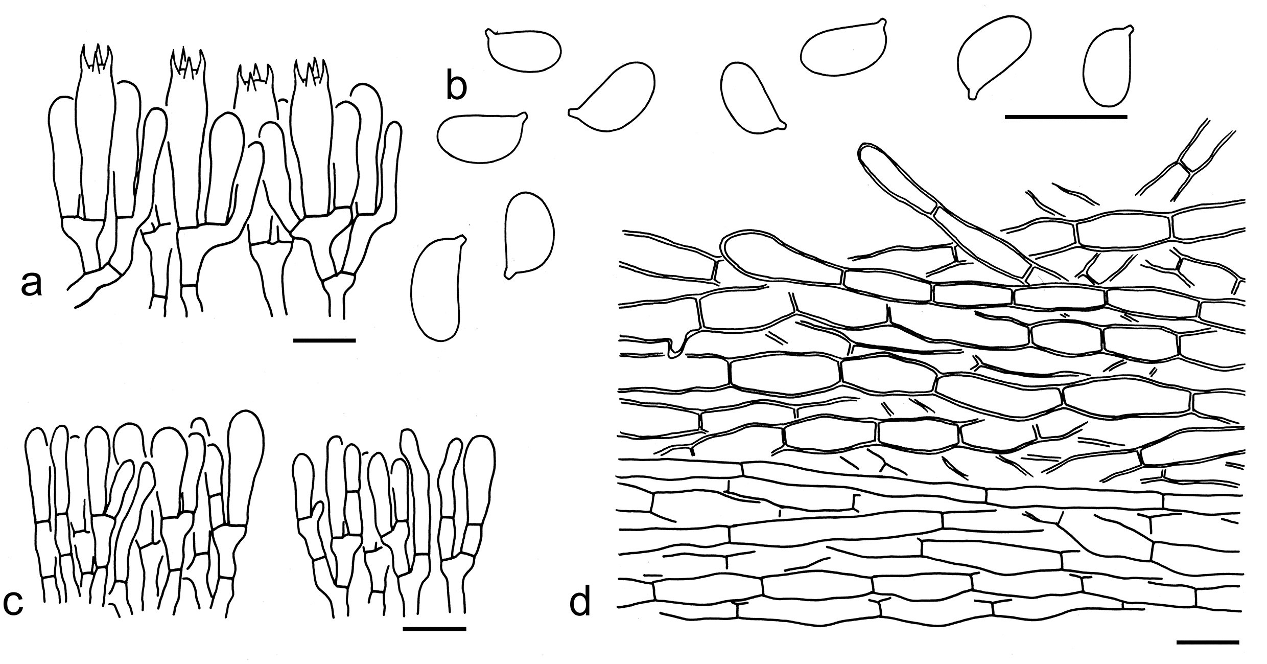
76. Tricholoma gansuense Y.Y. Cui & Zhu L. Yang, sp. nov. Figs. 86i, 95
Fungal Names: FN 572345
Etymology: gansuense referring to its type locality.
Diagnosis: Related to T. cingulatum, but differs in its poorly developed cortina present on the upper part of stipe in young basidioma only.
Type: CHINA. Gansu Province, Wuwei, Tianzhu Zangzu Autonomous County, in a temperate forest with Picea, altitude 2735 m, 29 Aug. 2018, Xue-Tai Zhu 2803 (holotype, KUN-HKAS106316).
Description: Basidioma small to medium-sized. Pileus 2–5 cm in diam., at first hemispherical to convex, then plano-convex to applanate, slightly umbonate or not; surface dry, greyish (2D1–3), grey (1D1–4)) to grey-brown (2E3–6), covered with grey-brown (2E3–6) to dark brown (4E2–4, 4F6–8) squamules; margin incurved when young, then straight to inflexed; context white (1A1). Lamellae sinuate to emarginate, white (1A1) to grey-white (1B1), crowded; edges entire, white (1A1) to grey-white (2E3–6). Stipe 4–8 cm long 0.5–1 cm wide, cylindrical, white (1A1) to grey-white (2E3–6), glabrous, cortina present on the upper part in young stage. Taste and odour not recorded.
Ecology: On soil in a temperate coniferous forest with Picea.
Known distribution: Currently only known from northwestern China.
Additional specimen examined: CHINA. Gansu Province, Xue-Tai Zhu 2793 (KUN-HKAS138831).
Notes: Tricholoma gansuense is well separated by its grey to grey-brown pileus, a white stipe often with white cortina on the upper part in young stage and small basidiospores (4.5–6 3–4 µm). This species is sister to T. cingulatum (Figs. 3, 4), but the latter species has a white, felty to cottony annulus on the upper part of stipe. Tricholoma inocybeoides and T. argyraceum resembles T. gansuense. However, T. inocybeoides differs by its larger basidiospores (6–8 3.5–4 µm) (this study). Tricholoma argyraceum has a developed veil often form a cobweb-like zone on the upper part of stipe, and conspicuous cheilocystidia. Tricholoma scalpturatum (Fr.) Quél. can be confused with T. gansuense, but the former species is mainly associated with deciduous trees and currently not found in China (Christensen & Heilmann-Clausen 2013).
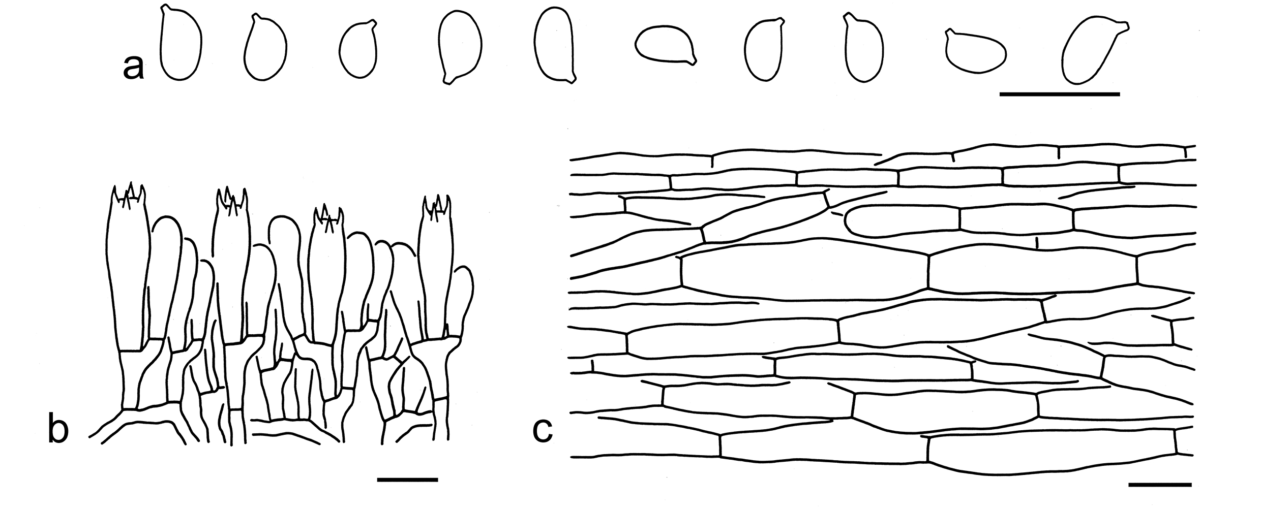
77. Tricholoma terreum (Schaeff.) P. Kumm., Führ. Pilzk. (Zerbst): 134 (1871). Figs. 86j, 96
Basionym: Agaricus terreus Schaeff., Fung. bavar. palat. nasc. (Ratisbonae) 4: 28 (1774).
Description: Basidiomata small to medium-sized. Pileus 3–8 cm in diam., at first convex, then plano-convex to applanate, often slightly umbonate at center; surface dry, greyish (1B1–3), grey (1D1–3), brownish (3B2–4) to brown (4C3–5), covered with grey (1C1–3), brown (4C3–5), grey-brown (4D1–4) to dark grey (1D1–3), felty to tomentose, sometimes fibrillose squamules; margin involute when young, straight to slightly inflexed when mature; context white (1A1), dirty white (1B1) to grey (1C1). Lamellae adnexed to sinuate, white (1A1) to greyish (1B1–3), becoming yellowish (3A2–3) when old or damaged, close to subdistant; edges entire or undulate to undate, white (1A1) to greyish (1B1–3), becoming brownish (3B2–4) to brown (4B2–4) when damaged or old. Stipe 4–7 cm long 0.3–1.5 cm wide, stuffed to hollow, cylindrical, white (1A1), dirty white (1B1) to greyish (1B2–4), glabrous or sometimes covered with greyish (1B1–3) to brownish (3B2–4) fibrils. Taste and odour not obvious.
Basidiospores [40/2/2] (5–) 6–7 3.5–5 μm (mean length = 6.2 µm, mean width = 4 µm), Q = (1.08–) 1.3–1.8 (–2), Qm = 1.55 ± 0.12, ellipsoid to elongate, non-amyloid, colorless, hyaline, thin-walled, smooth, apiculus relatively large. Basidia 20–30 6–9 μm, clavate, 4-spored, colorless, hyaline; sterigmata 4–6 μm long; basal septa without clamps. Pleurocystidia absent. Cheilocystidia 15–30 3–9 μm, poorly differentiated, cylindrical to clavate, thin-walled, colorless and hyaline. Subhymenium composed of 2–5 μm wide filamentous hyphae, clamps absent. Lamellar trama regular, composed of colorless to yellowish, thin-walled hyphae 4–15 μm wide. Pileipellis a cutis with transition to a trichoderm at regular intervals, composed of 4–15 μm wide, mostly thin-walled, sometimes thick-walled (ca. 1 μm), yellow brown filamentous hyphae; subpellis composed of radially arranged inflated cells, ellipsoid, broadly ellipsoid, elongate to cylindrical, 40–80 10–40 μm, thin-walled, colorless. Clamps absent in all parts of basidioma.
Ecology: Solitary to scattered on soil in coniferous or mixed forests with trees of Pinus and Fagaceae.
Known distribution: Known from Europe (Riva 1988, 2003; Christensen & Heilmann-Clausen 2013), North America (Bessette et al. 2013) and Asia.
Notes: Tricholoma terreum is characterized by its greyish, grey, brownish to brown pileus decorated with grey, brown, grey-brown to dark grey, felty to tomentose squamules, a differentiated subpellis with inflated cells, and ellipsoid to elongate basidiospores (6–7 3.5–5 μm, Q = 1.3–1.8, Qm = 1.55 ± 0.12). It is closely related to T. bonii in the five-gene phylogenetic analysis (Fig. 3). For comparison, see notes under that name.
Tricholoma argyraceum, T. inocybeoides and T. scalpturatum mophologically somewhat resemble T. terreum. However, the former three species have more or less developed veils and clearly farinaceous smell. The comparisons with T. argyraceum and T. inocybeoides have provided under each notes part. In addition, T. scalpturatum is different from T. terreum in its conspicuous cheilocystidia and a poorly differentiated subpellis with filamentous hyphae (Riva 1988; Christensen & Heilmann-Clausen 2013).
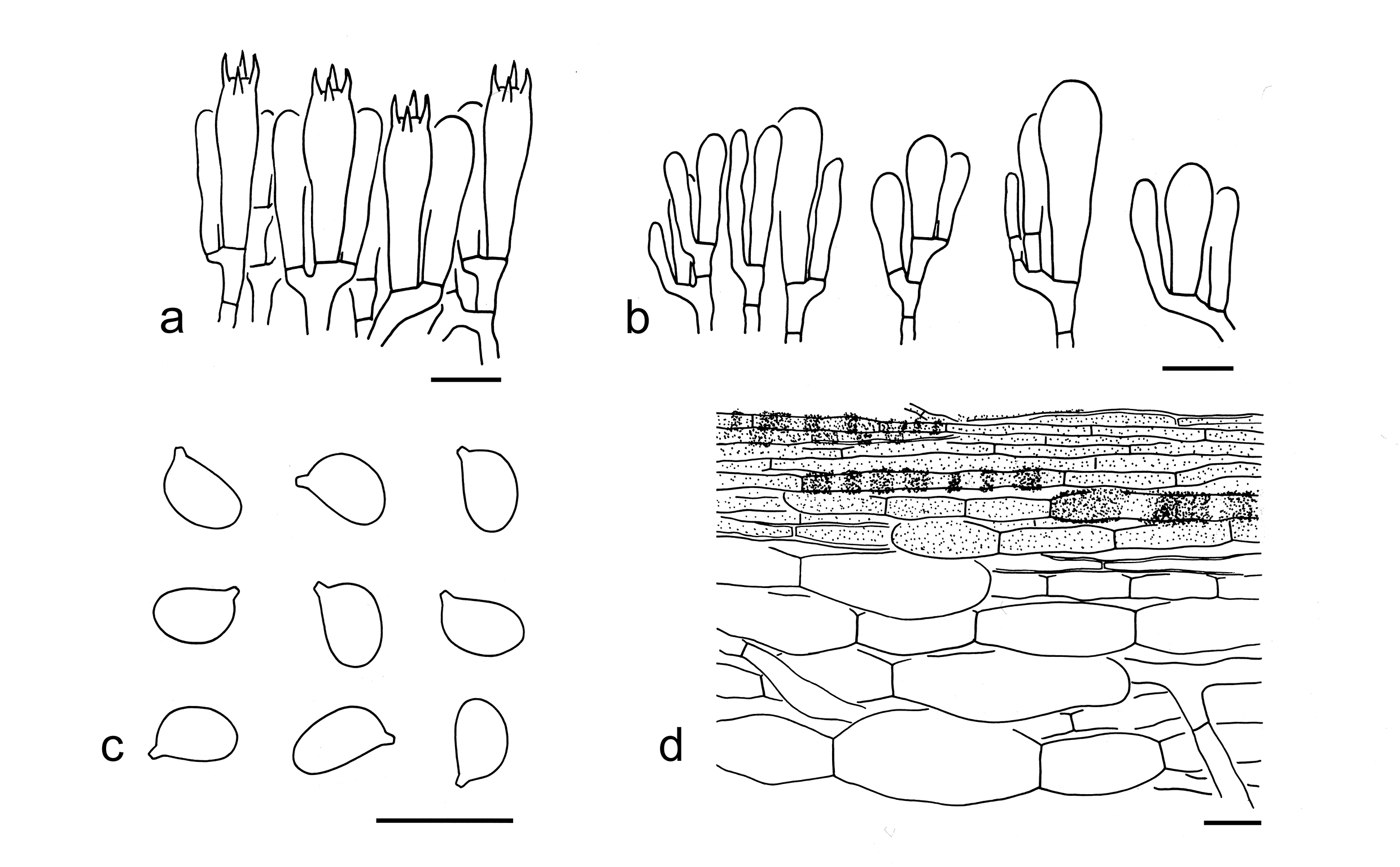
78. Tricholoma triste (Scop.) Quél., Mém. Soc. Émul. Montbéliard, Sér. 2 5: 79 (1872). Figs. 86k, 97
Basionym: Agaricus tristis Scop., Fl. carniol., Edn 2 (Wien) 2: 438 (1772).
Description: Basidioma very small to small. Pileus 2–5 cm in diam., convex to plano-convex, sometimes broadly umbonate at center; surface dry, black (2F6–8), dark grey (4E1–4) to grey (3D1, 2E1–3), densely covered with dark grey (4E1–4) to grey (3D1, 2E1–3), felty to woolly squamules, with a distinct, white (1A1) to dirty white (1B1, 2B1), woolly to fibrillose zone at margin; margin involute when young, later straight to inflexed; context white (1A1) to dirty white (1B1). Lamellae sinuate, dirty white (1B1) to greyish (3B1–2), sometimes with grey (2D1, 2E1) spots, with brown (3C2–4) color when old, subclose; edges serrulate, greyish (3B1–2), often with grey (2D1) to blackish (2E1) spots. Stipe 3–6 0.3–0.8 cm, cylindrical, hollow, white (1A1), dirty white (1B1) to greyish (2B1), covered with greyish (2B1) to brownish (1B2–4) fibrils. Taste not recorded. Odour fishy.
Basidiospores [100/5/2] 6–8 (–9) 4–5 µm (mean length = 7.1 µm, mean width = 4.5 µm), Q = (1.24–) 1.33–1.87 (–2), Qm = 1.59 ± 0.14, ellipsoid to elongate, non-amyloid, colorless, hyaline, thin-walled, smooth; apiculus relatively large. Basidia 25–35 6–8 µm, clavate, 4-spored, colorless, hyaline; sterigmata 3–5 µm long; basal septa without clamps. Pleurocystidia absent. Cheilocystidia 20–75 7–12 μm, conspicuous and abundant, clavate, sometimes septate, thin-walled, colorless and hyaline. Subhymenium composed of 2–4 µm wide filamentous hyphae, clamps absent. Lamellar trama regular, composed of colorless, thin-walled, filamentous to cylindrical hyphae 3–20 µm. Pileipellis a cutis, composed of radially arranged to interwoven, 5–15 µm wide, slightly thick-walled (ca. 1–2 µm), dark brown filamentous hyphae; subpellis composed of fusiform, ellipsoid to clavate inflated cells, 30–75 20–35 μm, thin-walled, colorless to yellow-brown. Clamps absent in all parts of basidioma.
Ecology: Solitary to scattered on soil in temperate coniferous forests.
Known distribution: Known from Europe (Riva 1988, 2003; Christensen & Heilmann-Clausen 2013) and Asia.
Specimens examined: CHINA. Xizang Autonomous Region, Xigaze, Gyirong County, the type of forest unknown, altitude 4700 m, 13 Aug. 2017, Ai-Guo Xu 2225 (AF0001608). Yunnan Province, Kunming, Xishan District, Qiongzhu Temple, the type of forest unknown, altitude 2200 m, 15 Jun. 2008, Zai-Wei Ge 2023 (KUN-HKAS54219); Lijiang, Yulong Naxi Autonomous County, in a coniferous forest with Pinus armandii, altitude 2400 m, 13 Jul. 2010, Yan-Jia Hao 40 (KUN-HKAS68012); same county, Lijiang Alpine Botanic Garden, in a coniferous forest, altitude 2700 m, 30 Sep. 2019, Jian-Wei Liu 2193 (KUN-HKAS135003).
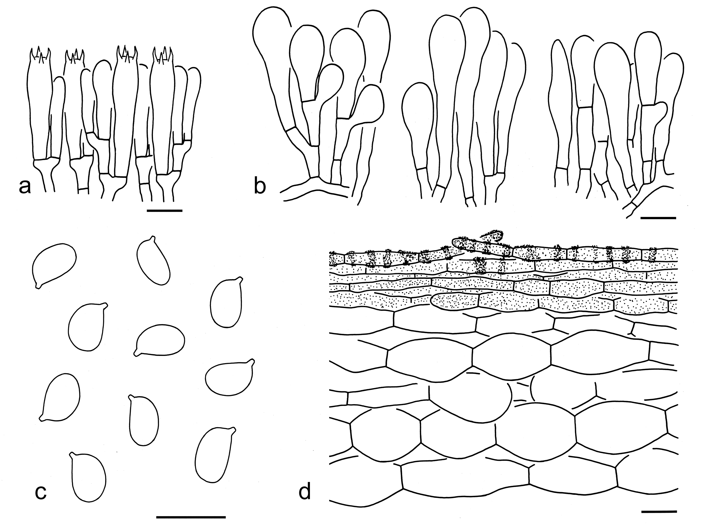
Notes: Tricholoma triste is well characterized by its dark colored pileus with a distinct, white to dirty white, woolly to fibrillose zone at margin, a well differentiated subpellis with inflated cells, and ellipsoid to elongate basidiospores (6–8 4–5 µm, Q = 1.33–1.87, Qm = 1.59 ± 0.14). In the five-gene phylogenetic analysis, it is closely related to the clade formed by T. bonii and T. terreum (Fig. 3). Indeed, they are similar to T. triste. However, T. bonii has a grey-brown, grey to dark grey pileus without a distinct zone at margin. Tricholoma terreum differs by its relatively paler colored pileus (grey, brown, grey-brown to dark grey) without a distinct zone at margin, poorly differentiated cheilocystidia, and relatively shorter basidiospores (6–7 3.5–5 μm, mean length = 6.2 µm) (Riva 1988; Bessette et al. 2013; Christensen & Heilmann-Clausen 2013; this study).
Species of Tricholoma with uncertain taxonomic position
Tricholoma clade A
Tricholoma clade A forms a monophyletic group with evident supports based on phylogenetic analyses (Figs. 2, 3, 4, 14). To date, only T. macrosporum Y.Y. Cui & Zhu L. Yang and T. vernaticum Shanks were detected in this clade, and are characterized by their robust basidioma with solid stipes, ellipsoid to elongate basidiospores and presence of clamps. Apart from clade A, sect. Terrea, sect. Atrosquamosa and clade B are found in subgen. Terrea based on the phylogenomic analysis (Figs. 2, 14). However, species in sect. Terrea differs from clade A by their felty, tomentose to somewhat fibrillose squamules on pileus, small to medium-sized basidiospores, and the absence of clamps. Members in sect. Atrosquamosa have densely squamules to scales on pileus and small basidiospores. Species from clade B can be separated from clade A by its glabrous pileus, broadly ellipsoid to ellipsoid basidiospores and the absent of clamps. Clade A may represent a novel section in subgen. Terrea. To date, only T. macrosporum was reported from China (see below).
79. Tricholoma macrosporum Y.Y. Cui & Zhu L. Yang, sp. nov. Figs. 86l, 98
Fungal Names: FN 572340
Etymology: macrosporum = large basidiospores, referring to its large basidiospores.
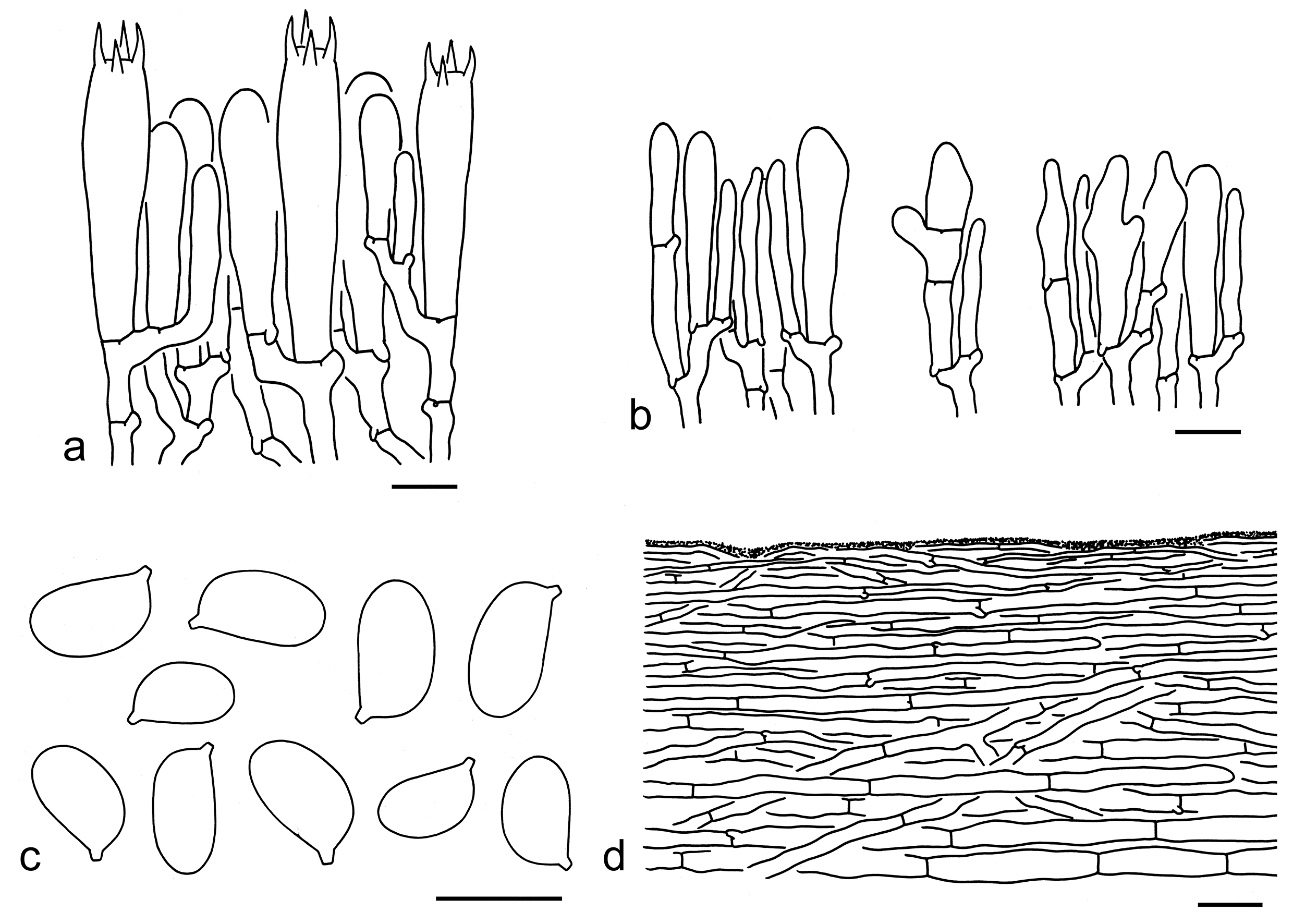
Diagnosis: Related to T. vernaticum Shanks, but differs by its greyish lamellae and a ringless stipe.
Type: CHINA. Sichuan Province, Ganzi Tibetan Autonomous Prefecture, Luhuo County, Renda Village, in a forest with Abies, altitude 3100 m, 1 Aug. 2019, Geng-Shen Wang 573 (holotype, KUN-HKAS107577).
Description: Basidioma medium-sized. Pileus ca. 6 cm in diam., conical to broadly conical, acutely umbonate; surface dry, grey-brown (2E4–6), greyish brown (2D2–4) to grey-white (1B1–3), glabrous to finely fibrillose; margin straight; context greyish (1B1–2, 1C1–2). Lamellae sinuate, greyish (1B1, 1C1), sometimes with brown (5E6–8) stains, subclose; edges concolor. Stipe 7–10 2–3.5 cm, subclavate, solid, dirty white (1B1–2) to pale brownish (5A2, 5B3), fibrillose, exannulate. Taste and odour not recorded.
Basidiospores [30/1/1] (7.5–) 8–11 5–6 µm (mean length = 9.4 µm, mean width = 5.7 µm), Q = (1.38–) 1.5–1.83 (–2), Qm = 1.66 ± 0.14, ellipsoid to elongate, non-amyloid, colorless, hyaline, thin-walled, smooth; apiculus relatively large. Basidia 40–50 9–11 µm, clavate, 4-spored, thin- to slightly thick-walled, colorless to yellowish, hyaline; sterigmata 4–7 µm long; basal septa with clamps. Pleurocystidia absent. Cheilocystidia 15–35 3–10 µm, clavate, cylindrical to irregular shaped, sometimes septate, thin-walled, colorless to yellowish, hyaline. Subhymenium composed of 3–5 µm wide filamentous hyphae, clamps present. Lamellar trama regular, composed of colorless to yellowish, thin-walled, ellipsoid, cylindrical, clavate to filamentous hyphae 5–25 µm wide. Pileipellis an ixo-cutis, gelatinized, composed of radially arranged, 2–10 µm wide, thin-walled, colorless to yellowish filamentous hyphae; subpellis poorly differentiated. Clamps present in all parts of basidioma.
Ecology: Solitary to scattered on soil in forests with Abies.
Known distribution: Known from Southwestern China.
Additional specimen examined: CHINA. Sichuan Province, Ganzi Tibetan Autonomous Prefecture, Luhuo County, Renda Village, in a forest with Abies, altitude 3000 m, 1 Aug. 2019, Geng-Shen Wang (KUN-HKAS154384).
Notes: Tricholoma macrosporum is characterized by its conical, grey-brown to grey-white pileus, ellipsoid to elongate basidiospores (8–11 5–6 µm, Q = 1.5–1.83, Qm = 1.66 ± 0.14), and the presence of clamps. In the phylogenetic analysis based on the ITS sequences, it is closely related to an undescribed species from USA and followed by T. vernaticum (Fig. 4). However, T. vernaticum differs by its white lamellae and a clavate stipe often with a median to superior ring (Shanks 1996; Bessette et al. 2013).
Morphologically, T. virgatum and T. guldeniae look like T. macrosporum. However, T. virgatum has a slender basidioma with a cylindrical, stuffed to hollow, somewhat twisted stipe, relatively shorter, broadly ellipsoid to ellipsoid basidiospores (7–10 5–7 µm, mean length = 7.9 µm), and the absence of clamps (Riva 1988, 2003; Christensen & Heilmann-Clausen 2013). Tricholoma guldeniae possesses a cylindrical stipe, heterogeneous basidiospores in size and shape (average 8–9.2 6–7.3 µm), and the absence of clamps (Christensen & Heilmann-Clausen 2013).
Tricholoma clade B
Tricholoma clade B forms a monophyletic group in phylogenetic analyses (Figs. 2, 3, 4, 14). This clade is sister to sect. Terrea (Figs. 2, 14), but can be easily separated by its glabrous pileus and broadly ellipsoid to ellipsoid basidiospores. Clade B may represent a novel section in subgen. Terrea. Species in this clade are characterized by its grey-brown and glabrous pileus often umbonate at center, close lamellae, absent of annulus, broadly ellipsoid to ellipsoid basidiospores, poorly differentiated cheilocystidia and absent of clamps. To date, only T. griseofulvum was collected from China (see below).
80. Tricholoma griseofulvum Y.Y. Cui & Zhu L. Yang, sp. nov. Figs. 86m, 99
Fungal Names: FN 572339
Etymology: griseofulvum = grey-brown, referring to its grey-brown pileus.
Diagnosis: Related to T. atratum Ovrebo, K.W. Hughes & Halling, but differs by its larger basidiospores and associations with trees of Picea.
Type: CHINA. Sichuan Province, Ganzi Tibetan Autonomous Prefecture, Kangding, Yala, Mugecuo, in a forest with Picea, altitude 3350 m, 17 Sep. 2017, Xiao-Xia Ding 310 (holotype, KUN-HKAS106652).
Description: Basidioma small to medium-sized. Pileus 3–5.5 cm in diam., convex to plano-convex, often acutely umbonate; surface dry, greyish brown (5E2–5) to reddish brown (6E3–6), sometimes grey-brown (6F5–7) at center, becoming greyish brown (5E2–5) to reddish brown (6E3–6) towards margin, glabrous; margin straight; context white (1A1). Lamellae sinuate, white (1A1) to dirty white (5B1), close to subdistant; edges concolor. Stipe 4.5–10 0.5–1 cm, subclavate, hollow when mature, greyish white (4D1) to grey white (4C1), exannulate. With strong soapy taste.
Basidiospores [45/2/2] 10–12 (–13) (7–) 7.5–9.5 (–10) µm (mean length = 10.8 µm, mean width = 8.3 µm), Q = 1.17–1.44, Qm = 1.3 ± 0.07, broadly ellipsoid to ellipsoid, non-amyloid, colorless, hyaline, thin-walled, smooth; apiculus relatively large. Basidia 50–65 12–15 µm, clavate, 4-spored, thin-walled, colorless, hyaline; sterigmata 6–9 µm long; basal septa without clamps. Pleurocystidia absent. Cheilocystidia 20–50 3–10 µm, poorly differentiated, clavate to subclavate, thin-walled, colorless, hyaline. Subhymenium composed of 3–5 µm wide filamentous hyphae, clamps absent. Lamellar trama regular, composed of colorless thin-walled, cylindrical, clavate to filamentous hyphae 3–15 µm wide. Pileipellis a cutis, composed of radially arranged, 5–15 µm wide, thin-walled filamentous hyphae; subpellis made of radially arranged, ellipsoid, clavate to cylindrical hyphae. Clamps absent in all parts of basidioma.
Ecology: Solitary to scattered on soil in forests with Picea.
Known distribution: Known from Southwestern China.
Additional specimen examined: CHINA. Sichuan Province, Ganzi Tibetan Autonomous Prefecture, Kangding, Yala, Mugecuo, in a forest with Picea, altitude 3350 m, 17 Sep. 2017, Xiao-Xia Ding 311 (KUN-HKAS106653).
Notes: Tricholoma griseofulvum is characterized by its greyish brown to reddish brown pileus often accurately umbonate at center, close to subdistant lamellae, broadly ellipsoid to ellipsoid basidiospores (10–12 7.5–9.5 µm) and the absence of clamps. Tricholoma atratum is closely related to this species in the tree generated from the ITS sequences (Fig. 4). However, T. atratum has smaller basidiospores (8–9 × 6–7.5 μm) and is distributed in Costa Rica in forests under Quercus copeyensis and Q. seemannii (Ovrebo et al. 2019). Morphologically, T. brunneiceps is similar to T. griseofulvum, but differs by its smaller basidiospores (6–8.5 × 5–6 μm), conspicuous cheilocystidia, a pileipellis with its lower part composed of inflated cells, and associations with trees of Fagaceae.
Tricholoma clade C
Tricholoma clade C clustered as an independent clade with evident support based on phylogenetic analyses (Figs. 2, 3, 4, 14), this clade is closely related to sect. Genuina in phylogenetic analysis (Figs. 2, 14). Presently only T. grave Peck and T. aurantiophyllum L.P. Tang et al. were detected in Clade C and are characterized by their robust basidioma and pale brown to brown lamellae. One species was found in China (see below).
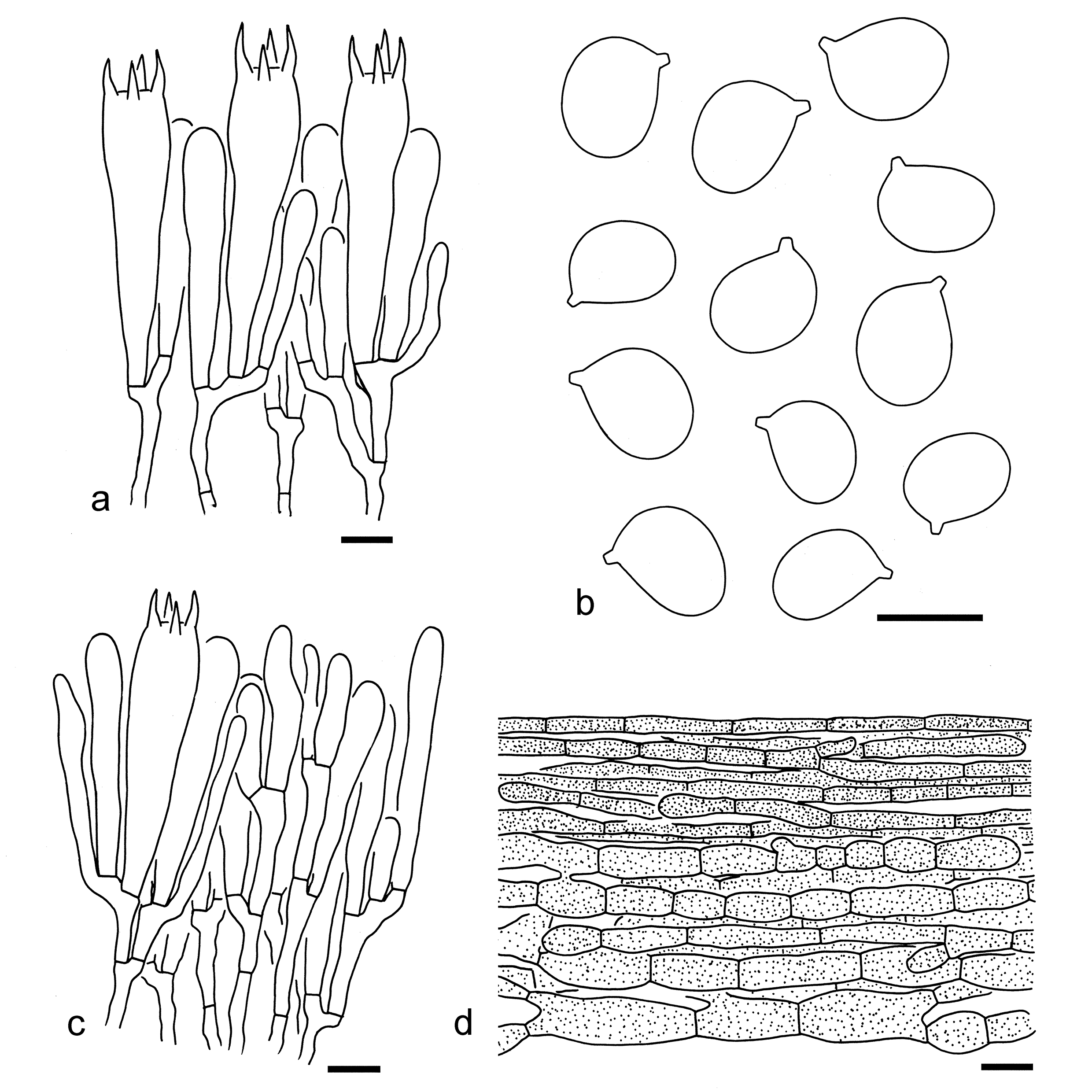
81. Tricholoma aurantiophyllum L.P. Tang, H.Y. Huang, F.Q. Yu & R. Wang, Phytotaxa 664 (3): 184. Figs. 86n, 100
Replaced synonym: Hygrophorus robustus F.Q. Yu, Mycotaxon 100: 170, Figs. 1–5 (2007).
Description: Basidioma medium-sized to large. Pileus 8–13 cm in diam., convex to plano-convex, without an umbo or depression at center; surface dry, brown, grey-brown (5B5–7) to brownish (5A3–5), often covered with brown (5B5–7), fibrillose squamules; margin straight; context white (1A1) to dirty white (5A1), up to 1.5 cm in thickness, stout. Lamellae adnate to sinuate, brown to pale brown (5C4–8), distant; edges concolorous or paler. Stipe 8–18 1.5–3.5 cm, cylindrical, dirty white (5A1–2) to brownish (5A3–5), fibrillose, exannulate or with an incomplete annulate zone near the apex of stipe, solid and compact. Taste and odour indistinct.
Basidiospores [20/1/1] 7–8 (–10) 5–6 µm (mean length = 7.7 µm, mean width = 5.3 µm), Q = 1.33–1.6 (–1.8), Qm = 1.47 ± 0.11, predominantly ellipsoid, sometimes elongate, non-amyloid, colorless, hyaline, thin-walled, smooth; apiculus relatively large. Basidia 35–60 9–11 µm, clavate, 4-spored, hyaline; sterigmata 4–6 µm long; basal septa without clamps. Pleurocystidia absent. Cheilocystidia 20–60 5–10 µm, clavate, cylindrical to irregularly shaped, sometimes septate, thin-walled, colorless to yellowish. Subhymenium composed of 2–4 µm wide filamentous hyphae, clamps absent. Pileipellis a cutis, non- to slightly gelatinized, composed of radially arranged, 3–15 µm wide, thin-walled, yellow brown filamentous hyphae; subpellis non-gelatinized, poorly differentiated. Clamps absent in all parts of basidioma.
Ecology: Solitary to scattered on soil in subtropical mixed forests with broad-leaved plants, and Pinus armandii or P. yunnanensis.
Known distribution: Known from Southwestern China.
Notes: Hygrophorus robustus was described from Southwestern China by Yu et al. (2007), and later transferred to Tricholoma as T. aurantiophyllum by Huang et al. (2024). According to our investigation, consumption of this species can cause headaches, eye pain, and severe vomiting in some individuals.
Tricholoma aurantiophyllum is characterized by its robust basidioma with a brown to grey-brown pileus, a rather stout context, subdistant and brown to brownish lamellae, mostly ellipsoid basidiospores (7–8 5–6 µm) and the absence of clamps. In the phylogenetic analysis based on the ITS sequences (Fig. 4), T. grave Bessette et al. is closely related to this species. However, T. grave has pale brown lamellae and is distributed in North America (Bessette et al. 2013; Landry et al. 2022).
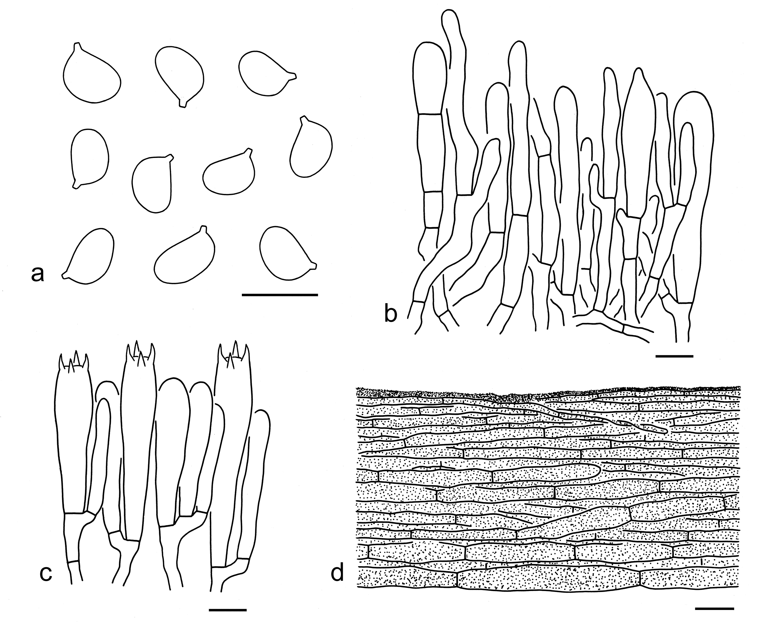
Tricholoma melleum clade
Tricholoma melleum represents an unassigned clade and may be a separate subgenus according to phylogenomic analysis (Figs. 2, 14). To date, only T. melleum Reschke et al. is known from this clade, as described below.
82. Tricholoma melleum Reschke et al., Mycologia110(6): 1103 (2018). Fig. 86o
Description: Basidioma medium-sized to large. Pileus 4–11 cm in diam., convex to plano-convex, more or less umbonate; surface dry, orange-brown (5B4–7), covered with orange-brown (5B4–7) to light orange (5A2–5), fibrillose squamules; margin involute when young, becoming straight when mature. Lamellae sinuate, cream (4A2), subclose; edges entire, cream (4A2). Stipe 6–15 1–2.5 cm, cylindrical, cream (4A2), yellowish (4A3–5) to orangish brown (5B4–7), exannulate. Taste and odour like dishwater.
Ecology: Solitary to scattered on soil in broad-leaved or mixed forests.
Known distribution: Known from southwestern China (Reschke et al. 2018).
Specimen examined: CHINA. Sichuan Province, Ganzi Tibetan Autonomous Prefecture, Kangding, altitude 3300 m, 21 August 2017, 513301MF0583 (KUN-HKAS103101).
Notes: Reschke et al. (2018) described T. melleum based on the holotype (KUN-HKAS93514) from China with an unassigned section. The holotype was not traced in the herbarium. We have collected another specimen (KUN-HKAS103101) which matches well with this species (Figs. 3, 4). Unfortunately, KUN-HKAS103101 has been used up and only macroscopic character of this species is given above. Its precise microscopic traits will be provided with more specimens available.
Discussion
Phylogeny of Tricholomataceae s.str.
Tricholomataceae in its narrow sense (Sánchez-García et al. 2014) is a monophyletic group and likely closely related to Callistosporiaceae Vizzini et al. (Vizzini et al. 2020a; He et al. 2023; Vizzini et al. 2024). Our study supports that members of this family have tricholomatoid basidioma with glabrous, squarrulose, squamose, or radially fibrillose pilei; adnate, adnexed, sinuate, emarginate to decurrent lamellae; cylindrical stipes mostly lacking an annulus; subglobose, broadly ellipsoid, ellipsoid to elongate basidiospores, sometimes amygdaliform, that are mostly smooth, occasionally verrucose, and either amyloid or inamyloid (Fig. 14).
Our phylogenetic analysis based on Dataset I (nrLSU/ITS/nrSSU/RPB2) revealed that Tricholomataceae s.str. comprises eleven genera (Fig. 1). However, the relationships among most genera were not well resolved. Among them, Pseudoporpoloma, proposed by Vizzini et al. (2016) to describe members of the ‘pes-caprae clade’ in Sánchez-García et al. (2014), was not included in the study by Sánchez-García et al. (2021) and Adamčíková et al. (2025). Our findings support the inclusion of Pseudoporpoloma as a valid genus within Tricholomataceae s.str. (Fig. 1, Supplementary Table S4).
Albomagister is closely related to two undetermined clades (‘undet small white clade’ and ‘undet grey scaly clade’) identified by Sánchez-García et al. (2014, 2021). Later, Matheny et al. (2024) reclassified these clades under Albomagister, which is also supported in this study (Fig. 1, Supplementary Table S4). The Corneriella-like clade in Sánchez-García et al. (2021) clustered with Corneriella in our analysis (Fig. 1), and we have treated them as Corneriella for now (Fig. 1, Supplementary Table S4). Regarding the Dermoloma-like clade, we follow the treatment of Adamčíková et al. (2025) as Neodermoloma (Fig. 1, Supplementary Table S4).
Phylogeny of Tricholoma and the key characteristics of the infrageneric divisions
Tricholoma is the most famous genus within Tricholomataceae s.str. and presents significant challenges for taxonomy. Its subdivisions have been a subject of ongoing debate. In recent years, various sequencing strategies have been employed to elucidate the phylogeny of this genus, yielding substantial progress (Heilmann-Clausen et al. 2017; Reschke et al. 2018; Landry et al. 2022; Trudell et al. 2022; Ding et al. 2023).
Our study compared phylogenetic analyses of Tricholoma based on ITS sequences, five gene fragments (ITS/RPB2/MCM7/EF1-α/mtSSU), and 1,477 SOGs (Fig. S1). The results indicate that ITS sequences effectively identify most species, except for those in sect. Rigida and sect. Matsutake. At the sectional level, only six sections (sect. Rigida, sect. Terrea, Clade A, sect. Sericella, sect. Genuina, and sect. Fucata) are supported by ITS sequences, and their phylogenetic relationships remain unresolved (Fig. S1). Phylogenetic analysis using five gene fragments provides better resolution at the section level and improves species identification but fails to clarify the boundaries of subgenera and relationships among major clades (Fig. S1). Notably, phylogenomic analyses based on 1,477 SOGs successfully resolved all subgenera and sections of Tricholoma (Fig. S1). Ding et al. (2023) provided deep insights into the phylogenetic relationships among four subgenera and 14 section-level clades of Tricholoma using a 50-locus dataset. However, the relationships between subgen. Terrea and subgen. Tricholoma, as well as between the clade formed by sect. Tricholoma and sect. Rigida, and the clade that includes sect. Muscaria, sect. Fucata and sect. Matsutake, were poorly resolved, particularly in the ML analysis (MLB=70% and MLB=66%) (Ding et al. 2023). In addition, within subgen. Terrea, the sister group relationship between sect. Atrosquamosa and clade A was not supported (MLB=42% and PP=0.93) (Ding et al. 2023). In our research, four subgenera and one subgenus-level clade, and 15 section-level clades were identified. The phylogenetic relationships among these subgenera and clades were highly resolved in the tree constructed with 1,477 SOGs (Fig. 2). Sect. Atrosquamosa was indicated to be sister to the clade formed by sect. Terrea and clade B. Furthermore, clade A was found to be closely related to the clade that includes the aforementioned two sections and clade B (Fig. 2). It could be concluded that our research, based on genomic data, successfully resolved the phylogeny of Tricholoma, providing an evolutionary perspective for future studies on Tricholoma taxonomy and evolution (Fig. 2).
Molecular and morphological evidence suggests that the diagnostic characteristics of Tricholoma include tricholomatoid basidioma, absence of pleurocystidia, a cutis pileipellis primarily composed of repent hyphae, and smooth inamyloid basidiospores. Vizzini et al. (2020b) proposed a novel standardized method (including heating the sample in Melzer’s reagent and different recipes for Melzer’s exist) to test for spore amyloidity in Tricholoma. They suggested that basidiospores of this genus are latently amyloid. However, our study followed traditional methods due to their simplicity and widespread use in fungal taxonomy. Using this approach, we observed visibly inamyloid basidiospores in all studied Tricholoma species. These findings align with recent observations by Lebeuf et al. (2024), which also support the inamyloid nature of Tricholoma basidiospores.
Combining phylogenomic and morphological evidence, we support the division of Tricholoma into 11 sections. These sections are characterized by features such as pileal center morphology, pileus color and squamules, lamellar density, presence or absence of an annulus, basidiospore size and shape, presence or absence of cheilocystidia, pileipellis structure, and clamp connections. While these characteristics have been highlighted by Bon (1984), Singer (1986), Riva (1988), and Noordeloos & Christensen (1999), their taxonomic significance varies. Heilmann-Clausen et al. (2017) emphasized that pileus color, pileipellis structure, clamp presence, and basidiospore size are conserved within sections, whereas annulus presence and host specificity are more variable. Our study highlights additional diagnostic features, such as the presence or absence of an umbo on the pileal center, which may distinguish sections. For instance, species in sect. Fucata and sect. Muscaria exhibit a prominent papilla. Similarly, cheilocystidia presence is a critical marker, as species in sect. Fucata and sect. Muscaria have conspicuous cheilocystidia, whereas those in sect. Lasciva have poorly differentiated cheilocystidia.
The pileipellis structure remains a valuable taxonomic characteristics, as previously emphasized by Bon (1984), Singer (1986), Riva (1988), and Noordeloos & Christensen (1999). For example, members of sect. Fucata have a cutis with a strongly differentiated subpellis composed of inflated cells, while species in sect. Atrosquamosa possess a pileipellis with somewhat erect, thick-walled hyphae forming squamules on the pileus. These findings deepen our understanding of Tricholoma classification from an evolutionary perspective and underscore the importance of integrating phylogenomic, phylogenetic, and morphological data in fungal taxonomy.
Species diversity of Tricholomataceae s.str. from China
Presently, approximately 500 species are regarded as members of the family Tricholomataceae in its narrow sense. In China, members of Tricholomataceae s.str. are found in only five genera (Tricholoma, Leucopaxillus, Pseudobaeospora, Albomagister, and Pseudotricholoma).
Due to morphological similarities and phenotypic plasticity, accurately identifying species in Tricholomataceae s.str. based on morphology alone is challenging. Some newly described species in this study were misidentified in the past. For instance, T. mastoideum was misidentified as T. virgatum, and T. sichuanense was mistaken for T. saponaceum. This highlights the necessity of using both morphological and molecular evidence to delimit species within this family. For herbarium collections, generating DNA sequences using Sanger sequencing can be difficult. Fortunately, next-generation sequencing (NGS) combined with optimized library construction processes has enabled the extraction of DNA from historical specimens, providing solid evidence for the accurate recognition of fungal species (Zeng et al. 2018; Wang et al. 2024).
In this study, we identified 82 species of Tricholomataceae s.str. distributed in China. Of these, 30 were originally described from Europe or the USA, five from Japan, and 47 from China. This indicates a rich diversity of Tricholomataceae s.str. in China, with over 60% of species originally described from Asia or even East Asia. This pattern hints at a potentially high level of endemism in East Asia, but further biogeographic studies are needed to confirm whether these species are truly restricted to this region. Additionally, several unique clades labeled as Tricholoma sp. 2 and sp. 3 in Figures 3 and 4 may represent unnamed species. However, further collections are required before these can be formally described. These findings provide a strong foundation for future studies on the diversity and distribution patterns of Tricholomataceae.
Edibility and toxicity of species of Tricholomataceae s.str.
The family Tricholomataceae s.str., particularly the genus Tricholoma, includes both edible and poisonous species (Bresinsky 1990; Anand et al. 2009; Bessette et al. 2013; Christensen & Heilmann-Clausen 2013; Chen et al. 2016; Laubner & Mikulevičienė 2016; Yang et al. 2021; Wan et al. 2023). Accidental ingestion of toxic species Tricholomataceae s.str. primarily leads to three types of poisoning symptoms: rhabdomyolysis, acute liver damage, and gastroenteritis-type poisoning (Bresinsky 1990; Anand et al. 2009; Christensen & Heilmann-Clausen 2013; Chen et al. 2016, Laubner & Mikulevičienė 2016; Wan et al. 2023). Rhabdomyolysis poisoning has only been reported in Europe following the consumption of large quantities of T. equestre (Anand et al. 2009; Laubner & Mikulevičienė 2016). No such cases have been reported in China. Gastroenteritis-type poisoning, however, is associated with species primarily distributed in sect. Fucata, sect. Genuina, sect. Sericella, sect. Lasciva, sect. Pardinicutis, and clade C (Bresinsky 1990; Christensen & Heilmann-Clausen 2013; Chen et al. 2016; Yang et al. 2021; Wan et al. 2023; Bau et al. 2025; this study). Here, we highlight T. aurantiophyllum in clade C, as ingestion of this species has been reported to cause headaches and severe vomiting in some individuals (Huang et al. 2024; this study). Notably, accidental ingestion of toxic species in sect. Pardinicutis can cause severe acute liver damage, as seen with T. nigrosquamosum and T. sinoportentosum, both found in southwestern China (Wan et al. 2023).
Despite these risks, many Tricholomataceae s.str. mushrooms are sold in markets across China, particularly in the southwestern areas. Our results, show that mushrooms of Tricholomataceae sold in markets can be grouped into four categories comprising 18 species. These are commonly known as: “songrong” (including T. matsutake, T. fulvocastaneum, T. bakamatsutake, T. xizangense, T. tianshanense and T. zangii), “qiaomianjun” (including T. citrinum, T. equestre, T. frondosae and T. qiaomianjun), “huihuijun” (including T. terreum, T. bonii, T. triste and T. virgatum), and “beifengjun” (including T. saponaceum, T. olivaceum, T. sichuanense and T. sinoportentosum). Despite their popularity, studies on the edibility and toxicity of these fungi remain limited. Further research is necessary to identify poisonous species, determine the types of toxins, and understand their distribution patterns within this family. Such investigations will raise public awareness of the safety of Tricholomataceae mushrooms and provide a scientific basis for food safety regulations.
Acknowledgments
We thank the curators of the herbaria HMAS, HMJAU, AF, MB, and HMUT for the loan of specimens. We are grateful to Dr. Zhuo Du (Institute of Microbiology, Chinese Academy of Sciences), Prof. Gerhard Kost (University of Marburg), Dr. Xiao-Juan Deng (North Minzu University), Mr. Ai-Guo Xu (Tibet Plateau Institute of Biology), Prof. Biao Xu (Zhongkai University of Agriculture and Engineering), and Dr. Xiao-Dan Yu (Shenyang Agricultural University) for their assistance with loan requests. We also appreciate the comments and advice from Prof. Bau Tolgor (Jilin Agricultural University), Prof. Tai-Hui Li, and Dr. Chao-Qun Wang (Institute of Microbiology, Guangdong Academy of Sciences). Special thanks to Mr. Zhi-Jia Gu (Kunming Institute of Botany, Chinese Academy of Sciences) for providing SEM images. Thanks are also due to the anonymous reviewers for their professional suggestions, important comments and valuable criticisms. The authors would like to express their gratitude to EditSprings (https://www.editsprings.cn) for the expert linguistic services provided. This work was supported by the Science and Technology Fundamental Resources Investigation Program, Ministry of Science and Technology of China (2023FY101303), the Joint Funds of the National Natural Science Foundation of China and Yunnan Provincial Government (U2202205), the Yunnan Revitalization Talent Support Program: Science & Technology Champion Project (202305AB350004), Yunnan Revitalization Talent Support Program "Young Talent" Project (XDYC-QNRC-2022-0509), Caiyun Postdoctoral Program, Yunnan Provincial High-Level Talents Support Program (YNWR-QNBJ-2020-295), the "Talent Introduction Program" of Kunming Institute of Botany, Chinese Academy of Sciences, the Second Tibetan Plateau Scientific Expedition and Research Program (2024QZKK02010303), and the Major Science and Technology Special Project of Yunnan Province (202502AE090044).
Author contributions
The authors confirm contribution to the paper as follows: Conceptualization: Cui YY, Yang ZL; Data curation: Cui YY, Li J, Fan XP, Che NJ; Formal analysis: Cui YY, Li J, Cai Q, Fan XP, Yang ZL, Che NJ; Funding acquisition: Yang ZL, Cui YY, Cai Q, Zhao Q; Investigation: Cui YY, Cai Q, Yan-Chun Li, Fan XP, Yang ZL, Liu JW, Ding XX, Xu X, Zhu XT, Deng CY, Guo T, Che NJ, Zhao Q; Methodology: Cui YY, Li J, Fan XP, Che NL, Cai Q; Project administration: Cui YY; Resources: Cui YY, Li J, Cai Q, Yan-Chun Li, Fan XP, Yang ZL, Liu JW, Ding XX, Xu X, Zhu XT, Deng CY, Guo T, Che NJ, Zhao Q; Software: Cui YY, Li J, Cai Q; Supervision: Yang ZL; Validation: Cui YY, Li J, Cai Q; Visualization: Cui YY, Li J, Che NJ, Cai Q; Writing—original draft preparation, Cui YY, Cai Q; Writing—review & editing: Cui YY, Yang ZL, Cai Q. All authors have read and agreed to the published version of the manuscript.
ORCID
Yang-Yang Cui: https://orcid.org/0000-0003-3990-8342
Yan-Chun Li: https://orcid.org/0000-0002-7109-9269
Zhu L. Yang: https://orcid.org/0000-0001-9745-8453
Qing Cai: https://orcid.org/0000-0001-5046-0484
Conflict of interest statement
The author list includes members of the Editorial Board of Fungal Diversity. They were not involved in the journal’s review of, or decisions related to, this manuscript. The authors declare no competing interests.
Supplementary Information
The online version contains supplemental information available at https://doi.org/10.65390/fdiv.2026.136010.
Supplementary File to this study.
Supplementary Figure S1 A comparison of the three Tricholoma trees inferred from Dataset II (1,477 SOGs), Datasets III (ITS/RPB2/MCM7/EF1-α/mtSSU) and IV (ITS).
Supplementary Table S1 Voucher information and GenBank accession numbers of the samples used in the phylogenetic analyses of Tricholomataceae (Dataset I). New Species described in this study are indicated in bold.
Supplementary Table S2 Voucher information of the samples used in the next-generation sequencing of Tricholomataceae (Dataset II).
Supplementary Table S3 Voucher information and GenBank accession numbers of the samples used in the phylogenetic analyses of Tricholoma (Datasets III and IV). New Species described in this study are indicated in bold.
Supplementary Table S4 Comparisons of several significant Tricholomataceae systems.
Supplementary Table S5 A comparison between the Chinese Tricholoma species reported in this work and Ding et al. (2023).
Rights and permissions
The Author(s) 2026. Published by BioAcademic Press on behalf of Kunming Institute of Botany,Chinese Academy of Sciences (CAS) and Mushroom Research Foundation. This is an open accessarticle under the Creative Commons Attribution license ( http://creativecommons.org/licenses/by/4.0), which permits use, distribution and reproduction in any medium, provided the originalwork is properly cited.
References
- Adamčík S, Jančovičová S (2011) Pseudobaeospora terrayi, a new species from Slovakia. Sydowia 63(2): 131–140.
- Adamčík S, Ripková S, Kučera V (2007) Re-evaluation of morphological variability of Pseudobaeospora group Celluloderma (Agaricales, Basidiomycota). Nova Hedwigia 85(3–4): 365–377.
https://doi.org/10.1127/0029-5035/2007/0085-0365 - Adamčíková K, Kiran M, Caboň M, Matheny BP, Sánchez-García M, Arnolds E, Caboňová M, Corriol G, Dima B, Friebes G, Griffith GW, Grootmyers D, Harries D, Karich A, Mešić A, Mihaljevič M, Moreau PA, Pošta A, Shapkin V, Tkalčec Z, Vizzini A, Vondrovicová L, Adamčík S, Jančovičová S (2025) A phylogenetic and morphological study of the genus Dermoloma (Agaricales, Tricholomataceae) in Europe and North America exposes inefficiency of opportunistic species descriptions. IMA Fungus 16: e157337.
https://doi.org/10.3897/imafungus.16.157337 - Ammirati JF, Ovrebo CL (1979) Type studies: some Cortinarius and Tricholoma species described by Charles Horton Peck. Mycotaxon 8(2): 353–371.
https://doi.org/10.5962/p.417895 - Anand JS, Chwaluk P, Sut M (2009) Acute poisoning with Tricholoma equestre. Przegl Lek 66(6): 339–340.
- Arauzo S (2012) Una nueva especie del género Porpoloma descubierta en el País Vasco. Errotari 9: 84–91.
- Arnolds E (1992) Notulae ad Floram agaricinam neerlandicam–XIX. A revision of Dermoloma (Lange J) Sing. –1. Persoonia 14(4): 519–532.
- Arnolds E (1993) Notulae ad Floram agaricinam neerlandicam–XX. A revision of Dermoloma (Lange J) Sing. –2. Persoonia 15(2): 187–196.
- Arnolds E, Leelavathy KM, Manimohan P (2004) Pseudobaeospora lavendulamellata: a new species from Kerala, India. Persoonia 18(3): 435–438.
- Bankevich A, Nurk S, Antipov D, Gurevich AA, Dvorkin M, Kulikov AS, Lesin VM, Nikolenko SI, Pham S, Prjibelski AD, Pyshkin AV, Sirotkin AV, Vyahhi N, Tesler G, Alekseyev MA, Pevzner PA (2012) SPAdes: a new genome assembly algorithm and its applications to single-cell sequencing. Journal of Computational Biology 19(5): 455–477.
https://doi.org/10.1089/cmb.2012.0021 - Bas C (1969) Morphology and subdivision of Amanita and a monograph of its section Lepidella. Persoonia 5(4): 285–573.
- Bas C (2002) A reconnaissance of the genus Pseudobaeospora in Europe I. Persoonia 18(1): 115–122.
- Bas C (2003) A reconnaissance of the genus Pseudobaeospora in Europe II. Persoonia 18(2): 163–199.
- Bau T, Yang ZL, Chen ZH, Li TH, Li HJ. 2025. Poisonous mushrooms of China. Science Press, Beijing.
- Bedry R, Baudrimont I, Deffieux G, Creppy EE, Pomies JP, Ragnaud JM, Dupon M, Neau D, Gabinski C, De Witte S, Chapalain JC, Godeau P, Beylot J (2001) Wild-mushroom intoxication as a cause of rhabdomyolysis. The New England Journal of Medicine 345(11): 798–802.
https://doi.org/10.1056/NEJMoa010581 - Bessette AE, Bessette AR, Roody WC (2013) Tricholomas of North America: a mushroom field guide. University of Texas Press, Austin.
- Bolger AM, Lohse M, Usadel B (2014) Trimmomatic: a flexible trimmer for Illumina sequence data. Bioinformatics 30(15): 2114–2120.
https://doi.org/10.1093/bioinformatics/btu170 - Bon M (1979) Taxons nouveaux. Documents Mycologiques 9: 39–44.
- Bon M (1984) Les tricholomes de France et d’Europe occidentale. Lechevalier, Paris.
- Bon M (1986) Novitates-validations et taxons nouveaux. Documents Mycologiques 17: 51–56.
- Bon M (1991) Flore mycologique d’Europe, vol. 2, Les tricholomes et ressemblants. St Valerysur-Somme, France.
- Bon M (1998) Novitates-Marasmiaceae et Dermolomataceae comb., st. et sp. nov. Documents Mycologiques 28: 6.
- Bougher NL (1987) A new species of Leucopaxillus Bours. (Agaricales) from western Australia. Sydowia 39: 17–21.
- Brandrud TE (2020) Tricholoma matsutake. The IUCN Red List of Threatened Species 2020: e.T76267712A76268018. Accessed 01 February 2026.
https://dx.doi.org/10.2305/IUCN.UK.2020-2.RLTS.T762677 12A76268018.en. - Bresinsky A (1990) A colour atlas of poisonous fungi: a handbook for pharmacists, doctors, and biologists. CRC Press, London.
- Caboňová M, Sanchez-Garcia M, Caboň M, Adamčíková K, Moreau PA, Vizzini A, Adamčík S, Jančovičová S (2025) Nomenclatural review of names published in the fungal genus Dermoloma (Basidiomycota, Agaricales, Tricholomataceae) based on morphological analyses of type specimens. Biodiversity Data Journal 13: e158080.
https://doi.org/10.3897/BDJ.13.e158080 - Cao ZM (2003) Phylogenetic reconstruction and biogeographic study of matsutake group. Dissertation, University of Chinese Academy of Sciences.
- Cao ZM, Yao YJ, Pegler DN (2003) Tricholoma zangii, a new name for T. quercicola M. Zang (Basidiomycetes: Tricholomataceae). Mycotaxon 85: 159–164.
https://doi.org/10.5962/p.414695 - Capella-Gutiérrez S, Silla-Martinez JM, Gabaldon T (2009) TrimAl: a tool for automated alignment trimming in large-scale phylogenetic analyses. Bioinformatics 25(15): 1972–1973.
https://doi.org/10.1093/bioinformatics/btp348 - Carbone M, Boccardo F, Calledda F (2020) Tricholoma ligusticum, una specie nuova dei querceti liguri. Neotipificazione e studio genetico di Tricholoma quercetorum. RdM 63: 197–214.
- Casas LR, Sánchez L, Gibert S, Palazón A (2015) Nuevas citas de Tricholoma quercetorum Contu para la Península Ibérica (Tricholomataceae: Agaricales). BVnPC 4: 43–50.
- Chapela IH, Garbelotto M (2004) Phylogeography and evolution in matsutake and close allies inferred by analyses of ITS sequences and AFLPs. Mycologia 96(4): 730–741.
https://doi.org/10.1080/15572536.2005.11832921 - Chen ZH, Yang ZL, Bau T, Li TH (2016) Poisonous mushrooms: recognition and poisoning treatment. Science Press, Beijing.
- Christensen M, Heilmann-Clausen J (2009) Two new boreal species of Tricholoma from Fennoscandia. Mycotaxon 107(1): 431–440.
https://doi.org/10.5248/107.431 - Christensen M, Heilmann-Clausen J (2013) The genus Tricholoma. Fungi of Northern Europe, vol. 4. Svampetryk, Denmark.
- Christensen M, Noordeloos ME (1999) Notulae ad Floram agaricinam neerlandicam–XXXVI. Tricholoma. Persoonia 17(2): 295–317.
- Comandini O, Haug I, Rinaldi AC, Kuyper TW (2004) Uniting Tricholoma sulphureum and T. bufonium. Mycological Research 108(10): 1162–1171.
https://doi.org/10.1017/s095375620400084x - Contu M (2004) Rare, critical and new species of the genus Tricholoma collected in Sardinia, with two new records for its mycoflora. Micologia e vegetazione mediterranea 18: 91–100.
- Contu M, Consiglio G, Setti L (2008) Studi sul genere Dermoloma (Basidiomycota, Tricholomataceae). Micologia e vegetazione mediterranea 22: 83–118.
- Corriol G, Jargeat P (2018) Albomagister virgineus sp. nov., a second species of Albomagister in Europe. Current Research in Environmental & Applied Mycology 8(2): 162–171.
https://doi.org/10.5943/cream/8/2/1 - Corriol G, Jargeat P (2019) Morphological and phylogenetical description of Dennisiomyces fibrillosus sp. nov. (Agaricales, Tricholomataceae) from the Pyrenees and the first record of the genus in Europe. Phytotaxa 405(5): 226–236.
https://doi.org/10.11646/phytotaxa.405.5.1 - Cosme M (2023) Mycorrhizas drive the evolution of plant adaptation to drought. Communications Biology 6: 346.
https://doi.org/10.1038/s42003-023-04722-4 - Craig S, Vaughan LJ, Holmes GD, May TW (2023) Pseudobaeospora taluna (Fungi: Agaricales) newly described from southern Australia. Australian Journal of Taxonomy 24: 1–16.
https://doi.org/10.54102/ajt.yuij6 - Cui YY, Ding XX, Kost G, Yang ZL (2022) Tricholoma sect. Tricholoma (Tricholomataceae) from China: molecular phylogeny and taxonomy. Mycological Progress 21(3): 35.
https://doi.org/10.1007/s11557-022-01788-y - Deng H (2005) Taxonomic and molecular systematic studies on Tricholoma in China. Dissertation, University of Chinese Academy of Sciences.
- Deng H, Yao YJ, Pegler DN (2004) An annotated checklist of Tricholoma from China. Journal of Fungal Research 2(1): 1–18.
- Dentinger BTM, Gaya E, O'Brien H, Suz LM, Lachlan R, Díaz‐Valderrama JR, Koch RA, Aime MC (2016) Tales from the crypt: genome mining from fungarium specimens improves resolution of the mushroom tree of life. Biological Journal of the Linnean Society 117(1): 11–32.
https://doi.org/10.1111/bij.12553 - Desjardin DE (2004) A new species of Pseudobaeospora from California. Mycotaxon 90(1): 69–76.
- Desjardin DE, Hemmes DE, Perry BA (2014) A ruby-colored Pseudobaeospora species is described as new from material collected on the island of Hawaii. Mycologia 106(3): 456–463.
https://doi.org/10.3852/13-030 - Ding XX, Cui YY, Yang ZL (2022) Two new species of Tricholoma sect. Genuina (Agaricales) from China based on molecular phylogenetic and morphological evidence. Mycological Progress 21: 37.
https://doi.org/10.1007/s11557-022-01797-x - Ding XX, Xu X, Cui YY, Kost G, Wang PM, Yang ZL (2023) A fifty-locus phylogenetic analysis provides deep insights into the phylogeny of Tricholoma (Tricholomataceae, Agaricales). Persoonia 50: 1–26.
https://doi.org/10.3767/persoonia.2023.50.01 - Gardes M, Bruns TD (1993) ITS primers with enhanced specificity for basidiomycetes-application to the identification of mycorrhizae and rusts. Molecular Ecology 2(2): 113–118.
https://doi.org/10.1111/j.1365-294x.1993.tb00005.x - Garrido N (1988) Agaricales s. l. und ihre mykorrhizen in den Nothofagus-Wäldern mittelchiles. J. Cramer, Berlin & Stuttgart.
- Gisotti D, Boccardo F, Zotti M, Piazza SD (2021) Molecular phylogeny and morphology of Pseudobaeospora cyanea. Italian Botanist 11(2): 121–129.
https://doi.org/10.3897/italianbotanist.11.64863 - Grgurinovic CA (1997) Larger fungi of south Australia. The Botanic Gardens of Adelaide and State Herbarium, Adelaide.
- Grubisha LC, Levsen N, Olson MS, Taylor DL (2012) Intercontinental divergence in the Populus‐associated ectomycorrhizal fungus, Tricholoma populinum. New Phytologist 194(2): 548–560.
https://doi.org/10.1111/j.1469-8137.2012.04066.x - Halbwachs H, Dentinger BTM, Detheridge AP, Karasch P, Griffith GW (2013) Hyphae of waxcap fungi colonise plant roots. Fungal Ecology 6(6): 487–492.
https://doi.org/10.1016/j.funeco.2013.08.003 - Halbwachs H, Easton GL, Bol R, Hobbie EA, Garnett MH, Peršoh D, Dixon L, Ostle N, Karasch P, Griffith GW (2018) Isotopic evidence of biotrophy and unusual nitrogen nutrition in soil-dwelling Hygrophoraceae. Environmental Microbiology 20(10): 3573–3588.
https://doi.org/10.1111/1462-2920.14327 - Hall TA (1999) BioEdit: a user-friendly biological sequence alignment editor and analysis program for Windows 95/98/NT. Nucleic Acids Symposium Series 41: 95–98.
- Hausknecht A, Zuccheiielli A (1999) Porpoloma juncicola, eine neue Art aus Ravenna (Italien). Österreichische Zeitschrift für Pilzkunde 8: 153–155.
- He J (2010) Globalised forest-products: commodification of the matsutake mushroom in Tibetan villages, Yunnan, southwest China. International Forestry Review 12: 27–37.
https://doi.org/10.1505/ifor.12.1.27 - He ZM, Chen ZH, Bau T, Wang GS, Yang ZL (2023) Systematic arrangement within the family Clitocybaceae (Tricholomatineae, Agaricales): phylogenetic and phylogenomic evidence, morphological data and muscarine-producing innovation. Fungal Diversity 123: 1–47.
https://doi.org/10.1007/s13225-023-00527-2 - He ZM, Yang ZL (2022) The genera Bonomyces, Harmajaea and Notholepista from Northwestern China: two new species and a new record. Mycological Progress 21: 26.
https://doi.org/10.1007/s11557-022-01786-0 - Heilmann-Clausen J, Christensen M, Frøslev TG, Kjøller R (2017) Taxonomy of Tricholoma in northern Europe based on ITS sequence data and morphological characters. Persoonia 38: 38–57.
https://doi.org/10.3767/003158517X693174 - Hesler LR, Smith AH (1963) North American species of Hygrophorus. University of Tennessee Press, Knoxville.
- Hibbett DS (1996) Phylogenetic evidence for horizontal transmission of group I introns in the nuclear ribosomal DNA of mushroom-forming fungi. Molecular Biology and Evolution 13(7): 903–917.
https://doi.org/10.1093/oxfordjournals.molbev.a025658 - Hoang DT, Chernomor O, von Haeseler A, Minh BQ, Vinh LS (2018) UFBoot2: improving the ultrafast bootstrap approximation. Molecular Biology and Evolution 35(2): 518–522.
https://doi.org/10.1093/molbev/msx281 - Hongo T (1959) The Agaricales of Japan I–(1). Memoirs of the Faculty of Education, Shiga University 9: 47–94.
- Hongo T (1960) Notes on Japanese large fungi (15). The Journal of Japanese Botany 35(3): 83–90. https://doi.org/10.51033/jjapbot.35_3_4491
- Hongo T (1974) Notes on Japanese large fungi (21). The Journal of Japanese Botany 49(10): 294–305.
- Hongo T (1988) On the genus Tricholoma of Japan. Transactions of the Mycological Society of Japan 29(4): 441–447.
- Hongo T (1991) Materials for the fungus flora of Japan (45). Transactions of the Mycological Society of Japan 32(4): 463–465.
- Hosen MI, Li TH, Li T, Zhong XJ, Chen Y (2016) Tricholoma sinoacerbum, a bitter species from Guangdong Province of China. Mycoscience 57(4): 233–238.
https://doi.org/10.1016/j.myc.2016.02.002 - Huang HY, Wang R, Zhang WH, Yu FQ, Tang LP (2024) Phylogeny and taxonomic revision of Hygrophorus robustus (Hygrophoraceae, Agaricales) from southwestern China. Phytotaxa 664(3): 181–190.
https://doi.org/10.11646/phytotaxa.664.3.3 - Hubregtse J (2019) Fungi in Australia, revision 2.2. Field Naturalists Club of Victoria Inc., Blackburn.
- Hubregtse V, Hubregtse J (2012) The fungus Leucopaxillus cerealis newly recorded from Australia. The Victorian Naturalist 129(5): 160–166.
- Iglesias P, Arauzo S, Fernandez J, Oyarzabal M, Undagoitia J (2021) Pseudotricholoma azoricum sp.nov., una especie localizada en la isla de Terceira y Sao Miguel (Islas Azores). Errotari 18: 277–287.
- Işık H, Türkekul İ (2018) Leucopaxillus lepistoides: a new record for Turkish Mycota from Yozgat Province. Süleyman Demirel Üniversitesi Fen Bilimleri Enstitüsü Dergisi 22(2): 402–405.
https://doi.org/10.19113/sdufbed.04130 - Jargeat P, Martos F, Carriconde F, Gryta H, Moreau PA, Gardes M (2010) Phylogenetic species delimitation in ectomycorrhizal fungi and implications for barcoding: the case of the Tricholoma scalpturatum complex (Basidiomycota). Molecular Ecology 19(23): 5216–5230.
https://doi.org/10.1111/j.1365-294X.2010.04863.x - Jordal J, Olariaga Ibarguren I (2019) Pseudotricholoma metapodium. The IUCN Red List of Threatened Species 2019: e.T70401892A70401908. Accessed 08 October 2025.
https://dx.doi.org/10.2305/IUCN.UK.2019-2.RLTS.T7040189 2A70401908.en. - Kalamees K (2001) Taxonomy and ecology of the species of the Tricholoma equestre group in the Nordic and Baltic countries. Folia Cryptogamica Estonica 38: 13–23.
- Katoh K, Standley DM (2013) MAFFT multiple sequence alignment software version 7: improvements in performance and usability. Molecular Biology and Evolution 30(4): 772–780.
http://dx.doi.org/10.1093/molbev/mst010 - Kearse M, Moir R, Wilson A, Stones-Havas S, Cheung M, Sturrock S, Buxton S, Cooper A, Markowitz S, Duran C, Thierer T (2012) Geneious basic: an integrated and extendable desktop software platform for the organization and analysis of sequence data. Bioinformatics 28(12): 1647–1649.
http://dx.doi.org/10.1093/bioinformatics/bts199 - Kibby G, Henrici A, Spooner B (2009) Leucopaxillus tricolor (Peck) Kühner new to Britain. Field Mycology 10(3): 86–89.
https://doi.org/10.1016/S1468-1641(10)60597-X - Ko PY, Seok SJ, Jeun YC (2012) Four new records of Agaricales from Halla mountain of Jeju island in Korea. The Korean Journal of Mycology 40(3): 127–131.
https://doi.org/10.4489/KJM.2012.40.3.127 - Kornerup A, Wanscher JH (1981) Taschenlexikon der Farben. Muster-Schmidt Verlag, Göttingen.
- Kraisitudomsook N, Ahrendt S, Riley R, LaButti K, Lipzen A, Daum C, Barry K, Grigoriev IV, Rämä T, Martin F, Smith ME (2024) On the origin of bird’s nest fungi: phylogenomic analyses of fungi in the Nidulariaceae (Agaricales, Basidiomycota). Molecular Phylogenetics and Evolution 193: 108010.
https://doi.org/10.1016/j.ympev.2024.108010 - Kudo S, Nagasawa E (1998) Tricholoma cingulatum new to Japan. Reports of the Tottori Mycological Institute 36: 16–20.
- Kühner R (1980) Les Hyménomycètes agaricoïdes (Agaricales, Tricholomatales, Pluteales, Russulales). Étude générale et classification. Bulletin Mensuel de la Société Linnéenne de Lyon 49 (numéro spécial): 1–1027.
- Læssøe TL, Petersen JH (2019) Fungi of Temperature Europe (in 2 volumes). Princeton University Press, Princeton.
- Landry J, Lamoureux Y, Lebeuf R (2022) Répertoire des tricholomes du Québec. Québec, Mycoquébec.
- Laubner G, Mikulevičienė GA (2016) A series of cases of rhabdomyolysis after ingestion of Tricholoma equestre. Acta Medica Lituanica 23(3): 193–197.
https://doi.org/10.6001/actamedica.v23i3.3385 - Lebeuf R, Landry J, Saar I, Russell SD, Ovrebo CL (2024) New species of Tricholoma in sections Genuina and Fucata from eastern North America. Botany 102(10): 395–420.
https://doi.org/10.1139/cjb-2024-0057 - Manni M, Berkeley MR, Seppey M, Simão FA, Zdobnov EM (2021) BUSCO update: novel and streamlined workflows along with broader and deeper phylogenetic coverage for scoring of Eukaryotic, Prokaryotic, and Viral Genomes. Molecular Biology and Evolution 38(10): 4647–4654.
https://doi.org/10.1093/molbev/msab199 - Matheny PB, Lebeuf R, Sánchez-García M, Graddy MG, Trudell SA, Wood MG, Vellinga EC (2024) Four new species of Albomagister (Agaricales) from eastern North America. Botany 102(9): 355–365.
https://doi.org/10.1139/cjb-2024-0058 - Matheny PB, Wang Z, Binder M, Curtis JM, Lim YW, Nilsson RH, Hughes KW, Hofstetter V, Ammirati JF, Schoch CL, Langer E (2007) Contributions of rpb2 and tef1 to the phylogeny of mushrooms and allies (Basidiomycota, Fungi). Molecular Phylogenetics and Evolution 43(2): 430–451.
https://doi.org/10.1016/j.ympev.2006.08.024 - Moreau PA, Bellanger JM, Biancardini S, Richard F (2015) Albomagister alesandrii sp. nov., a new element of the naturel heritage of Corsica. Bulletin Semestriel de la Fédération des Associations Mycologiques Méditerranéennes 48: 7–14.
- Murata Y, Minamide T (1989) Occurrences of Tricholoma matsutake. Hokkaido Hoppo Ringyo 41: 293–299.
- Murata Y, Takahashi Y, Horahira K, Adachi Y (2001) Productivity of Matsutake in a natural forest of Todo-fir and environmental improvement for its occurrence. Bulletin of the Hokkaido Forestry Research Institute 38: 1–22.
- Murrill WA (1914) North American flora, volume 10, part 1: Agaricales, Agaricaceae (pars). New York Botanical Garden, New York.
- Nguyen LT, Schmidt HA, von Haeseler A, Minh BQ (2015) IQ-TREE: a fast and effective stochastic algorithm for estimating maximum-likelihood phylogenies. Molecular Biology and Evolution 32(1): 268–274.
https://doi.org/10.1093/molbev/msu300 - Noordeloos ME, Christensen M (1999) Tricholoma. In: Bas C, Kuyper TW, Noordeloos ME, Vellinga EC (ed) Flora Agaricina Neerlandica: critical monographs on families of agarics and boleti occurring in the Netherlands, vol 4. A.A.Balkema, Netherlands, pp 107–148.
- Ota Y, Yamanaka T, Murata H, Neda H, Ohta A, Kawai M, Yamada A, Konno M, Tanaka C (2012) Phylogenetic relationship and species delimitation of matsutake and allied species based on multilocus phylogeny and haplotype analyses. Mycologia 104(6): 1369–1380.
https://doi.org/10.3852/12-068 - Overall A (2021) Tricholoma quercetorum new to Britain. Field Mycology 22(1): 18–21.
https://doi.org/10.1016/j.fldmyc.2021.02.008 - Ovrebo CL (1973) Taxonomy of the genus Tricholoma in the Pacific Northwest. Dissertation, University of Idaho.
- Ovrebo CL (1980) A taxonomic study of the genus Tricholoma (Agaricales) in the Great Lakes Region. Dissertation, University of Toronto.
- Ovrebo CL (1986) Three new species of Tricholoma with a description of Tricholoma luteomaculosum. Mycologia 78(3): 418–425.
https://doi.org/10.1080/00275514.1986.12025264 - Ovrebo CL (1989) Tricholoma, subgenus Tricholoma, section Albidogrisea: North American species found principally in the Great Lakes region. Canadian Journal of Botany 67(11): 3134–3152.
https://doi.org/10.1139/b89-393 - Ovrebo CL, Hughes KW, Halling RE (2019) Three new species of Tricholoma from Costa Rica. Phytotaxa 392(1): 33–44.
https://doi.org/10.11646/phytotaxa.392.1.3 - Peck CH (1904) New species of fungi. Bulletin of the Torrey Botanical Club 31(4): 177–182.
- Peck CH (1912) Report of the state botanist 1911. The University of the State of New York, New York.
- Qu H, Cai Q, Redhead SA, Chen X, Ge ZW, Yang ZL (2025) Exploring criteria for constructing suprageneric classifications of fungi in the genomic era: a case study of suborders Agaricineae, Pluteineae, and Tricholomatineae (Agaricales). Fungal Diversity 132: 127–150.
https://doi.org/10.1007/s13225-025-00557-y - Raj KA, Latha KD, Sánchez-García M, Manimohan P (2015) A new species of the genus Corneriella from India supported by morphological and molecular data. Phytotaxa 213(2): 102–112.
https://doi.org/10.11646/phytotaxa.213.2.3 - Rehner SA, Buckley E (2005) A Beauveria phylogeny inferred from nuclear ITS and EF1-alpha sequences: evidence for cryptic diversification and links to Cordyceps teleomorphs. Mycologia 97(1): 84–98.
https://doi.org/10.3852/mycologia.97.1.84 - Reschke K, Popa F, Yang ZL, Kost G (2018) Diversity and taxonomy of Tricholoma species from Yunnan, China, and notes on species from Europe and North America. Mycologia 110(6): 1081–1109.
https://doi.org/10.1080/00275514.2018.1512295 - Riva A (1988) Tricholoma (Fr.) Staude. Fungi Europaei, vol 3. Candusso Edizioni, Italy.
- Riva A (2003) Tricholoma (Fr.) Staude. Fungi Europaei, supplement 3A. Candusso Edizioni, Italy.
- Ronquist F, Teslenko M, van der Mark P, Ayres DL, Darling A, Höhna S, Larget B, Liu L, Suchard MA, Huelsenbeck JP (2012) MrBayes 3.2: efficient bayesian phylogenetic inference and model choice across a large model space. Systematic Biology 61(3): 539–542.
https://doi.org/10.1093/sysbio/sys029 - Sánchez-García M, Adamčíková K, Moreau PA, Vizzini A, Jančovičová S, Kiran M, Caboň M, Matheny PB, Adamčík S (2021) The genus Dermoloma is more diverse than expected and forms a monophyletic lineage in the Tricholomataceae. Mycological Progress 20: 11–25.
https://doi.org/10.1007/s11557-020-01651-y - Sánchez-García M, Matheny PB (2017) Is the switch to an ectomycorrhizal state an evolutionary key innovation in mushroom-forming fungi? A case study in the Tricholomatineae (Agaricales). Evolution 71(1): 51–65.
https://doi.org/10.1111/evo.13099 - Sánchez-García M, Matheny PB, Palfner G, Lodge DJ (2014) Deconstructing the Tricholomataceae (Agaricales) and introduction of the new genera Albomagister, Corneriella, Pogonoloma and Pseudotricholoma. Taxon 63(5): 993–1007.
https://doi.org/10.12705/635.635.3 - Sanmee R, Lumyong S, Dell B, Lumyong P (2007) First record of Tricholoma fulvocastaneum from Thailand. Mycoscience 48(2): 131–133.
https://doi.org/10.1007/S10267-006-0341-5 - Sawada K (1931) Descriptive catalogue of the Formosan fungi V. Report of the Department of Agriculture Government Research Institute of Formosa 51: 1–131.
- Sayyari E, Mirarab S (2016) Fast coalescent-based computation of local branch support from quartet frequencies. Molecular Biology and Evolution 33(7): 1654–1668.
https://doi.org/10.1093/molbev/msw079 - Sayyari E, Whitfield JB, Mirarab S (2017) Fragmentary gene sequences negatively impact gene tree and species tree reconstruction. Molecular Biology and Evolution 34(12): 3279–3291.
https://doi.org/10.1093/molbev/msx261 - Schwarz C (2012) Pseudobaeospora deckeri sp. nov.–a new agaric from central California. Mycotaxon 119(1): 459–465.
https://doi.org/10.5248/119.459 - Shanks KM (1996) New species of Tricholoma from California and Oregon. Mycologia 88(3): 497–508.
https://doi.org/10.1080/00275514.1996.12026676 - Singer R (1942) Type studies on agarics. Lloydia 5: 97–135.
- Singer R (1951) Thaxterogaster–a new link between Gastromycetes and Agaricales. Mycologia 43(2): 215–228.
https://doi.org/10.1080/00275514.1951.12024121 - Singer R (1952) The agarics of the Argentine sector of Tierra del Fuego and limitrophous regions of the Magallanes area. I. White and pink spored groups. Sydowia 6(1–4): 165–226.
- Singer R (1955a) New species of Agaricales from Pernambuco. Anais da Sociedade de
- Singer R (1955b) Type studies on Basidiomycetes VIII. Sydowia 9: 367–431.
- Singer R (1956) New genera of fungi. VII. Mycologia 48(5): 719–727.
https://doi.org/10.1080/00275514.1956.12024585 - Singer R (1962a) Diagnoses fungorum novorum agaricalium II. Sydowia 15: 45–83.
- Singer R (1962b) Type studies on agarics IV. Sydowia 15: 133–151.
- Singer R (1975) The Agaricales in modern taxonomy. J. Cramer, Vaduz.
- Singer R (1986) The Agaricales in modern taxonomy. Koeltz Scientific Books, Koenigstein.
- Singer R (1989) New taxa and new combinations of Agaricales (Diagnoses fungorum novorum Agaricalium IV). Fieldiana Botany 21: 1–133.
https://doi.org/10.5962/bhl.title.2537 - Singer R, Smith AH (1943) A monograph on the genus Leucopaxillus Boursier. Papers of the Michigan Academy of Science 28: 85–132
- Singer R, Smith AH (1947) Additional notes on the genus Leucopaxillus. Mycologia 39(6): 725–736.
https://doi.org/10.1080/00275514.1947.12017649 - Smith AH, Walters MB (1943) A new species of Tricholoma. Mycologia 35(4): 477–479.
- Stamatakis A (2014) RAxML version 8: a tool for phylogenetic analysis and post-analysis of large phylogenies. Bioinformatics 30(9): 1312–1313.
https://doi.org/10.1093/bioinformatics/btu033 - Steenwyk JL, Buida TJ, Labella AL, Li YN, Shen XX, Rokas A (2021) PhyKIT: a broadly applicable UNIX shell toolkit for processing and analyzing phylogenomic data. Bioinformatics 37(16): 2325–2331.
https://doi.org/10.1093/bioinformatics/btab096 - Struck TH (2014) TreSpEx-detection of misleading signal in phylogenetic reconstructions based on tree information. Evolutionary Bioinformatics 10: 51–67.
https://doi.org/10.4137/EBO.S14239 - Tedersoo L, May TW, Smith ME (2010) Ectomycorrhizal lifestyle in fungi: global diversity, distribution, and evolution of phylogenetic lineages. Mycorrhiza 20(4): 217–263.
https://doi.org/10.1007/s00572-009-0274-x - Tominaga Y, Xian M, Tang L (1988) On the matsutake mushroom in the Republic of China, II. Matsutake mushroom in Barkan prefecture and Xiaojin prefecture of Sichuan province. Bulletin of the Hiroshima Agricultural College 8(3): 559–570.
- Trudell SA, Matheny PB, Parker AD, Gordon M, Dougil DB, Cline ET (2022) Pacific Northwest Tricholomas: Are We Using the Right Names? Published by the authors, Seattle.
- Trudell SA, Parker AD (2021) Nomenclatural novelties. Index Fungorum no. 502. Accessed 25 November 2021.
https://www.indexfungorum.org/Publications/Index%20Fungorum%20no.502.pdf. - Trudell SA, Xu J, Saar I, Justo A, Cifuentes J (2017) North American matsutake: names clarified and a new species described. Mycologia 109(3): 379–390.
https://doi.org/10.1080/00275514.2017.1326780 - Turland NJ, Wiersema JH, Barrie FR, Greuter W, Hawksworth DL, Herendeen PS, Knapp S, Kusber WH, Li DZ, Marhold K, May TW (2018) International Code of Nomenclature for algae, fungi, and plants (Shenzhen Code) adopted by the Nineteenth International Botanical Congress Shenzhen, China, July 2017. Koeltz Botanical Books, Glashütten.
- Ushijimaa S, Nagasawaa E, Endo N, Yamada A (2021) Tricholoma olivaceonigrum, a new species of the section Tricholoma (Agaricales) from Japan. Mycoscience 62(4): 233–238.
https://doi.org/10.47371/mycosci.2021.03.005 - Vaario, LM, Yang X, Yamada A (2017) Biogeography of the Japanese gourmet fungus, Tricholoma matsutake: a review of the distribution and functional ecology of Matsutake. In: Tedersoo L (ed) Biogeography of mycorrhizal symbiosis. Ecological Studies, vol 230. Springer, Cham., pp 319–344.
- Vellinga EC (2009) Pseudobaeospora aphana, a new species from California. Mycologia 101(2): 243–246.
https://doi.org/10.3852/07-204 - Vesterholt J (2008) Dermoloma (Lange JE) Singer. In: Knudsen H, Vesterholt J (ed) Funga Nordica, agaricoid, boletoid and cyphelloid genera. Nordsvamp, Copenhagen, pp 403–405.
- Vesterholt J (2012) Dermoloma (J.E. Lange) Singer. In: Knudsen H, Vesterholt J (ed) Funga Nordica, agarioid, boletoid, clavarioid, cyphelloid and gastroid genera, 2nd edn. Nordsvamp, Copenhagen, pp 464–465.
- Vilgalys R, Hester M (1990) Rapid genetic identification and mapping of enzymatically amplified ribosomal DNA from several Cryptococcus species. Journal of Bacteriology 172(8): 4238–4246.
https://doi.org/10.1128/jb.172.8.4238-4246.1990 - Vizzini A, Alvarado P, Consiglio G, Marchetti M, Xu J (2024) Family matters inside the order Agaricales: systematic reorganization and classification of incertae sedis clitocyboid, pleurotoid and tricholomatoid taxa based on an updated 6-gene phylogeny. Studies in Mycology 107: 67–148.
https://doi.org/10.3114/sim.2024.107.02 - Vizzini A, Consiglio G, Adamčíková K, Setti L, Adamčík S (2025) Pseudobaeosporoideae, a new subfamily within the Tricholomataceae for the genus Pseudobaeospora (Agaricales, Tricholomatineae) based on morphological and molecular inference. IMA Fungus16: e144994.
https://doi.org/10.3897/imafungus.16.144994 - Vizzini A, Consiglio G, Ercole E, Setti L (2016) Pseudoporpoloma, a new genus for Agaricus pes-caprae (Agaricales, Tricholomataceae). Phytotaxa 243(3): 271–280.
https://doi.org/10.11646/phytotaxa.243.3.5 - Vizzini A, Consiglio G, Marchetti M, Alvarado P (2020a) Insights into the Tricholomatineae (Agaricales, Agaricomycetes): a new arrangement of Biannulariaceae and Callistosporium, Callistosporiaceae fam. nov., Xerophorus stat. nov., and Pleurocollybia incorporated into Callistosporium. Fungal diversity 101: 211–259.
https://doi.org/10.1007/s13225-020-00441-x - Vizzini A, Consiglio G, Setti L (2020b) Testing spore amyloidity in Agaricales under light microscope: the case study of Tricholoma. IMA Fungus 11(1): 24.
https://doi.org/10.1186/s43008-020-00046-8 - Vizzini A, Contu M (2009) The North American Leucopaxillus monticola (L. cerealis complex) newly recorded from Italy. Mycotaxon 109(1): 469–475.
https://doi.org/10.5248/109.469 - Vizzini A, Ercole E, Contu M (2012) A contribution to the ITS-LSU phylogeny of the genus Leucopaxillus (Tricholomatoid clade, Agaricales), with three new genera and notes on Porpoloma. Mycosphere 3(1): 79–90.
https://doi.org/10.5943/mycosphere/3/1/10 - Voto P (2009) Proposta di una sistematica infragenerica del genere Pseudobaeospora, fondata su basi morfologiche, e note su due specie non ancora descritte. Rivista di Micologia 52: 291–311.
- Wan R, Liu ZT, Li HJ (2023) Color atlas of Yunnan wild poisonous mushrooms. Yunnan Science and Technology Press, Kunming.
- Wang GS, Cai Q, Hao YJ, Bau T, Chen ZH, Li MX, David N, Kraisitudomsook N, Yang ZL (2024) Phylogenetic and taxonomic updates of Agaricales, with an emphasis on Tricholomopsis. Mycology 15(2): 1–30.
https://doi.org/10.1080/21501203.2023.2263031 - Wang XH, Liu PG, Yu FQ (2004) Color atlas of wild commercial mushrooms in Yunnan. Yunnan Science and Technology Press, Kunming.
- Wang Y, Yu F, Zhang CX, Li SH (2017) Tricholoma matsutake: an edible mycorrhizal mushroom of high socioeconomic relevance in China. Revista mexicana de micología 46: 55–61.
https://doi.org/10.33885/sf.2017.46.1169 - White TJ, Bruns T, Lee SB, Taylor JW, Innis MA, Gelfand DH, Sninsky J (1990) Amplification and direct sequencing of fungal ribosomal RNA genes for phylogenetics. In: Innis MA, Gelfand DH, Sninsky JJ, White TJ (ed) PCR protocols: a guide to methods and applications. Academic Press, San Diego, pp 315–322.
- Wu SY, Li JJ, Zhang M, Yu XD, Lv SX, Cao DX (2017) Pseudobaeospora lilacina sp. nov., the first report of the genus from China. Mycotaxon 132(2): 327–335.
https://doi.org/10.5248/132.327 - Xu X, Cui YY, Yang ZL (2020) Two new species of Tricholoma sect. Genuina (Agaricales) from China. Phytotaxa 443(2): 155–166.
https://doi.org/10.11646/phytotaxa.443.2.3 - Yang JJ, Xu YY, Lv JC, Fan L (2023) Tricholoma lishanense (Tricholomataceae, Agaricales), a new species from China revealed by morphology and phylogenetic analysis. Phytotaxa 598(1): 89–96.
https://doi.org/10.11646/phytotaxa.598.1.7 - Yang X, He J, Li C, Ma JZ, Yang Y, Xu J (2008) Matsutake trade in Yunnan Province, China: an overview. Economic Botany 62: 269–277.
https://doi.org/10.1007/s12231-008-9019-6 - Yang ZL, Ding XX, Kost G, Rexer KH (2017) New species in the Tricholoma pardinum complex from eastern Himalaya. Phytotaxa 305(1): 1–10.
https://doi.org/10.11646/phytotaxa.305.1.1 - Yang ZL, Wu G, Li YC, Wang XH, Cai Q (2021) Common edible and poisonous mushrooms of southwestern China. Science Press, Beijing.
- Yu FQ (2007) Studies on the taxonomy and phylogeny of Tricholoma matsutake group (Tricholomataceae, Agaricales) and its allied species. Dissertation, University of Chinese Academy of Sciences.
- Yu FQ, Wang Y, Liu PG (2006) Tricholoma lavendulophyllum, a new species from Yunnan, China. Mycotaxon 95: 305–308.
- Yu FQ, Xu GB, Liu PG (2007) A new and noteworthy species of Hygrophorus from Yunnan, China. Mycotaxon 100: 169–175.
- Yu XD (2011) Taxonomic and molecular systematic studies on Tricholomataceae. Dissertation, University of Chinese Academy of Sciences.
- Zang M (1990) A taxonomic and geographic study on the Songrong (matsutake) group and its allied species. Mycosystema 9: 113–127.
- Zdobnov EM, Kuznetsov D, Tegenfeldt F, Manni M, Berkeley M, Kriventseva EV (2021) OrthoDB in 2020: evolutionary and functional annotations of orthologs. Nucleic Acids Research 49(D1): D389–D393.
https://doi.org/10.1093/nar/gkaa1009 - Zeng CX, Hollingsworth PM, Yang J, He ZS, Zhang ZR, Li DZ, Yang JB (2018) Genome skimming herbarium specimens for DNA barcoding and phylogenomics. Plant Methods 14: 43.
https://doi.org/10.1186/s13007-018-0300-0 - Zhang C, Rabiee M, Sayyari E, Mirarab S (2018) ASTRAL-III: polynomial time species tree reconstruction from partially resolved gene trees. BMC Bioinformatics 19 (Suppl 6): 153.
https://doi.org/10.1186/s12859-018-2129-y - Zhang C, Sayyari E, Mirarab S (2017) ASTRAL-III: increased scalability and impacts of contracting low support branches. In: Meidanis J, Nakhleh L (ed) Comparative Genomics. Springer, Cham., pp 53–75.
- Zhao ZX, Song JG, Senanayake IC, Wu DS, Wang GQ, Xu B (2022) Tricholoma tianshanense sp. nov., in Tricholoma sect. Caligata from Tianshan Mountains in China evidenced by morphological characters and phylogenetic analyses. Phytotaxa 549(1): 22–30.
https://doi.org/10.11646/phytotaxa.549.1.2 - Copyright: The Author(s) 2026. Published by BioAcademic Press on behalf of Kunming Institute of Botany, Chinese Academy of Sciences (CAS) and Mushroom Research Foundation. This is an open access article under the Creative Commons Attribution license (http://creativecommons.org/licenses/by/4.0), which permits use, distribution and reproduction in any medium, provided the original work is properly cited.