Abstract
Table of contents
Botrytis cinerea Pers. - Contributed by Madhushan A & Maharachchikumbura SSN
Pyricularia oryzae Cavara - Contributed by Madhushan A & Maharachchikumbura SSN
Rhizoctonia solani J.G. Kühn - Contributed by Dissanayake LS
Fusarium oxysporum Schltdl. - Contributed by Lateef AA
Phytophthora infestans (Mont.) de Bary - Contributed by Dissanayake LS
Zymoseptoria tritici (Roberge ex Desm.) Quaedvl. & Crous - Contributed by Mahadevakumar S, Danteswari C & Podile AR
Blumeria graminis (DC.) Speer - Contributed by Mahadevakumar S, Sarma PVSRN & Kumar S
Puccinia recondita Roberge ex Desm. - Contributed by Mahadevakumar S, Sarma PVSRN & Danteswari C
Puccinia striiformis Westend. - Contributed by Mahadevakumar S, Sarma PVSRN & Danteswari C
Sclerotinia sclerotiorum (Lib.) de Bary - Contributed by Prasannath K
Fusarium graminearum Schwabe - Contributed by Ilyukhin E
Ustilago maydis (DC.) Bref. - Contributed by Mahadevakumar S, Chandranayaka S. & Podile AR
Erysiphe necator Schwein. - Contributed by Mahadevakumar S, Chandranayaka S & Kumar S
Phakopsora pachyrhizi Syd. & P. Syd. - Contributed by Mahadevakumar S, Danteswari C & Sarma PVSRN
Verticillium dahliae Kleb. - Contributed by Gunasinghe N
Fulvia fulva (Cooke) Cif. - Contributed by Ilyukhin E
Colletotrichum gloeosporioides (Penz.) Penz. & Sacc. - Contributed by Prasannath K
Alternaria alternata (Fr.) Keissl. - Contributed by Dissanayake LS
Leptosphaeria maculans Ces. & De Not. - Contributed by Madhushan A & Maharachchikumbura SSN
Podosphaera fuliginea (Schltdl.) U. Braun & S. Takam. - Contributed by Pandey AK
Neocosmospora solani (Mart.) L. Lombard & Crous - Contributed by Ilyukhin E
Venturia inaequalis (Cooke) G. Winter - Contributed by Dissanayake LS
Plasmopara viticola (Berk. & M.A. Curtis) Berl. & De Toni - Contributed by Lateef AA
Puccinia graminis Pers. - Contributed by Mahadevakumar S, Danteswari C & Sarma PVSRN
Hemileia vastatrix Berk. & Broome - Contributed by Mahadevakumar S, Danteswari C & Sarma PVSRN
Puccinia hordei G.H. Otth - Contributed by Mahadevakumar S, Sarma PVSRN & Danteswari C
Aspergillus flavus Link - Contributed by Lateef AA
Podosphaera xanthii (Castagne) U. Braun & Shishkoff - Contributed by Mahadevakumar S, Kumar S & Chandranayaka S
Agroathelia rolfsii (Sacc.) Redhead & Mullineux - Contributed by Mahadevakumar S, Kumar S & Chandranayaka S
Macrophomina phaseolina (Tassi) Goid. - Contributed by Mahadevakumar S, Danteswari C & Sarma PVSRN
Microbotryum violaceum (Pers.) G. Deml & Oberw. - Contributed by Mahadevakumar S, Sarma PVSRN & Podile AR
Globisporangium ultimum (Trow) Uzuhashi, Tojo & Kakish. - Contributed by Prasannath K
Ustilaginoidea virens (Cooke) Takah. - Contributed by Pandey AK
Phytophthora cinnamomi Rands - Contributed by Pandey AK
Penicillium digitatum (Pers.) Sacc. - Contributed by Pandey AK
Phytophthora capsici Leonian - Contributed by Pandey AK
Fusarium verticillioides (Sacc.) Nirenberg - Contributed by Ilyukhin E
Bipolaris sorokiniana Shoemaker - Contributed by Madhushan A & Maharachchikumbura SSN
Pyrenophora tritici-repentis (Died.) Drechsler - Contributed by Dissanayake LS
Puccinia coronata Corda - Contributed by Mahadevakumar S, Danteswari C. & Sarma PVSRN
Colletotrichum acutatum J.H. Simmonds - Contributed by Madhushan A & Maharachchikumbura SSN
Erysiphe pisi DC. - Contributed by Mahadevakumar S, Kumar S. & Chandranayaka S
Phytophthora sojae Kaufm. & Gerd. - Contributed by Lateef AA
Plasmodiophora brassicae Woronin - Contributed by Lateef AA
Uromyces appendiculatus (Pers.) Steud. - Contributed by Mahadevakumar S, Danteswari C & Sarma PVSRN
Phytophthora ramorum Werres, De Cock & Man in 't Veld - Contributed by Prasannath K
Austropuccinia psidii (G. Winter) Beenken - Contributed by Mahadevakumar S, Danteswari C & Sarma PVSRN
Parastagonospora nodorum (Berk.) Quaedvlieg - Contributed by Prasannath K
Cronartium ribicola J.C. Fisch. - Contributed by Maharachchikumbura SSN
Hyaloperonospora parasitica (Pers.) Constant. - Contributed by Maharachchikumbura SSN
Introduction
Fungal and oomycete pathogens pose a significant threat to global food security, agricultural sustainability, and ecosystem resilience (Chakraborty & Newton 2011, Bhunjun et al. 2024, Lahlali et al. 2024). The global value of fungi was estimated at 54.57 trillion USD (Niego et al. 2023), and pathogens make up a large proportion of this. With the global population projected to exceed 9 billion by 2050 (Béné et al. 2015), according to United Nations estimates, the demand for food production is set to intensify. This adds pressure to agricultural systems already strained by climate change, land degradation, and limited resources. Fungal diseases are especially damaging as they can sharply reduce yields, compromise food safety and quality, and threaten the financial stability of farming worldwide (Evans & Walle 2010, Hyde et al. 2014). Fungal pathogens impact a range of ecosystems and wildlife populations. In addition to agriculture, they negatively affect biodiversity, ecosystem services, and human health (Ghelardini et al. 2016, Janbon et al. 2019, Chen et al. 2023). A thorough understanding of the taxonomy, biology, distribution and management of fungal and oomycetes pathogens is essential for developing effective strategies to mitigate their impacts and ensure the sustainability of global food systems (Jayawardena et al. 2025).
For many years, fungal taxonomy employed dual nomenclature in which the same species could have two names (one for its asexual morph and another for its sexual morph) (Wingfield et al. 2012). Historically, this system has caused confusion, particularly in plant pathology and medical mycology (Jayasiri et al. 2015, Crous et al. 2021a). As fungal taxonomy has evolved with the use of molecular techniques, many familiar names have been replaced by updated ones based on phylogenetic data, which has caused considerable confusion. For instance, taxonomists now use Colletotrichum gloeosporioides, whereas plant pathologists may refer to the same species as Glomerella cingulata. Similarly, Zymo-septoria tritici has replaced Septoria tritici and Pyricularia oryzae is the updated name for Magnaporthe oryzae (Index Fungorum 2025). This dual naming system has resulted in considerable challenges in communication among plant pathologists, mycologists, and other researchers, particu¬larly in the fields of disease management, research, and regulatory policies. For instance, agricultural practitioners may be familiar with the common names found in older literature, whereas scientists who stay up-to-date with the latest taxonomic updates might employ completely different names, leading to potential gaps in knowledge transfer and confusion in research collaborations. To tackle this issue, this paper presents both the commonly used names (familiar to plant pathologists) and the most up-to-date, taxonomi¬cally accepted names used by mycologists.
Accurate identification and classification rely mainly on type material and other authentic specimens (Ariyawansa et al. 2014, Yurkov et al. 2021). These materials serve as definitive references for species names, ensuring taxonomic consistency and providing a benchmark for comparisons. In addition to providing the details of the type or authentic materials for these 50 most studied fungal and oomycete plant pathogens, we include key gene regions used for their identification. We also provide DNA sequences from type or authenticated material, which are vital for reliable identifications and downstream studies, given that many public-database sequences are incorrectly annotated or from misidentified strains (Renner et al. 2024). Providing verified sequences from type strains ensures that researchers can employ accurate and reliable data, thereby avoiding the pitfalls of incorrect or misleading information.
Understanding the host range and geographic distribution of a fungal pathogen is essential for several reasons. Recognising the host range enables researchers, agricultural professionals, and policymakers to assess the potential impact of the pathogen on various crops or ecosystems (Cai et al. 2011, Shaw & Osborne 2011). A broad host range may indicate that a pathogen can infect multiple species, thereby increasing the risk of widespread damage. For instance, Fusarium ox¬ysporum has a wide host range, causing diseases in various crops, including tomatoes, bananas, and cotton (Edel-Hermann & Lecomte 2019). This knowledge assists farmers in implementing crop rotation and other strategies to reduce the spread of the disease. Likewise, awareness of the geographical distribution of a pathogen is crucial for tracking its spread, particularly in the context of global trade and climate change (Singh et al. 2023).
Some fungal pathogens are highly localised, while others are distributed globally, and checking this information enables targeted control measures. For instance, Phytophthora infestans, the cause of late blight in potatoes that led to the Irish potato famine, has since spread to many parts of the world (Turner 2005). Monitoring its distribution allows regions to take precautionary measures and manage outbreaks effectively. Host range and geographical distribution inform quarantine and biosecurity protocols (De Silva et al. 2017, Drenkhan et al. 2020). For example, pathogens with a narrow host range but significant economic impact, such as Venturia inaequalis, may require localised control, whereas a pathogen with a broad host range and wide geographical spread needs more extensive biosecurity measures (Charest et al. 2002, Bebber et al. 2014, Lucas 2017). In this paper, we discuss the distribution and host range of these pathogens, which shape decisions regarding disease management, resistance breeding, and even international trade regulations to ensure that interventions are both effective and efficient.
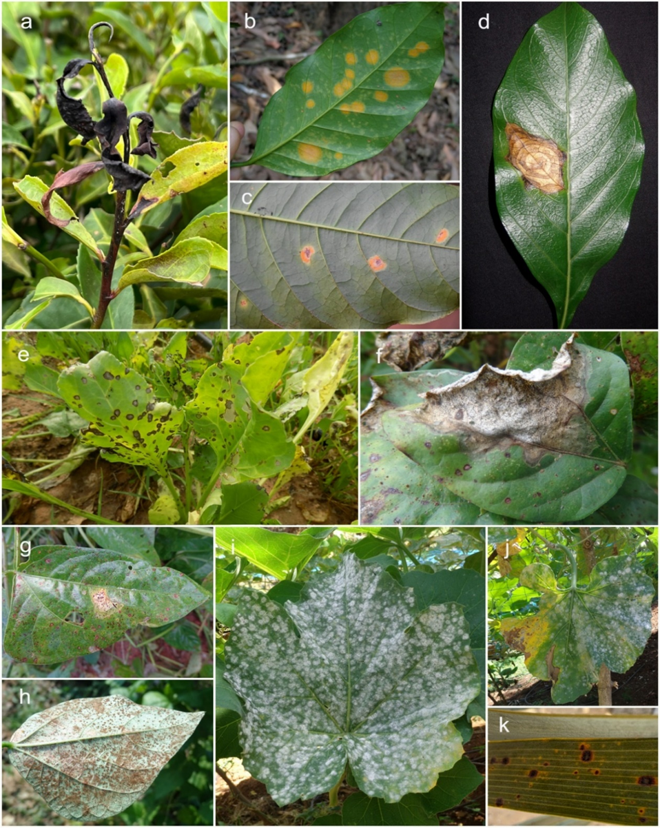
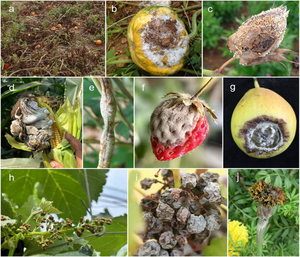
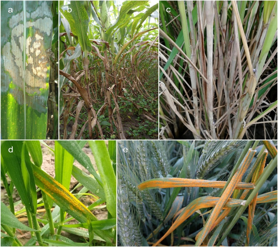
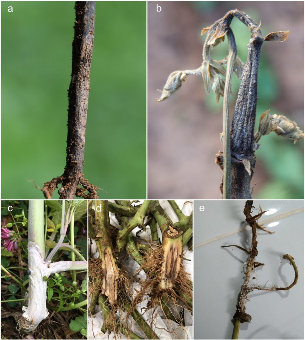
Detailed information on the symptoms and life cycles of fungal pathogens is essential for effective disease management (Dean et al. 2012, Palmieri et al. 2022). Early detection of symptoms enables timely intervention (Fig. 1–5). For instance, Puccinia striiformis causes yellow-orange pustules on wheat leaves, whereas Phytophthora infestans, responsible for late blight in potatoes, leads to water-soaked lesions that ultimately result in plant collapse (Bolton et al. 2008, Ivanov et al. 2021). Grasping these symptoms enables timely interventions, including fungicide application. Puccinia graminis possesses a complex life cycle, with sexual reproduction occurring on barberry (Barnes et al. 2020). Removing barberry disrupts the cycle, thereby reducing the spread of disease. Phytophthora infestans spreads through asexual zoospores in moist conditions, while its sexual oospores can survive in the soil, making its management complicated (Tiwari et al. 2021, Koshariya et al. 2023). Similar strategies apply to Plasmopara viticola, in which sporangia spread in humid conditions, and oospores survive in plant debris (Kennelly et al. 2007, Koledenkova et al. 2022). Other pathogens (i.e., Melampsora lini) complete their life cycle with a telial stage on an alternate host, providing another target for control by removing this host (Barrett et al. 2008). The survival structures of Sclerotinia sclerotiorum (Bolton et al. 2006) and oospores in Phytophthora species (Judelson & Blanco 2005) enable them to persist in the environment, making knowledge of their life cycles crucial for long-term control strategies, such as the timing of fungicide applications or soil treatments.
Quantifying pathogen impact enables researchers, policymakers, and agricultural professionals to prioritise interventions and allocate resources to the most destructive threats (Jeger et al. 2021, Ristaino et al. 2021). The economic impact of a pathogen is directly correlated with its ability to cause yield losses, reduce crop quality, and increase management costs, such as the application of fungicides or the breeding of resistant cultivars (Savary et al. 2012, Mano-harachary & Kunwar 2014, Singh et al. 2016). Understanding the impact of these pathogens helps guide the development of disease-resistant crop varieties and informs strategic decisions in breeding programmes (Chen et al. 2023). It also underpins the implementation of targeted biosecurity measures, preventing the spread of highly destructive pathogens to new regions (Kaundal et al. 2006). By recognising the significant economic cost associated with pathogens such as Phytophthora infestans in potatoes or Pyricularia oryzae in rice, efforts can concentrate on monitoring, early detection, and effective control strategies, ultimately reducing losses. Highlighting the economic impact of each pathogen also enhances understanding of the wider effects on trade, food prices, and the livelihoods of farmers (Vurro et al. 2010, Udomkun et al. 2017).
Management strategies are important for reducing the economic and environmental impacts of fungal diseases (Heydari & Pessarakli 2010, Juroszek & Von Tiedemann 2011). Various methods, including cultural practices, biological control agents, chemical treatments, and biotech¬nological advancements, are available for combating fungal pathogens (Agrios 2005). By examining existing management methods and new technologies, we aim to highlight strategies for sustainable disease control, focusing mainly on integrated pest management and holistic crop protection approaches. Additionally, understanding the complex interactions among host plants, pathogens, and environ¬mental factors is essential for developing long-term solutions (Wille et al. 2019, Jeger et al. 2021, Singh et al. 2023).
Breeding disease-resistant plants is a key strategy for managing crop health. This approach is becoming even more important as we gain deeper insights into the diversity of fungal pathogens. For example, anthracnose disease in chilli (Capsicum spp.) was once thought to be caused by three species, Colletotrichum gloeosporioides. However, work by Mongkolporn & Taylor (2018) showed that the disease is actually caused by 24 different Colletotrichum species. This finding is important for breeding programs, as it demonstrates that developing resistance against the wrong species may not effectively protect the plants. A similar situation has been reported for tropical fruits, where what was once considered Colletotrichum gloeosporioides has now been separated into several closely related species, each with different characteristics (Phoulivong et al. 2010a, Udayanga et al. 2013). These examples show how the correct identification of the fungus is essential when developing resistant plant varieties. As new studies continue to refine the classification of plant pathogens, it becomes increasingly important to link accurate fungal identification with efforts to breed disease-resistant crops.
Alongside addressing immediate challenges, this paper aims to provide a forward-looking perspective on future research concerning fungal and oomycete pathogens and their management. It explores emerging trends, innovative technologies, and novel approaches that could transform the field in the years to come. By identifying gaps in existing knowledge and highlighting areas requiring further investigation, this paper aims to inspire a new wave of research that anticipates future needs in fungal and oomycete pathogen control and sustainable management strategies.
Materials and methods
Data collection
We conducted a comprehensive literature search to identify the most extensively studied fungal plant pathogens. The primary source for this search was the Web of Science Core Collection database (http://apps.webofknowledge.com). Data collection encompassed various formats of scholarly publications, including articles, book chapters, book reviews, data papers, editorial materials, letters, meeting abstracts, news items, proceeding papers, and review articles. The study period was from 1900 to 2023, providing a broad historical perspective on research trends. The final search was conducted on December 31, 2023.
Search strategy
The search strategy was designed to capture all relevant literature on fungal plant pathogens. We employed a combination of search terms within several bibliographic fields: topic (TS), title (TI), abstract (AB), author keywords (AK), and Keyword Plus® (KP). This was augmented with Boolean operators "and" and "or" to refine and expand the search results. The primary search query was structured as ((((TS=(fungal plant pathogen)) OR TI=(fungal plant pathogen)) OR AB=(fungal plant pathogen)) OR AK=(fungal plant pathogen)) OR KP=(fungal plant pathogen). To ensure thorough coverage, additional keywords such as 'Fungal Pathogens', 'Plant Pathogen', 'Plant Pathogens', 'Fungal Plant Pathogen', 'Plant Disease', 'Fungal Phytopathogens', 'Phytopathogen', 'Phytopathogens', 'Powdery Mildew', 'Rust', 'Smut', and 'Fungi-like Pathogen' were also included. This strategy resulted in the retrieval of 95,256 research publications containing approximately 72,072,280 words.
Data export and analysis
The search results were exported as Tab Delimited Files and downloaded as Text Documents (.txt) for further analysis. Our custom export selections targeted specific bibliographic information including 'Author(s)', 'Title', 'Source', 'Conf.Info/Sponsors', 'Times Cited Count', 'Accession Number', 'Abstract', 'Addresses', 'Document Type', 'Keywords', 'Cited References', 'Usage Count', 'Hot Paper' and 'Highly Cited'. Due to database export limitations, which cap downloads at 1000 records per session, we downloaded the data in sequential blocks (i.e. 1–1000, 1001–2000, 2001–3000), until all 95,256 articles were acquired.
Network analysis
The network analysis was conducted using VOSviewer version 1.6.20, a tool for constructing and visualizing bibliometric networks. This software was employed to extract and analyze data regarding total link strength, occurrences, and the number of citations for each keyword. The analysis mode was set to 'Co-occurrence', using 'Full counting' as the counting method and 'All keywords' as the unit of analysis. We applied a threshold to exclude terms with fewer than 100 occurrences, allowing us to concentrate on the most relevant and frequently mentioned terms. The initial analysis identified the top 100 most studied fungal pathogens. Subsequently, synonyms were consolidated, and their scores were recalculated using Microsoft Excel. Based on these revised scores, the 50 most studied fungal plant pathogens were ranked and listed for further detailed examination.
In-depth search
Following the network analysis, a comprehensive literature search was undertaken on the top-ranked fungal pathogens to collect detailed information on their taxonomy, distribution, host range, impact, life cycle, management strategies, and future outlook. This detailed examination made use of several academic databases, including ScienceDirect, Google Scholar, ResearchGate, and Web of Science, to ensure comprehensive coverage of each fungal profile and its significance in plant pathology.
Fungal and oomycete list
The list below details the 50 most studied fungal and oomycetous plant pathogens, ranked from top to bottom. Each entry includes comprehensive notes on taxonomy, distribution, host range, impact, life cycle, management strategies, and future outlook. If certain information (i.e. holotype, ex-type, DNA barcodes from ex-type or authenticated strains) was not available, it was recorded as NA (not available). Classification follows Hyde et al. (2024a) and Thiyagaraja et al. (2025).
Result
Botrytis cinerea Pers., Syn. meth. fung. 2: 690. 1801.
Synonyms: Index Fungorum (2025) lists 41 species as synonyms, including the commonly used name Botryotinia fuckeliana
Classification: Fungi, Ascomycota, Pezizomycotina, Leotiomycetes, Helotiales, Sclerotiniaceae
Holotype: (On rotten Cucurbita and stems of Brassica oleracea)
Ex-type: MUCL87; VKM F-85 (Type of Botrytis cinerea f. lini J.F.H. Beyma, from seeds of Linum usitatissimum in Netherlands)
Diagnostic DNA barcodes: NEP1, NEP2 (Staats et al. 2007)
Growth conditions: Hay agar (HAY), potato-carrot agar (PCA), 24°C (Westerdijk Fungal Biodiversity Institute https://wi.knaw.nl/details/80/21746)
Host range: The fungus infects approximately 1,606 plant hosts (Singh et al. 2024), including fruits, vegetables, and cut flowers (Williamson et al. 2007, Romanazzi & Feliziani 2014).
Geographical distribution: Argentina, Armenia, Australia, Austria, Bangladesh, Barbados, Belgium, Brazil, Bulgaria, Canada, Chile, China, Colombia, Costa Rica, Cuba, Cyprus, Czech Republic, Denmark, Dominican Republic, Ecuador, Egypt, El Salvador, England (UK), Ethiopia, France, Georgia, Germany, Greece, Greenland, Guatemala, Honduras, Hong Kong, Hungary, India, Iran, Israel, Italy, Japan, Jordan, Kazakhstan, Kenya, South Korea, Libya, Lithuania, Malawi, Malaysia, Mauritius, Mexico, Morocco, Nepal, Netherlands, New Caledonia, New Zealand, Nicaragua, Norway, Pakistan, Panama, Papua New Guinea, Peru, Poland, Portugal, Puerto Rico (USA), Romania, Russian Federation, Scotland (UK), Serbia and Montenegro, Sierra Leone, Slovakia, South Africa, Spain, Sri Lanka, Sweden, Switzerland, Tanzania, Thailand, Turkey, Ukraine, United Kingdom, United States, Uruguay, Uzbekistan, Venezuela, and Zimbabwe.
Disease symptoms: Botrytis cinerea causes diseases during both pre- and post-harvest stages, producing a wide range of symptoms. The fungus induces soft rot, leading to water-soaked lesions on the tissues after infection, followed by the formation of grey conidial masses (commonly referred to as grey mould). On thick-peeled fruits, symptoms appear as dark, water-soaked areas inside the fruit. The fungus infects attached, decaying flowers on various fruits and vegetables, leading to soft rot symptoms developing from the blossom ends (known as blossom-end rot). The fungus causes minute brown spots to develop into large-scale soft rotting on flower petals (Botrytis blight). Additionally, it can cause stems to rot, starting at pruning wounds, particularly in herbaceous plants such as tomatoes grown in greenhouses (Williamson et al. 2007, Schumacher 2022).
Life cycle: The life cycle of Botrytis cinerea involves both sexual and asexual stages. During the asexual phase, the fungus forms clusters of conidia on the irregularly branched terminals of conidiophores that arise from either mycelium or sclerotium (Romanazzi & Feliziani 2014). The sclerotia are overwintering structures created by the fusion of mycelial branches and are found within decaying host tissues or soil (Elmer & Michailides 2007, Romanazzi & Feliziani 2014). The asexual cycle includes chlamydospores that result from the transformation of hyphal structures and subsequent disintegration (Holz et al. 2007). The sexual cycle of Botrytis cinerea involves haploid ascospores, which are produced in eight sets within each ascus originating from the apothecia. The apothecia are formed by the spermatization of sclerotia (Williamson et al. 2007, Romanazzi & Feliziani 2014). These asexual and sexual structures serve as the primary inoculum in the Botrytis cinerea life cycle, initiating infections on seedlings, flower petals, senescent flowers, leaves, and wounded tissues (Agrios 2005). These are dispersed through various means, such as air currents (Jarvis 1962a), water droplets (Jarvis 1962b), and/or insects (Elmer & Michailides 2007). Botrytis cinerea is a necrotrophic fungus that initially kills host cells and subsequently colonises the dead tissues (Amselem et al. 2011). When the fungus infects small fruits, it may remain in a quiescent stage for a considerable period without damaging tissues until the fruit matures (Williamson et al. 2007). When the fungus is infected, senescent flowers attached to fruits also persist until the fruit ripens as a saprobe (Williamson et al. 2007).
Impact: Botrytis cinerea causes significant economic losses, affecting both qualitative aspects (such as the taste, aroma, and oxidative stability of wine) and quantitative factors, including reduced yields of fruit, vegetable crops, and ornamental plants (De Miccolis Angelini et al. 2016). Each year, Botrytis cinerea is responsible for at least 30% of global crop production losses (Hao et al. 2017a, Liu et al. 2018a, Ullah et al. 2024). Globally, the economic impact of Botrytis cinerea is estimated to range from USD 10 to 100 billion (Hua et al. 2018, Roca-Couso et al. 2021), with an annual cost of approximately USD 1 billion on fungicides for its control (about 10% of the global fungicide market) (De Long et al. 2020). Botrytis cinerea causes substantial postharvest losses (between 15–50%) in fruits and vegetables, particularly in developing countries in Africa and Asia, owing to limited technologies for prolonging storage life (Romanazzi & Feliziani 2014). During storage, the fungus can infect surrounding healthy fruits, leading to the spoilage of entire batches. Furthermore, the pathogen can thrive even at low temperatures (0–5°C), particularly on fruits with diminished resistance (Romanazzi & Feliziani 2014). Nevertheless, Botrytis cinerea has a beneficial role in certain wine regions around the world. Under particular environmental conditions, Botrytis cinerea can induce noble rot in grapes, which is vital for producing sweet botrytised wines or select high-quality dry wines (Modesti et al. 2024).
Control and management strategies: The application of synthetic fungicides in the field remains a conventional method for managing grey mold infections, with preventive measures advised before disease symptoms manifest (Romanazzi & Feliziani 2014). Postharvest treatments, including the application of fungicides such as fluodioxonil, boscalid, cyprodinil, fenpyrazamine, fluazinam, and fluopyram, are approved in certain regions (Romanazzi & Feliziani 2014). Natural compounds, including plant extracts and essential oils (Antunes & Cavaco 2010, Feliziani et al. 2013a), as well as inorganic salts like bicarbonates (Sanzani et al. 2009), have demonstrated potential in controlling the infections. Resistance inducers such as chitosan and benzothiadiazole can activate plant defence mechanisms (Terry & Joyce 2004, Feliziani et al. 2013b, Romanazzi et al. 2013). Physical treatments, such as heat, UV-C light, exposure to modified atmospheres, edible coatings, and packaging, are effective against the pathogen (Romanazzi & Feliziani 2014, De Simone et al. 2020). The use of biological control methods, such as bioactive substances derived from plants and antagonistic microorganisms, offers benefits in mitigating grey mold decay (Chen et al. 2023). Biofungicides based on microorganisms, i.e. Bacillus subtilis, Cryptococcus albidus, and Pseudomonas syringae, provide environmentally friendly alternatives for disease management (Romanazzi & Feliziani 2014). In the cut flower industry, the routine application of fungicides is a common practice. Measures such as sanitation, nutrition, plant regulators, botanical extracts, and biological control have been incorporated to improve efficacy in ornamental production systems (Bika et al. 2021).
Research and development: Genomic and transcriptomic analyses of Botrytis cinerea have illuminated the genetic basis of its pathogenicity by identifying key genes involved in the infection process (Zhang et al. 2020a, Fernández-Morales et al. 2024, Singh et al. 2024). Currently, this species has over 50 genome sequences, and studies on virulence factors, such as secreted enzymes and secondary metabolites, have clarified their roles in host colonisation (Pontes et al. 2020). Investigations into plant immune responses reveal the molecular mechanisms of resistance, encompassing the involvement of reactive oxygen species and various signalling pathways (Li & Cheng 2023, Singh et al. 2024). Advancements in genetic resistance through breeding and genetic engineering, including CRISPR/Cas9, are enhancing crop resilience (Wang et al. 2018a, Leisen et al. 2020, Su et al. 2023). Recent developments in managing Botrytis cinerea further involve the use of RNAi techniques, which include exogenous application of small RNA molecules via spray-induced gene silencing (SIGS), providing an efficient and environmentally friendly approach to combat grey mould (Wang et al. 2017, Duanis-Assaf et al. 2022, Singh et al. 2024).
Future outlook: As temperatures and humidity levels fluctuate, the lifecycle and virulence of the pathogen may be altered, potentially leading to more frequent and severe outbreaks. Therefore, improving predictive models and early detection systems, including remote sensing and automated monitoring technologies, will be essential for the timely and accurate management of grey mold in the face of an ever-changing climate. Future research on Botrytis cinerea should evaluate transgene expression and resistance in subsequent progenies and across multiple growing seasons in perennial ornamentals, as breeding alone may be inadequate due to the ability of the pathogen to develop new virulent strains under favourable environmental conditions (Bika et al. 2021). A comprehensive understanding of the epidemiology and infection processes of Botrytis cinerea will also be crucial for developing integrated management strategies to mitigate the effects of the pathogen under changing environmental conditions (Rhouma et al. 2022). Future research on Botrytis cinerea should concentrate on exploring the genetic and molecular diversity among a wide range of isolates, as different strains display varying levels and mechanisms of virulence. With over 5,000 unannotated genes and fewer than 500 fully studied, a thorough investigation of these largely unexplored genes is vital for understanding their roles in pathogenicity and enhancing disease management strategies (Singh et al. 2024). Future studies should examine epigenetic modifications, phase separation, and other emerging regulatory mechanisms in plant defence responses against Botrytis cinerea (Li & Cheng 2023). These could offer novel insights and innovative approaches for improving crop resistance.
Notes: In instances where plant defences exceed the attack of the pathogen, Botrytis cinerea infections may develop into 'quiescent' lesions, remaining symptomless within a few cells until the plant tissue senesces or ripens (Rajaguru & Shaw 2010). This asymptomatic presence highlights the endophytic nature of the pathogen within the plant (Barnes & Shaw 2003, Sowley et al. 2010). This complicates its control by delaying symptom expression, evading plant defences, and rendering detection and timely intervention more difficult.
Pyricularia oryzae Cavara, Fung. Long. Exsicc. 1: 49. 1892.
Synonyms: Index Fungorum (2025) lists seven species as synonyms, including the commonly used names Magnaporthe oryzae, Magnaporthe grisea, and Pyricularia grisea
Classification: Fungi, Ascomycota, Pezizomycotina, Sordariomycetes, Sordariomycetidae, Magnaporthales, Pyriculariaceae
Holotype: BPI:841383 (on cross of strains from Oryza sativa and Eleusine, Guyana), isotype TRTC 52742
Epitypus: NA
Ex-epitype: NA (most certain strain: CBS 657.66 from Klaubauf et al. 2014)
Diagnostic DNA barcodes: LSU, ITS, RPB1, ACT, CAL
DNA barcodes from ex-epitype: LSU: KM485003, ITS: KM484893, RPB1: KM485113, ACT: KM485194, CAL: KM485265
Growth conditions: Cornmeal agar (CMA), oatmeal agar (OA), 2% potato dextrose agar (PDA), and 2% malt extract agar (Klaubauf et al. 2014)
Host range: Anthoxanthum spp., Avena fatua, A. sativa, Brachiaria spp., Bromus tectorum, Bromus unioloides, Ctenanthe oppenheimiana, C. setosa, Cynodon dactylon, Cyperus rotundus, Digitaria spp., Echinochloa spp., Eleusine spp., Eragrostis curvula, Eragrostis spp., Eremochloa ophiuroides, Eriochloa villosa, Festuca spp., Hakonechloa macra, Hordeum spp., Leersia hexandra, Leptochloa chinensis, Lolium spp., Luziola sp., Melinis minutiflora, Oryza spp., Panicum spp., Paspalum spp., Phalaris spp., Phleum pretense, Phyllostachys sp., Rottboellia spp., Saccharum officinarum, Sasaella sp., Setaria spp., Stenotaphrum secundatum, Triticosecale sp., and Triticum spp.
Geographical distribution: Widespread across the rice-growing regions of the world, it has been reported in over 85 countries (Zhang et al. 2016a).
Disease symptoms: Symptoms can manifest on all parts of the plant at various stages of growth and development (Zhang et al. 2016a, Agbowuro et al. 2020). Symptoms observed on rice and wheat leaves include initial white to grey-green water-soaked lesions or spots with dark green borders that later develop into elliptical, spindle-shaped, or eye-shaped necrotic lesions with whitish to grey centres (TeBeest et al. 2007, Islam et al. 2016, Zhang et al. 2016a). Symptoms on the rice collar manifest as necrosis at the junction of the leaf blade and sheath, subsequently extending to the entire leaf and a few millimetres around the sheath (TeBeest et al. 2007). Infection in the neck region of the rice plant shows rotting of the stem portion beneath the panicle, leading to either no or partial grain filling (referred to as seed blanking) or the panicle detaching (TeBeest et al. 2007, Agbowuro et al. 2020). Symptoms of panicle infection in rice and wheat include complete or partial grey-brown discolouration of spikes and grains (TeBeest et al. 2007, Islam et al. 2016).
Life cycle: Pyricularia oryzae has a hemibiotrophic lifestyle, commencing with an initial biotrophic phase that suppresses the immune system of the plant, followed by a necrotrophic phase leading to plant cell death (Fernandez & Kim 2018). The pathogen inoculum for Pyricularia oryzae may originate from various sources, including host plant residues, seeds, soil, equipment, and alternative hosts (Agbowuro et al. 2020). The fungal mycelia can survive on rice straws for over three years at 18–32ºC, while asexual spores (conidia) develop when moist. They can persist for over a season in tropical and subtropical regions (Agbowuro et al. 2020). The life cycle begins when conidia land on the surface of a host plant. Conidia are typically dispersed by wind, rain, or irrigation water (Zhang et al. 2014a, Agbowuro et al. 2020). Upon encountering a susceptible host, the conidia adhere to the leaf surface and germinate under suitable conditions (Fernandez & Kim 2018). Once germinated, the fungal hyphae form specialised infection structures known as appressoria at the tips of the germ tubes (Fernandez & Kim 2018). The mature appressoria subsequently develop penetration pegs, which enable the fungus to infiltrate host plant cells by breaching the cuticle and cell wall (Zhang et al. 2016a, Chethana et al. 2021a). After penetrating the plant surface, the fungus forms invasive hyphae that grow intracellularly within the plant cells, resulting in visible symptoms (Agbowuro et al. 2020). As the fungus colonises the plant tissue, it produces new conidia on the surface of the lesions within 7 days (Talbot et al. 1996, Zhang et al. 2016a). The newly formed conidia are primarily dispersed to new host plants through wind and rain splash, repeating the cycle as they land and initiate new infections (Ou 1985, Talbot 2003). The life cycle of Pyricularia oryzae also involves sexual reproduction, although this is less common than asexual reproduction and is primarily confined to its centres of origin (Saleh et al. 2012). During the sexual cycle, the fungus forms sexual spores, known as ascospores, within the asci, which are contained within the perithecia. These perithecia develop when compatible mating types of the fungus come into contact and undergo sexual reproduction (Valent 2021). Once the perithecia mature, they release ascospores that can disperse to new host plants. When landing on the host surface, ascospores produce appressoria for plant penetration, grow vegetatively, and generate conidia (Valent 2021).
Impact: Pyricularia oryzae causes blast disease, posing a significant threat to global food security by annually destroying approximately 10–30% of the worldwide rice harvest, which could otherwise nourish about 60 million people (Pennisi 2010, Fernandez et al. 2014, Fernandez & Kim 2018). The pathogen has caused severe rice blast epidemics in China, Korea, Japan, Vietnam, and the United States, with China alone losing 5.7 million hectares of rice between 2001 and 2005 (Wilson & Talbot 2009). The disease has been reported in approximately 85 countries worldwide, with some regions experiencing up to 100% crop damage (Agarwal et al. 1989). Annual losses due to blast disease hinder rice production, especially in developing countries, and climate change is likely to exacerbate the spread of pathogens into new areas (Fernandez et al. 2014). Traditional breeding and chemical methods have proven ineffective in controlling the disease since Pyricularia oryzae can swiftly adapt and mutate, developing resistance to multiple rice cultivars (Pennisi 2010). In addition to rice, host-adapted lineages (pathotypes) of Pyricularia oryzae have been found to cause blast disease in other cereal crops (Valent 2021). The Magnaporthe oryzae Triticum (MoT) lineage causes wheat blast disease, which can lead to yield losses of up to 100% under favourable disease conditions, with reported outbreaks in South America and Bangladesh (Islam 2020a). Pathotypes of Pyricularia oryzae have limited the production of millets, including finger millet and foxtail (Italian) millet, which have been subsistence crops cultivated for thousands of years by low-resource farmers in Africa and Asia (Valent 2021). In addition to crop plants, Pyricularia oryzae has been reported to cause significant damage to forages and grasses grown on golf courses, resulting in outbreaks (Bain et al. 1972, Landschoot & Hoyland 1992).
Control and management strategies: Among the strategies implemented for controlling Pyricularia oryzae, breeding for resistant varieties is considered the most sustainable and cost-effective approach (Fang et al. 2017). However, this is limited by the rapid evolution of the pathogen, which frequently overcomes host resistance (Ou 1980, Zeigler et al. 1994). Transgenic methods are also employed in developing resistant varieties (Pokhrel et al. 2021, Jin et al. 2024). Biological control is also a cost-effective and environmentally friendly method for managing Pyricularia oryzae. Studies have demonstrated that various bacterial genera, including Bacillus, Chryseobacterium, Pseudomonas, Rhizobacteria, and Streptomyces, as well as fungal genera such as Aspergillus, Curvularia, Fusarium, Penicillium, and Trichoderma, are effective in controlling Pyricularia oryzae, particularly in vitro settings (Chakraborty et al. 2021). However, their effectiveness has been inadequately established in commercial-scale, long-term field trials. Chemical control is widely employed to manage blast pathogens, primarily applied in two stages: seed treatment to prevent initial seedling infection and fungicidal sprays to protect leaves and panicles during the growing season (Maciel 2012). Studies have demonstrated that fungicides such as azoxystrobin, benomyl, carbendazim, carpropamid, coumoxystrobin, diclocymet, edifenphos, fenoxanil, iprobenfos, isoprothiolane, metominostrobin, probenazole, prochloraz, propiconazole, pyraclostrobin, tebuconazole, thiophanate-methyl, and tricyclazole are effective against blast disease (Agbowuro et al. 2020, Xin et al. 2020). Several cultural practices are adopted by farmers to control Pyricularia oryzae, including the use of healthy seeds, burning diseased straw before the next season, water management through flooding, early planting during rainy seasons, and nutrient management (Bonman 1992, Reis et al. 1995, Filippi & Prabhu 1997, Agbowuro et al. 2020).
Research and development: Recent research on Pyricularia oryzae has led to major advancements in managing this key agricultural threat. Genomic and molecular studies have mapped the genome of the pathogen, identifying critical genes involved in its pathogenicity and life cycle (Korinsak et al. 2019). Currently, this species has over 400 genomes. Studies on resistance (R) genes and quantitative trait loci (QTL) against Pyricularia oryzae have identified several genetic markers from various rice genetic resources (Younas et al. 2024). These findings have facilitated the development of resistant rice varieties through both traditional breeding and genetic engineering (Devanna et al. 2022). NGS-enabled comparative, pan-genome and meta¬genomic analyses have explained the frequent emergence of new races and improved insight into host–pathogen interactions, supporting more effective rice blast manage¬ment (Devanna et al. 2022, Younas et al. 2024). Advance¬ments in molecular biology, computational biology, biotechnology, and nanotechnology have led to the development of highly sensitive and specific diagnostic methods for Pyricularia oryzae, including nucleic acid-based protocols, enhanced amplification platforms, quantitative PCR, DNA barcoding, next-generation sequencing, imaging techniques, and nanomaterial-based sensors, all of which have improved accuracy and reduced costs (Kumar et al. 2021).
Future outlook: Due to climate change, Pyricularia oryzae has expanded its distribution and invaded new areas (Rezvi et al. 2023), and it remains possible for it to continue spreading and cause epidemics. This underscores the need for innovative disease management strategies to tackle these emerging challenges. The future innovative disease management strategies could involve exploring broad-spectrum disease-resistance genes, releasing and rotating blast-resistant cultivars based on the AVR genotype of the field population, implementing microbiome-based biological control strategies, early pathogen monitoring, and optimising prevention and control measures, utilising rapid diagnostic methods in plant quarantine to restrict pathogen spread and detect diseases early in fields, and providing timely weather forecasting and alerts to farmers (Zhang et al. 2022a). Despite recent advancements, many details remain ambiguous, particularly concerning how the fungus regulates the gene expression of effector proteins and the subsequent stages of lesion development. Comprehending these mechanisms will be vital in identifying vulnerabilities within its life cycle, thus facilitating the design of resistant plants or innovative disease management strategies (Valent 2021). Given the ability of Pyricularia oryzae to rapidly overcome R genes through AVR gene deletion and transfer, future research could focus on understanding how genomic location influences AVR effector gene dynamics and the durability of R genes (Valent 2021). Research on Pyricularia oryzae should aim to discover effective R genes and understand disease mechanisms for other blast diseases while leveraging insights from the diverse evolutionary stages of blast pathotypes to enhance our understanding of host adaptation and improve control strategies (Valent 2021).
Notes: Pyricularia oryzae is an ideal model organism for studying plant-pathogenic fungi because of its ability to be cultured on defined media, its well-established transfor¬mation system, relatively small genome, extensive genetic mapping data, and the availability of a draft sequence of the rice genome (Zhang et al. 2016a).
Rhizoctonia solani J.G. Kühn, Ann. Sper. agr., N.S.: 224 (1858)
Synonyms: Species Fungorum (2025) lists 39 species as synonyms.
Classification: Fungi, Basidiomycota, Agaricomycotina, Agaricomycetes, Cantharellales, Ceratobasidiaceae
Typ. cons.: CBS 739.95
Diagnostic DNA barcodes: ITS
DNA barcodes from type: ITS: MH862557
Growth conditions: The highest radial growth of the tested fungus was observed on PDA, followed by Carrot Meal Agar. Optimal colony growth occurred at 25°C, with the fungus demonstrating its best performance at a pH range of 5.5 to 7 (Nuri et al. 2021).
Host range: Rhizoctonia solani exhibits a wide range of pathogenicity, infecting approximately 250 plant species across various families, including Amaranthaceae, Araceae, Asteraceae, Brassicaceae, Fabaceae, Linaceae, Malvaceae, Moraceae, Poaceae, Rubiaceae, and Solanaceae (Chahal et al. 2003, Yang et al. 2022a).
Geographical distribution: Rhizoctonia solani is found worldwide, across Africa, Asia, Australia and Oceania, Europe, North America, and South America.
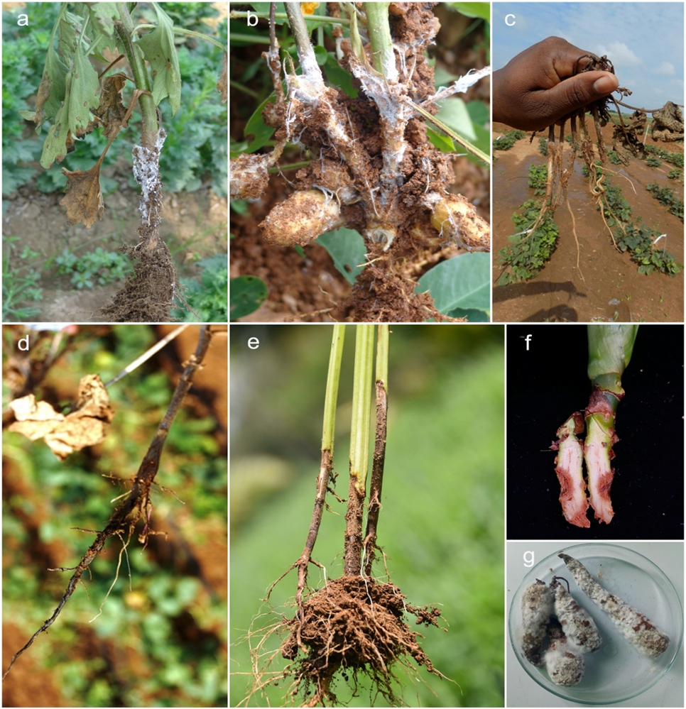
Disease symptoms: Rhizoctonia solani is recognised for causing a variety of symptoms in different crops, including sheath blight, foliar blight, leaf blight, web blight, head rot, bottom rot, and brown patch. In rice, the pathogen primarily affects leaf sheaths and blades (Fig. 3), with symptoms typically appearing within 24–72 hours following infection, depending on environmental conditions (Rangaswami & Mahadevan 1998). The susceptibility of rice increases during the tillering stage (Singh et al. 1988). The fungal mycelium triggers lesion formation, which initially appears as greenish-grey, water-soaked patches on the leaf sheath. These lesions often expand into irregularly shaped areas with grey-white centres encircled by brown margins (Ou et al. 1973). Lesions may converge, encircling the stem and spreading to the upper leaf sheaths and blades, resulting in sheath rot and leaf desiccation (Singh et al. 2016). In severe instances, the infection may extend to the panicle, hindering grain filling and causing seed discolouration. Acute infections can lead to the death of entire leaves, tillers, or plants (Hollier et al. 2009). The disease is also called ‘snake skin disease’, ‘mosaic foot stalk’ and ‘rotten foot stalk’ because of its symptoms (Zhang et al. 2019a, Molla et al. 2020, Li et al. 2021a).
Life cycle: Rhizoctonia solani, present in both seeds and soil, thrives in tropical environments, surviving through sclerotia and mycelia within infected seeds or in the soil. In these areas, soil-borne sclerotia, typically originating from rice or weed hosts, act as the primary carriers. In temperate climates, sclerotia found in the soil and on crop residues serve as the main sources of inoculum. These sclerotia promote the spread of the fungus through irrigation water, transferring between fields (Kozaka 1970). Under favourable conditions, sclerotia germinate to produce mycelia, which develop infection structures and enable penetration into plant tissues upon contact with rice surfaces. In some cases, infection can also occur through stomata without the formation of these structures (Marshall & Rush 1980). The pathogen spreads both vertically and horizontally, with rates of horizontal spread reaching up to 20 cm per day under field conditions (Savary et al. 1995). Disease transmission occurs through float sclerotia and mycelia, carried by rainfall and irrigation runoff. Infected seeds serve as primary inoculum sources, exhibiting infection rates ranging from 4.6% to 14% under field conditions (Sivalingam et al. 2006). Additionally, wind disperses basidiospores to new fields, where the basidia hymenium continuously acts as a source of secondary inoculum.
Impact: Sheath blight is recognised as the second most harmful fungal disease affecting rice, surpassed only by rice blast (Pan et al. 1999). Rhizoctonia solani, the pathogen responsible for this disease, exhibits two developmental stages: an anamorph stage and a teleomorph stage, the latter being known as Thanatephorus cucumeris. Rice sheath blight leads to yield losses up to 45% and seriously threatens global food security (Nadarajah et al. 2017, Zhang et al. 2021a, Yang et al. 2022a). Rhizoctonia solani can infect rice at any stage of its growth (Dath 1990). Early maturing, semi-dwarf, high-tillering, and densely planted cultivars are particularly susceptible to severe infections (Bhunkal et al. 2015). The severity of the disease tends to increase as the rice plants mature (Singh et al. 2004a). There is also noticeable variability in resistance among different rice genotypes, with variations observed between mature plants and seedlings (Dath 1990). The progression of sheath blight is slow during early growth stages but accelerates during the tillering phase and subsequent growth stages (Thind et al. 2008).
Control and management strategies: Currently, the management of sheath blight in rice mainly involves the use of fungicides, along with the incorporation of genetic resistance or tolerance and various cultural practices. Biological control methods are also strategically employed. Although no rice varieties, landraces, weedy types, or wild relatives have been identified as immune or fully resistant to Rhizoctonia solani infection, some genotypes have demonstrated partial resistance to the disease (Senapati et al. 2022).
Research and development: Current global research concentrates on genomic and comparative genomic studies, incorporating transcriptomic, proteomic, and metabolomic analyses to elucidate the genetic mechanisms underlying its pathogenicity and to identify potential targets for disease management. Whole-genome sequencing of various Rhizoctonia solani anastomosis groups (AGs) has been pivotal in pinpointing genes related to host range, pathogenicity, overwintering capability, competitive saprobic behaviour, aggressiveness, and epidemiological fitness (Senapati et al. 2022), and currently, Rhizoctonia solani has over 40 genomes. Diagnostic techniques for detecting Rhizoctonia solani include fatty acid profiling, pectin enzyme analysis, allozyme polymorphism, and serological methods (Banniza & Rutherford, 2001). Furthermore, a novel and highly sensitive LFD-based LAMP assay has been developed to enhance the detection of this pathogen. Additional strategies to develop resistant germplasms include the use of host-derived RNA interference and transgenic technology to disarm essential pathogenicity factors in Rhizoctonia solani, manipulating the expression of plant defence-associated genes, and pyramiding quantitative trait loci for resistance to rice sheath blight (Li et al. 2021a).
Future outlook: Enhancing our understanding of Rhizoctonia solani is vital for future advancements in taxonomy, population biology, and pathogenicity research. Given the considerable genetic diversity among rhizoctonia-like fungi, thorough studies are essential to clarify taxonomic relationships within this group. Utilising genome sequence data will enhance our understanding of fungicide sensitivity, assist in preventing the development of resistance to fungicides, and facilitate the creation of new, environmentally friendly fungicides. Insights into the mating habits, gene flow, and geographical distribution of Rhizoctonia solani genetic variants will be enriched through population genetics studies. Emerging technologies, such as next-generation sequencing and whole-genome sequencing, provide unique and effective detection and diagnostic approaches. Although currently underutilised in the diagnostics of Rhizoctonia solani, these methodologies are anticipated to gain prominence in detecting and diagnosing this pathogen. With these advancements, we anticipate developing more effective and sustainable strategies for managing Rhizoctonia solani, thereby enhancing disease control measures.
Notes: Rhizoctonia solani produces phytotoxins that adversely affect plants, especially potatoes. This pathogen induces symptoms on both the above-ground portions of the plant and, in severe cases, on the roots (Kankam et al. 2021).
Fusarium oxysporum Schltdl., Fl. Berol. 2: 139. 1824.
Synonyms: Crous et al. (2021b) list 108 species as synonyms.
Classification: Fungi, Ascomycota, Sordariomycetes, Hypocreomycetidae, Hypocreales, Nectriaceae
Holotype: HAL 1612 F (on Solanum tuberosum Germany, Berlin)
Epitype: CBS H-23620 (designated in Lombard et al. 2019)
Ex-epitype: CBS 144134
Diagnostic DNA barcodes: RPB2, TEF (Lombard et al. 2019)
DNA barcodes from ex-epitype: cmdA: MH484771, IGS: MH484862, RPB2: MH484953, TEF: MH485044, TUB: MH485135
Growth conditions: Generally, grows well in PDA, SNA, and CLA (Lombard et al. 2019).
Host range: The fungus associated with over 500 hosts.
Geographical distribution: Distributed across approximately 108 countries.
Disease symptoms: Fusarium oxysporum can penetrate plants through the root system and colonise the xylem, causing wilting, vascular discolouration, chlorosis, dwarfism and premature plant death (Davis et al. 2006, Gauthier et al. 2022, Hao et al. 2023). The fungus causes a vascular wilt disease known as Fusarium wilt, primarily affecting the vascular system of plants and disrupting the transport of water and nutrients (Nehra et al. 2021). Symptoms include wilting, yellowing, and stunted growth, with the lower leaves being the first to be affected (Zhang et al. 2024a).
Life cycle: Fusarium oxysporum can persist in the soil and crop residue for extended periods as spores or mycelia, and occasionally as resilient asexual chlamydospores. The fungus overwinters as spores or mycelia in crop residue and also produces robust, thick-walled asexual chlamydospores that resist dehydration (Ploetz 2015). The plant is infected at the roots, and the pathogen subsequently translocates to the above-ground parts, where it obstructs the vascular tissues. Within the vascular tissues, Fusarium oxysporum proliferates as mycelia and spores, prompting the plant to secrete gums to halt the spread, ultimately resulting in the wilting of the affected areas.
Impact: Fusarium oxysporum poses management challenges and currently affects over 100 essential crops, including cotton, tomatoes, bananas, cucumbers, and beans (Yan et al. 2023). During the Gros Michel era, it resulted in losses estimated at around USD 2.4 billion (Ploetz 2015). Fusarium oxysporum exhibits high pathogenic complexity, primarily due to the presence of numerous host-specific formae speciales that are adapted to infect distinct plant species. For instance, F. oxysporum f. sp. lycopersici infects tomato, f. sp. cubense causes Panama disease in banana, and f. sp. vasinfectum affects cotton (Gordon & Martyn 1997). This host specialisation reflects the evolutionary adaptability of the species and presents challenges for disease management in diverse cropping systems.
Control and management strategies: Crop rotation is an essential farming practice for controlling Fusarium wilt. To disrupt the disease cycle, farmers frequently alternate tomato crops with non-host plants such as grains or legumes (Haque et al. 2023). Effective strategies also encompass mulching to inhibit weeds, rotating crops with non-solanaceous species, and intercropping maize with tomatoes. Minimising plant handling practices is crucial for preventing Fusarium wilt (Haque et al. 2023). Disinfectants such as sodium hypochlorite, hydrogen peroxide, and ozone are effective oxidisers that reduce the presence of pathogens in seeds. In tomatoes, fungicides like bromuconazole and prochloraz are applied as soil drenches (McGovern 2015). For banana Fusarium wilt, a fungicide containing thiophanate-methyl is used. Managing fumigation with alternatives, such as 1,3-dichloropropene combined with chloropicrin, has also shown effectiveness.
Biological control agents, particularly fungi such as Trichoderma and other microorganisms like non-pathogenic Fusarium and Penicillium, as well as bacteria including Pseudomonas and Bacillus, serve as beneficial antagonists against pathogens (Lecomte et al. 2016, Ayaz et al. 2023, Yao et al. 2023). Moreover, plant extracts and essential oils are employed for their control properties (Bolouri et al. 2022, Mohd Israfi et al. 2022). Soil pre-fumigation can effectively enhance the disease suppressiveness of biofertilizer against banana Fusarium wilt by modifying the soil microbiome (Shen et al. 2018).
Breeding programmes in crops such as cotton, potatoes, and cucurbits like watermelon focus on developing inherent resistance to minimise the need for chemicals (Ajaharuddin et al. 2024). However, due to pathogenic variability, the effectiveness of many resistant cultivars is limited to only a few years (de Vallavieille-Pope 2004).
Research and development: Over 26 virulence and pathogenicity-related genes were analysed functionally, revealing the prominence of the zinc finger transcription factor (TF) family in the pathogenesis pathway (Zuriegat et al. 2021), and it has over 730 genomes. A master regulator of pathogenic development (Michielse et al. 2009) and a conserved nitrogen response pathway that governs invasive growth functions (López-Berges et al. 2008) have been recognised as significant advancements in understanding the pathogenicity of Fusarium oxysporum.
Future outlook: Additional pathogenicity-related systems and transcription factors require functional characterisation to ensure a comprehensive and systematic analysis of the regulation of pathogenicity in Fusarium oxysporum. The genetic basis of host specificity in Fusarium oxysporum is poorly understood. Strains that infect a particular plant species are not necessarily more closely related to each other than to strains that infect other hosts (Lievens et al. 2008).
Phytophthora infestans (Mont.) de Bary, J. Roy. Agric. Soc. England, ser. 2 12: 240 (1876)
Synonymy: Species Fungorum (2025) lists five species as synonyms.
Classification: Fungus-like, Oomycota, Oomycetes, Peronosporales, Peronosporaceae
Holotype: FUSION94490 in PC (by H. Montagne, 18 August 1845)
Epitype: CBS H-24657 (Designated by Chen et al. 2022, Stud. Mycol. 101: 417-564)
Ex-epitype: CBS 147289
Diagnostic DNA barcodes: ITS, TUB, tigA, COX1
DNA barcodes from ex-epitype: ITS: MZ753914, TUB: MZ736454, tigA: MZ736481, COX1: MZ736428
Growth conditions: the most suitable media for Phytophthora infestans are PDA or rye agar at 25°C (Tumwine et al. 2000). Some researchers suggest that using rye agar at 20°C in the dark enhances the production of gametangia (Brasier 1967, Erwin & Ribeiro 1996, Jung et al. 1999, Scanu et al. 2014).
Host range: The species most affected are those of the Solanaceae family, particularly the potato (Solanum tuberosum) and the tomato (S. lycopersicum), which hold significant agricultural value. Ornamental Solanaceae, such as Calibrachoa spp. and Petunia spp., as well as wild species like Solanum dulcamara and S. sarrachoides, can also host Phytophthora infestans (Ivanov et al. 2021). In addition to Solanaceae, Phytophthora infestans has also been reported in other plant families, including Apiaceae, Asteraceae, Convolvulaceae, Geraniaceae, Malvaceae, Nyctaginaceae, Polygonaceae, Rosaceae, and Sapindaceae.
Geographical distribution: The distribution encompasses a broad spectrum of geographical locations spanning all continents, indicating a global presence. This includes countries from tropical, subtropical, and temperate climates, highlighting the adaptability and extensive range of Phytophthora infestans across diverse environmental conditions. Among these countries, the majority of records were from the United States, Mexico, Peru, and Ecuador, respectively (Farr & Rossman 2025).
Disease symptoms: Phytophthora infestans causes late blight in many species of Solanaceae, which is characterised by water-soaked lesions frequently surrounded by a halo of white, downy sporangia. These sporangia develop on sporangiophores that emerge from the stomata of the leaves. The initial symptoms feature dark green spots that progress to brown and black patches on the foliage and stems, particularly near the tips or edges where water or dew gathers. The sporangia and sporangiophores are visible as white structures on the lower surface of the foliage. In instances of tuber blight, white mycelium often becomes apparent on the surface of the tubers (Birch & Whisson 2001).
Life cycle: The asexual life cycle of Phytophthora infestans involves alternating phases of hyphal growth, sporulation, sporangial germination, and the re-establishment of hyphal growth. Sporangia, dispersed by wind or water, facilitate the movement of Phytophthora infestans between various host plants. Additionally, there is a sexual cycle in which Phytophthora infestans produces oospores as the sexual spores. These oospores can disperse along water films on leaves or in soil. While sporangia are generally short-lived, oospores can remain viable for many years, offering a stark contrast in longevity. Sporangia develop on the leaves and can spread through the crop when temperatures exceed 10°C and humidity rises above 75–80% for two days or more. Under optimal conditions, Phytophthora infestans completes its life cycle on Solanaceae species in approximately five days (Fry 2008). Rain can wash spores into the soil, where they infect young tubers. Additionally, spores can be carried over long distances by the wind.
Impact: The potato is the fourth most produced non-cereal crop worldwide. Among various biotic stresses, late blight, caused by Phytophthora infestans, emerges as the most devastating disease. This disease affects both the foliage of potato plants in the field and the tubers in storage, and it can completely destroy a crop, potentially causing a 100% yield loss (Goutam et al. 2018). Diseases caused by Phytophthora infestans account for losses ranging from 20% to 40% of total tomato production (Ali et al. 2020). The annual worldwide potato crop losses due to late blight in 2008 are conservatively estimated at USD 6.7 billion (Haverkort et al. 2008, Haas et al. 2009).
Control and management strategies: Advancements in modern sequencing technologies, molecular genetic markers, and computer data processing have significantly enhanced the ability to monitor genetic changes in populations of Phytophthora infestans. This understanding is essential for developing targeted responses, including the creation of predictive models that could lead to the development of effective fungicides with a reduced risk of resistance (Rodenburg et al. 2018). An integrated approach that combines cultural controls, resistant cultivars, and careful fungicide application ensures the health and productivity of crops.
Cultural controls offer primary protection by reducing the survival, reproduction, and spread of pathogens. Key practices include using disease-free seed tubers, destroying cull and volunteer potatoes, minimising overhead irrigation, ensuring good soil coverage, and employing proper harvesting and storage techniques. Mulching enhances soil health and plant vigour by improving nutrient uptake and moisture retention, as well as supporting beneficial soil microbes (Aryantha et al. 2000, Lazarovits et al. 2001). Proper storage conditions and the use of fertilisers further enhance plant resistance to diseases (Draper et al. 1994, Garrett & Dendy 2001, Davis et al. 2009, Kirk 2009). The application of fungicides remains a global standard for managing Phytophthora infestans. Fungicides such as the Bordeaux mixture are effective, but their excessive use may lead to resistance. Mixtures containing broad-spectrum fungicides are recommended to minimise resistance risks (Thind 2015). Innovations such as Zorvec™ have demonstrated promise in providing lasting protection and enhancing yields under various climatic conditions (Bhaik & Trivedi 2015). Biological control presents an economical and environmentally friendly alternative. Agents like Bacillus subtilis var. amyloliquefaciens and Purpureocillium lilacinum have shown potential in suppressing the growth of Phytophthora through direct antagonism (Arnold et al. 2003, Wang et al. 2016). Identifying new biocontrol agents remains a priority for sustainable disease management.
Using resistant varieties is the most effective and environmentally safe way to manage diseases such as late blight. Research has shown variations in resistance among different potato varieties, with some exhibiting useful resistance to foliage blight but limited resistance to tuber blight, and vice versa (Njualem et al. 2001). While most resistant varieties are not entirely immune to late blight, they do display varying degrees of resistance to different races of the pathogen (Popokova 1972). Nonetheless, the resistance in existing potato varieties is often race-specific and can be overcome by other compatible races of Phytophthora infestans, rendering the varieties susceptible to the pathogen in a short timeframe (Shtienberg et al. 1994).
Research and development: Recent advancements in the research and development of strategies against Phytophthora infestans demonstrate significant progress in plant pathology and genetic engineering. One notable discovery is using β-aminobutyric acid (BABA), a non-proteinogenic amino acid, as a potent inducer of Systemic Acquired Resistance (SAR) in plants. BABA effectively triggers SAR against various plant pathogens, including Phytophthora infestans, enhancing plant defences without relying on chemical fungicides (Cohen 2002, Baider & Cohen 2003, Ton & Mauch-Mani 2004, Ton et al. 2005, Andreu et al. 2006, Dubreuil-Maurizi et al. 2010, Worrall et al. 2012, Janus et al. 2013). Innovations in genetic engineering, such as transcriptional gene silencing (TGS), have proven effective. TGS entails adding extra copies of a gene to the host plant, silencing the native gene locus, and providing a stable and efficient defence mechanism against pathogens. For instance, complete resistance in the potato cultivar Desiree against a specific isolate of Phytophthora infestans was achieved by silencing only five specific genes (Sun et al. 2016a, b).
Introducing R-genes from wild Solanum species into potato cultivars is considered an effective and environmentally friendly strategy for combating Phytophthora infestans (Simko et al. 2007). Over 20 functional R-genes have been cloned from species such as Solanum bulbocastanum and S. demissum, integrating these genes into susceptible cultivars to confer resistance (Li et al. 2011, Kim et al. 2012). Previous studies have identified 24 quantitative trait loci (QTLs) for late blight resistance, and candidate gene approaches have led to the identification of diagnostic markers for quantitative resistance (Goutam et al. 2015, Mosquera et al. 2016). Genome-wide association studies (GWAS), based on single-nucleotide polymorphisms (SNPs) across the genome, have facilitated the discovery of additional markers associated with resistance (Goutam et al. 2015, Mosquera et al. 2016). This species has six genomes in databases.
Future outlook: The current understanding of combating Phytophthora infestans is being revolutionised by rapid advances in computer technology, meteorology, and molecular biology, enabling an unprecedented level of observation and control of this pathogen. Molecular genetic markers, which have long been used to precisely identify clonal lineages of Phytophthora infestans (Lees et al. 2006), now lay the groundwork for the next phase of research. This phase involves analysing both established and emerging lineages for their resistance to fungicides and R-genes, as well as closely monitoring their distribution and potential recombination events.
The exogenous use of RNA emerges as a promising strategy. Its effectiveness depends on a comprehensive understanding of its mechanisms of action and the careful experimentation and refinement of its applications (Dubrovina et al. 2019). There is an expectation that the costs associated with conventional fungicides and exogenous dsRNA treatments present a more viable option. However, the efficiency of dsRNA applications varies significantly among different fungal and oomycete species, and comprehensive data specifically concerning Phytophthora infestans remain limited. As research continues to evolve, developing refined application strategies will be critical to maximising the potential of this innovative approach to effectively managing late blight.
Notes: When Phytophthora infestans invades a host, it responds by producing a variety of antifungal agents, such as phytoalexins. These phytoalexins enhance the resistance of the host to the pathogen, although their mechanisms of inhibition are generally non-specific. The production of phytoalexins in response to Phytophthora infestans is well documented. Compounds like Bion (acibenzolar-S-methyl), which is an analogue of salicylic acid, have been shown to induce systemic acquired resistance (SAR) in the host, thereby improving resistance against Phytophthora species (Erwin & Ribeiro 1996, Ali et al. 2000).
Zymoseptoria tritici (Roberge ex Desm.) Quaedvl. & Crous, Persoonia 26: 67 (2011)
Synonyms: Species Fungorum (2025) lists 25 species as synonyms, including the commonly used names Septoria tritici and Mycosphaerella graminicola.
Classification: Fungi, Ascomycota, Pezizomycotina, Dothideomycetes, Mycosphaerellales, Mycosphaerellaceae
Holotype: Pl. Crypt., edit. 1, no. 1169, edit. 1, no. 669
Epitype: IPO 323 = CBS 115943
Ex-epitype: CBS 144134 (Quaedvlieg et al. 2011)
Diagnostic DNA barcodes: ACT, CAL, ITS, TUB, RPB2
DNA barcodes from ex-epitype: ACT: JF701061, CAL: JF701129, ITS: AF181692, TUB: JF700993, RPB2: JF700824
Growth conditions: Yeast sucrose broth or defined minimal media at pH 5.8 (Francisco et al. 2019).
Host range: Bread and durum wheat (Triticum aestivum L. and T. turgidum ssp. durum L.) are the common hosts. Aegilops tauschis, Avena sp., Calamagrostis sp., Triticale sp., Triticum repens.
Geographical distribution: USDA database records indicate that pathogens have been reported in 30 countries, including Algeria, Australia, the Czech Republic, Denmark, Ethiopia, France, Germany, Hungary, Iran, Ireland, Israel, Italy, Kenya, Mexico, Morocco, the Netherlands, New Zealand, Peru, Poland, Portugal, Romania, Sweden, Switzerland, Syria, Tunisia, Turkey, the United Kingdom, the USA, Uruguay and Uzbekistan.
Disease symptoms: The first signs of the disease appear as yellowish or chlorotic specks on leaves, particularly those in contact with the soil. These dark to reddish-brown specks develop into asymmetrical sores. As the lesions mature, the centres become slightly bleached, with tiny, dark brown to black specks (pycnidia) dispersed throughout.
Life cycle: The fungus Zymoseptoria tritici can alternate between hyphal and yeast-like development in response to its environment. Hyphae from germinated ascospores, pycnidiospores, or blastospores are required to penetrate wheat leaves through the stomata and colonise the apoplastic region. Following a prolonged asymptomatic phase (typically lasting 8–11 days, which varies by wheat genotype and fungal strain), the necrotrophic phase begins with the development of lesions, host tissue disintegration, and asexual fruiting bodies (Duncan & Howard 2000, Kema et al. 2000, McDonald et al. 2015). Hyphae expand into the cavities of the virgin stomata and begin to fill them with fungal matter. The necrotrophic phase, marked by the first signs of leaf chlorosis, commences at this developmental stage (Francisco et al. 2020, Fantozzi et al. 2021). Ascospores in the air contribute to the epidemics of Zymoseptoria tritici. Pycnidiospores, which can persist in pycnidia on contaminated stubble for months, may offer additional inoculum. Under high humidity, ascospores and pycnidiospores are released. Ascospores are expelled from mature pseudothecia throughout the year, initially from infected wheat debris and volunteer plants, and subsequently from within the crop, leading to a heterogeneous genetic population. Pycnidiospores can be transported to leaves by ‘splashy’ rain that elevates inoculum from debris or lower crop leaves to the upper canopy leaves or neighbouring plants. Hence, the structure of the canopy influences disease progression in the upper leaves of the crop, which incur the most damage (Palmer & Skinner 2002).
Impact: Leaf blotch disease is currently a significant and ongoing threat to wheat growers worldwide (Zhan et al. 2005, Ponomarenko et al. 2011). Yield losses of approximately 30–54% have been recorded in susceptible cultivars during severe epidemics (Ponomarenko et al. 2011, Berraies et al. 2014). Ethiopia experienced a 25–82% decline in wheat output due to Zymoseptoria tritici (Bekele 1985, Takele et al. 2015). Outbreaks of Septoria leaf blotch disease can reduce yields by 30–40% (Eyal et al. 1987). In 1998, economic losses from this disease in the UK alone reached £35.5 million (Hardwick et al. 2001). The pathogen is particularly harmful in humid and temperate regions, where yield losses can be as high as 50%. It is estimated that around 70% of fungicides used on wheat in Europe target Zymoseptoria tritici (Torriani et al. 2009). The threats posed by serious plant pathogens have created a market for cereal fungicides in Europe valued at over USD 2.4 billion, of which USD 1.7 billion (€1.3 billion) was allocated to wheat, with an estimated 70% (USD 1.2 billion) primarily directed towards the management of Zymoseptoria tritici (Torriani et al. 2015).
Control and management strategies: The disease is mainly controlled using a combination of resistant cultivars and fungicides. Rapid advancements in disease control, particularly in resistance breeding, are broadening management options (Orton et al. 2011). Host resistance to Zymoseptoria tritici is complex, and no resistance genes have been identified, except for certain types possessing a single dominant gene. Other varieties exhibit several genes with additive effects, and their combined expression to specific races of Zymoseptoria tritici diminishes susceptibility, typically by inhibiting pathogen growth during the latent stage. Some cultivars are resistant; however, they produce lower yields compared to susceptible cultivars treated with fungicides. Therefore, antifungal agents are used. Several chemical fungicides are designated for managing Zymoseptoria tritici, including cyproconazole and epoxiconazole (14-demethylase inhibitors of sterol biosynthesis), as well as broad-spectrum, systemic strobilurin fungicides like azoxystrobin. Key cultural practices for managing Zymoseptoria tritici involve crop rotation and avoiding the planting of wheat in fields with high levels of stubble-borne inoculum. Implementing two to three years of crop rotation, tilling, and removing volunteers is crucial for minimising the leaf blotch disease. Several biocontrol agents have been reported to reduce infections caused by Zymoseptoria tritici. Lynch et al. (2016) noted that Lactobacillus brevis JJ2P, Lactobacillus arizonensis R13, and L. reuteri R2 effectively controlled Zymoseptoria tritici. Trichoderma harzianum and Gliocladium roseum were employed as biological controls both in the greenhouse and in vitro (Perelló et al. 1997). However, there is currently no evidence or reports supporting the successful management of biocontrol agents.
Research and development: A considerable amount of knowledge has accrued regarding the epidemiology and population dynamics of Zymoseptoria tritici; however, the biochemical and genetic factors that govern pathogenicity remain poorly understood. Although the first genome of Zymoseptoria tritici was published in 2011 (Goodwin et al. 2011), exploring the advanced features of molecular breeding has yet to be accomplished. Over 60 genomes for Zymoseptoria tritici is available. A limited understanding of the genetic and metabolic roots of pathogenicity, including host resistance and infection pathways, has hindered the control of the disease. Zymoseptoria tritici evades host defences for a considerable time during its dormant stage. To investigate Zymoseptoria tritici in susceptible and resistant wheat, Seybold et al. (2020) employed coinfection tests, comparative metabolomics, and microbiome profiling. They demonstrate that Zymoseptoria tritici inhibits immune-related metabolites in a sensitive cultivar, which spreads internally and to other leaves, causing “systemic induced susceptibility”. The broad Zymoseptoria tritici-resistant Stb gene was identified by Tidd et al. (2023). Given their historical use, wheat genotypes with several Stb genes exhibited stronger resilience than anticipated. Disease resistance governed by various Stb genes was linked to different levels of chlorosis, with some genotypes displaying high resistance to fungal pycnidia development and significant early chlorosis. This suggests multiple resistance mechanisms. Mathieu et al. (2024) developed SeptoSympto, a Python image analysis software for Zymoseptoria tritici, which has yet to be used to quantify the severity of the disease. A recent review by Ababa (2023) highlights advancements in research and gaps in understanding Zymoseptoria tritici and Blotch disease in wheat.
Future outlook: Zymoseptoria tritici is a major destructive fungal pathogen impacting wheat. Despite the importance of this fungus, the underlying mechanisms of plant-pathogen interactions remain poorly understood. A consistent host genotype should be selected within the Zymoseptoria tritici community to facilitate comparative studies of effector searches across different laboratories. Although there has been extensive sequencing work, comparative genomics and transcriptomics have not definitively identified any genes necessary for virulence. By pinpointing the genes essential for its transitional phases, it would be possible to determine the plant defence pathways that are targeted, potentially leading to new control methods for this pathogen (McDonald et al. 2015).
Blumeria graminis (DC.) Speer, Sydowia 27(1–6):2, 1975 [1973–1974]
Synonyms: Species Fungorum (2025) lists four species as synonyms, including the commonly used name Erysiphe graminis. However, Liu et al. (2021) included nine synonyms.
Classification: Fungi, Ascomycota, Pezizomycotina, Leotiomycetes, Helotiales, Erysiphaceae
Holotype: NA
Neotype: G 00122110/MUMH1707 (On Triticum aestivum CHE) (Liu et al. 2021b)
Diagnostic DNA barcodes: ITS, LSU, CHS1
DNA barcodes from type/authentic material: MUMH1707 – ITS: AB273542, TUB: AB273608, CHS1: AB273580. More details regarding additional DNA barcodes of Blumeria graminis, along with voucher and sequence accession numbers, are available in Inuma et al. (2007).
Growth conditions: Obligate parasite on Poaceae members
Host range: Poaceae primarily includes the tribe Triticeae, encompassing Aegilops, Dasypyrum, Elymus (including Hystrix), Hordeum, Secale, and Triticum. It also includes the tribe Poeae, with Milium and Phleum, and occasionally covers tribe Brachypodieae, specifically Brachypodium (Liu et al. 2021b). In the USDA Host-Fungus Database, there are over 2,700 entries associated with Blumeria graminis and its synonyms from 499 hosts across 41 countries.
Geographical distribution: Africa: Angola, Canary Islands, Ethiopia, Kenya, Libya, Malawi, Morocco, South Africa, Sudan, Tanzania, Zambia, Zimbabwe, Asia: Afghanistan, China, India, Iran, Iraq, Israel, Japan, Yemen, Kazakhstan, Korea, Kyrgyzstan, Lebanon, Myanmar, Nepal, Pakistan, Russia (Siberia, Far East), Saudi Arabia, Thailand, Turkey, Turkmenistan, Uzbekistan, Australia, Caucasus: Azerbaijan, Armenia, Georgia, Europe: throughout the continent, New Zealand, North America: Canada, Mexico, USA, Central & South America: Argentina, Brazil, Chile, Colombia, Ecuador, El Salvador, Guatemala, Nicaragua, Peru, Uruguay (Cowger et al. 2012, Cowger & Brown 2019, Liu et al. 2021b).
Disease symptoms: The fungal pathogen initially appears as isolated wefts of fine, grey to white spore masses (conidia) and hyphal growth on the upper surface of grass leaves. The fungal growth eventually becomes dense and may cover the entire leaf, giving it a gray-white appearance. In severe outbreaks, entire turf stands and crop sections may appear dull white. Portions of older leaves that have been infected turn yellow, but plants rarely die. Occasionally, tiny dark brown or black structures known as cleistothecia can be observed within the white powdery growth on leaf surfaces. These structures represent the sexual stage of the fungal pathogen and contain ascospores. The degree of disease occurrence is affected by temperature, humidity and rainfall (Liu et al. 2015b, Matić et al. 2018, Xu et al. 2025). High temperature and humidity promote disease development (Lobell et al. 2012).
Life cycle: Blumeria graminis overwinters as mycelium or spores in dead grass and in infected living grass. The spores of the fungus are dispersed by wind, mowing, or foot traffic to the leaves of other plants, triggering new infections. These infections are superficial, with the fungus deriving nutrients from leaf cells without causing damage to the stem, crown, and root tissues. The asexual cycle of Blumeria graminis is characterised by the germination of haploid conidia on the leaf surface of graminaceous plants, followed by the formation of haustoria and hyphal growth. Conidia are produced on conidiophores and are dispersed by wind. The sexual cycle involves anastomosis between hyphae of different mating types, resulting in a very brief dikaryon stage. This dikaryon stage is succeeded by the formation of cleistothecia and the development of ascospores. The rapid spread and adaptation of the pathogen are enhanced by its short life cycle, the ease with which airborne spores can be spread over long distances, and the possibility of sexual recombination leading to the generation of new virulent strains (Jankovics et al. 2015, Mapuranga et al. 2022).
Impact: Blumeria graminis is an obligate biotroph that causes powdery mildew, inflicting severe damage on various cereal crops and leading to significant yield losses. The actual losses, which depend on the timing and severity of the outbreak, may cause yield reductions of 10–40% and in extreme cases, even up to 50–60% (Oerke et al. 1994, Parlange et al. 2015, Zhang et al. 2017, Mapuranga et al. 2022). Powdery mildew is prevalent in all regions of the world where cereals are cultivated, however, it is not always considered a serious threat. Although this disease has the potential for substantial harm, it appears to be more destructive at temperate latitudes, particularly in the northern hemisphere, where wheat and barley are more commonly grown (Cowger et al. 2012). Nevertheless, powdery mildew may also limit cereal production in tropical and subtropical regions. Estimating yield losses from powdery mildew is typically challenging and depends on several factors, including the year, environment, cropping system, grain species, and cultivar.
Control and management strategies: The cultural management method for Blumeria graminis involves eradicating volunteers and disposing of them as part of cultural management practices, given that volunteer cereals can overwinter as inoculum and that stubble and crop debris may harbour chasmothecia. Autumn-sown and spring-sown cereals should be grown separately to mitigate the risk of infection. Excessive nitrogen fertiliser should be avoided, as it encourages luxuriant crop growth and the development of mildew. The highest level of fungal protection in wheat is achieved by treating seeds with difenoconazole, flutriafol, triticonazole, and triadimenol (Reis et al. 2008). Numerous fungicides are used to prevent powdery mildew in cereals. Key chemicals in the management of Blumeria graminis include QoI or strobilurins, fenpropidin, and DMIs (tebuconazole and cyproconazole). Host-plant resistance is crucial for managing cereal powdery mildew. Wheat, barley, and oats exhibit a broad spectrum of powdery mildew resistance; therefore, resistant plant materials should be employed in regions where mildew is prevalent. In warmer climates, even moderate host resistance, or adult-plant resistance, is often sufficient to preclude the need for fungicides, as the development of powdery mildew halts when daytime high temperatures exceed 26°C. Some research indicates that biological control agents (e.g., Bacillus tequilensis) may also be effective against Blumeria graminis, which is considered highly safe, unaffected by fungicide resistance, and effective (Bi et al. 2025).
Research and development: Earlier studies suggested that Blumeria graminis is associated with a single host genus. Consequently, eight formae speciales were established, specifically: 1) ff. spp. tritici (Triticum and Aegilops spp.), 2) hordei (Hordeum), 3) avenae (Avena sativa), 4) secalis (Secale cereale), 5) agropyri (Agropyron and Elymus), 6) bromi (Bromus spp.), 7) poae (Poa spp.), and 8) dactylidis (Dactylis spp.). Most recently, Blumeria graminis was identified on triticale (× Triticosecale), a wheat-rye hybrid, and named f. sp. triticale (Troch et al. 2012, Menardo et al. 2016). Subsequent studies have demonstrated that the host ranges extend to plants from more than one genus and even to other tribes. Troch et al. (2014) suggest that the concept of forma specialis should no longer be applied to Blumeria graminis found in most wild grasses, as there is not a strong correlation between evolution and host specialisation as there is with domesticated cereal hosts. The host species of origin would only be indicated when necessary to clarify the origin of an isolate, as the f. sp. concept would no longer apply to Blumeria graminis. Recent global genomic analyses revealed that international trade is responsible for the extensive expansion of Blumeria graminis distribution. A global sample of 172 mildew genomes was employed (currently 8 genomes available for Blumeria graminis) to examine its distribution and evolution. After spreading across Eurasia, colonisation facilitated its transport to USA, where it hybridised with unidentified grass mildew species. Analysis indicates that recent commercial activities have brought USA and European strains to Japan and China (Sotiropoulos et al. 2022).
Future outlook: The resistance to Blumeria graminis is pursued through cloning and molecular breeding of powdery mildew-resistant genes. Recent advanced molecular platforms are examining pathogen genomics, secretomes, and effector protein structures to enhance understanding of pathogenesis and host-pathogen interactions, thereby aiding in the development and breeding of resistance. Since then, new Pm genes have been identified from common wheat and its relatives. Although over 100 powdery mildew resistance genes/alleles across 63 loci (Pm1-Pm66) have been documented (McIntosh et al. 2019, Zhang et al. 2019b), only a limited number have been cloned and characterised so far (Mapuranga et al. 2022). Rye possesses 23 powdery mildew resistance genes, most of which are located in wheat-rye translocation lines developed for wheat improvement. Additionally, rye has a novel powdery mildew-resistant locus (Vendelbo et al. 2021). Contemporary research focuses on identifying novel genes that are resistant. These tools enable accurate identification of resistance-associated haplotype blocks and scanning for trait-associated genes. The development of new resistant cultivars necessitates streamlining the process from identifying resistance-associated loci to isolating the R gene for diseases such as Blumeria graminis, which displays considerable evolutionary potential plasticity.
Puccinia recondita Roberge ex Desm., Bull. Soc. bot. Fr. 4: 798 (1857)
Synonyms: Species Fungorum (2025) lists 70 species as synonyms, including the commonly used name Puccinia triticina.
Classification: Fungi, Basidiomycota, Pucciniomycetes, Pucciniomycotina, Pucciniales, Pucciniaceae
Holotype: anon. s.n. (Desmazières, Pl. crypt. Fr., Ser. 2, no. 252) (On leaves of Secale France)
Ex-type: NA
DNA barcodes: ITS, TEF
DNA barcodes from authentic material: Puccinia triticina (S) Reg.nr.F180131 – ITS: JX533571, BP 88134 – ITS: HM147357, PUR N1253 – HM057146l, Puccinia recondita PUR F15509 – ITS: JX533547, TEF: JX533488 (Liu et al. 2013).
Growth conditions: Obligate plant pathogen
Host range: Triticum aestivum, T. turgidum var. durum, T. dicoccon and T. dicoccoides, Aegilops speltoides, A. cylindrica, and A. triticale (× Triticosecale) are the primary hosts (Kolmer 2005). Thalictrum speciosissimum (=T. flavum-glaucum) and Isopyrum fumaroides serving as potential alternate hosts (spermogonia/aecial hosts) (Bolton et al. 2008). A total of 119 entries, belonging to 36 hosts for Puccinia triticina, and 527 host records, distributed across 66 countries for Puccinia recondita, are listed in the USDA host-fungus database.
Geographical distribution: Armenia, Australia, Belgium, Brazil, Canada, Chile, China, Cyprus, Denmark, Finland, France, Hungary, India, Iran, Israel, Japan, Kazakhstan, Lithuania, Madagascar, Malawi, Mexico, Morocco, New Caledonia, New Zealand, Norway, Oman, Pakistan, Poland, Portugal, Romania, Russia, South Africa, Spain, Sudan, Sweden, Uganda, Ukraine, UK, USA, Zambia and Zimbabwe.
Disease symptoms: The fungus forms small, round orange-brown pustules on the upper surfaces of wheat leaves. These pustules, known as uredinia, are eruptive, spherical to ovoid, and can be up to 1.5 mm in diameter. They are dispersed across both the upper and lower leaf surfaces of the primary host. The uredinia produce sub-globose, orange-brown urediniospores averaging 20 µm in diameter, each with thick, echinulate walls and up to eight scattered germ pores. Yellow halos often develop around young pustules, and as the disease progresses, leaves turn brown and dry from the tips downward. In later stages, black telia form on the leaves, indicating the end of the infection cycle. Severe infections reduce green leaf area, leading to lower photosynthesis and yield losses. Occasionally, pustules may also occur on leaf sheaths, glumes, and awns (Bolton et al. 2008).
Life cycle: Wheat leaf rust spreads via airborne spores, with five types produced during its life cycle: urediniospores, teliospores, and basidiospores develop on wheat plants, while pycniospores and aeciospores form on alternate hosts (Hyde et al. 2014). Puccinia recondita possesses both asexual and sexual phases in its life cycle. To complete its sexual phase, it requires a second host, Thalictrum flavum subsp. glaucum, for overwintering. In regions where Thalictrum does not thrive, such as Australia, the pathogen will solely undergo its asexual life cycle, overwintering as mycelium or uredinia. Germination requires moisture and temperatures between 15 and 20°C, with symptoms becoming visible on wheat leaves approximately 10 to 14 days post-infection as the fungus begins to sporulate. Urediniospores (dikaryotic) can be wind-disseminated and infect host plants hundreds of kilometres from their source, potentially resulting in wheat leaf rust epidemics on a continental scale (Anikster et al. 2005a). As the host plant matures and uredinial infections develop, dikaryotic, brown–black, two-celled teliospores are initially generated in uredinia. In Mediterranean climates, teliospores enable the rust to survive hot, dry summers and infect the alternative hosts in autumn. Teliospores with diploid nuclei undergo meiosis to produce haploid basidiospores in groups of four via promycelium, which are expelled from sterigmata to infect the alternate hosts, where spermogonia and aecia will develop.
Impact: Leaf rust, caused by Puccinia recondita, is the most prevalent rust disease affecting wheat. Puccinia recondita is heteroecious and completes its life cycle on two distinct hosts. This trait complicates disease management and heightens the potential for widespread outbreaks. In severe cases, it can cause a yield loss of more than 40% (Huerta-Espino et al. 2011, Savary et al. 2019, Zhao et al. 2023, Song et al. 2025). The ability of the pathogen to persist and infect crops in various regions underscores the necessity for monitoring and developing effective strategies to manage wheat leaf rust, as it can severely impact crop production and food security in the affected areas. The fungus can continue to produce infectious urediniospores as long as infected leaf tissue remains alive. The fungus causes significant crop losses across vast areas (Goswami & Kistler 2004, Leonard & Szabo 2005, Kolmer 2005, Marasas et al. 2004). Puccinia recondita was introduced to North America with the cultivation of wheat in the early 17th century (Chester 1946). However, it was often overlooked as a significant disease because it did not affect grain quality as severely as stem rust disease. Nevertheless, it is well established that Puccinia recondita infections reduce wheat yield by diminishing the number of kernels per head and their weight.
Control and management strategies: Understanding the biology and life cycle of Puccinia recondita is crucial for farmers and agricultural scientists in combating this persistent threat to wheat crops. Currently, the use of resistant cultivars is the most promising strategy. Additionally, controlling volunteer wheat, adjusting seeding dates, and applying fungicide sprays are the main measures. Panthi et al. (2024) demonstrated that plant-derived peptides reduce the severity of leaf rust in bread wheat. Early diagnosis will aid in effective management through cultural practices and fungicidal treatment applications.
Research and development: Major wheat-growing countries are investing substantial amounts in research and development activities, focusing on achieving resistance to rust and enhancing yields. Scientists are exploring all possible avenues to understand the biology of the pathogen, including pathogenesis, host-pathogen interactions, and utilising proteomic, genomic, and metabolomic approaches, and over 20 genomes are available for this species. Identifying resistant genes and ensuring their stability across many generations is an immediate requirement. As the pathogens evolve due to changing conditions, it becomes increasingly difficult to maintain resistance genes for extended periods. Researchers are investigating genes encoding resistant traits, associated proteins, and secretomes to leverage for crop improvement and disease management resistance.
Puccinia recondita races and virulence traits exhibit global variation. Approximately 70 leaf rust races are identified annually in the USA across 20 distinct lines (Kolmer et al. 2005). In France, 30–50 races are reported each year (Goyeau et al. 2006). Australia identifies 10–15 races annually (Park 2007). Due to the widespread use of wheat cultivars with race-specific resistance genes, virulent leaf rust races proliferate rapidly in the USA. The vast leaf rust population produces sufficient random mutations to generate virulent races. The race-specific resistance genes in Australia confer lasting protection. This is likely because the lower number of susceptible cultivars has diminished the population size of Puccinia recondita and reduced the selection pressure for virulent mutants. Australia first cultivated Lr24 cultivars in 1983 (Park et al. 2002), but races with pathogenicity to this gene were observed in 2000. A few years after cultivars with this gene were introduced to the USA market, races exhibiting Lr24 pathogenicity emerged.
Small peptides with antibacterial and antifungal properties inhibit the growth of pathogens and activate the plant immune system to combat fungal infections. Foliar treatment with β-purothionin, Purothionin-α2, and Defensin-2 reduced leaf rust severity and increased defence gene expression for pathogen resistance in wheat seedlings (Panthi et al. 2024). Over 30 Lr genes are available in the USA; however, most varieties contain only a few. The leaf rust fungus must overcome all its Lr genes to infect a variety. Distinct wheat types with different Lr genes continually change the frequency of various rust races. Understanding the sensitivity of a wheat variety is crucial, as new fungal races can emerge.
Labuschagne et al. (2021) provided a detailed historical overview of Puccinia recondita in South Africa (SA), identifying five subpopulations, three of which represented the original SA races introduced during European settlement, while the other two were recent exotic introductions. However, the original populations were eliminated by employing resistant wheat cultivars. No sexual reproduction of Puccinia recondita was observed in SA. The genetic structure and pathogenicity of 98 Canadian Puccinia recondita isolates from 2018 to 2020 revealed that isolates from Saskatchewan, Manitoba, and Ontario were strongly correlated with P. triticina isolates from the three genetic clades, which exhibited distinct virulence profiles. Additionally, SNP genotypes corresponded with virulence, suggesting that RAD genotyping-by-sequencing SNPs may be utilised to monitor the genetic and virulence dynamics of this disease in Canadian wheat (Wang et al. 2024a). Kudinova et al. (2024) illustrated a paradigm shift in the population of an obligate parasite towards increased virulence in response to the selection pressure imposed by cultivars with race-specific resistance in Puccinia recondita.
Future outlook: Among plant pathogens, Puccinia recondita has a relatively long historys of population studies, with nationwide race surveys for this rust commencing in the USA in 1926, in Canada in 1931, and in Australia in 1920 (Bolton et al. 2008). The wheat cultivars Malakof (Lr1), Webster (Lr2a), Carina (Lr2b, LrB), Loros (Lr2c), Brevit (Lr2c, LrB), Hussar (Lr11), Democrat (Lr3), and Mediterranean (Lr3) have been designated as the International Standard set of leaf rust differentials and were employed in early race identification studies.
Future strategies to combat Puccinia recondita should concentrate on developing resistant wheat varieties, enhancing surveillance systems, and improving integrated disease management practices. Ongoing research into the genetics of the pathogen and life cycle will be crucial for developing effective control measures and ensuring sustainable wheat production in affected areas.
Notes: The brown rust is a significant disease that leads to substantial yield losses for rye and wheat growers. Although there have been numerous changes in the taxonomy of the species responsible for brown rust in rye, the currently accepted name for wheat leaf rust is Puccinia recondita. The complex life cycle of brown rust relies on environmental factors as well as primary and secondary hosts. To better understand the biology, distribution, and harmfulness of Puccinia recondita, further research is necessary. Puccinia recondita is a fungal disease that primarily affects the stems, leaves, and grains of wheat, barley, and rye. It is especially destructive to winter wheat in temperate regions, as the pathogen can survive through the winter. Infections can lead to yield losses of up to 20%. As a member of the Puccinia rust fungi, Puccinia recondita is the most widespread of all wheat rust diseases, found in nearly every wheat-growing area worldwide. It is notorious for causing severe epidemics in North America, Mexico, and South America, and presenting a significant seasonal threat in India.
Puccinia striiformis Westend., Bull. Acad. R. Sci. Belg., Cl. Sci. 21(no. 2): 235 (1854)
Synonyms: Species Fungorum (2025) lists nine species as synonyms.
Classification: Fungi, Basidiomycota, Pucciniomycetes, Pucciniomycotina, Pucciniales, Pucciniaceae
Lectotype: NY 14766
Ex-type: NA
DNA barcodes: ITS, TUB, RPB2
DNA barcodes from authentic material: PUR F15509–ITS: HM057123, RS480–ITS: HM057137, PSH17–ITS: DQ417394, PUR N5378–ITS: HM057109, DAOM 240071–ITS: HM057121, TUB: HM067991, RPB2: HM147369 (Liu & Hambleton 2010).
Growth conditions: Obligate plant pathogen.
Host range: Puccinia striiformis is known to infect a wide range of hosts, primarily wheat. So far, a total of 157 hosts are listed in the USDA Host-Fungus Database. A large number of hosts belong to the grass family, but their role in crop epidemics remains unclear in many parts of the world (Chen 2020, Bhunjun et al. 2022).
Geographical distribution: Puccinia striiformis is widely distributed and is known to occur in over 60 countries across all major wheat-growing regions (Chen 2005, 2020).
Disease symptoms: Typical symptoms include the formation of yellow uredinia arranged in linear rows along the axis of the leaf. Under extreme epidemic circumstances, infections can also develop on the wheat spikes and stems. Even slight infections on the spike can lead to significant yield losses. In the early stages of infection, wheat leaves will exhibit yellow tissues that may develop chlorotic lines along their length, often without any visible spores. Eventually, the urediniospores will burst through the surface of the leaf, stem, or spike tissues when the infection eventually overwhelms the host (Evans et al. 2008).
Life cycle: The life cycle of Puccinia striiformis involves five distinct spore stages on two different host plants: a cereal host (primary/asexual host) and Berberis spp. (alternate/sexual host). Initially, dikaryotic urediniospores form within uredinia on the primary host, breaking through the epidermis and leading to yellow pustules. These asexual spores can cause widespread epidemics on cereal crops, creating characteristic stripes on leaves within 10 to 18 days. As infected leaves begin to senesce, Puccinia striiformis produces telia, resulting in two-celled teliospores that undergo karyogamy to form diploid nuclei. The germination of these teliospores produces haploid basidiospores, which infect the alternate host, forming spermatia and dikaryotic aeciospores. The aeciospores then infect the primary host, where they generate urediniospores. The asexual phase on cereals relies on urediniospores that penetrate through stomata, forming haustoria to extract nutrients and water from the host. The life history of Puccinia striiformis, the stripe (or yellow) rust pathogen, remained a mystery until 2010 due to the lack of details regarding alternate (or aecial) hosts. Since the fungus resembles many other rusts that possess macrocyclic and heteroecious properties, scientists were searching for an alternate host to deduce the complete life cycle. Jin et al. (2010) discovered the complete life cycle of Puccinia striiformis f. sp. tritici with the identification of Berberis as an alternate host. Environmental conditions, such as high humidity and low temperatures, exacerbate the infection, leading to rapid disease progression and substantial crop loss (Chen et al. 2014, Beddow et al. 2015, Asghar et al. 2025).
Impact: Welling (2011) describes the unpredictable nature of stripe rust epidemics and the resulting crop losses worldwide. These epidemics mainly stem from susceptible hosts, suitable environments, and viable pathogen inoculum. The pathogen exhibits a higher genetic diversity due to sexual recombination, which primarily occurs in the Himalayan and neighbouring regions. Its long-distance dispersal across continents and rapid local adaptation weaken the resistance of wheat cultivars, leading to subsequent epidemics (Brown & Hovmøller 2002, Hovmøller et al. 2011, Schwessinger 2016).
The principal outcome of stripe rust epidemics is a reduction in grain yield and quality. Cromey (1989) in New Zealand reported losses of 11% in grain weight, and the timing of infection, along with the duration of moisture during the flowering period, determines the extent of loss in commercial fields. Murray & Brennan (2009) in Australia estimated stripe rust losses at 17.82 AUD per hectare, which serves as a reference for the economic assessment of its impact on wheat production on a global scale. The management of wheat stripe rust costs at least USD 1 billion annually worldwide (Chen 2020).
Control and management strategies: Historically, major race-specific resistance (R) genes have been used in wheat varieties to manage disease effectively (Aggarwal et al. 2018). Fungicides, appropriate cultural practices, and the development of resistant cultivars are efficient methods for managing stripe rust. Breeding for resistance has been the primary strategy employed in the battle against stripe rust. Generally, cultivars resistant to the local pathogen races are identified and introduced by wheat breeders and rust pathologists. Cultural techniques can mitigate stripe rust; however, climatic conditions and pathogen behaviour limit their effectiveness (Chen & Kang 2017). The development of effective fungicide formulations aids in managing Puccinia striiformis. Over 40 chemical fungicide formulations are available for use against stripe rust (Chen & Kang 2017), and numerous fungicides come with labels specifically intended to manage stripe rust. Quilt and other tilt and strobilurin fungicides containing propiconazole are very effective.
Research and development: Progressive genetic investigations have described and characterised resistance genes (McIntosh et al. 1995, Singh et al. 2004b, Boyd 2005, McIntosh et al. 2008), many of which are available in genetic stocks for research and breeding. Singh et al. (2004b) summarised resistance genes associated with molecular markers. Singh et al. (2004b) and Boyd (2005) reviewed minor gene resistances, including QTLs and molecular markers. The genomes of three common Indian Puccinia striiformis pathotypes, Pst110S119, Pst46S119, and Pst78S84, were found to be largely heterozygous after whole-genome resequencing by Yadav et al. (2022). Recently, Wang et al. (2024b) reported the haplotype-resolved genome analysis (75.59 Mb and 75.91 Mb with contig N50 of 4.17 Mb and 4.60 Mb). With the rapid advancement of sequencing technologies, genome sequences of Puccinia striiformis are now available, enabling researchers to better understand its aetiology (Cuomo et al. 2017, Kiran et al. 2017). Increased genome sequencing has led to the identification of more potential effector genes in Puccinia striiformis. From 2,999 projected secreted protein (SP) genes, Cantu et al. (2013) discovered five Puccinia striiformis effector genes. Comparative genomics and association analysis identified 25 Puccinia striiformis Avr candidate genes from 2,146 predicted SPs, as found by Xia et al. (2017). Avr candidate genes in Puccinia recondita, the wheat leaf rust disease, were identified using similar methods (Li et al. 2020). The genomic resources will assist in advancing research on the evolution of rust fungi and in molecular breeding, and 25 genomes are available for Puccinia striiformis.
Future outlook: Wheat stripe rust has a significant impact on global wheat production. Despite the recurring and considerable effects of the disease, there seems to be limited international capacity to respond to epidemics (Welling 2021). Although the disease is old, it frequently reemerges and spreads to new areas. Massive outbreaks of stripe rust occur when the pathogen population develops new races resistant to specific resistance genes or when highly favourable weather conditions for the disease develop. Progress has been achieved in research on host-pathogen interactions, epidemiology, disease management, and the biology, genetics, and evolution of the disease pathogen.
Notes: The alternate hosts of Puccinia striiformis are known, but their roles in virulence diversity, phenotypic and genotypic changes, and sexual recombination remain unclear. The aecial host, barberry, may play a crucial role in the genetic diversity of the wheat stripe rust pathogen through sexual recombination. However, the specific contribution of barberry species to sexual recombination and genetic diversity has yet to be fully established (Mehmood et al. 2020). The impact of the disease on cultivated cereals varies in significance depending on weather conditions, the amount of inoculum present, and the susceptible varieties. Advances in pathogen biology have revealed levels of specialisation among and within host groups, which have had diverse effects on the hosts concerned (Welling 2011).
Sclerotinia sclerotiorum (Lib.) de Bary, Vergl. Morph. Biol. Pilze (Leipzig): 56 (1884)
Synonyms: Species Fungorum (2025) lists ten species as synonyms
Classification: Fungi, Ascomycota, Pezizomycotina, Leotiomycetes, Helotiales, Sclerotiniaceae
Ex-epitype: NA
Diagnostic DNA barcodes: RPB2, HSP60, G3PDH
DNA barcodes from type/authentic material: Strain 484 – RPB2: AJ745716, HSP60: AJ716048, G3PDH: AJ705044, Strain 1980 – HSP60: JQ036098, G3PDH: JQ036048. Strains 484 and 1980 are considered type specimens of Sclerotinia sclerotiorum (Garfinkel 2021).
Growth conditions: PDA is the most suitable medium for the growth of mycelia and the formation of sclerotia of Sclerotinia sclerotiorum (Sharma et al. 2023).
Host range: Sclerotinia sclerotiorum is a necrotrophic fungal pathogen with a broad host range, encompassing many important crops, such as oilseed rape, soybeans, and various vegetable crops (Allan et al. 2019). The fungus targets over 400 plant species from numerous families, including Brassicaceae (Cruciferae), Fabaceae (Leguminosae), Solanaceae, Asteraceae, and Apiaceae (Umbelliferae) (Boland & Hall 1994, Bolton et al. 2006).
Geographical distribution: Argentina, Australia, Bangladesh, Bermuda, Bolivia, Brazil, Bulgaria, Burundi, Canada, Central America, Chile, China, Cook Islands, Costa Rica, Cuba, Cyprus, Czechoslovakia, Egypt, El Salvador, England, Ethiopia, Europe, Fiji, France, Germany, Greece, Guatemala, Honduras, Hong Kong, Iceland, India, Iran, Italy, Sicily, Japan, Kenya, Korea, Libya, Mauritius, Mexico, Nepal, Netherlands, New Zealand, Nicaragua, Nigeria, Norway, Pakistan, Panama, Poland, Portugal, Romania, Russia, Rwanda, Scotland, South Africa, South Korea, Spain, Sri Lanka, Switzerland, Tanzania, Tonga, Tunisia, Turkey, United Kingdom, United States, USSR, Venezuela, Viet Nam, West Indies, Yugoslavia, Zimbabwe
Disease symptoms: The first above-ground symptom of Sclerotinia root rot, basal stalk rot, and wilt is the sudden wilting of sunflower plants before or during flowering, with wilted plants often found in clumps. Symptoms of Sclerotinia stem rot can manifest at any stage after the seedling growth phase of sunflowers, although the disease predominantly occurs during the middle to late part of the crop growing season. Initially, small, water-soaked lesions develop on the plants near the soil line. As the disease advances, additional symptoms may appear, including wilting, bleaching, and shredding of the plant stem. Sclerotinia head rot disease can occur before or after flowering. Symptoms include dark, water-soaked lesions on the underside of sunflower heads or the presence of white mycelial growth that covers the developing seeds. As the disease progresses, Sclerotinia sclerotiorum rots the inside of the head, causing large sclerotia to fill the head beneath the seed layer and around the seeds. As the disease progresses, the sunflower head disintegrates and shreds, leaving behind large sclerotia (12 cm or more in diameter). The head resembles a straw broom and is easily visible from a distance in the field (Markell et al. 2015).
Life cycle: The substantial reproductive potential and long-term survival capabilities make sclerotia central components in the epidemiology of Sclerotinia sclerotiorum diseases. Sclerotia can germinate carpogenically or myceliogenically, depending on environmental conditions, leading to two distinct categories of diseases. Sclerotia that germinate myceliogenically produce hyphae that can directly attack plant tissues. Conversely, sclerotia that germinate carpogenically produce apothecia, which subsequently produce ascospores that infect the above-ground parts of host plants. Although no asexual conidia are formed (Amselem et al. 2011), microconidia are produced on hyphae or the apothecial hymenium. Nonetheless, these microconidia do not germinate, and their role in the biology of the fungus remains unknown. Most diseases caused by this pathogen are initiated by ascospores. The apothecium, or fruiting body, of Sclerotinia sclerotiorum, which produces ascospores, forms following the carpogenic germination of a sclerotium at or near the soil surface under specific environmental conditions. Ascospores can germinate on the surfaces of healthy tissue, but cannot infect the plant without an external nutrient source and a film of water. Therefore, senescent or necrotic tissues generally serve as the nutrient source to initiate ascospore germination, leading to mycelial infection of the host plant. Flowering is regarded as a critical host factor associated with most ascospore-initiated diseases because senescing flower parts serve as the primary nutrient source as they fall onto the leaves, petioles, or stems. Diseases caused by myceliogenic germination occur in only a few crops, such as sunflowers and certain vegetables, where mycelia can directly infect susceptible root tissues. Myceliogenic germination of sclerotia produces mycelia that can directly invade plant tissue. In sunflowers, infection usually begins through the roots and progresses upward into the stem. Since sclerotia are the primary inoculum in the development of Sclerotinia wilt in sunflowers, soil inoculum density is directly linked to the extent of the disease. In vegetables like carrots and snap beans, the mycelium may continue to develop after harvest, leading to storage rot (Bolton et al. 2006).
Impact: Severe crop losses are caused by Sclerotinia sclerotiorum, leading to millions of pounds lost each year, mainly from reduced yields and quality (Purdy 1979). This pathogen can infect plants at different growth stages, especially targeting flowers, stems, and leaves (Yang et al. 2025a). The diseases linked to Sclerotinia sclerotiorum in sunflowers include Sclerotinia root rot, basal stalk rot, wilt, Sclerotinia stem rot, and Sclerotinia head rot, all of which cause significant yield losses in the USA and other sunflower-growing countries worldwide. For example, yield losses of 10–20% have been noted for Sclerotinia head rot and 5–70% for Sclerotinia wilt/basal stalk rot in commercial sunflower fields (Gulya et al. 2019). Besides affecting yield, Sclerotinia head rot can also reduce seed quality by decreasing oil content by 10–15% and increasing free fatty acids, which can cause the oil to become rancid (Gulya et al. 2019). The development of Sclerotinia stem rot can lead to yield losses of up to 70% in rapeseed (Brassica napus subsp. napus) cultivation (Mei et al. 2020, Koch et al. 2007, Del Río et al. 2007, Bolton et al. 2006, Chittem et al. 2020, Starzycka-Korbas et al. 2021, Yang et al. 2025a).
Control and management strategies: Field soil should be sterilised before use in growing media. Susceptible crops should not be cultivated in areas with a history of white mould problems. Additionally, maintaining good sanitation is crucial to restrict the spread. Control weeds in production areas, as some weeds act as hosts to Sclerotinia sclerotiorum. Fungicide drenches can be used to protect plants from infection. A rotational break of three to five years, using non-hosts such as wheat (Triticum), sorghum (Sorghum bicolor), and corn (Zea mays), can reduce the number of sclerotia (Harveson 2011). Excessive nitrogen application in sunflower fields should be avoided, as excessive nitrogen can encourage dense canopies and foster a microclimate favourable to disease development (Harveson et al. 2016). Fungicides (applied via ground, aerial, and/or irrigation systems) are used in the USA for disease management. Currently, sunflower farmers are restricted to fungicides with active ingredients in the FRAC Groups 3 (e.g., metconazole and tebuconazole), 7 (e.g., boscalid, fluopyram, and penthiopyrad), and/or 11 (e.g., azoxystrobin and pyraclostrobin) for managing these diseases (Seiler et al. 2017).
Research and development: Complete genome sequences are available for strains 1980 UF-70 from the USA (Derbyshire et al. 2017) and WH6, isolated from diseased rape (Zhang et al. 2021b). Over 30 whole genomes are presently available for this species. Alongside cell wall-degrading enzymes and effector proteins, oxalic acid plays a central role in the pathogenesis of Sclerotinia sclerotiorum (Rollins & Dickman 2001). Five intracellular necrosis-inducing effectors have been identified from Sclerotinia sclerotiorum, displaying differing host subcellular localization patterns and designated intracellular necrosis-inducing effectors 1–5 (SsINE1–5) (Newman et al. 2023). Recent results from Ouyang et al. (2025) indicate that ceramide-1-phosphate plays a key role in resistance to Sclerotinia sclerotiorum through metabolic regulation and signal transduction in Brassica napus.
Future outlook: The role of SsINE effectors in the virulence of Sclerotinia sclerotiorum should be explored through the development of knockout or knockdown strains, followed by phenotyping assays to determine any reduction in pathogenicity. There is likely some functional redundancy among necrosis-inducing proteins; therefore, a lack of phenotypic change in knockout or knockdown strains would not necessarily mean that the gene is not involved in the infection process. Furthermore, future research should investigate secretion from Sclerotinia sclerotiorum during infection and the subsequent localisation within host tissue to validate the findings from SsINE overexpression.
Fusarium graminearum Schwabe, Flora Anhalt 2: 285 (1839)
Synonymy: Species Fungorum (2025) lists 22 species as synonyms, including the commonly used names Gibberella zeae and Fusarium roseum.
Classification: Fungi, Ascomycota, Sordariomycetes, Hypocreomycetidae, Hypocreales, Nectriaceae
Lectotype: MBT 10000689 (on Triticum sp., Germany)
Epitype: CBS 136009 (designated in Crouse et al. 2021b)
Ex-epitype: CBS 136009
Diagnostic DNA barcodes: TEF, RPB1, RPB2
DNA barcodes from ex-epitype: TEF: MW928838, RPB1: MW928810, RPB2: MW928826
Growth conditions: Fusarium graminearum can be easily cultured and maintained using universal media (e.g., corn meal, PDA) under normal conditions of temperature and light. However, several selective and semi-selective media have been developed to isolate Fusarium spp., including Fusarium graminearum (Thompson et al. 2013, Jung et al. 2013a, Ashiq et al. 2023). Some studies use modified culturing protocols to observe their sexual reproduction (Lu & Edwards 2018).
Host range: The fungus can cause head blight (scab) on many economically important cereal crops such as wheat (Triticum), barley (Hordeum), rice (Oryza), and oats (Avena) (Desjardins et al. 2000, Lee et al. 2012, Leslie & Summerell 2006, Jung et al. 2013a). Other diseases linked to the species include Gibberella stalk and ear rot on maize (Zea). Plant species from more than twenty genera (e.g., Medicago, Poa, Trifolium) can also become infected by Fusarium graminearum while plants often stay symptomless (Goswami & Kistler 2004, Harris et al. 2013, Lofgren et al. 2018).
Geographical distribution: The geographical distribution of species occurrences across continents shows a pronounced concentration in North America, particularly in the USA, with others from Canada, Mexico and various countries in Central America. South America sees notable entries primarily from Brazil, with additional, albeit fewer, occurrences in countries like Argentina and Uruguay. In Europe, the distribution is more widespread, with Poland, Bulgaria, Italy, Germany, and Portugal leading, alongside minor occurrences scattered across many other European countries, including those in the Nordic region. In Asia, it is largely distributed in China, with significant occurrences also in Korea, Nepal, Japan and Sri Lanka, among others. The African continent shows a lower frequency of occurrences, with South Africa leading, followed by other countries scattered across the continent from Malawi to Nigeria and Egypt. Oceania is represented by Australia and New Zealand, accounting for the majority, alongside smaller island nations like Fiji and Papua New Guinea.
Disease symptoms: The disease affecting cereal crops (most common) appears in the head, grain, and peduncle. The primary symptom is the bleaching of some or all the spikelets. Pinkish spore masses may become visible on the infected areas. Infected kernels are shrivelled, discoloured, and lightweight (Kim et al. 2018, Kannangara et al. 2024). A typical symptom of corn ear root is white to pink or salmon-coloured cottony mould that occurs on single or multiple kernels scattered or clustered on the ear (Li et al. 2019a).
Life cycle: Fusarium graminearum is haploid for most of its life cycle and is also characterised as a homothallic species. Sexual reproduction is critical for disease development. Infection is initiated by airborne spores landing on flowering spikelets, germinating, and entering the plant through natural openings such as the base of the lemma and palea or through degenerating anther tissues. The fungus then grows intercellularly and asymptomatically, spreading through the xylem and pith. Behind the infection front, the fungus spreads radially, resulting in necrosis as the growth progresses intracellularly. Following water soaking, colonised tissue becomes bleached (Guenther and Trail 2005). The life cycle has not been thoroughly studied in natural conditions. Under controlled conditions, it takes about two weeks for mature asci to release spores (Trail et al. 2002).
Impact: Fusarium head blight (FHB) affects kernel development and can devastate cereal crops. The production of Fusarium-damaged kernels and the accumulation of mycotoxins cause considerable losses in both grain quantity and quality. It is regarded as a significant limiting factor for the production of wheat, barley, and oats, as well as for associated industries in Europe, North America, and Asia (Dahl and Wilson 2018, Fernando et al. 2020, Islam et al. 2021, Bakker et al. 2024, Jayathissa et al. 2024). Crops infected with this disease often showed significant yield losses during the severe epidemic years (Zhu et al. 2015). Economic losses can exceed 1 billion USD annually for major wheat-producing countries, such as the USA (Wilson et al., 2018). Losses resulting from the 1991–1996 FHB epidemics in the USA were estimated to be around USD 7.67 billion, the most expensive loss to date (McMullen et al. 2012, Mielniczuk & Skwaryło-Bednarz 2020, Powell & Vujanovic 2021). In 2010, parts of Ohio reported a 60% incidence of FHB in wheat fields, which is typical of fields worldwide when environmental conditions are conducive to the disease (McMullen et al. 2012, Moonjely et al. 2023). In China, although a 5–10% yield loss is common due to FHB, it can reach up to 100% in epidemic years, affecting around 7 million hectares of wheat fields (Cheng et al. 2012, Khan et al. 2020). In view of current climate change, the disease could have an even greater effect on the cultivation of important crops (Timmusk et al. 2020).
Control and management strategies: Cultivating genetically based resistant cultivars is the most cost-effective and sustainable method for controlling FHB. Chemical compounds (e.g., propiconazole and tebuconazole) also serve as effective means to combat the disease (Bian et al. 2021, Chen et al. 2022, Jayawardana et al. 2024). Proper agronomic practices, including crop rotation with non-host species, tillage, fertilisation, and sowing periods, can further enhance resistance to Fusarium graminearum. Trichoderma spp., functioning as biological control agents, have demonstrated promising results in suppressing the pathogens in infected plants (Matarese et al. 2012, Alukumbura et al. 2022).
Research and development: The availability of high-quality genome sequences (128) for the species stimulates pangenome studies. The analyses uncovered non-synonymous mutations in gene clusters involved in trichothecene biosynthesis (Alouane et al. 2021). Several secreted proteins that promote adaptation and rapid responses during infection have also been identified. Analysis of structural variants in Fusarium graminearum genomes demonstrated that structural rearrangements considerably influence pathogen-host interactions (Dhakal et al. 2024). Zhu et al. (2015) showed that inhibition of phospholipase C (FgPLC) resulted in significant alterations of mycelial growth, conidiation, conidial germination, perithecium formation and expressions of Tri5 and Tri6 genes of Fusarium graminearum. In recent studies, Wen et al. (2025) identified an autophagy gene, FgAtg27, from Fusarium graminearum and investigated its possible roles in regulating morphogenesis and pathogenicity. Their results showed that the deletion of of the gene did not impact the growth phenotype of Fusarium graminearum, but significantly reduced its pathogenicity and resistance to Ca2+ stress by affecting the autophagic process.
Future outlook: Fungicide resistance presents challenges for producers of many economically significant crops. The species has acquired this resistance over the years to the main agricultural fungicides used to combat FHB over the year. Research has shown that the widespread application of fungicides may lead to a greater degree of resistance in fungal populations over time (Becher et al. 2010, de Chaves et al. 2022, Jayawardana et al. 2024). Genomic analysis can reveal the mechanisms underlying this phenomenon (Guo et al. 2024). For instance, sequencing Fusarium graminearum strains with varying sensitivity to fungicides, along with further analysis, enables the identification of gene mutations that contribute to this resistance (Zheng 2015).
Notes: The Fusarium graminearum species complex includes at least sixteen species that can be species-specific to different hosts (Boutigny et al. 2011, Sarver et al. 2011, Hao et al. 2017b).
Ustilago maydis (DC.) Corda, Icon. fung. (Prague) 5: 3 (1842)
Synonyms: Species Fungorum (2025) lists 14 species as synonyms, including the commonly used name Mycosarcoma maydis.
Classification: Fungi, Basidiomycota, Ustilaginomycotina, Ustilaginomycetes, Ustilaginales, Ustilaginaceae
Holotype: NA
Neotype: DSM 14603 (MBT374099). Because no original specimens or illustrations exist that could serve as a lectotype for Ustilago maydis, McTaggart et al. (2016a) designated DSM 14603 as the neotype. This strain represents a typical corn smut isolate and was previously used for the published genome sequence of Ustilago maydis (Kämper et al. 2006).
Diagnostic DNA barcodes: ITS, LSU
DNA barcodes from type/authentic material: ITS: AY345004, LSU: AF453938
Growth conditions: The defined medium for culturing Ustilago maydis is YEPS medium, which contains 10 g/L yeast extract, 10 g/L peptone, and 10 g/L sucrose. The fungus is haploid and grows by budding, forming compact colonies on plates that can be replica-plated. In addition to varying glucose and buffer concentrations (2-(N-morpholino) ethanesulfonic acid (MES, pH 6.5)), this medium includes 0.8 g/L NH4Cl, 0.2 g/L MgSO4·7H2O, 0.01 g/L FeSO4·7H2O, 0.5 g/L KH2PO4, 1 mL/L vitamin solution, and 1 mL/L trace element solution, and is widely used to screen for secondary metabolite production by Ustilago maydis.
Host Range: Zea mays, Z. mays subsp. mexicana, Z. mays var. rugosa
Geographical distribution: Argentina, Austria, Bolivia, Brazil, Brunei Darussalam, Bulgaria, Cambodia, Canada, Canary Islands, Chile, China, Colombia, Costa Rica, Croatia, Cuba, Czech Republic, Dominican Republic, El Salvador, Eritrea, Fiji, France, Georgia, Germany, Greece, Guatemala, Guadeloupe, Haiti, Hungary, India, Iran, Israel, Jamaica, Libya, Malawi, Mexico, Mongolia, Nicaragua, Nepal, Nigeria, North America, Panama, Pakistan, Poland, Portugal, Puerto Rico, Russia, Sicily, South Africa, South Asia, Spain, Sweden, Thailand, Trinidad and Tobago, Uganda, Uruguay, Venezuela, Virgin Islands.
Disease symptoms: Ustilago maydis causes common smut in maize, which is characterised by tumour formation in the aerial parts of the plant. The development of thick, fleshy galls containing spores is a distinctive feature. Although the fungus infects the plants systemically, the disease remains inconspicuous until symptoms appear. Tumours result from the de novo cell division of highly developed bundle sheath cells and subsequent cell enlargement. It infects all aerial organs of maize, grows locally, causes tumuor formation, and produces massive amounts of teliospores. Consequently, Ustilago maydis manipulates plant cell proliferation and creates additional space for tumours formation (Lanver et al. 2018, Zuo et al. 2019, 2023).
Life cycle: The two-stage life cycle of this fungus is closely connected to its infection process, as shown by numerous studies. Initially, haploid spores undergo saprobically and germinate on specific substrates to form yeast-like colonies. However, this form is not pathogenic. Then, the compatible haploid cells form a conjugation tube and fuse to produce an invasive binucleate mycelium (Kahmann & Schirawski 2007). In the early stage of infection, the tips of the binucleate hyphae of Ustilago maydis, known as appressoria, swell and begin to penetrate the epidermal cells of growing maize (Lanver et al. 2014, Snetselaar & Mims 1993). These filaments differentiate into appressoria. After the epidermal layer of the plant is penetrated, the cell cycle arrest ceases, and clamp-like structures ensure the correct separation of the two different nuclei, maintaining the dikaryotic state in the growing hyphae (Lanver et al. 2017). The extracellular mycelium grows between cells without causing visible disease, while the intracellular mycelium is tightly enclosed by the plant plasma membrane, forming a living nutrient interface that facilitates the exchange of nutrients and signalling molecules, including various proteins (Matei & Doehlemann 2016). Subsequently, the mycelium proliferates massively in the foliar tissue of plant, vascular system, and surrounding cavities, leading to the development of plant tumours. Following this, extracellular hyphae form large clusters within the cavities between tumour cells, the nuclei of binucleate mycelium cells fuse, and the mass of growing hyphae breaks off to form pigmented spore aggregates (Matei & Doehlemann 2016). When the tumours dry out and burst, the spores are released and germinate under appropriate conditions. The nuclei of diploids undergo meiosis and germinate to produce promycelium and haploid spores. The entire lifecycle of Ustilago maydis is strictly reliant on the plant and typically lasts around two weeks (Lanver et al. 2018).
Impact: The smut fungi Ustilaginales have been found to cause extreme changes in host tissue morphology (Luttrell 1981). Ustilago maydis has emerged as a model microorganism for studying the mechanisms of interactions between biotrophic fungi and plants (Bölker, 2001). Regarded as one of the top 10 plant fungal pathogens, Ustilago maydis can infect all aboveground organs of the maize plant, including seedlings, ears, and adult leaves, leading to the formation of tumours (commonly known as maize black truffles) and posing a considerable threat to modern maize productivity (Dean et al. 2012). The parasitism of Ustilago maydis does not cause maize plants to die. However, in cases of severe infection, maize fails to produce ears and instead develops extensive tumour tissue. To effectively colonise hosts, Ustilago maydis has evolved various strategies, including evasion of host recognition, interference with plant defence responses, and reprogramming of host metabolism (Redkar et al. 2017). Common smut, caused by Ustilago maydis, occurs worldwide and can cause yield losses in dent corn ranging from a trace to 10% (White 1999). Galling has spread to most maize-growing regions and can lead to crop losses of up to 20% and sometimes reaching 30% to 40% (Sade 2001, Brefort et al. 2009). The number, size, and location of smut galls directly influence the extent of yield losses. In some sweet maize fields, losses may reach nearly 100% due to maize smut (Agrios 2004). In Azerbaijan, the average maize yield reduction caused by blister smut disease from Ustilago maydis across three local varieties (Gurur, Umid, and Fakhri) was estimated at 43.19% in 2022 and 60.08% in 2023 (Ramazanova et al. 2024).
Control and management strategies: The disease is usually considered economically important, however, certain fields may experience considerable losses due to widespread disease following severe weather events. Unfortunately, there are no management recommendations for corn smut in the field. Field corn hybrids are less susceptible to the disease than sweet corn hybrids.
Research and development: As the most frequently used model for plant pathogenic basidiomycetes, the Ustilago maydis-maize pathosystem represents one of the rare instances of a true biotrophic association that persists throughout the fungal development within the host plant. The highly developed genetic systems of both the pathogen and its host, the ability of Ustilago maydis to multiply in axenic culture, and its exceptional capacity to cause noticeable disease signs (tumours) on all aerial parts of maize in less than a week, form the basis for this. Although it poses no economic threat, the maize smut pathogen will always serve as a model for similar obligate biotrophic fungi. The dimorphic life cycle of crop smut is inherently linked to the process of infecting maize, beginning with the adhesion of appressoria to the maize surface and culminating in the formation of new spores within tumour tissue. This aspect of the cycle of parasitising maize plants illustrates the mycelial structure. The interaction process between the mycelium-like smut and maize has been examined at the cellular and molecular levels. Recent researchers are increasingly focused on understanding the molecular basis of disease development, host-pathogen interactions, genomic features, and effectors. Yu et al. (2023) provided a detailed review of advances in pathogenesis research concerning Ustilago maydis. The genome analysis of Ustilago maydis revealed it to be lean, with minimal repetitive DNA within its genome. Plant responses and the most significant fungal developmental stages have defined distinct transcriptional patterns. This has led to the advancement of reverse genetics techniques, enabling the identification of clustered genes that encode secreted effectors vital for host colonisation and the identification of tissues susceptible to infection (Dean et al. 2012). Recent investigations have discovered several genes essential for the pathogenicity of Ustilago maydis. A transcription factor implicated in pathogenicity in Ustilago maydis, Ztf1 (Velez-Haro et al. 2020), and the effector Sta1, a conserved protein found in the cell wall of fungi necessary for pathogenicity (Tanaka et al. 2020), were also discovered. Ustilago maydis can grow axenically on a nitrate-only medium, relying on efficient reductases and transporters, with its pathogenicity diminished in mutants (Khanal et al. 2021). Fukada et al. (2021) reported that Lep1, a new cell adhesin, works with other surface-active proteins to promote the proliferation of diploid hyphae and spore production. In Ustilago maydis, Tec1, a transcription factor belonging to the TEA family, plays a role in basidiocarp development and pathogenicity (León-Ramírez et al. 2022). Of the 33 phenotype-free mutants, 13 possess sequence-different, structurally comparable paralogs. Seven uncharacterised single-core effectors and one effector family contribute to pathogenicity (Schuster et al. 2024). The oxidative stress burst response in Ustilago maydis is optimal, and increased H2O2 resistance does not enhance virulence (Cuamatzi-Flores et al. 2024). The functional requirements of Ustilago maydis appressoria necessitate Row1, a new family of conserved fungal proteins involved in infection (Pejenaute‐Ochoa et al. 2024). The fungal pathogen Ustilago maydis modulates host transcription via RELK2 to cause tumors (Huang et al. 2024).
Notes: Although Ustilago maydis was transferred to the genus Mycosarcoma by Vánky (2001), resulting in the name M. maydis. The current accepted name in Index Fungorum (2025) is Mycosarcoma maydis. However, MycoBank (2025) and Hyde et al. (2024a) still recognise Ustilago maydis as the valid name. Furthermore, Google Scholar search results from 2020 to June 21, 2025, yield only 72 hits for Mycosarcoma maydis, while Ustilago maydis returns 8,120 results, reflecting its continued dominance in scientific literature. Ustilago maydis is a pathogenic basidiomycete fungus that infects maize. The disease results in stunted plant growth and reduces yield, leading to significant economic losses (Martinez-Espinoza et al. 2002). Ustilago maydis is dimorphic and grows as saprobic yeast in its haploid phase. Sexual development is initiated by the fusion of two haploid cells. The resulting filamentous dikaryon invades plant cells through a specialized infection structure known as an appressorium. During penetration, the host plasma membrane invaginates and envelops the invading hypha. An interaction zone forms between the plant and fungal membranes that is characterized by fungal deposits produced by exocytosis (Bauer et al. 1997). Although hyphae traverse plant cells, there is no evident host defence response, and the plant tissue remains alive until late in the infection process. The most characteristic symptom of the disease is large tumours, which result from fungus-induced alterations in plant growth. The fungus proliferates and differentiates within tumour tissue, producing masses of black diploid spores. Upon germination, spores undergo meiosis and produce the haploid phase (Banuett 1995). Ustilago maydis is an important model organism for the study of reproduction, infection pathways, virulence, and cellular signaling in fungi (Bakkeren et al. 2006, Brefort et al. 2009, McTaggart et al. 2016b). As an example, the thick-walled diploid teliospores of Ustilago maydis were used as a model for studying fungal spore dormancy and germination (Seto et al. 2024).
Erysiphe necator Schwein., Trans. Am. phil. Soc., New Series 4(2): 270 (1832) [1834]
Synonyms: Species Fungorum (2025) lists ten species as synonyms, including the commonly used names Uncinula necator and Oidium tuckeri.
Classification: Fungi, Ascomycota, Pezizomycotina, Leotiomycetes, Helotiales, Erysiphaceae
Holotype: Schweinitz 2495
Neotype: FH 01131078 (on Vitis vinifera, Colorado, Denver Botanical Garden, Denver)
Epitypus: NA
Ex-epitype: NA
Diagnostic DNA barcodes: ITS, LSU, CAM, GAPDH, GS, RPB2, TUB2
DNA barcodes from type/authentic material: Voucher UC1512311 – ITS: AF011325
Ex-neotype sequences: FH01131078 – ITS+28S: ON073862, CAM: ON101648, GAPDH: ON075643, GS: ON075680, RPB2: ON119155, TUB: OQ830817 (Bradshaw et al. 2022)
Growth conditions: Obligate parasite on living hosts.
Host range: The pathogen is obligate parasitic on genera within the Vitaceae family, including Vitis, Cissus, Parthenocissus, and Ampelopsis (Pearson and Gadoury 1992). The most economically important host is grapevine Vitis vinifera, which is highly susceptible to powdery mildew. Ampelopsis brevipedunculata, Anacardium occidentale, Carica papaya, Cissus rhombifolia, Hevea brasiliensis, Vitis arizonica, V. flexuosa, V. labrusca, V. vinefera.
Geographical distribution: Australia, Belgium, Brazil, Bulgaria, Czech Republic, Denmark, Finland, France, Germany, Greece, Hungary, India, Israel, Italy, Japan, Korea, Netherlands, Peru, Poland, Romania, Russia, Serbia and Montenegro, Spain, Sweden, Switzerland, Thailand, Turkey, United Kingdom and USA.
Disease symptoms: It can infect the shoots, leaves, buds and berries of grape plants, but is most commonly found on leaves (Gadoury et al. 2012). Ascospore colonies are most commonly found on the lower surface of the earliest-formed leaves near the bark of the vine and may be accompanied by a similarly shaped chlorotic spot on the upper surface. Young colonies appear whitish. Infected leaves show reduced photosynthesis and often experience premature senescence and abscission. Stem infections initially produce symptoms similar to those seen on leaves. Still, colonies on shoots are eventually destroyed as periderm forms, resulting in a dark, web-like scar on the cane (Gadoury et al. 2011). Early berry infections cause berries to crack, and the overall impact on the crop includes decreased yields, increased acidity, and reduced anthocyanin and sugar content in mature fruit (Calonnec et al. 2004). Even low levels of powdery mildew infection on the berries can result in ruined table grapes and wines with negative sensory attributes and diminished varietal character (Calonnec et al. 2004, Stummer et al. 2005).
Life cycle: After germination, the spores of Erysiphe necator produce mycelium that extends over the epidermis. Periodically, specialised projections penetrate downward into epidermal cells. These generate short extensions within the cell, called haustoria, establishing close contact with the cytoplasm. Nutrients extracted by the haustoria support the ongoing growth and sporulation of the surface mycelium. The underlying palisade cells undergo the greatest physiological disruption, soon becoming necrotic. This probably results from the redirection of nutrients from these and neighbouring cells to the infected epithelial cells, causing their starvation. Most of the fungal mycelium remains external to the vine.
Fungal overwintering typically depends on dormant hyphae that remain as early infections on the inner scales of buds. Sporulation may begin within the bud and cause infection when the bud breaks. In cooler climates, survival may also involve microscopic, round, reddish-black resting structures called cleistothecia (or chasmothecia). Mature cleistothecia, washed from diseased tissue, might become lodged in bark crevices. In this position, they are well placed to start infections in spring (Gubler & Ypema 1996). After rain, overwintering cleistothecia swell, rupture, and release ascospores. These can be washed or carried by wind onto young tissues, leading to early infections following bud break.
Impact: Erysiphe necator is the most damaging pathogen affecting Vitis vinifera worldwide. Even low levels of infection can negatively impact grape quality. Smaller, diseased berries can lead to a potential yield reduction of up to 45% (Calonnec et al. 2004). This issue may also compromise the export quality of the grapes (Rusjan et al. 2012, Pinar et al. 2017 a,b). Additionally, Erysiphe necator may increase susceptibility to other diseases, pests, and spoilage organisms (Gadoury et al. 2007). This pathogen impacts crop yield and fruit quality, as well as modifying sugar content, acidity levels, and anthocyanin concentrations (Gadoury et al. 2012, Calonnec et al. 2004). Additionally, it also influences the sensory qualities of wine, such as reducing vanilla-like aromas in red wines and tropical fruit-like aromas in Sauvignon Blanc (Calonnec et al. 2004, Lopez Pinar et al. 2017).
Control and management strategies: Disease management mainly relies on sanitation and fungicidal sprays, especially sulfur and/or DMI fungicides. Since spore dispersal during the season is limited, removing overwintering fungal tissue from leaves, stems, and fruit helps reduce early disease onset. Delaying the initial development can often postpone more severe disease outbreaks until after harvest. Elemental sulfur was the first effective fungicide recommended for vineyards in 1848 to control powdery mildew, and it remains widely used, primarily because of its effectiveness and low cost (Caffi et al. 2011). Wettable sulfur dust is applied during the growing season as a preventative and curative agent, acting on external fungal tissue exposed to the fungicide. While effective against powdery mildew due to its multi-site mode of action, sulfur has limitations: phytotoxicity at high temperatures, the need for frequent protective applications, potential off-flavours in wine, and the risk of unintended environmental consequences. Demethylation-inhibiting (DMI) fungicides (e.g., Bayleton®, Rally®, and Rubigan®), or strobilurin fungicides (e.g., Abound®, Flint®, and Sovran®) are more effective, less phytotoxic. Agents such as silicon, bicarbonates, oils, cinnamic aldehyde, and phosphate fertilizer may also effectively manage powdery mildew. Chitosan (a deacetylated derivative of chitin) activates chitin/chitosan receptors, which can induce a series of systemic acquired resistance factors, providing effective control even under conditions of high disease pressure (Iriti et al. 2011). Most of these agents are available in commercial form. All systemic fungicides used for managing powdery mildew are susceptible to disease resistance and should be used in rotation as a component of an organized, integrated pest management programme. Ampelomyces quisqualis and other mycoparasitic fungi have been employed as biological control agents. Regalia (Reynoutria spp.) offers moderate to good control. Recently, a Brevibacillus brevis CP-1 bacterial formulation was evaluated against the pathogen (Avan et al. 2023). Other cultural practices in disease management include shoot-thinning and leaf removal of open canopies, which allow sunlight, heat, and ventilation to reach their interiors and reduce microclimate humidity. These methods may also enhance fungicide spray coverage. When possible, irrigation management controls canopy growth and transpiration. Poor environmental conditions will also encourage the shift from dispersal (conidia spore release) to survival (chasmothecia formation) if an infection arises succeeds.
Research and development: Erysiphe necator has a highly repetitive genome with regular structural alterations that may help it respond to fungicide stress (Jones et al. 2014), and 7 genome sequences are available for this speices. Zaccaron et al. (2021) cloned and sequenced full-length cDNA to create a high-quality mitochondrial gene annotation for Erysiphe necator. They identified a 188,577 bp circular DNA with 14 mitochondrial protein-coding genes, ribosomal subunit genes, a ribosomal protein S3, and 25 mitochondrial-encoded transfer RNAs. Succinate dehydrogenase inhibitors (SDHIs) are frequently used against Erysiphe necator. However, fungicide resistance hampers effective control. DNA-based monitoring enables the identification of resistance. In vitro fungicide resistance tests demonstrated that Erysiphe necator isolates carrying sdhB-A794G were resistant to Boscalid. Seress et al. (2024) reported a novel CAPS assay, which revealed a high prevalence of a boscalid resistance marker and its co-occurrence with an azole resistance marker in Erysiphe necator. Several qPCR assays were developed (ARMS-SYBR Green method, TaqMAN assay) to detect resistance against various fungicides used in treating grape powdery mildew (DMI) and to identify multiple mutations. Pintye et al. (2023) provided a comprehensive analysis of fungicide resistance markers and the genetic structure within Erysiphe necator populations. It is always risky to rely more heavily on fungicide-based disease management, as new resistance mechanisms have emerged in both the host and the pathogen in response to the use of several new-generation fungicides.
Future outlook: Erysiphe necator is an obligate biotrophic fungus from the Erysiphaceae family that causes grape powdery mildew, a widespread and destructive vineyard disease (Gadoury et al. 2012). Early prevention is key to controlling disease spread before severity increases. Yield loss correlates with initial infection. The development and extensive use of disease-risk models improve fungicide application timing and effectiveness of fungicide application while reducing the quantities required. Chemical fungicides typically control foliar fungi, but resistance, pesticide residues, and registration withdrawals have prompted efforts to develop biocontrol agents pathogens.
The resistance of grapevine powdery mildew to various classes of fungicides presents a significant issue across all grapevine growing regions. However, the development of novel sensitive techniques that could be routinely employed for the early detection of resistant isolates and for enhancing resistance management is still progressing slowly. This challenge is compounded by the fact that Erysiphe necator is an obligate biotroph, making research even more difficult (Kunova et al. 2021).
Phakopsora pachyrhizi Syd. & P. Syd., Annls mycol. 12(2): 108 (1914)
Synonyms: Species Fungorum (2025) lists nine species as synonyms, including the commonly used names Uredo sojae and Phakopsora sojae.
Classification: Fungi, Basidiomycota, Pucciniomycetes, Pucciniomycotina, Pucciniales, Phakopsoraceae
Holotype: Fujikuro 37 (On leaves of Pachyrhizus angulatus: Taiwan region)
Isotype: PUR-66727
Ex-epitype: BPI871755
Diagnostic DNA barcodes: SSU, LSU, CO3
DNA barcodes from ex-epitype: BPI871755 - LSU: DQ354537, SSU: DQ354536
Growth conditions: An obligate biotroph on living hosts.
Host range: Phakopsora pachyrhizi has a broad host range and can infect on 60 species across 26 genera of leguminous plants.
Geographical distribution: Phakopsora pachyrhizi is prevalent in Asia and Oceania. It has also been noted in Sri Lanka, India, South America, North America, and China. The more aggressive Asian-Australian strain, Phakopsora pachyrhizi, was discovered in Hawaii in 1994 (Killgore et al. 1994) and was also identified in several locations in southern and central Africa between 1997 and 2001 (Levy 2005). Before 2001, Argentina, Brazil, Bolivia, Paraguay, and the American continent were the only regions without Phakopsora pachyrhizi (Freire et al. 2008). In 2004, Phakopsora pachyrhizi was reported from USA (Stokstad 2004, Schneider et al. 2005).
Disease symptoms: Phakopsora pachyrhizi soybean rust initially appears as small brown or brick-red spots on leaves. In the field, these patches usually begin in the lower canopy at or after flowering, although in some cases, seedlings may also develop the infection. Lesions measure 2–5 mm wide but increase in number as the disease progresses. Uredinia develop on the lower leaf surface and produce numerous urediniospores. Although the lesions are small, each often contains several pustules (uredinia). Lesions can become completely covered in urediniospores when the pustules are active. Soybean rust urediniospores range from pale yellow-brown to colourless and have short spines.
Life cycle: Phakopsora pachyrhizi is a microcyclic rust that produces only urediniospores and teliospores. Urediniospores have surface ornamentation and vary in colour from salmon to pale yellow-brown. They are the only known spore stage capable of infecting plants as hosts. Using a 20x hand lens, one can observe urediniospore masses within the pustules on the underside of leaves. Although teliospores form in old lesions, they do not germinate in nature and lack alternate hosts, aecia, or spermogonia. Teliospores are black, and their role in the disease cycle is unclear. Epidemics of Phakopsora pachyrhizi begin with the arrival of airborne inoculum, in the form of urediniospores. Unique among rusts, this pathogen has several alternate hosts that can supply inoculum and facilitate the wind-dispersal of urediniospores. Spores can connect hosts and overwinter, with many such hosts available. Once the viable spore lands on leaves, environmental conditions determine the infection and subsequent epidemic growth. Infection usually develops when leaves are damp and temperatures range from 8 to 28°C. Urediniospore-containing lesions and pustules appear within 7 to 8 days of infection, initiating the next infection cycle (Rupe & Sconyers 2008). Urediniospores of Phakopsora pachyrhizi germinate through an equatorial pore, producing a germ tube with an appressorium that the fungus uses to enter the host directly or through a stoma.
Impact: One of the most important soybean diseases worldwide is soybean rust. As a major crop in USA and other countries, soybeans are rich in protein and vegetable oil (20% and 40%, respectively), providing 68% of the vegetable protein and 57% of the vegetable oil consumed globally. Soybean rust has long been considered a significant threat to soybean production in both North and South America due to the lack of plant resistance, the rapid spread of the disease, and the considerable potential for yield losses (30% to 80%). Crop yields in China have decreased, ranging from 30% to 50%. The extent of the reduction depends on the amount of rainfall and the severity of the infestation (Yu et al. 1994). Phakopsora pachyrhizi was not a significant disease in India before 1977, but after 1993, it frequently caused yield losses of 10% to 90% (Miles et al. 2003). Since the early 1960s, the Taiwan region has faced significant economic impacts, with yield losses estimated to reach as high as 80% (Chen 1989, Miles et al. 2003). The rust posed such a major threat to soybeans that the 2002 USA Bioterrorism Act classified it as one of the "select agents," alongside biological pathogens causing haemorrhagic fever and anthrax (Rupe & Sconyers 2008). Phakopsora pachyrhizi is a major obstacle to soybean cultivation across Asia, as evidenced by the severe yield reductions reported in Bangladesh, Thailand (100%), Korea (22.3% to 68.7%), Indonesia (90%), and the Philippines (up to 80%) (Hossain & Yamanaka 2018, Sumartini & Sari 2022). Between 2002 and 2003, the disease spread through Brazil, resulting in losses estimated at USD 2 billion in 2003 (Yorinori et al. 2005, Goellner et al. 2010). Variations in yield loss largely depend on factors such as the timing of infection, the crop genotype, and the prevailing climatic conditions (Hossain et al. 2024).
Control and management strategies: Three fundamental management strategies for Phakopsora pachyrhizi include fungicides, genetic resistance, and cultural practices that can help reduce outbreaks of soybean rust. Currently, fungicides are the most successful strategy; however, host resistance, along with advancements in fungicides and cultural practices, will become increasingly important for long-term control. The application of fungicides is essential for disease management and boosting crop yield. Selecting the appropriate fungicide is crucial for managing disease effectively (Menino et al. 2024, Leal et al. 2025). Asia has traditionally been the primary focus for most research on fungicidal management of soybean rust (Miles et al. 2003). The main fungicide classes approved for treating soybean rust are triazoles, strobilurins, and chloronitriles. Mancozeb was the first and most effective fungicide against Phakopsora pachyrhizi, succeeded by Bayleton 25 WP, Bavistin C-65, benomyl, and chlorothalonil. Most of these fungicides function as protectants, requiring application before infection and remaining on the leaf. Several experimental disease forecasting and early warning systems are being developed. These models connect spore mobility, spore deposition, and infection to various meteorological, agricultural, and disease-related factors. They incorporate a range of variables, including inoculum sources, wind speed and direction, temperature, humidity, leaf wetness, sunlight exposure, and crop growth stage. Currently, these models are used to recommend areas and timings for increased scouting activity.
Research and development: The main areas of research on Phakopsora pachyrhizi include studying host range, epidemiology, and evaluating yield loss and control methods. The resistance source for each known race of the rust pathogen remains unidentified. Using microsatellite markers (Twizeyimana et al. 2011) and DNA sequencing analysis (Freire et al. 2012, Zhang et al. 2012), several studies have uncovered the population structure and genetic diversity of Phakopsora pachyrhizi. Using microsatellite markers presents challenges for obligate biotroph population genetics. Single urediniospore isolates have been employed to examine the molecular variation of Phakopsora pachyrhizi through microsatellite marker analysis (Ordoñez and Kolmer, 2007). A phylogenetic study of ITS sequences from a global collection of soybean rust isolates identified six clades, while ADP-ribosylation factor (ARF) sequences revealed only two in Asia. The phylogenies based on ITS and ARF sequencing overlapped considerably (Zhang et al. 2012), emphasising the need for integrating multi-gene phylogenies to mitigate bias. Most clades included isolates from various countries, showing that genetic diversity is as variable at the national level as it is across Asia. Recently, advanced molecular platforms have been utilised to investigate genomic characteristics. To date, three isolates of soybean rust have been sequenced and annotated. Further information must be extracted from molecular sequence signatures, which aid in genomic breeding for disease resistance. Despite the impact of this fungus, the exceptional size and complexity of its genome have hindered the generation of an accurate genome assembly. Gupta et al. (2023) reported the sequence of three independent Phakopsora pachyrhizi genomes and revealed a genome of up to 1.25 Gb comprising two haplotypes with a transposable element (TE) content of approximately 93%. Structural and phylogenetic analysis of 3,082 soybean accessions based on 30,314 SNPs was reported by Xiong et al. (2023). The UDP-glucosyltransferase BRT1 (UGT84A2), a component of the phenylpropanoid pathway, has been identified as essential and specific to the post-invasion mesophyll resistance of Arabidopsis to Phakopsora pachyrhizi (Langenbach et al. 2013).
Future outlook: It is essential to identify the key components of the fungal infection process and potential intervention points to develop innovative plant protection methods. Assessing fungal gene expression at different stages of the plant-pathogen interaction is a significant step in this process. To understand the molecular basis of its lifecycle, research should focus on gene transcripts that are particularly up-regulated during appressorium development, epidermal penetration, invasive growth, and notably, haustorium formation. However, the challenge of manipulating rust fungi and utilising well-established reverse and forward genetics techniques creates a methodological bottleneck. The success in mitigating the threat of Asian soybean rust in major soybean-growing regions will likely depend on how these molecular technologies are implemented. Alternative approaches may include traditional methods or more advanced techniques such as capability studies combined with multi-line formation and molecular tools like genetic transformation and marker-assisted selection. Additionally, modern biotechnological methods, such as multiplex CRISPR/Cas9 systems and genome editing, could be used to enhance host resistance at the genetic level of the host crop.
Notes: Soybean rust is a major disease affecting soybeans, causing defoliation, leaf blotches, and potential yield losses. It can occur anytime but is most common during or after flowering, especially in vulnerable reproductive stages. Favourable conditions include prolonged rain, moderate temperatures, and high humidity, leading to outbreaks in subtropical regions with summer rainfall. While it cannot infect during hot summers, it persists year-round in overwintering sites. Temperature influences development: 17- 27°C promotes rust, above 30°C hinders it. For detailed pathogen info, see Goellner et al. (2010), and a comprehensive review by Hossain et al. (2024).
Verticillium dahliae Kleb., Mycol. Centbl. 3: 66 (1913)
Synonyms: Species Fungorum (2025) lists five species as synonyms
Classification: Fungi, Ascomycota, Pezizomycotina, Sordariomycetes, Glomerellales, Trichosphaeriaceae
Holotype: HBG (on Dahlia sp. cv. Geiselher, Flensburg, Germany)
Epitype: CBS 130341, NRRL 54785 (designated in Inderbitzin et al. 2011a)
Ex-epitype: CBS 130341
Diagnostic DNA barcodes: ITS, LSU
DNA barcodes from ex-epitype: ITS: LR026889, LSU: LR026028
Growth conditions: Verticillium dahliae can be isolated and maintained on PDA at optimal temperatures of 22–27 °C, however, growth is limited at temperatures above 32 °C. Spore production can be maximized by culturing mycelia in liquid KM media (Hill and Keifer 2001), shaking at room temperature in the dark.
Host range: Verticillium dahliae has wide host range that includes different plant families (Aceraceae, Amaranthaceae, Anacardiaceae, Araliaceae, Asteraceae, Brassicaceae, Cucurbitaceae, Fabaceae, Linaceae, Malvaceae, Oleaceae, Papaveraceae, Rosaceae, Solanaceae). Some species from the families listed include economically important crops (e.g., almond, canola, cotton, olives, potato, cabbage) (Yanna et al. 2001, Johnson & Dung 2010, Inderbitzin et al. 2011a,b, Nouri et al. 2012, Hwang et al. 2017, Walftor Dumin et. al. 2021, Choi et al. 2023).
Disease symptoms: The fungus causes Verticillium wilt (or leaf mottle) in several fruit, vegetable, and ornamental plants. Yellowing of leaves, which become brown and necrotic, sudden wilting, brown or black streaks underneath the bark of woody plants, discoloured vascular tissue on the stems, and branch dieback are the primary symptoms of the disease (Taylor 2019). Wilting often occurs in the upper parts of a plant due to water stress from spring through to autumn (Keykhasaber et al. 2018a, b, Nair et al. 2019). Infected plants are stunted, mature early, or die before flowering (García-Ruiz et al. 2014).
Life cycle: The species causes monocyclic disease when a single cycle of disease and inoculum production occurs during a growing season. During the dormant phase, the germination of fungal structures is inhibited through microbiostasis or mycostasis. Root exudates stimulate the germination of microsclerotia. Growing hyphae are typically directed by nutrient gradients to reach potential host plants (Heinz et al. 1998). The fungus then infects susceptible plants at the root tip or at the sites of lateral root formation. After the endodermis colonisation, it enters the vascular tissues, followed by the formation of conidia. The conidia are located in trapping sites, where they germinate and penetrate adjacent vessel elements to promote infection. Sporulation occurs within 2–4 days to initiate another infection cycle. The initial sporulation in the root thought colonization of stem vessels leads to rapid fungal biomass accumulation. When large amounts of microsclerotia are produced, the fungus enters a saprobic stage during tissue necrosis. These fungal structures can survive in the soil within decomposing plant material for several years (Heinz et al. 1998, Kowalska 2021). In potatoes, seed tubers infected with Verticillium dahliae are an important source of spreading the disease in newly planted fields (Nair et al. 2019).
Impact: Verticillium wilt presents a significant economic threat and is among the most widespread plant diseases worldwide. This disease is estimated to affect 300–400 species of both herbaceous and woody plants (Klosterman et al. 2009). It can significantly reduce the yield and quality of key crops such as cotton (Erdogan 2006), potato (Johnson & Dung 2010, Dung et al. 2013), and mango (Baeza-Montañez et al. 2010), among others. In some years, the disease can spread rapidly between tree groves, with an incidence rate reaching up to 20% (Levin et al. 2014). In China, around 50% of the total planting area is infested with Verticillium wilt, causing direct damage valued at approximately USD 250–310 million (Wang et al. 2016). The average yield loss of cotton caused by verticillium wilt is roughly 10%–35% (Li et al. 2019b, Xu et al. 2022).
Control and management strategies: There are no curative measures for Verticillium wilt, so control strategies focus on preventing the spread of the disease. Soil fumigation with chemicals like metam sodium can reduce inoculum and lower infection rates in some plants. However, soil fumigation has limitations, including restrictions on registered products, environmental concerns, and health risks. Nevertheless, simple techniques can effectively prevent disease spread. Fertilising with optimal levels of nitrogen and phosphorus helps plants become more resistant. Limiting watering during the growing season can significantly reduce disease severity. Sanitation methods such as removing infected plants and debris after harvesting, flaming, and proper pruning also decrease the inoculum returned to the soil (Carroll et al. 2018). Additionally, resistant cultivars and rootstocks can greatly diminish the impact of Verticillium wilt. Crop rotation with non-host species and the use of soil amendments like compost, green manure, and biochar further minimise the occurrence of Verticillium wilt (Hills et al. 2020, Ogundeji et al. 2021).
Research and development: Identifying and characterising potential biocontrol agents (BCA) is currently a priority in the study of Verticillium wilt. Recent findings indicate that some strains of Bacillus amyloliquefaciens can significantly reduce disease incidence, and the efficacy of the BCA slightly outperforms that of a chemical fungicide (Pei et al. 2022). Other potential BCAs, such as Paenibacillus alvei or non-pathogenic strains of Fusarium oxysporum, have also effectively prevented the development of Verticillium wilt symptoms (Angelopoulou et al. 2014). Despite the promising results, factors such as effectiveness in field conditions, product preservation conditions, and application methods must be considered when selecting BCA candidates against Verticillium dahliae (Deketelaere et al. 2017). Studies indicated that increased lignin deposition, an enhanced burst of reactive oxygen species (ROS), and activation of phenylpropanoid biosynthesis defense response pathways all contributed to a reduced colonization by Verticillium dahliae (Zhang et al. 2019c). Xu et al. (2022) demonstrated that the overexpression of the fumonisin B1 inhibitor and Verticillium dahliae both downregulated the gene GhIQD10, which enhanced resistance to verticillium wilt by promoting the expression of brassinosteroid and anti-pathogen genes. Currently over 50 genomes are available for Verticillium dahliae. Novel methods involving near-infrared spectroscopy and machine learning are being developed for early detection of Verticillium wilt of potatoes (Shin et al. 2023).
Future outlook: Functional and comparative genomics analyses enable the identification of genes that contribute to the virulence of Verticillium dahliae on various hosts. These genes can also serve as genetic markers to distinguish between virulent and avirulent races of the species (Wang et al. 2021a). Transcriptome analysis may lead to the discovery of genes and pathways associated with disease resistance. Studies have been conducted to identify candidate genes for breeding cotton cultivars resistant to Verticillium dahliae infection using genetic engineering techniques (Zhang et al. 2020b). Similar investigations for other plant hosts should be conducted to develop improved control strategies for Verticillium wilt.
Notes: The disease (wilt) can be caused by two species (Verticillium dahliae, Verticillium albo-atrum) simultaneously. The species affect plants in a similar manner, although there are some differences in their life cycles (e.g., sclerotium formation), the size of the conidiophore, and conidia (Fradin & Thomma 2006). Verticillium tricorpus also causes wilt in potatoes (Nair et al. 2015).
Fulvia fulva (Cooke) Cif., Atti Ist. bot. Univ. Lab. crittog. Pavia, sér. 5 10(1): 246 (1954)
Synonyms: Species Fungorum (2025) lists four species as synonyms, including the commonly used names Cladosporium fulvum (basionym) and Passalora fulva.
Classification: Fungi, Ascomycota, Pezizomycotina, Dothideomycetes, Mycosphaerellales, Mycosphaerellaceae
Lectotype: BPI 426698 (on Solanum lycopersicum, South Carolina, USA)
Epitype: CBS H-22950 (designated in Videira et al. 2017)
Ex-epitype: CBS 142314
Diagnostic DNA barcodes: ITS, LSU, RPB2
DNA barcodes from ex-epitype: ITS: MF951317, LSU: MF951163, RPB2: MF951498
Growth conditions: Fulvia fulva is a non-obligate biotrophic fungus that can be cultured and maintained using universal media (e.g., PDA).
Host range: The fungus is known to be associated with tomato (Solanum lycopersicum L.) plants (Thomma et al. 2005).
Disease symptoms: Fulvia fulva causes tomato leaf mould that can significantly impact the foliage of tomatoes, particularly those cultivated in greenhouses. The primary symptoms of the disease include pale green or yellowish diffuse spots on the upper side, accompanied by grey-brown mould growth appearing beneath the leaf surface (Oliver et al. 2000).
Life cycle: Conidia are primarily spread by water splashes. They germinate on the abaxial side of a leaf at higher-than-normal relative humidity (around 85%). They develop thin hyphae that grow unidirectionally across the leaf surface. The growth extends from the substomatal cavity into the intercellular space between apoplast cells, resulting in long and branched hyphae. Close contact between hyphae and host cells is necessary. Occasionally, slight indentations can be observed where fungal hyphae contact host cells when the fungus can obtain nutrients. After ten days of infection, it produces aerial mycelium with conidiophores protruding through stomata, forming chains of two-celled conidia (Ackerveken et al. 1994, Wubben et al. 1994). During infection, Fulvia fulva resides in the apoplastic space between the leaf mesophyll cells, where it secretes an arsenal of effector proteins (virulence factors) to promote host colonization and disease development (de Wit 2016, Rocafort et al. 2020, Mesarich et al. 2023, de la Rosa et al. 2024).
Impact: The disease causes wilting and abscission of floral organs during the flowering stage. As the disease progresses, photosynthesis decreases, negatively affecting nutrient accumulation and yield. In years with significant disease spread, total yield losses can surpass 50% (Zhao et al. 2022a). Outbreaks continue in countries where tomato cultivars lack Cf resistance genes, as well as in regions with intensive year-round cultivation of resistant tomato plants, which can lead to some fungal strains overcoming Cf genes (de Wit et al. 2009). Severe outbreaks of leaf mould, affecting up to 100% of the plants, have occurred in greenhouse tomatoes (Latorre & Besoain 2002).
Control and management strategies: Disease management mainly depends on common growing methods, including good ventilation, temperature regulation, avoiding leaf watering, maintaining proper row spacing, and applying fungicides. It is known that prolonged use of chemical pesticides can lead to increased resistance in pathogens. Alternatively, certain microbes, such as Bacillus subtilis, can be used as biological control agents (Wang et al. 2018b).
Research and development: The fungicides derived from the natural compound strobilurin A represent a relatively new class of compounds. They inhibit electron transport between cytochrome b and cytochrome c1 in the mitochondrial respiratory chain, which disrupts the production of ATP. These compounds have been tested on various pathogenic species of Oomycetes, Ascomycetes, Basidiomycetes, and Deuteromycetes, and have been found to be effective (Bartlett et al. 2022). The application of trifloxystrobin significantly reduced the sporulation of Fulvia fulva on infected tomato leaves. Fungicides can be employed to control the disease in greenhouses.
The secreted effector proteins (Avr and Ecp) from Fulvia fulva were identified as avirulence determinants in tomato accessions. Recognition by the corresponding tomato Cf resistance genes triggers a hypersensitive response that prevents further ingress of the fungus into host tissue (Zaccaron et al. 2022).
Future outlook: The functions, such as chitin-binding, of certain specific effectors have been recently identified in the fungus. Considering that the genome of Fulvia fulva can contain around 350 effectors, there remains substantial scope for the functional study of these genes. Obtaining conclusive evidence for gene-for-gene relationships is complicated by the limited availability of tools for studying plant-pathogen interactions (Thomma et al. 2005). The application of techniques such as RNA-seq or CRISPR-Cas9 can provide additional data that will establish Fulvia fulva as a model species for further research. Currently, four genomes are available for this species.
Notes: The first analyzed genome of the species contains a high content of repetitive DNA, which has affected assembly statistics (De Wit et al. 2012). Different races of the species with varying virulence characteristics have been identified (lida et al. 2015).
Colletotrichum gloeosporioides (Penz.) Penz. & Sacc., Atti Reale Ist. Veneto Sci. Lett. Arti., Serie 6, 2: 670. 1884
Synonyms: Species Fungorum (2025) lists 87 species as synonyms, including the commonly used names Glomerella cingulata and Vermicularia gloeosporioides.
Classification: Fungi, Ascomycota, Pezizomycotina, Sordariomycetes, Hypocreomycetidae, Glomerellales, Glomerellaceae
Ex-type: CBS 273.51 = ICMP 19121
Ex-epitype: IMI 356878 = CBS 112999 = ICMP 17821
Diagnostic DNA barcodes: GAPDH, CAL, ACT, CHS-1
DNA barcodes from ex-epitype: ITS: JX010152, GAPDH: JX010056, ACT: JX009531, CHS-1: JX009818, GS: JX010085, SOD2: JX010365, TUB2: JX010445
Growth conditions: Maximum mycelial growth of Colletotrichum gloeosporioides can be obtained in PDA (Cannon et al. 2008, Pandey et al. 2012)
Host range: Colletotrichum gloeosporioides is regarded as one of the most significant pathogens globally, infecting at least 1,000 plant species. However, the identification of this species has primarily relied on morphological characteristics. In their research, Phoulivong et al. (2010a) analyzed 25 isolates from tropical fruits and found that none were identified as Colletotrichum gloeosporioides, suggesting that this species is not common in these tropical environments. Colletotrichum gloeosporioides has been linked to over 400 host genera and is recognized as a prevalent tropical fruit pathogen responsible for anthracnose (Cannon et al. 2012, Jayawardena et al. 2021a). Some of the important hosts include, Adhatoda, Aloe vera, Almond, Avocado, Banana, Bamboo, Boehravia, Cacao, Camellia, Chili, Citrus, Coffee, Eggplant, Gleditsia, Guava, Hevea, Jasminum, Macadamia, Mango, Mangroves, Magnolia, Mulberry, Olive, Papaya, Passion fruit, Pedilanthus, Plumeria, Pomegranate, Sweet pepper, Stylosanthes, Tomato, Vanilla, Vitis, Walnut, Yam, Zinnia (Abang et al. 2002, Photita et al. 2004, 2005, Than et al. 2008, Prihastuti et al. 2009, Phoulivong et al. 2010a,b, Promputtha et al. 2010, Liu et al. 2011, 2013, 2015a, Su et al. 2011, Weir et al. 2012, Yang et al. 2012, Doyle et al. 2013, Huang et al. 2013, Moraes et al. 2013, Peng et al. 2013, Udayanga et al. 2013, Schena et al. 2014, Vieira et al. 2014, Ramos et al. 2016, Rhaiem & Taylor 2016, Mongkolporn & Taylor 2018, Samarakoon et al. 2018, Bhunjun et al. 2019, Jayawardena et al. 2021a, Wang et al 2021,2024, Armand et al. 2023, Zhang et al. 2023, Aumentado et al. 2024, Khan et al. 2025, Zhou et al. 2025).
Geographical distribution: Algeria, Andaman Islands, Angola, Antigua, Argentina, Armenia, Australia, Bangladesh, Barbados, Belize, Benin, Bolivia, Botswana, Brazil, Brunei Darussalam, Bulgaria, Cambodia, Cameroon, Canada, Chile, China, Colombia, Congo, Costa Rica, Cote d'Ivoire, Cuba, Cyprus, Dominican Republic, East Germany, East Indies, Ecuador, Egypt, El Salvador, Eritrea, Ethiopia, Fiji, France, Germany, Ghana, Greece, Guatemala, Guyana, Honduras, Hong Kong, Hungary, India, Indonesia, Iran, Israel, Italy, Jamaica, Japan, Kenya, Korea, Madagascar, Madeira Islands, Malawi, Malay Peninsula, Malaysia, Malta, Mauritius, Mexico, Montenegro, Morocco, Mozambique, Myanmar, Nepal, Netherlands, New Caledonia, New Guinea, New Zealand, Nicaragua, Nigeria, Pakistan, Panama, Papua New Guinea, Paraguay, Peru, Philippines, Poland, Portugal, Puerto Rico, Romania, Russia, Scotland, Sierra Leone, Slovenia, Solomon Islands, Somalia, South Africa, Southern Africa, Spain, Sri Lanka, Sudan, Suriname, Sweden, Tanzania, Thailand, Trinidad and Tobago, Tunisia, Turkey, Uganda, Ukraine, United Kingdom, United States, Uruguay, Venezuela, Viet Nam, Virgin Islands, West Indies, Yugoslavia, Zambia, Zimbabwe (Talhinhas & Baroncelli 2023).
Disease symptoms: Colletotrichum gloeosporioides causes a variety of symptoms depending on the host species and the infected tissue. Black or brown lesions, such as on pods, scabs, and pits, are common on the fruits. Infection of the inflorescence leads to blight, necrosis, and lesions with flecks and streaks. Infected leaves show abnormal colours and patterns, featuring dark, necrotic, angular, or irregular areas. Dieback and discolouration, along with gummosis and resinosis, occur on infected stems. Occasionally, cankers are also seen on the infected stem. Fungal sporulation produces acervuli, which appear as pinkish, pinhead-sized structures when humidity is high. The acervuli form a concentric pattern around the necrotic tissue. The fruiting bodies may appear as black flecks within the infected tissue. Initial symptoms of anthracnose caused by Colletotrichum gloeosporioides are described as rounded to oval, water-soaked, and sunken spots, which develop as the disease progresses and eventually result in tissue necrosis or death (Hyde et al. 2009, Jayawardena et al. 2021a).
Life cycle: Colletotrichum gloeosporioides colonises dead twigs and damaged plant tissues, forming an abundance of acervuli and conidia (Sutton 1992, Cannon et al. 2008, De Silva et al 2017). Conidia can disperse over relatively short distances through rain splash or overhead irrigation. Ascospores are airborne and play a crucial role in long-distance dispersal. When conidia come into contact with leaves, twigs, and fruit, they germinate to produce appressoria and quiescent infections, leading to tissue necrosis. This tissue is then colonised, with acervuli formed, thereby completing its life cycle. Dead wood and plant debris are primary sources of inocula. Fruits with quiescent infections remain asymptomatic before harvesting. Injuries and tissues weakened by other factors promote further development of quiescent infections, resulting in lesions during post-harvesting (Arauz 2000). The conidia of Colletotrichum gloeosporioides can spread to susceptible hosts through cross-infection in several ways, including irrigation, light rain, heavy dew and fog (Lakshmi et al. 2011, Rahman et al. 2015, Choub et al. 2025).
Impact: Anthracnose, caused by the fungus Colletotrichum gloeosporioides, is the most widespread and serious postharvest disease affecting many crops (Shane & Sutton 1981, Dean et al. 2012, Weir et al. 2012, Udayanga et al. 2013, Siddiqui & Ali 2014, Jayawardena et al. 2016, 2021a). It causes significant losses to young shoots, flowers, and fruits under favourable conditions with high humidity, frequent rains, and temperatures ranging from 24 to 32 ℃. Anthracnose can lead to losses of 30–60%, which may increase to as much as 100% in fruits produced in very wet or highly humid conditions. It is also known as Bird’s eye disease (leaf spot), blossom blight, or fruit rot (Prakash et al. 1996). For example, strawberry plants can show severe anthracnose symptoms on all parts, resulting in 45–80% of plant losses in nurseries and over 50% of fruit losses in the field, respectively (Smith et al. 2008, Zhang et al. 2016b, Ciofini et al. 2022). Onion anthracnose, also called severe curl disease, is presently the most damaging disease affecting onions, with estimated yield losses of 80–100% depending on the severity and growth stage of the crop (Chawda & Rajasab 1996, Alberto et al. 2001, 2019, Dutta et al. 2024). It is also the most aggressive pathogen causing anthracnose, die back, and petal drop in Citrus species worldwide (Wang et al. 2021c,2024c).
Control and management strategies: Cultural practices to reduce disease prevalence include pruning dead wood and removing infected plant debris to limit the dispersal of fungal inocula, avoiding injury to fruit during transport, packaging, and storage, applying insecticides prior to harvest to control fruit-damaging pests, and treating postharvest with registered fungicides. If degreening (artificial ripening) is necessary, it is important to maintain proper ethylene concentrations and the correct duration of degreening. Elevated levels of ethylene exposure significantly increase anthracnose. Delaying harvest to allow better natural fruit colour development will minimise the time needed for degreening and consequently reduce disease incidence.
Several synthetic fungicides, including azoxystrobin, benomyl, benzovindiflupyr, mancozeb, propineb, prochloraz, mefentrifluconazole, metiram, copper oxychloride, pyraclostrobin, and thiabendazole, have been used to control anthracnose in various crops (Sundravadana et al. 2007, Yokosawa et al. 2017, Piccirillo et al. 2018, Patrice et al. 2021, Ishii et al. 2022, Yang et al. 2022b). Recent investigations also report the use of biocontrol agents such as biocoatings and biofilms, whether reinforced with extracts and essential oils or not, alongside antagonistic microorganisms. This aligns with sustainable approaches for post-harvest anthracnose management (Peralta-Ruiz et al. 2023). The essential oils of savoury and thyme were found to be the most effective in inhibiting the growth of Colletotrichum gloeosporioides, achieving 100% inhibition of mycelial growth under in vitro conditions (Sarkhosh et al. 2018). Essential oils from the Lamiaceae family (Oregano and Thyme) were found to be effective in the management of this fungus (Horst et al. 2025). Cinnamon essential oil nanoemulsion has also been successfully applied to control Colletotrichum gloeosporioides (Pongsumpun et al. 2020). Biodegradable polymers, alongside chitosan, have been tested for anthracnose control. Polymers such as aloe vera gel (comprising mainly polysaccharides), methylcellulose, starch, and gum arabic have demonstrated the ability to suppress fungal decay, thus helping to preserve the quality of the fruit (Bill et al. 2014). Bacillus spp., Pseudomonas spp., Stenotrophomonas spp., Streptomyces spp., and various other species are emerging as promising biological control agents for managing Colletotrichum gloeosporioides (Kim & Chung 2004, Prapagdee et al. 2008, Mochizuki et al. 2012, Alvindia & Acda 2015, Yánez-Mendizábal & Falconí 2021, Choub et al. 2025, Ge et al. 2025). Bacillus amyloliquefaciens, B. pumilus, and B. thuringiensis exhibited the highest inhibitory activity (>80%) in mango (Alvindia & Acda 2015).
Research and development: Currently, about 20 Colletotrichum gloeosporioides genomes, including CgDa01, CgLc1, SMCG1#C, Cg01, and Cg-14, infecting yam, tulip tree, Chinese fir, toothed clubmoss, and avocado, are in the NCBI database, with sizes from 53.2 Mb to 62.7 Mb (Alkan et al. 2013, Fu et al. 2020a, Huang et al. 2019, Kang et al. 2019, Wang et al. 2023a). The pathogenicity of Colletotrichum gloeosporioides is determined by a multitude of genes involved in the infection process. Through molecular cloning, numerous pathogenicity genes have been identified, including the Colletotrichum hard surface-induced protein 1 gene, which functions during surface contact (Liu and Kolattukudy 1998), the cap20 gene, active during appressorium formation, the nitrogen starvation-induced gene CgDN3, which is involved in the biotrophic phase of primary infection (Stephenson et al. 2000), and CgCTR2 (copper transporter), which manages cellular copper balance for optimal germination (Barhoom et al. 2008). These genes may interact with one another and form regulatory gene networks that fine-tune the pathogenicity of Colletotrichum gloeosporioides in various host plants under diverse environmental conditions. Gong et al. (2025) reveal that allelic variation in JrWDRC2A9 and JrGPIAP confers resistance against Colletotrichum gloeosporioides, providing a genetic basis for future walnut disease resistance breeding. Malahlela et al. (2025) studied the efficacy of air and oxygen micro-nano bubble (MNB) waters against Colletotrichum gloeosporioides, demonstrating that MNB water causes cellular damage to the pathogen.
Future outlook: Due to the sequence recognition mechanism, both RNAi-based approaches, host-induced gene silencing (HIGS) and spray-induced gene silencing (SIGS), are characterised by high specificity towards target pathogens; however, the lack of broad-spectrum efficacy can be a limiting factor, especially for protecting crops susceptible to multiple Colletotrichum species. Therefore, the target gene or sequence selection process should focus on identifying regions that effectively silence related pathogens species.
Notes: The gloeosporioides species complex is polymorphic consisting of several subgroups, displaying varying levels of pathogenicity, host specificity, and genetic homogeneity (Hyde et al. 2009). Weir et al. (2012) discovered that species previously classified as Colletotrichum gloeosporioides belong to different lineages, with some still recognised as Colletotrichum gloeosporioides sensu stricto, based on molecular markers. Udayanga et al. (2013) pointed out that, despite the narrow host range of Colletotrichum gloeosporioides sensu stricto, many species within the complex are the primary causes of anthracnose in tropical Asia, emphasising the importance of molecular identification. Notably, Colletotrichum siamense and Colletotrichum gloeosporioides are linked to the broadest range of host species worldwide.
Alternaria alternata (Fr.) Keissl., Beih. bot. Zbl., Abt. 2 29: 434 (1912)
Synonyms: Species Fungorum (2025) lists 11 species as synonyms, including the commonly used name Alternaria tenuis.
Classification: Fungi, Ascomycota, Pezizomycotina, Dothideomycetes, Pleosporomycetidae, Pleosporales, Pleosporaceae
Neotype: L 910.262-129 (Designated by Simmons, Mycologia 59(1): 67-92. 1967)
Epitype: IMI 254138 (designated by de Hoog & Horre 2002)
Ex-epitype: CBS 916.96 = ATCC 66981 = EGS 34.016
Diagnostic DNA barcodes: GAPDH, TEF, RPB2, Alt a 1, endoPG, OPA10-2, KOG1058, KOG1077
DNA barcodes from ex-epitype: SSU: KC584507, LSU: DQ678082, ITS: AF347031, GAPDH: AY278808, TEF: KC584634, RPB2: KC584375, Alt a 1: AY563301, endoPG: JQ811978, OPA10-2: KP124632, KOG1058: KP125233, KOG1077: KP125281
Growth conditions: Alternaria alternata can grow on potato dextrose agar (PDA, 39 g/L distilled water, DifcoTM potato dextrose, Montreal, Canada) or malt extract agar (MEA, 33.6 g/L sterile distilled water, DifcoTM malt extract, Montreal, Canada) media at temperatures ranging from 18 to 25°C (Li et al. 2023).
Host range: Alternaria alternata is present in a diverse array of host families (i.e. Adoxaceae, Arecaceae, Asteraceae, Betulaceae, Brassicaceae, Caprifoliaceae, Fagaceae, Lamiaceae, Malavaceae, Orobanchaceae, Pinaceae, Poaceae, Rubiaceae, Resedaceae, Rosaceae, Sapindaceae, Solanaceae, and Urticaceae) as well as humans (Guo et al. 2004, Woudenberg et al. 2013, 2015, Tao et al. 2014, Ariyawansa et al. 2015, Li et al. 2023).
Geographical distribution: Currently, the Global Biodiversity Information Facility (GBIF, https://www.gbif.org/, accessed on 20 April 2024) contains 11,350 georeferenced records of Alternaria alternata reported globally, encompassing countries such as Argentina, Austria, Australia, the Bahamas, Belgium, Brazil, China, Chile, Cyprus, Denmark, Estonia, France, Georgia, Germany, India, Iraq, Italy, Japan, Kenya, South Korea, Mexico, New Zealand, Russia, Slovakia, Spain, Sweden, Switzerland, Tanzania, Tonga, Thailand, Turkey, Ukraine, UK, Uruguay, the USA, Uzbekistan, and Zimbabwe.
Disease symptoms: Alternaria alternata is regarded as a weak and opportunistic pathogen that employs various routes to infect plant tissue, including wounds, natural openings such as lenticels, stem ends, pedicels, and direct penetration of the host cuticle. This allows the pathogen to enter immature tissue and remain dormant until the fruit ripens (Mersha et al. 2013, Troncoso-Rojas et al. 2014. Alternaria alternata causes Brown Spot or Alternaria Leaf Spot, which is particularly harmful in warm, humid regions such as southern China, Malawi, Zimbabwe, Argentina, and northern Brazil. Initial disease symptoms include round lesions with concentric rings surrounded by a yellow halo. Subsequently, necrotic lesions expand, merge into irregular shapes, and can cover the entire tissue, leading to premature defoliation in severe cases (Olmez et al. 2023).
Life cycle: Conidia are dispersed by wind or water, settling on suitable environments such as plant parts, i.e. leaves, fruits, or seeds. Spores begin to germinate in favourable moisture and temperatures of 31–32 °C. They start to produce from the tips of the hyphae, known as conidiophores. Conidiophores can be pale or dark brown, appearing as straight elongated chains or with a flexuous form. They generate brownish conidia with short beaks (Chung 2012).
Impact: Alternaria alternata causes black spots on many fruits and vegetables worldwide. It is a latent fungus that develops during cold storage of fruit and becomes visible during the marketing period, resulting in significant postharvest losses (Rotem 1994, Thomma 2003, Tsuge et al. 2013). It causes leaf and fruit spot disease in crops, leading to yield losses of 35–80%. Basal stem lesions on seedlings, stem lesions on mature plants, leaf and fruit spot disease, and fruit rot can all contribute to these reductions (Chaerani et al. 2006, Troncoso-Rojas & Tiznado-Hernández 2014, Kaur et al. 2020, Tripathi et al. 2024). Lesions on stems may cause seedling mortality rates ranging from 20% to 40% in the field (Chaerani et al. 2006).
Control and management strategies: Alternaria alternata is a fungus that primarily infects fruits through wounds or natural openings. Managing Alternaria rot depends on careful handling during picking, washing, and packing to prevent physiological diseases and injuries that favour infection. Warmer temperatures promote the development of rot, so prompt storage and cooling of the fruits are essential.
Several fungicides are used before and after harvest to prevent or control the development of Alternaria alternata. Spalding & King (1980) reported that dipping tomatoes and bell peppers in an aqueous solution of 50–250 μg of imazalil for 10 seconds inhibited rot caused by Alternaria alternata. In another study, imazalil controlled Alternaria rot on wound-inoculated apples and naturally inoculated pears during 0°C storage for 6 months (Prusky & Ben-Arie 1981).
Recently, the effects of five different fungicides (i.e. prochloraz, deconil, carbendazim, thiabendazole, and mancozeb) were evaluated as dip treatments for black spot decay caused by Alternaria alternata in mangoes stored at 20°C. The results demonstrated that mancozeb and prochloraz were the most effective, reducing lesion diameter by 67.43% and 64.25%, respectively (Mohsan et al. 2011). There are also alternatives to synthetic chemical fungicides for preserving fruits and vegetables during storage and their shelf life, such as biological control methods and the application of natural compounds, including chitosan, essential oils, isothiocyanates, elicitors, and shortwave radiation ozone.
Several microbial antagonists have been identified to control various postharvest diseases affecting fruits and vegetables. Bacterial antagonists, such as Bacillus subtilis, have been shown to be effective against Alternaria alternata in citrus, litchi, and muskmelon (Jiang et al. 2001). Similarly, the yeast Pichia guilliermondii and the Trichoderma harzianum have demonstrated success in managing Alternaria alternata in tomatoes (El-Katatny & Emam 2012). Zhang et al. (2024b) demonstrated the biocontrol potential and growth-promoting effects of the endophytic fungus Talaromyces muroii SD1-4 against potato leaf spot disease caused by Alternaria alternata. While commonly used for combating fungal diseases, heat treatments are not widely employed for postharvest decay caused by Alternaria alternata. However, Prusky et al. (1999) found that hot water spray and fruit brushing treatments reduced the incidence of Alternaria alternata by 60% and maintained the quality of mango fruit for a longer period duration. Chen et al. (2025) verified that citral can effectively inhibit the growth of Alternaria alternata and reduced the severity of spot diseases on pears.
Modified atmosphere packaging (MAP), combined with refrigeration, has been used for over a century to enhance fruit quality during prolonged storage. Ben-Arie et al. (1991) discovered that packing persimmon fruits in low-density polyethylene bags significantly delayed the development of black rot disease. The CO2 concentration required to inhibit mycelial growth varies by fungal species. For Alternaria alternata, mycelial growth decreased linearly with increasing CO2 concentrations from 10% to 45%, achieving total inhibition (Wells & Uota 1970). Natural compounds have displayed promising results in controlling plant pathogens. Their antifungal effects depend on their chemical characteristics, fungal species, host nature, and storage conditions (Cota et al. 2007, Mahdavi et al. 2013). Certain natural compounds, such as essential oils, isothiocyanates, and chitosan, have been tested for controlling Alternaria alternata diseases during the postharvest period of fruits and vegetables. A study by Abo-El-Seoud et al. (2005) examined the antimicrobial activities of essential oils from fennel, peppermint, caraway, eucalyptus, geranium, and lemongrass against several plant pathogens, including Alternaria alternata. Cota et al. (2007) assessed the control of black rot in tomatoes and the effect of benzyl isothiocyanate (BITC) on postharvest physiology and quality.
Research and development: Recent genome and comparative studies reveal extensive biosynthetic gene clusters (BGCs) and strain-level diversity in secondary metabolites. Pan-genome/mining efforts across Alternaria alternata genomes have mapped numerous BGCs, clarifying links between metabolite repertoires and virulence (Witte et al. 2022; Kim & Dettman 2025). A strain of Alternaria alternata (Y784-BC03) isolated from ‘Hongyang’ kiwifruit causes black spot on fruit. Its genome totals 33,869,130 bp (32.30 Mb) across 10 chromosomes, encoding 11,954 genes; 2,180 putative virulence factors were predicted (Huang et al. 2021). Genes encoding the polypeptides for Alternaria alternata host-selective toxins (HSTs) reside on a dispensable chromosome, helping explain rapid shifts in host range and virulence (Hatta et al. 2002). Alternaria alternata exhibits notable flexibility and uniqueness in signalling pathways to cope with environmental cues and host niches (Chung 2012). In the tangerine pathotype, the SLT2-type MAP kinase pathway (AaSLT2) governs diverse physiological, developmental, and pathogenic processes (Yago et al. 2011). Cyclic AMP-dependent protein kinase A negatively regulates conidiation in this pathotype (Tsai et al. 2013). The Alternaria alternata HOG1 orthologue (AaHOG1) carries the TGY phosphorylation motif implicated in osmotic-stress signaling (Kültz 1998). Targeted disruption of AaHOG1 renders mutants highly sensitive to oxidants (tert-butyl-hydroxyperoxide, H₂O₂, menadione), salts and additional stressors (TIBA, CHP) (Lin & Chung 2010). By contrast, strains lacking FUS3 (Δfus3) grow faster than wild type under KCl/NaCl, underscoring distinct roles of MAPK pathways in stress adaptation (Lin et al. 2010). Screening 234 isolates from seven potato fields in China showed sensitivity profiles to mancozeb and difenoconazole and revealed cross-resistance between these fungicides with different modes of action, evidenced by a strong positive correlation in tolerances (Yang et al. 2019). Gao et al. (2022) successfully isolated a marine strain of Alternaria alternata capable of effectively colonising and degrading polyethylene (PE) by creating numerous holes throughout the film. Using scanning electron microscopy, Fourier transform infrared spectroscopy, and X-ray diffraction, they confirmed typical indicators of degradation, including colonisation, scission, and micro-destruction of the PE film by this strain of Alternaria alternata. DeMers (2022) demonstrated that all lineages of Alternaria alternata are capable of both endophytism and mild pathogenicity. The extensive suite of metabolites characteristic of Section Alternaria likely supports diverse host interactions and nutritional modes. There is no evidence suggesting that specific lineages of Alternaria alternata are genetically constrained to endophytism or parasitism on particular plants, aside from specificity imparted by host-specific factors toxins. He et al. (2025) developed an RPA-CRISPR/Cas12a platform to detect Alternaria alternata. Compared with the traditional qPCR method, the platform is more suitable for field tests.
Future outlook: Advances in genomic sequencing will improve our understanding of its genetic diversity and pathogenic mechanisms, leading to better species delimitation and management strategies. Currently, 86 whole genomes are available for this species. Sustainable biocontrol methods, utilising microbial antagonists and natural compounds, are expected to reduce reliance on synthetic fungicides. Research on environmental adaptations will be vital for predicting its impact on climate change. Understanding host-specific toxins will assist in developing targeted treatments for crops. Exploring the potential of Alternaria alternata in biodegradation could aid environmental remediation. Integrated disease management strategies combining genetic resistance, cultural practices, and novel treatments will be crucial for sustainability control.
Notes: Alternaria alternata is cosmopolitan and can be found in both outdoor and indoor environments, contributing to clinical diseases. The most significant diseases caused by Alternaria are allergic conditions. This fungus is commonly isolated from plants as both an endophyte and a pathogen. Despite its current classification based on morphological, genetic, and genomic analyses, doubts remain regarding its scope within the genus due to varied symbiotic interactions and a broad host range. The history of unstable taxonomy in Alternaria, stemming from limited morphological characters and host specificity linked to toxins, contributes to these uncertainties. Woudenberg et al. (2015) characterised Alternaria alternata based on whole-genome sequences and multi-locus phylogeny, synonymising most of its previous pathotypes and morphologically similar taxa. More recently, Armitage et al. (2020) referred to this group as a species complex (the ‘tenuissima clade’), but the redefinition by Woudenberg et al. (2015) remains an adequate representation of the taxonomy. Alternaria alternata is a cosmopolitan species with a broad host range and multiple nutritional modes. To date, fifteen Alternaria alternata allergens have been identified, twelve of which are recorded in the official database of the WHO/IUIS Allergen Nomenclature Subcommittee (Abel-Fernández et al. 2023).
Leptosphaeria maculans Ces. & De Not., Comm. Soc. crittog. 4: 235. 1863.
Synonyms: Species Fungorum (2025) lists 29 species as synonyms, including the commonly used names Phyllosticta brassicae, Sphaeria maculans (basionym), and Phoma lingam.
Classification: Fungi, Ascomycota, Dothideomycetes, Pleosporomycetidae, Pleosporales, Leptosphaeriaceae
Holotype: NA
Isotype: France, Desmazières (1784), no herbarium specimen
Epitype: CBS H-24655, MBT 10001723
Ex-epitype: CBS 260.94 = PD 78/989 = CCM F-700
Diagnostic DNA barcodes: SSU, LSU, ITS, RPB2, TEF, ACT (Ariyawansa et al. 2015)
DNA barcodes from ex-epitype: LSU: JF740307, ITS: JF400235, ACT: JF740116, TUB: MZ073915, TEF: MZ073954
Growth conditions: Oatmeal agar (OA), cornmeal agar (CMA) 18°C (Westerdijk Fungal Biodiversity Institute https://wi.knaw.nl/details/80/36505), nonclarified V8 agar (Liban et al. 2016)
Host Range: Alliaria officinalis, A. petiolata, Arabis glabra, Argemone mexicana, Astragalus adsurgens, Avena sativa, Beta vulgaris, Brassica × napus-rapa, B. campestris, B. chinensis, B. hirta, B. juncea, B. kaber, B. napobrassica, B. napus, B. narinosa, B. nigra, B. oleracea, B. pekinensis, B. rapa, B. tournefortii, Capsella bursa-pastoris, Cardamine bellidifolia, Cardaria draba, Cheiranthus cheiri, Clematis vitalba, Diplotaxis siifolia, D. virgata, Echinops sp., Eucalyptus globulus, Gentiana cruciate, Hibiscus rosa-sinensis, Iberis spp., Ledum palustre, Lepidium virginicum, Lobularia maritime, Lolium perenne, Lupinus sp., Matthiola incana, Matthiola tristis, Populus × canadensis, Raphanus raphanistrum, Raphanus sativus, Rorippa curvisiliqua, Scirpus lacustris, Secale cereale, Sinapis alba, Sisymbrium spp., Thlaspi arvense, Turritis glabra.
Geographical distribution: Argentina, Australia, Belgium, Brazil, Bulgaria, Canada, Chile, China, Costa Rica, Denmark, El Salvador, England (U.K.), Finland, France, Georgia, Germany, India, Iran, Italy, Korea (South Korea), Malaysia, Mexico, Netherlands, New Zealand, Pakistan, Panama, Philippines, Poland, Portugal, Romania, Russia, Scotland (U.K.), South Africa, Spain, Sweden, Switzerland, Thailand, Tunisia, Turkey, Ukraine, United Kingdom, USA, Uruguay, and Zimbabwe (Fitt et al. 2006).
Disease symptoms: Leptosphaeria maculans can infect various parts of plants, including leaves, stems, and roots. Infection may occur at any stage of the plant (Fernando et al. 2007, Guo et al. 2005). On leaves and cotyledons, the disease manifests as round or irregular grey lesions featuring black pycnidia that release pycnidiospores (Rouxel & Balesdent 2005, Travadon et al. 2009, Bousset et al. 2018, Guo et al. 2005, Fernando et al. 2007). As the infection advances, it leads to dry necrosis in crown tissues and blackened stem cankers, which can result in plant lodging (Howlett et al. 2001, Rouxel & Balesdent 2005, Fernando et al. 2007). Furthermore, under certain environmental conditions, Leptosphaeria maculans can cause seedling damping-off and premature ripening (Rouxel & Balesdent 2005).
Life cycle: The life cycle of Leptosphaeria maculans begins with the production of both ascospores in pseudothecia and conidia in pycnidia on infested host stubble, which serve as the primary sources of infection (Williams 1992, Howlett et al. 2001, West et al. 2001, Marcroft et al. 2004, Ghanbarnia et al. 2007). This hemibiotrophic pathogen has a complex life cycle closely linked to its host plant, alternating between different nutritional modes (Rouxel & Balesdent 2005). Initially, it survives and grows as a saprobe on infected crop residues, where sexual reproduction takes place (Noah et al. 2024). It has been reported that the Leptosphaeria maculans can survive in stubble for five years (Petrie 1995). Colonised crucifer seeds can also act as primary inoculum (Williams 1992). The ascospores and conidia are released during rainfall, typically coinciding with the sowing period (Williams 1992, Howlett et al. 2001). Seedlings become infected when the ascospores and conidia invade cotyledons and young leaves through stomata or wounds (Howlett et al. 2001, Van de Wouw et al. 2021). Initially, the fungus colonises the tissue as a symptomless biotroph, but as it progresses, it becomes necrotrophic, producing pycnidia in the dead tissue (Hammond et al. 1985, Hammond & Lewis 1987, Williams 1992). The conidia generated act as secondary inoculum, spreading by rain splash to other leaves and neighbouring plants (Howlett et al. 2001). However, the conidia can act as primary inoculum in several regions, including Canada (Ghanbarnia & Fernando 2007, Fernando et al. 2016). Throughout the growing season, ascospores from remaining stubble cause leaf and stem lesions, often starting with a symptomless phase before causing visible damage (Hammond et al. 1985, Howlett et al. 2001). The fungus eventually invades the stem cortex, leading to blackened cankers that can girdle the stem base, causing the plant to lodge (Howlett et al. 2001, Fernando et al. 2007).
Impact: Leptosphaeria maculans is the pathogen responsible for blackleg, dry rot, and canker diseases, representing the most significant threat to oilseed rape (Brassica napus) worldwide (Williams 1992, Howlett 2004, Fitt et al. 2006). This disease causes substantial yield losses, especially through stem cankering, which leads to severely infected plants lodging and dying without producing seeds (Howlett et al. 2001). On average, this pathogen results in up to 37% yield loss and an estimated global loss of 1.6 billion USD (Cai et al. 2018, Hearfield et al. 2025). Blackleg is a major economic concern in key oilseed rape regions such as Australia, Canada, and Europe, with estimated global losses exceeding USD 900 million per growing season (West et al. 2001, Fitt et al. 2008). Furthermore, depending on the susceptibility of the cultivar, yield reductions can reach up to 80–90% (Marcroft et al. 2004, Zhang & Fernando 2018, Van de Wouw et al. 2022). Historically, blackleg epidemics have severely impacted countries such as France in the 1950s and Australia in the 1970s during the development of the Brassica napus industry (Gugel & Petrie 1992, Salisbury et al. 1995). In Canada, the first severe blackleg epidemics were reported in the 1980s, with a second wave occurring between 2010 and 2016 (Zhang & Fernando 2018). Blackleg disease, caused by the fungus Leptosphaeria maculans, is found worldwide except in China, and causes annual yield losses of 5%–20% in Europe, Canada, and Australia, with some localised epidemics resulting in losses of up to 90%.
Control and management strategies: Controlling and managing Leptosphaeria maculans requires a multifaceted approach. Sanitary practices in seed and seedling production are essential to prevent the long-distance spread of primary inoculum and local infestation (Williams 1992). Effective strategies include seedbed sanitation, stubble destruction, crop rotation, and seed treatment with fungicides (Williams 1992, Howlett et al. 2001, West et al. 2001, Rashid et al. 2022a, Peng et al. 2020, Padmathilake et al. 2022), all of which are important for managing Leptosphaeria maculans in the field. Adjusting cropping practices, such as changing sowing dates and managing plant density, can also help to mitigate disease severity (Aubertot et al. 2006). Another key strategy is the use of resistant cultivars; however, resistance can be overcome by new pathogen races (Gladders et al. 2006, Long et al. 2011, Rouxel et al. 2024, Zhang & Fernando 2018, Van de Wouw et al. 2014, Zhang et al. 2016c). R gene labelling is practised in several countries, including Australia and Canada, by grouping resistant cultivars into various resistant groups (Rouxel et al. 2024). Rotating R genes is also beneficial to delay resistance breakdown by Leptosphaeria maculans (Cornelsen et al. 2021, Rashid et al. 2022a). Additionally, biological control agents such as Erwinia herbicola, Paenibacillus polymyxa strain PKB1, Cyathus striatus, Pseudomonas chlororaphis, and Trichoderma harzianum have shown promise in experimental settings (Chakraborty et al. 1994, Maksymiak & Hall 2000, Yang 2001, Beatty & Jensen 2002, Hysek et al. 2002, Ramarathnam et al. 2011). Yet, they are not widely adopted by farmers (Aubertot et al. 2006). In addition to seed treatment, chemical control measures include fungicide sprays during the leaf spot phase or on stubble, as well as coated fertilizer granules, depending on the disease epidemiology and crop economy (Aubertot et al. 2006, Fitt et al. 2006). Fungicides used for seed treatments comprise carbathin, thiram, fluopyram, fluquinconazole, and iprodione (West et al. 2001, Peng et al. 2020, Marcroft & Potter 2008). For stubble application, effective fungicides include fluquinconazole, flutriafol (technical grade), and glyphosate-ammonium (Aubertot et al. 2006). Flutriafol also coats fertilizer granules to protect young seedlings (West et al. 2001). Furthermore, legislative measures such as crop isolation and quarantine can further reduce the risk of infection (West et al. 2001).
Research and development: Molecular and genetic studies have identified key virulence factors and host resistance genes, enabling the development of resistant crop varieties (Sonah et al. 2016, Ma et al. 2018, Cantila et al. 2020, Balesdent et al. 2024). The complete genome sequencing of Leptosphaeria maculans has provided insights into its pathogenicity mechanisms and evolutionary dynamics, facilitating the identification of molecular markers for disease resistance (Van de Wouw & Howlett 2020). Rouxel et al. (2011) speculated that the Leptosphaeria maculans genome, which is distinctly divided into GC-equilibrated and AT-rich blocks of uniform nucleotide composition, was reshaped by a massive invasion of transposable elements (TEs), followed by their subsequent degeneration. Researchers are also focusing on understanding the interactions between Leptosphaeria maculans and its host plants at the molecular level, revealing new targets for disease control (Borhan et al. 2022). As a result, on studying host-pathogen interactions, several Avr genes have been identified and 12 AvrLm genes have been cloned in Leptosphaeria maculans (Rouxel et al. 2024). Larkan et al. (2013) reported the high-resolution mapping of the Leptosphaeria maculans LepR3 locus on linkage group A10 of Brassica napus, examined the collinearity of the LepR3 region among B. napus, B. rapa and Arabidopsis thaliana, and successfully cloned the LepR3 gene, which encodes a receptor-like protein. To expedite the screening processes on screening new resistant sources in canola, a virulent Leptosphaeria maculans isolate (umavr7) for was developed through CRISPR/Cas9 system (Zou et al. 2020). Advances in fungicide formulations and application techniques, such as soil and seed treatments and foliar sprays, have improved the efficacy of chemical control measures (Fraser et al. 2020, Peng et al. 2020). Studies on fungicide resistance in Leptosphaeria maculans are also being conducted to develop more effective management strategies (Van de Wouw et al. 2017, Wang et al. 2020a). Additionally, biological control agents are being explored for their potential to suppress Leptosphaeria maculans infections in a sustainable manner (Hanif 2021).
Future outlook: Despite considerable progress in understanding the virulence of Leptosphaeria maculans, many aspects of its virulence remain unclear. Future research should focus on clarifying the mechanisms of effector protein delivery, determining whether these proteins operate inside or outside host cells, and investigating post-translational modifications as well as potential effector oligomerisation (Borhan et al. 2022). Comprehending the mechanisms behind resistance breakdown is crucial for developing more sustainable disease management strategies (Balesdent et al. 2024). This includes investigating how the pathogen overcomes host resistance and exploring ways to reinforce and extend the efficacy of resistance genes. Future initiatives should also incorporate extensive applications of genomics, pangenomics, and superpangenomics in Brassica research to facilitate the identification, cloning, and deployment of novel resistance (R) genes (Cantila et al. 2020). Mapping the Brassica-Leptosphaeria interactome and identifying quantitative resistance genes will aid in the development of disease-resistant crops (Borhan et al. 2022, Rouxel et al. 2024). Furthermore, transcriptomics will reveal key interactions and participants in pathogenicity and resistance, creating opportunities for gene editing techniques such as CRISPR to enhance disease management (Cantila et al. 2020). Given the limited research on co-infection dynamics between fungal and viral pathogens in agricultural settings, studying the interactions between Leptosphaeria maculans and viruses like turnip mosaic virus in Brassica napus could provide valuable insights into varying pathogen resistances and susceptibilities (Abidin et al. 2025).
Notes: Studying and diagnosing Leptosphaeria maculans infections poses challenges due to its latent phase, during which the fungus remains asymptomatic within plant tissues. In the early biotrophic stage, the pathogen successfully avoids triggering host defences and can resemble an endophyte, existing without causing immediate damage. However, this phase is temporary, as the pathogen eventually transitions to a necrotrophic lifestyle, causing plant damage and disease symptoms. Taxonomically, Leptosphaeria maculans is currently regarded as a synonym of Plenodomus lingam following the revision by de Gruyter et al. (2013), and MycoBank (2025) lists P. lingam as the accepted name. Index Fungorum (2025), however, still lists Leptosphaeria maculans as the current name. Despite the updated taxonomy, Leptosphaeria maculans remains overwhelmingly used in scientific literature: a Google Scholar search (21 June 2025) returned only 178 results for Plenodomus lingam, compared to 4,180 for Leptosphaeria maculans. Consequently, the older name remains more widely adopted in research publications, disease reports, and applied contexts.
Podosphaera fuliginea (Schltdl.) U. Braun & S. Takam., Schlechtendalia 4: 29 (2000)
Synonyms: Species Fungorum (2025) lists 29 species as synonyms, including the commonly used names Erysiphe fuliginea and Sphaerotheca fuliginea.
Classification: Fungi, Ascomycota, Pezizomycotina, Leotiomycetes, Helotiales, Erysiphaceae
Holotype: Jaczewski 1927 (on Veronica longifolia, Germany Berlin)
Lectotype: Schlechtendal (HAL) (Erysibe fuliginea on Veronica spicata L.)
Epitypus: Not available, but voucher FH00941252 is used in a recent publication (Bradshaw et al. 2022)
Diagnostic DNA barcodes: ITS, LSU, RPB2
DNA barcodes from type/authentic material: Voucher FH00941252 – RPB2: ON119181, ITS & LSU: ON073893
Growth conditions: The fungus is an obligate biotrophic parasite, predominantly unculturable and typically grows on the plant surface. Relative humidity above 50% and an optimal temperature range of 10–32°C favour the growth of the pathogen under natural conditions (Pérez-García et al. 2009).
Host range: The pathogen infects cucurbits such as muskmelon (Cucumis melo), pumpkin (Cucurbita pepo), watermelon (Citrullus lanatus (Thunb.) Matsum. & Nakai), sponge gourd (Luffa aegyptiaca Mill.), and ridge gourd (Luffa acutangular L. (Roxb.)) (Patel et al. 2023). Besides, adaptation to the cosmos (Hirata & Takamatsu 2001) and a few Lamiaceae plants (Bradshaw et al. 2022) have also been reported.
Geographical distribution: Australia, China, Germany, India, Italy, Jordan, Kiribati, Libya, Malawi, New Zealand, Samoa, Sudan, USA, Uzbekistan (Tang & Liu 2023, Farr & Rossman 2025).
Disease symptoms: The fungus leads to powdery mildew disease, which is easily identified by the presence of white, powdery masses on the leaf surface, young stems, and petioles, primarily composed of mycelia and conidia (Patel et al. 2023). Under favourable conditions, the fungal colonies may coalesce, covering the entire upper surface of the affected area. The fungus deprives the plant of nutrients, reduces photosynthesis, and induces yellowing, sometimes resulting in the death of leaves. In severe cases of infection, affected plants may perish (Zitter et al. 1996).
Life cycle: The asexual cycle of this fungus resembles that of other pathogens causing powdery mildew. Following the infection of a susceptible host, conidia generate a germ tube, culminating in a primarily differentiated appressorium, from which a primary haustorium develops within the epidermal cell. From the primary appressorium, a first hypha emerges, producing secondary appressoria, from which secondary haustoria are formed (Pérez-García et al. 2001). The cells of primary hyphae divide and give rise to secondary hyphae, from which distinct structures of conidiophores emerge vertically. Five to ten ovoid-shaped conidia are produced in chains at the tip of each conidiophore. The conidia, along with a mat of secondary hyphae, create the white mycelium on the plant surface, the characteristic visible symptom of powdery mildews (Pérez-García et al. 2001).
The fruiting bodies, chasmothecia, are generally regarded as the source of primary inoculum. In the case of cucurbit powdery mildew, chasmothecia have rarely, if ever, been observed in several of the world's most significant cucurbit-growing regions (McGrath 1994). For this reason, the question of the prevalence and epidemiological significance of the sexual stage of the pathogen remains largely unresolved. The fungus typically spreads in spring through mycelium from an infected plant or through ascocarps. Generally, signs appear 3–7 days post-infection under favourable conditions. The mycelium usually develops during warm summers with temperatures ranging from 10–32°C (Patel et al. 2023). Both moderate and high humidity promote disease development. Conidia are dispersed through the air, allowing for long-distance spread. The mycelium can also overwinter in the buds of infected hosts (Jarvis et al. 2002).
Impact: The fungus causes a complete reduction in yield after infection as it reduces both the number and size of the fruits. Affected fruits also show lower quality in terms of nutrients (Eskandari & Sharifnabi 2020). The fruits of cucurbit plants are not directly affected by this fungus, but they may become malformed, sunburned, or ripen prematurely or incompletely due to the early senescence of infected leaves (Pérez-García et al. 2009). Powdery mildew on cucurbits can lead to yield losses of over 50% in Illinois, USA (Babadoost 2016, Babadoost et al. 2020). Powdery mildew on cucumber generally can cause a 10–40% reduction in yield (Eichmann & Hückelhoven 2008, Li et al. 2024, Wu et al. 2025).
Control and management strategies: The pathogen can be managed through an integrated management approach. Cultural practices such as sanitation and ensuring good air circulation can help reduce powdery mildew. Removing heavily infected and old diseased leaves improves air circulation and reduces inoculum. Using resistant cultivars is also an economical option to control this pathogen; however, not all genetically tolerant or resistant varieties can withstand every race of the pathogen (Nuñez-Palenius et al. 2006). Microbial agents like Ampelomyces quisqualis, Bacillus subtilis, and Trichoderma harzianum have been found effective in reducing powdery mildew in cucumbers caused by Podosphaera fuliginea (Abo-Elyousr et al. 2022). Furthermore, applying azoxystrobin, mancozeb, propiconazole, triadimefon (Goswami & Thind 2012), Quinoxyfen, myclobutanil, cyflufenamid, and flutriafol has also provided effective control of the genus Podosphaera (Hendricks & Roberts 2023). N-cyclohexyl-α-isocyano-β-phenylpropionamide (3c) was demonstrated to be an effective anti-Podosphaera fuliginea agent in a field test using cucumber plants (Takiguchi et al 1989).
Research and development: Resistance to fungicides has been identified in various species. For instance, Podosphaera fuliginea has exhibited resistance to thiophanate-methyl (Hendricks & Roberts 2023). The whole-genome sequence of Podosphaera fuliginea, with isolate code YZU573 (PRJNA913294), associated with cucumber, was studied in China, revealing a total genome size of 152.7 Mb and a 43.27% GC content (Wang et al. 2023b). The nature of resistance has also been examined in melon, which indicates that leaf resistance is linked to a dominant gene, CmPMRl (MELO3C002441), while stem resistance is associated with a recessive gene, CmPMrs (MELO3C012438), with the dominant gene exhibiting an epistatic effect on the recessive gene (Cui et al. 2022). As this fungus is an obligate biotrophic parasite unable to grow on culture media, a fact that has significantly limited its genetic manipulation. Vela-Corcía et al. (2015) reported a protocol based on the electroporation of fungal conidia.
Future outlook: The pathogen poses a significant threat to global vegetable production, especially cucurbit crops. Despite extensive breeding efforts and ongoing development of new fungicides, effective disease control remains challenging. Future research should also focus on identifying the emergence of fungicide-resistant isolates caused by climate change. Although only a few resistant cultivars currently exist, future work could aim to develop more cultivars resistant to multiple strains of the pathogen. Breeding strategies should also involve understanding host-pathogen interactions through omics approaches such as genomics and transcriptomics. Recent omics techniques, including genome selection, speed breeding, and CRISPR/Cas9, could enable precise and rapid editing of genomic regions linked to powdery mildew resistance in crops. Additionally, omics approaches will help elucidate the physiological and molecular mechanisms underlying the pathogenicity and biology of Podosphaera fuliginea, as well as the complex molecular dialogue between host and pathogen. This knowledge will support the development of more targeted and effective strategies for managing powdery mildew and controlling the disease.
Notes: The pathogen contains only one ascus bearing eight ascospores or sexual spores inside the cleistothecium (McGrath 1994), which differentiates it from other powdery mildew fungi, such as Eryshiphe sp.
Neocosmospora solani (Mart.) L. Lombard & Crous, Stud. Mycol. 80: 228 (2015)
Synonymy: Species Fungorum (2025) lists 41 species as synonyms, including the commonly used name Fusarium solani.
Classification: Fungi, Ascomycota, Pezizomycotina, Sordariomycetes, Hypocreomycetidae, Hypocreales, Nectriaceae
Holotype: Germany, from dry-rotten potato, von Martius (1842), taf. III, f. 29
Lectotype: Fusisporium solani from tuber of Solanum tuberosum, Slovenia (designated in Shroers et al. 2016)
Epitype: CBS H-22335,
Ex-epitype: CBS 140079 = NRRL 66304 (designated in Sandoval-Denis et al. 2019)
Diagnostic DNA barcodes: acl1, CMD, RPB1
DNA barcodes from ex-epitype: ITS: NR_163531, acl1: MW218042, CMD: MW218088, RPB1: MW218134, RPB2: KT313623, TEF1: KT313611.
Growth conditions: The species representing the genus Neocosmospora (formerly Fusarium solani species complex) can be cultured and maintained using commonly available media (e.g., PDA) under standard conditions. Leaf-piece agar is also frequently employed for species isolation (Chehri et al. 2015).
Host range: The fungus has a very broad host range, causing symptomatic diseases in plant species across 66 families. It is subdivided into formae speciales based on host affiliation. Phylogenetic analysis showed that formae speciales represent biologically and phylogenetically distinct species (Coleman 2016). The species is often associated with diseases of economically important plants: potato (Solanum tuberosum), soya bean (Glycine max), common bean (Phaseolus vulgaris), onion (Allium cepa), pea (Pisum sativum), sweet potato (Ipomoea batatas), lemon (Citrus sp.), cacao (Theobroma cacao), eggplant (S. melongena), tomato (Lycopersicon esculentum), pepper (Capsicum annuum) (Sandoval-Denis et al. 2019, Navasca et al. 2025).
Geographical distribution: The geographic distribution of Neocosmospora solani across many continents. In Africa, it is found in Burundi, Cameroon, Egypt, Gambia, Ghana, Guinea, Ivory Coast, Kenya, Libya, Madagascar, Malawi, Nigeria, Reunion, Rwanda, South Africa, Sudan, Tanzania, Tunisia, Uganda, West Africa, Zambia and Zimbabwe. In Asia, the species is present in Armenia, Bahrain, China, India, Indonesia, Iran, Iraq, Israel, Japan, Malaysia, Nepal, Oman, Pakistan, Palestine, Qatar, Saudi Arabia, South Korea, Sri Lanka, Thailand, Turkey, Uzbekistan, Vietnam and Yemen. In the Caribbean, Barbados, the Dominican Republic, Haiti, Jamaica, Trinidad and Tobago and the West Indies. In Europe, its range includes Belgium, Bulgaria, Cyprus, Estonia, Finland, France, Germany, Greece, Hungary, Italy (including Sicily), the Netherlands, Poland, Russia, Serbia, Slovenia, Sweden, the United Kingdom and Ukraine. In North America, it can be found in Canada, Mexico, Puerto Rico, the United States and the Virgin Islands. In Oceania, it appears in Australia, Fiji, New Caledonia, New Zealand, Papua New Guinea and Samoa. In South America, the species is widespread in Argentina, Brazil, Chile, Colombia, Ecuador, French Guiana, Peru and Venezuela.
Disease symptoms: This pathogen is known for causing a range of rot diseases, including dry rot in potato stems (Goss 1940) and various fruit rots in pumpkin (Rampersad 2009), sweet pepper (Ramdial & Rampersad 2010) and strawberries (Mehmood et al. 2017). It also leads to crown rot in cucumbers and strawberries (Li et al. 2010a, Pastrana et al. 2014, Villarino et al. 2019). Root rots have been observed in peas (VanEtten 1978, Gibert et al. 2022), sweet potatoes (Wang et al. 2014a), strawberries (Pastrana et al. 2014, Villarino et al. 2019), okra (Li et al. 2016), eggplants (Li et al. 2017), tobacco (Yang et al. 2020) and olives (Perez et al. 2011). Neocosmospora solani also affects ornamental plants, causing bulb rot in tulips (Nisa et al. 2021), soft rot and wilt in orchids (Han et al. 2017, Xie et al. 2024) and cankers in sweet potatoes and English Walnut (Wang et al. 2014a, Chen & Swart 2000, Mulero-Aparicio et al. 2019, Tuerdi et al. 2023). Additional symptoms include gummosis in rubber trees (Huang et al. 2016), wilt in cotton (Zhu et al. 2019), leaf-sheath rot in bush lilies (Sun et al. 2022) and leaf spot in pineapples (Zhang et al. 2024c). The common symptoms that facilitate the identification of disease caused by the fungus include reddish-brown necrotic lesions on stems and primary roots. Stunting and yellowing of leaves appear above ground after 1–2 weeks of infection. For woody plants, it can lead to wilting leaves and twigs in the crown, as well as annual cankers on trunks and branches. Infected plants are rarely killed by the disease (Vujanovic et al. 1999, Šišić et al. 2018).
Life cycle: The life cycle of Neocosmospora solani can be succinctly described as the infection of a young plant, accompanied by the formation of spore-like structures necessary for overwintering. The pathogen typically infects hosts via growing roots. Following rapid unidirectional growth, the fungus generally produces asexual macroconidia, microconidia, and chlamydospores, which may be dispersed by wind and rain. This usually occurs after wilting and plant collapse in cases of severe disease progression. The pathogen can survive in the soil for 5 to 10 years as a saprobe in dead or decaying plant material (Coleman 2016).
Impact: The species of Neocosmospora are capable of causing disease in numerous agriculturally important crops. The fungus can considerably alter the biochemical composition of seeds (e.g., Hibiscus sabdariffa), contributing to losses in germination and seed quality (Tahmasebi et al. 2023). Although the species is often regarded as a weak pathogen, it can still adversely impact the quality and yield of vital crops, such as potato (Tiwari et al. 2023) and apple (Yan et al. 2018). In May 2019, approximately 70% of grafted seedlings in a newly established Chandler walnut orchard in Bursa province, Turkey, succumbed to stem cankers at the grafting sites (Polat et al. 2020). Root rot in pea caused by the fungal pathogen Neocosmospora solani can result in a 15–60% reduction in yield (Williamson-Benavides et al. 2020).
Control and management strategies: Numerous studies indicate that Neocosmospora solani can be managed using other microbes. The strains of Trichoderma harzianum and Trichoderma longibrachiatum notabaly reduced the mean disease severity index in peanut fields infected with the pathogen (Rojo et al. 2007). Furthermore, the endophytic Bacillus siamensis has also emerged as a promising biocontrol agent (BCA) for addressing Neocosmospora solani infections in chickpea (Gorai et al. 2023). When comparing the efficacy of biological and chemical control methods, it was demonstrated that fungicides (e.g., Carbendazim) still outperform BCA applications in terms of disease incidence, intensity, and maximum yield (Nazir et al. 2022).
Research and development: The genome sequences of the fungus isolated from various hosts (both plant and animal) have been widely used in comparative analyses. Through both transcriptomic and comparative studies, a new animal model for fungal pathogens has been established (Hoh et al. 2022). This species was also used to develop various techniques for functional genomics analyses. The protocols were developed or modified for Agrobacterium-mediated transformation (Nielsen et al. 2022), CRISPR/Cas9-mediated gene replacement (Lightfoot & Fuller 2019), and RNAi-induced gene silencing of the species (Shanmugam et al. 2017). A study utilizing qRT-PCR revealed elevated expressions of 14-3-3 genes in Lycium barbarum when affected by Neocosmospora solani-induced root rot. This gene expression is believed to be crucial for the resistance of Lycium barbarum to root rot disease (Zhao et al. 2025a). Abdelaziz et al. (2025) explored the effectiveness of Claroideoglomus etunicatum and Trichoderma harzianum in promoting the growth and enhancing the resistance of Olea europaea against the Neocosmospora solani wilt disease.
Future outlook: Endophytic strains of this fungus can be employed as beneficial organisms that promote plant growth in certain Lotus spp., which hold significant agricultural and biological importance, while contrasting effects can be observed (Nieva et al. 2019). A relationship among the fungus, nematode, and insect was also investigated. It has been demonstrated that it can attract entomopathogenic nematodes, which infect root weevils, ultimately leading to the control of pest insects in citrus fields (Wu et al. 2018). The impact of the fungus on the growth and survival of other pest insects (e.g., Anoplophora glabripennis) has been assessed recently (Wang et al. 2023c). However, the symbiotic relationship between these organisms has not been fully evaluated.
Venturia inaequalis (Cooke) G. Winter, in Thümen, Mycoth. Univ., cent. 3: no. 261 (1875)
Synonyms: Shen et al. (2020) list 21 species as synonyms, including the commonly used names Sphaerella inaequalis (basionym) and Fusicladium dendriticum.
Classification: Fungi, Ascomycota, Pezizomycotina, Dothideomycetes, Pleosporomycetidae, Venturiales, Venturiaceae
Holotype: UK, England, Surrey, Shere, on Sorbus aria (Rosaceae), Apr. 1866, Herb. Cooke
Lectotype: K(M) 237177 (designated by Rossman et al. 2018)
Isolectotypes: BPI 798917, K(M) Nos. 237173, 237174, 237175, 237176, 237178
Epitype: MBT391376 (specimen designated here as metabolically inactive culture)
Ex-epitype: CBS 120627
Diagnostic DNA barcodes: ITS, LSU, RPB2, TEF, TUB
DNA barcodes from ex-epitype: ITS: NR_170757, LSU: MK810868, RPB2: MK887865, TEF: MK888804, TUB: MK926538
Growth conditions: Venturia inaequalis can be cultured on MEA, OA, PDA, and SNA and incubated at 25°C under continuous near-ultraviolet light to induce sporulation (Crous et al. 2019).
Host range: Venturia inaequalis infects members of the Maloideae subfamily, including Malus communis, M. domestica, M. pumila, M. sylvestris and Pyrus malus, causing apple scab, the most important apple disease worldwide. In addition to apple, Venturia inaequalis infects other hosts such as Cotoneaster aitchisonii, C. integerrima, Crataegus spp., Eriobotrya japonica, Heteromeles arbutifolia, Pyracantha coccinea, Pyrus aria, P. communis, P. ioensis, P. lantana, P. prunifolia, P. soulardi, P. torminalis, Sorbus aria, S. aucuparia and S. terminalis.
Geographical distribution: Australia, Belgium, Brazil, Bulgaria, Canada, Chile, China, Cyprus, Czech Republic, Czechoslovakia, Denmark, England, Ethiopia, Finland, France, Germany, Greece, India, Italy, Japan, Kenya, Korea, Libya, Mexico, Nepal, Netherlands, New Zealand, Pakistan, Poland, Romania, Russia, Scotland, South Africa, Spain, Sweden, Switzerland, Turkey, Ukraine, United Kingdom, USA, Uzbekistan and Zimbabwe.
Disease symptoms: The fungus mainly affects apples, showing symptoms on leaves and fruit. Other parts, such as petioles, flowers, sepals, pedicels, young shoots, and bud scales, are also vulnerable to infection. The disease, commonly called apple scab, mostly impacts the upper parts of the plant. Early signs appear on younger leaves as olive-coloured, velvety patches resulting from asexual spore production. These leaf lesions may crack, causing leaves to fall prematurely from the trees, potentially leading to their weakening. Symptoms on fruit include blister-like, scabby marks with clear borders. Initial symptoms on fruit start as water-soaked spots, which quickly turn into velvety patches ranging from green to olive-brown. Heavily affected fruits may drop from the tree earlier than expected.
Life cycle: Once leaves infected by the fungus fall from the trees, they become completely colonised by fungal mycelia. Sexual reproduction occurs in early spring. During rainy periods, the asci expand through the ostiole, releasing ascospores that are carried by the wind and rain. These ascospores infect blossoms and young leaves when there is sufficient moisture. In the asexual cycle, lesions produce asexual conidia within 9 to 30 days. These conidia are carried to healthy leaves and developing fruits, causing secondary infections. A single lesion can generate up to 100,000 conidia (Gauthier 2018). The rate of spread for primary and secondary infections depends on factors such as temperature, host tissue characteristics, genotype, and age. Under cooler conditions and in more resistant cultivars, lesions expand more slowly and tend to be smaller. The occurrence and production of spores in infections are closely linked to moisture levels. The duration of wetness on leaves or fruits, combined with the average temperature, plays a vital role in infection rates. For example, at a typical temperature of 18°C, a mild infection might occur if wetness persists for 9 hours. If the temperature remains at an average of 18°C, lesions can start producing conidia in 9 days; however, this extends to 17 days at cooler temperatures, averaging around 8°C.
Impact: Infection by Venturia inaequalis (apple scab) significantly affects apple production by making fruit unmarketable (MacHardy 1996). Most commercial apple cultivars are susceptible to this disease. As a result, managing apple scab in orchards requires extensive and costly disease control measures to lower infection rates (Gessler et al. 2006, Holb 2007). Apple scab can cause economic losses of up to 70% in apple yield (Biggs et al. 1990, MacHardy 1996, Jha et al. 2009).
Control and management strategies: Fungicides have become the primary method for managing diseases in apple cultivars, with limited exploration of alternative strategies on a commercial scale (Carisse & Dewdney 2005). This heavy reliance on fungicides leads to significant costs for growers and negative environmental impacts (Porsche et al. 2017). There is increasing focus on integrating less harmful control methods to decrease fungicide dependence. Such a shift aims to reduce expenses for growers and address environmental problems caused by widespread fungicide use in apple cultivation (Padder et al. 2021). Typically, management practices for apple scab focus on breaking the disease cycle, particularly by targeting key reproductive stages (i.e., spore germination).
Cultural control and sanitation measures are crucial for reducing scab infection. These include leaf shredding, pruning, burning, or burying fallen leaves, along with applying a 5% urea solution to accelerate decomposition (Xu et al. 2013, Belete & Boyraz 2017). Overhead irrigation systems, which can increase leaf wetness, are linked to higher severity of apple scab. In contrast, drip irrigation systems, which deliver water directly to the roots without wetting the foliage, keep leaves dry. This makes them less prone to infection and helps reduce disease development (Nu et al. 2019). The findings from Boualleg et al. (2024) emphasise that removing fallen leaves to cut down the inoculum source is an effective strategy that complements fungicide or biological control applications. The implementation of mixed-cultivar orchards, which mimic natural diversity, shows promise in lowering scab incidence (MacHardy et al. 2001). By strategically combining different cultivars within or between rows, barriers are established to hinder inoculum spread, thus reducing the number of susceptible tissues and improving disease control (Blaise & Gessler 1994). However, a significant concern with such mixtures in commercial horticulture is the potential development of a scab 'super race'. This race could combine virulence factors capable of overcoming most or all resistance genes in the host cultivars within the mixture, making the strategy ineffective for managing scab (Xu et al. 2013, Stewart et al. 2023). The current strategy for cultivating scab-resistant apple varieties involves integrating Rvi resistance (R) genes, which bolster the defence mechanisms of plants against Venturia inaequalis (Stewart et al. 2023). To date, twenty R genes (Rvi1–Rvi20) have been identified, most of which were discovered in wild Malus species and landraces (Papp et al. 2016, Stewart et al. 2023).
Despite the benefits, the broader adoption of scab-resistant cultivars like Topaz, Prima, and Florina in commercial orchards is hindered by concerns over fruit quality and the uncertain durability of resistance (Švara et al. 2021). Challenges also arise from the presence of multiple races of Venturia inaequalis and the emergence of virulent strains that can infect even resistant wild Malus species or genotypes, such as 'Golden Delicious,' which remains highly susceptible despite carrying the Rvi1 gene (Belete & Boyraz 2017, Stewart et al. 2023). Due to variations in weather, disease history, and the unique traits of different apple varieties, forecasting models are becoming increasingly essential for managing apple scab (Garofalo et al. 2016, Shuttleworth 2021). The availability of biological control products for controlling apple scab is limited; many are less effective than traditional fungicides. However, Gouit et al. (2024) have reported promising findings regarding the use of Trichoderma isolates. Their study showed that these isolates effectively inhibit the mycelial growth and conidial germination of pathogenic Venturia inaequalis in vitro.
A deeper understanding of constitutive versus induced pathogenesis-related gene expression and salicylic acid-mediated immunomodulation pathways in apples offers a foundation for sustainable improvements in crop protection against apple scab disease Mohamed et al. (2025).
Research and development: The widespread use of fungicides to control Venturia inaequalis has resulted in the development of fungicide-resistant populations. A study involving 418 single-spore isolates from three major apple-producing regions was conducted to evaluate resistance to eight different fungicides from unrelated chemical groups (Chatzidimopoulos et al. 2022). Chatzidimopoulos et al. (2022) reported high resistance to trifloxystrobin in 92% of the isolates, along with moderate resistance to cyprodinil (75%), dodine (28%), difenoconazole (36%), boscalid (5%), and fludioxonil (7%). Reduced sensitivity was noted for captan (8%) and dithianon (6%), marking the first record of such resistance profiles in Greece.
The first comprehensive RNA-seq transcriptome of Venturia inaequalis during apple colonization was generated by Rocafort et al. (2022). This analysis identified five distinct temporal waves of gene expression peaking at various stages of the infection process: early, mid and mid-late. Notably, while the presence of genes encoding secreted, non-enzymatic proteinaceous effector candidates (ECs) fluctuated across these waves, the majority were associated with the mid-late infection peak. Further investigation through spectral clustering based on sequence similarity showed that most ECs belonged to expanded protein families. Recent developments in apple cultivation have seen many commercial apple-growing regions conducting research to discover apple varieties resistant to Venturia inaequalis. A study by Zelmene et al. (2022) involved analyzing apple hybrid samples to assess the inheritance of resistance genes. These samples were categorized into different populations based on the resistance genes (Rvi6 and Rvi5) present in the parent genotypes and their combinations. The research highlighted that field resistance to apple scab is determined by the specific resistance genes within the genotype and the broader genetic background of the apple cultivar. Factors such as the overall health and resistance of the trees to other diseases also play critical roles in shaping resistance levels to apple scab (Zelmene et al. 2022).
The study by Steiner & Oerke (2024) provided new microscopic evidence and biochemical data, including insights into the secretome of Venturia inaequalis, which collectively support the development of a comprehensive model of its lifecycle. Their findings indicate that Venturia inaequalis does not undergo a necrotrophic phase, marking a departure from typical fungal pathogen behaviour. Passey et al. (2018) published an annotated Venturia inaequalis whole-genome sequence of 72 Mb, assembled into 238 contigs, with 13,761 predicted genes.
Future outlook: The future of managing apple scab through biocontrol appears promising, due to significant advancements in research technologies. Innovations in next-generation sequencing and functional genomics are offering insights into the metabolic pathways of potential fungal and bacterial antagonists. This improved understanding is essential for developing more effective biocontrol agents against Venturia inaequalis. The development of omics approaches presents new possibilities for integrating biocontrol strategies into the broader management of apple scab disease.
A thorough understanding of the molecular mechanisms behind the biocontrol activities of agents against plant pathogens is essential. This knowledge provides a solid basis for further experimental work using functional genetics approaches (Okoro et al. 2024). Despite extensive research on microbial antagonists, developing effective biocontrol agents to manage apple scab outbreaks remains difficult. Going forward, continual research, innovative strategies, and collaboration are vital. These efforts will address current knowledge gaps and improve our understanding and use of biocontrol methods. Future studies might benefit from further exploring how multispectral imaging systems can be integrated with deep learning classification models as cost-effective tools for early detection of plant diseases in commercial settings. Bleasdale & Whyatt (2025) have already demonstrated the potential of this approach by successfully classifying stages of apple scab infection, from early to late, within a multispectral time series dataset.
Notes: Venturia inaequalis produces dark-pigmented spores and partially melanised infection structures. Melanin significantly improves its ability to penetrate the cuticle and release conidia. Furthermore, it contributes to the rigidity of the fungal cell walls and the shape of conidia, and it plays a role in fungicide tolerance. Therefore, melanin is a crucial virulence factor for the apple scab pathogen (Steiner & Oerke 2023).
Researchers should focus on identifying effective R genes and elucidating the mechanisms of disease for various blast diseases, utilising insights from the diverse evolutionary stages of blast pathotypes to deepen our understanding of host adaptation and refine control strategies (Valent 2021).
Plasmopara viticola (Berk. & M.A. Curtis) Berl. & De Toni, in Berlese, De Toni & Fischer, Syll. fung. (Abellini) 7(1): 239 (1888)
Synonyms: Species Fungorum (2025) lists nine species as synonyms, including the commonly used name Peronospora viticola (basionym).
Classification: Fungus-like, Oomycota, Oomycetes, Peronosporales, Peronosporaceae
Holotype: NA
Reference strain: INRA-PV221
Diagnostic DNA barcodes: ITS, COX2, TUB, LSU, NADH1
DNA barcodes from type/authentic material: NA
Growth conditions: Generally, it grows well on PDA (Si Ammour et al. 2020).
Host range: Ampelopsis brevipedunculata, Ampelopsis cordata, Ampelopsis sp., Cissus hypoglauca, Cissus rhombifolia, Cissus sp., Parthenocissus quinquefolia, Parthenocissus tricuspidate, Psedera quinquefolia, Vitis adstricta, V. aestivalis, V. amurensis, V. arizonica, V. bicolor, V. betulifolia, V. californica, V. cinerea, V. coignetiae, V. cordifolia, V. ficifolia, V. ficifolia var. sinuate, V. flexuosa, V. girdiana, V. labrusca, V. labruscana, V. lanata, V. piasezkii, V. quinquangularis, V. riparia, V. romanetii, V. rotundifolia, V. rupestris, V. thunbergia, V. thunbergii var. adstricta, V. tiliifolia, V. vinifera var. sylvestris, V. vinifera var. vinifera, V. vulpine.
Geographical distribution: Argentina, Australia, Brazil, Bulgaria, Canada, China, Cuba, Cyprus, Egypt, England, France, Germany, Greece, Hungary, India, Italy, Japan, Korea, Madagascar, Mauritius, Mexico, Morocco, Poland, Portugal, Puerto Rico, Quebec, Romania, Russia, South Korea, Spain, Switzerland, Tanzania, United Kingdom, USA, Uruguay, Venezuela.
Disease symptoms: The fungus causes Downy mildew in grapes, attacking all green parts of the plant, particularly the leaves. It forms angular, yellowish lesions on the leaves that can sometimes look oily, located between the veins. Later, white mycelial growth appears on the underside of the leaves (Koledenkova et al. 2022). In severe cases, defoliation occurs, and infected berries drop.
Life cycle: Sporangia of Plasmopara viticola are dispersed by wind to wet grape leaves, where they release zoospores (Kiefer et al. 2002, Gessler & Pertot 2012). When they land on moist leaf surfaces, these zoospores swim toward and enter the stomata, where they germinate and extend hyphae into the leaf tissue (Wu et al. 2025). Inside the leaf, the infection takes hold as hyphae form haustoria that penetrate host cells to extract nutrients. The environment within the infected leaf favours the production of new sporangia, especially when temperatures exceed 10°C (Gessler & Pertot 2012). If sporulation is hindered by adverse conditions or as leaves begin to senesce, Plasmopara viticola shifts to a sexual phase, producing oospores through a heterothallic mode of reproduction (Dussert et al. 2020). These oospores overwinter within fallen leaf debris, maturing and remaining dormant during colder months. When warmer, wet conditions resume in spring or summer, the oospores germinate to form new sporangia, continuing the cycle of infection (Gessler & Pertot 2012).
Impact: Downy mildew, primarily caused by Plasmopara viticola, severely affects agriculture, leading to significant economic losses, especially to grapevines. Historically, the disease has been a major concern; for example, German vineyards experienced a 33% decline in output from 1907 to 1916 due to downy mildew, while France losing 70% of its grapevine production in 1915 and 20 million litres of wine in 1930 (Cadoret 1931). Italian vineyards experienced significant periodic losses in several years, including 1889, 1890, 1903, 1910, 1928, 1933, and 1934 (Müller 1938). More recent assessments suggest that the pathogen mainly affects the young parts of the crop, such as leaves, shoots, twigs, and fruits, causing yield reductions of up to 75% in some instances (Koledenkova et al. 2022), and can destroy between 40% and 90% of the plant (Toffolatti et al. 2018). Downy mildews also impact a wide range of other hosts; Wiemann et al. (2013) reported that 12% of affected plants include cucurbits, followed by lettuce (8%), leeks and onions (6%), and smaller percentages among peas, brassicas, sugar beet, soybeans, maize, hops, and sunflowers. In 1960, an epidemic of tobacco downy mildew affected 11 countries, resulting in a 30% loss of tobacco plants globally and costing USD 25 million (Koledenkova et al. 2022). The disease also caused substantial losses in Texas in 1969, where field incidences reached up to 90%, and in Taiwan region, where sugarcane downy mildew from 1960 to 1964 reduced crop yields by 70% (Payak 1975). In Europe, the disease had a devastating impact on cucumber crops in 1985, especially in Czechoslovakia, where cucumber yields fell by 80–90% (Koledenkova et al. 2022). In Africa, epidemics of sorghum and maize downy mildew occurred in 1977, 1989, 1992, 1993, and 1995, causing crop losses ranging from 10% to 100% (Jeger et al. 1998).
Control and management strategies: Fungicide application remains a crucial control method for Plasmopara viticola, with commonly used fungicides including captan, copper, mancozeb, and ziram. These are typically applied four times: just before bloom, 7–10 days after the initial application, another 10–14 days later, and finally 21 days after the third application. Farm sanitation practices, such as removing infected shoots and fallen leaves that can serve as sources of inoculum, are also important. Xie et al. (2025) showed that Streptomyces atratus could be used as a potential biocontrol agent to control grapevine downy mildew.
Research and development: Recent discoveries in the management of downy mildew diseases caused by Plasmopara viticola include the genetic identification of resistance factors in American and Asian wild accessions of Vitis vinifera (Fu et al. 2020b, Schneider et al. 2024). Aziz et al. (2003) demonstrated that β-1,3-glucan laminarin, derived from the brown algae Laminaria digitata, effectively triggers defence responses in grapevine cells and plants, reducing the development of these pathogens on infected vines. Liu et al. (2018b) employed an Agrobacterium-mediated heterologous expression strategy to study 83 candidate PvRXLR effectors, providing insights into the pathogenic mechanisms of Plasmopara viticola. Bellin et al. (2009) dissected the phenotypic and genetic aspects of downy mildew resistance in grapevine 'Bianca', derived through backcrossing with 'Villard Blanc' and subsequently transmitted to its offspring when crossed with 'Chardonnay'. Feechan et al. (2013) focused on the positional cloning and functional characterisation of a resistance locus from Muscadinia rotundifolia, showing that the genes MrRUN1 and MrRPV1 from this locus confer strong resistance to downy mildew in susceptible Vitis vinifera wine grape cultivars. Wu et al. (2010) identified a series of candidate genes and pathways that contribute to downy mildew resistance in grapes, demonstrating the efficacy of Solexa-based tag-sequencing for gene expression analysis in control and treated grape samples. Recent studies also include the work of Puccioni et al. (2025), who found that yeast-based bioproducts act as elicitors, boosting grapevine immunity and reducing dependence on synthetic inputs. Semunyana et al. (2025) explored the roles of KPvRxLR27, an RxLR effector from Plasmopara viticola JN-9, in inducing cell death in non-host Nicotiana benthamiana and a hypersensitive response in resistant grape cultivars through Agrobacterium-mediated transformation, showcasing its potential in breeding disease-resistant grapevines. In terms of diagnostic advances, Yang et al. (2025b) utilised a quantitative real-time PCR TaqMan assay to measure the infection levels of Plasmopara viticola in grapevines under controlled conditions. Their findings highlight the robustness and speed of the assay, marking a significant improvement in the methods available for assessing grapevine susceptibility to downy mildew.
Future outlook: The effect of climate change may influence the spread and range of this fungal host. The increasing availability of high-quality genome assemblies of Plasmopara viticola has enhanced the understanding of the mechanisms involved in its adaptation to biotic and abiotic selective pressures. This advancement facilitates a better understanding of the population genomics of this pathogen (Gouveia et al. 2024).
Puccinia graminis Pers., Neues Mag. Bot. 1: 119 (1794).
Synonyms: Species Fungorum (2025) lists 36 species as synonyms
Classification: Fungi, Basidiomycota, Pucciniomycetes, Pucciniomycotina, Pucciniales, Pucciniaceae
Holotype: Persoon (l.c., tab, fig. 31) (on leaf of Poaceae, Germany)
Lectotype: L 910.263-499 (Designated by Jørstad, Blumea 9(1): 14. 1958)
Diagnostic DNA barcodes: ITS
DNA barcodes from type/authentic materials: Voucher TF NZ 5(03) – ITS: HQ012437, Strain BPI 803290 – ITS: HM131359, Strain PUR 66554 – ITS: HM131358, HQ012443 (PUR N115), HQ012444 (TF SA5040)
Growth conditions: Biotrophic pathogens grow on living hosts. However, sustained axenic growth has been successfully achieved under laboratory conditions. According to Foudin & Wynn (1972), this particular strain can grow on a defined medium that includes 1% agar to solidify the medium, 3% glucose as a carbon source, Czapek's minerals for essential nutrients, Burkholder and Nickell's trace elements for micronutrient supplementation, and a blend of 16 amino acids mirroring the composition found in purified casein hydrolysate.
Host range: The rust Puccinia graminis has a wide host range, mainly affecting grasses. The USDA Host-Fungus database lists 613 host species.
Geographical distribution: Puccinia graminis is widely spread across wild grasses and cereals. The USDA Host-Fungus database includes details about the fungus found in 74 countries.
Disease symptoms: Stem rust causes erumpent pustules on wheat stems and leaf sheaths. Initial infection shows no sign on leaves and stems. After 7–14 days of infection, pustules of uredinia develop on the leaf surface, and powdery, brick-red urediniospores break through the epidermis. Pustules may be numerous on the leaves and stems of grass hosts. Infected grass species develop black teliospores pustules later in the season. Teliospores are two-celled and thick-walled (Schumann & Leonard 2000).
Life cycle: Puccinia graminis is a heteroecious, macrocyclic rust with five spore stages. Harder (1984) described the ultrastructural features and ontogeny of each spore stage. The uredinial stage begins with the germination of urediniospores on its grass host, followed by penetration, intracellular mycelium development with intracellular haustoria, and uredinia sporulation to produce new spores. The fungus spreads epidemics by renewing the uredinial stage. As infected plants mature, the synthesis of urediniospores ceases, and teliospore formation commences in the same or different fruiting structures. At this point, the infections turn black, hence the name black rust. Teliospores develop similarly to urediniospores but remain attached. Because they are constitutionally dormant, teliospores enable the fungus to endure severe cold or drought. The only diploid state is the mature teliospore. The production and features of teliospores are detailed by Mendgen (1984). Teliospores germinate by forming a promycelium and generate haploid basidiospores via meiosis. Each basidium produces four basidiospores, two of each mating type. If basidiospores land on the alternate host (usually Berberis vulgaris), they germinate, penetrate the epidermis, and form haploid mycelium. Berberis is most susceptible to the fungus when its leaves are young and sensitive. Basidiospore infection results in the creation of spermogonia, the fruiting structures. Clusters of spermogonia form on the adaxial leaf surface. Spermogonia generate flexuous (receptive) hyphae and haploid spermatia. Spermatia are released in nectar from the terminal ends of sporophores. The nectar attracts insects, which along with splash raindrops, deliver spermatia to the flexuous hyphae of opposite mating types of spermogonia for fusion. Spermatial nuclei travel to the protoaecium, where mitosis occurs, dikaryons develop, and the aecial structure forms. Elongated, cylindrical Puccinia graminis aecia produce decorated, dikaryotic aeciospores in chains. The fungal life cycle is completed when aeciospores infect a grass host.
Barberry, the alternate host, is the most noxious temperate plant, and it provides the primary inoculum of stem rust. If barberry grows near wheat fields, it will provide aeciospores that can cause spring wheat disease. At the end of the growing season, wheat and other grass hosts produce black, thick-walled, diploid teliospores that overwinter. Teliospores undergo karyogamy (the fusion of two haploid nuclei to form a diploid nucleus) and meiosis (a reduction division that produces four haploid basidiospores). Each teliospore, produced in a telium, gives rise to thin-walled, colorless, haploid basidiospores in spring. The basidiospores germinate and infect the alternate host, such as common barberry.
Impact: Puccinia graminis, known as wheat stem rust, has historically inflicted the most damage to wheat. This disease can turn a healthy crop into a mass of black stalks and shrivelled grains, leaving it weakened after harvest. Under optimal conditions, yield losses can exceed 70%, reaching up to 100% (Saari & Prescott 1985, Beard et al. 2004). Wheat stem rust spreads quickly through wind or inadvertent human transmission, such as via contaminated clothing or plant material. Saari & Prescott (1985) identified ten major epidemiological zones for cereal rusts, including South Asia, Western Asia, South Africa and the Sahara, North Africa, the Far East, South East Asia, North America, South America, Australia and New Zealand, as well as Europe and Central Asia. These main zones usually contain one or more subzones, shaped by geography or distance. Epidemics also frequently occur in Africa, China, and Asia (Anikster & Wahl 1979, Saari & Prescott 1985). Accurately assessing losses proves challenging, resulting in frequent underreporting of losses documented.
Due to resistant cultivars, wheat stem rust has been controlled for over 30 years. However, Ug99, a new aggressive strain of stem rust, was discovered in Ugandan wheat fields in 1999. North American scientists refer to Ug99 as race TTKSK. Its unique virulence patterns make race TTKSK a cause for concern. No other stem rust race has overcome so many wheat resistance genes, including Sr31. By 2007, the wind had carried race TTKSK from East Africa to Yemen and Iran. With 80–90% of worldwide wheat cultivars vulnerable, TTKSK and its variations pose a threat to wheat production (Singh et al. 2011).
The genomic distribution of predicted effector-encoding genes, along with patterns of selection and genetic differentiation between Puccinia graminis isolates from various geographic regions, suggests that effectors are more likely to be targets of regional adaptation than other gene groups. They exhibited selection signatures around effector-encoding genes based on virulence specificity or geographic region, indicating that effectors may assist wheat cultivars in adapting to new environmental conditions and genotypes (Guo et al. (2022).
The factors driving the evolution of its virulence and adaptation remain poorly characterised. Using ‘Puccinia graminis’ haplotypes as a reference, Guo et al. (2022) characterised the structural variants and single-nucleotide polymorphisms in a diverse panel of isolates of P. graminis. Szabo et al. (2022) developed a diagnostic assay for differentiating various genetic clades of Puccinia graminis isolates. Recently, Esmail et al. (2024) discovered 24 races of Puccinia graminis from Egypt. Among them, seven had broad distribution, virulence, and a diverse range. TTKSK and Digalu races of Puccinia graminis are the most virulent, rendering many resistant genes ineffective.
Control and management strategies: Stem rust can be most effectively managed through genetic resistance. The barberry eradication programme has significantly decreased the number of races in North America. Removing secondary and/or alternate hosts is crucial to contain the disease. It took time to recognise the importance of stem rust epidemics spreading across continents. After overwintering in northern Mexico and southern USA wheat fields, urediniospores are carried northward by air along what is now called the "Puccinia pathway." Urediniospores will arrive in northern wheat-growing regions in time and sufficient quantities if the weather favours stem rust development in the south. Fungicide application after prompt or early diagnosis can slow the rust outbreak and prevent substantial financial losses. However, fungal pathogens can produce new races that overcome these fungicides. Fungicide return rates have also increased due to high crop production potential. The relevant state departments include disease forecasts for rusts, resistant varieties, and related data, along with information on chemical and cultural practices management.
Research and development: Most research and development activities related to Puccinia graminis concentrate on identifying additional hosts, geographical factors, and alternative hosts. This is followed by the development of resistant varieties for effective management and understanding of host-pathogen interactions, as well as the molecular mechanisms involved in pathogenesis and disease development. Several genes linked to resistance in wheat have been identified. At least 50 genes for race-specific (vertical) stem rust resistance have been discovered in wheat or derived from wild relatives through extensive crosses. However, not all resistance genes are beneficial. Many have been swiftly excluded from wheat breeding programmes due to the presence of virulent races capable of overcoming their resistance within the fungal population. Some resistances were initially effective, but other aggressive fungal races emerged within a few years. TTKSK and other newly identified wheat stem rust strains pose a threat to global food security. Li et al. (2019) uncovered genomic evidence suggesting that TTKSK originated from somatic hybridisation and nuclear exchange between dikaryons. Genomics and DNA proximity analysis indicate that TTKSK possesses one haploid nucleus genotype, which is related to a much older African lineage of Puccinia graminis, exhibiting neither recombination nor chromosome reassortment. These findings imply that nuclear exchange between dikaryotes can enhance genetic diversity within asexual fungal populations and support the emergence of new lineages grow.
Two significant genes conferring resistance to TTKSK have been successfully cloned. Saintenac et al. (2013) identified and cloned Sr35 from Triticum monococcum, a diploid wheat species that is less commonly cultivated. In a related study, Periyannan et al. (2013) extracted and cloned Sr33 from Aegilops tauschii, a diploid wild grass integral to the hexaploid genome of modern cultivated wheat. Both genes encode proteins that possess characteristics typical of other disease resistance proteins, providing a valuable opportunity to decelerate the spread of TTKSK. Vishwakarma et al. (2023) reported reactive oxygen species production as major defense mechanism in Lr28 and Sr24-mediated defence mechanism against stem rust. Wang et al. (2020b) demonstrated that proteins from the homoeologous group 3, specifically TaNPR1, play a role in regulating the transcription of salicylic acid-responsive PR genes in wheat. Furthermore, their research uncovered a novel aspect of NPR1 action within wheat at the Ta7ANPR1 locus. This involves an NB-ARC–NPR1 fusion protein that acts to negatively regulate the defense response against stem rust infection.
Future outlook: Further research on pathogen effectors is necessary to understand how infections like rusts have developed their virulence mechanisms. The public database currently contains several published genomic research studies and resources. Characterising rust pathogen biology, along with clarifying virulence mechanisms and related studies, is facilitated by these genomic resources, which will improve our understanding of the evolution and adaptation of this important pathogen. New genomic technologies have sped up rust fungal research over the past 20 years. Rust fungi have larger and more complex genomes than other fungi, with highly diverse haplotypes and genome sizes that range from 87 Mb to 2 Gb. Their genomes include numerous repetitive elements and large gene families, including extensive secretomes vital for their biotrophic lifestyle. This may lead to new discoveries that boost the use of host R genes. Despite significant progress in generating genomic resources, there are no "gold standard" haplophased and chromosome-based genome assemblies with comprehensive gene annotations for rust fungi. Classical genetic experiments through defined crosses remain a valuable approach and should be revisited given the genomic resources now available. Much of the effector research has concentrated on genes encoding small secreted proteins (Bakkeren & Szabo 2020).
Hemileia vastatrix Berk. & Broome, Gard. Chron., London: 1157 (1869)
Synonyms: Wardia vastatrix J.F. Hennen & M.M. Hennen
Classification: Fungi, Basidiomycota, Pucciniomycetes, Pucciniomycotina, Pucciniales, Zaghouaniaceae
Holotype: K(M) 102467 (On leaf of Coffea: Sri Lanka)
Ex-type: NA
Diagnostic DNA barcodes: LSU, SSU
DNA barcodes of type/authentic material: BRIP 61233 – LSU: KT199399, SSU: DQ354565, CO3: KT199410 (Aime 2006, McCook & Vandermeer 2015)
Growth conditions: Since it is an obligate parasite, culturing is not possible.
Host range: Coffea arabica (arabica coffee) and C. canephora (robusta coffee), the two most important commercial coffee species.
Geographical distribution: Brazil, China, Cuba, Fiji, India, New Zealand, Panama, Papua New Guinea, the Philippines, South Africa, Spain, Sri Lanka, the West Indies, and the USA (Farr and Rossman 2025). The disease is present in every coffee-growing country.
Disease symptoms: Coffee leaves are infected by Hemileia vastatrix. The first signs appear as small, pale-yellow spots. Larger patches of orange urediniospores become visible underneath these patches. Because the fungus sporulates through the stomata rather than the epidermis, it does not form the larger pustules typical of many rusts. The orange-yellow to reddish-orange powdery lesions on the underside of leaves vary significantly by region. Although the lesions can occur anywhere on the leaf, they are more often found on the margins, where dew and rain collect (Arneson 2000). The centres of the spots gradually dry out and turn brown as the lesions enlarge and release more urediniospores. Early in the season, the earliest lesions typically appear on the lowest leaves, and the infection then slowly travels upwards through the tree. When diseased leaves drop prematurely, long sections of twigs without leaves are left behind. The urediniospores produced by Hemileia vastatrix are reniform. The rust also produces teliospores with hyaline, smooth walls measuring 1 mm in thickness. Two Hemileia species are associated with coffee (Hemileia vastatrix and H. coffeicola). Hemileia coffeicola can be distinguished by its sori scattered across the leaf surface and its urediniospores, which have fewer but larger spines. Hemileia coffeicola is less severe and more geographically limited compared to Hemileia vastatrix (Talhinhas et al. 2017).
Life cycle: Hemileia vastatrix is a hemicyclic fungus known to produce three spores in its lifecycle: urediniospores, teliospores, and basidiospores. Spermatia and aeciospores have not been identified. Within the same sorus, urediniospores and teliospores are formed at distinct times. The asexual cycle is represented by urediniospores, which are dikaryotic and can reinfect leaves whenever conditions are favourable. Rarely, teliospores develop in situ and form a promycelium that gives rise to four basidiospores (Chinnappa & Sreenivasan 1965, Rodrigues et al. 1980, Coutinho et al. 1995, Fernandes et al. 2009). Although no alternate host plant has been identified, basidiospores cannot infect coffee (Rodrigues et al. 1980, Kushalappa & Eskes 1989). However, according to several accounts (Rajendren 1967, Rodrigues et al. 1980, Carvalho et al. 2011), H. vastatrix might be described as a primitive autoecious rust lacking spermogonia and aecial stages (Hennen & Figueiredo 1984). Urediniospores would, therefore, function as teliospores. On the other hand, as most adaptations for survival would have occurred in the uredinial stage, H. vastatrix may during evolution have lost the ability to produce sexual spores. According to Berndt (2012), the presence of uredinial stages in short-cycled rust species is thought to be an adaptation to the short growing seasons and varied vegetation of the tropic environments.
Impact: Coffee leaf rust (CLR) disease was first documented by an English explorer in 1861 near Lake Victoria (East Africa) on wild Coffea species. This rust is among the primary challenges limiting Arabica coffee production worldwide, resulting in annual losses of one to two billion USD (McCook 2006). Historical records indicate that Sri Lanka (Ceylon) suffered its coffee crop loss due to coffee rust, which had devastating social and economic consequences (Morris 1880). Premature defoliation caused by coffee rust reduces photosynthetic capability and weakens the tree. Rust infection can negatively impact crop yield of the following season since berries for that season develop on the shoots of current season. Yields can fluctuate dramatically from season to season based on the climate, yield and the level of infection from the previous season. Severe infection may annihilate trees and induce twig dieback. Coffee rust remains the most significant coffee-related disease worldwide, and financially, coffee is the most valuable agricultural commodity traded internationally. In countries where economies are entirely dependent on coffee exports, even a minor decline in coffee yields or a slight increase in production costs due to rust can substantially impact coffee growers, support services, and even banking institutions.
Following the first instance of coffee rust in Sri Lanka, the fungus spread across Asian and Oceanian coffee-producing countries, including Australia, Fiji, India, Indonesia, Mauritius, Pakistan, Papua New Guinea, the Philippines, and Vietnam. The coffee disease was subsequently reported to have returned to its continent of origin, Africa, with widespread outbreaks initially occurring in the East (e.g., Uganda, Kenya, and Mozambique) and later in the West (e.g., Ivory Coast). Latin American coffee production remained unaffected by CLR until the 1970s, when the first report appeared from Brazil. The disease was dubbed the “big rust” following a significant outbreak beginning in 2008 in Colombia (Avelino et al. 2015, McCook & Vandermeer 2015). The epidemic spread northwards to Central America and Mexico by 2012–2013 and, since 2014, has impacted coffee farms in Ecuador and Peru. Yield losses reached up to 35%, directly affecting the income and livelihoods of hundreds of thousands of farmers and labourers (Talhinhas et al. 2017). In 2020, H. vastatrix arrived in Hawaii, the only region cultivating coffee that had managed to avoid CLR for more than a century. At high incidence, coffee leaf rust can cause defoliation of up to 50% and yield losses between 30 and 50% (Bhat et al. 2000, Capucho et al. 2013, Zambolim 2016), with economic losses estimated at between 1–2 billion USD annually (Talhinhas et al. 2017). Today, CLR affects all coffee-producing countries, though to varying degrees depending on climatic conditions, on-farm resources, deforestation activities (where trees once served as buffer zones between farms), and the overall health of coffee plants.
Control and management strategies: Complex coevolutionary histories with their host plants significantly influence the biology and epidemiology of rust fungi (Figueroa et al. 2020). Crop diseases evolve and spread rapidly in managed agroecosystems due to the imposed natural selection of the host, which tends to be stronger than in unmanaged systems (Möller & Stukenbrock 2017). Proper pruning and training of the coffee plant help prevent overcropping and maintain the vigour of the plant, thereby reducing its susceptibility to rust (Araaf et al. 2024). However, the use of chemical fungicides is not promising for effectively managing coffee rust disease. Some common and widely used fungicides, including copper-containing and dithiocarbonate (organic, protective) fungicides, are very effective in controlling coffee rust. Resistance to coffee rust in wild Coffea species has existed for some time (Arneson 2000). One challenge for breeders is to combine rust resistance with good agronomic traits and high-quality coffee. The next challenge is to deploy these resistance genes in a way that they are not immediately overcome by new races of Hemileia vastatrix. The use of resistant cultivars is considered the most efficient and long-lasting method of disease control, even though the application of fungicides is one of the recommended immediate treatment measures. The employment of resistant cultivars, developed through breeding initiatives in several countries, has proven to be the most effective disease management strategy. The fungus has more than 50 documented physiological races worldwide, which contribute to the challenges of overcoming the resistance of newly released cultivars, exacerbating this disease (Gichuru et al. 2012, Zambolim 2016, Talhinhas et al. 2017). The discovery of "Híbrido de Timor" has produced sources of resistance that, after being employed in numerous breeding programmes for over 30 years, have proved to be reliable and durable (Sofia et al. 2022). Use of the potential biocontrol bacterium Paenibacillus sp. NMA1017, might help reduce the application of chemical fungicides in managing coffee leaf rust, making coffee a more sustainable crop and providing management options for organic growers (Gómez-de la Cruz et al. 2024). β-aminobutyric acid not only impacts the germination of Hemileia vastatrix urediniospores but also enhances the ability of the host plant to cope more effectively with infections by Hemileia vastatrix (Brás et al. 2025). This makes it a promising alternative for controlling coffee leaf rust.
Research and development: Coffee breeding for rust resistance has been one of the most effective and sustainable strategies for controlling the disease. However, the frequent pathotype shifts of Hemileia vastatrix have steadily undermined resistance in some varieties.
Future outlook: Despite its destructive nature, global spread, and significant economic impact on coffee production, Hemileia vastatrix has received relatively less research attention compared to other rust fungi. The rust–coffee relationship is a biologically unique pathosystem that can be studied from historical, economic, and epidemiological perspectives. The haustorial invasion of stomatal subsidiary cells before tissue colonisation, along with the induction of a hypersensitive response as early as the appressorial stage, is characteristic of the complex, developmentally regulated infection process of Hemileia vastatrix. Pathogenicity and virulence mechanisms remain poorly understood at the molecular level. Nonetheless, the emergence of new rust races that may compromise resistance underscores the need for further research into the evolution of Hemileia vastatrix pathogenicity and the discovery of new resistance sources. Molecular diversity studies have not revealed any population genetic structure or significant phenotypic variation. Large-scale demographic and evolutionary genomic research will clarify the approximately 800-Mbp genome of Hemileia vastatrix populations, shedding light on their origins and evolutionary signatures. Improved genome annotation would also enhance molecular research on Hemilelia vastatrix (Porto et al. 2019). To address these critical issues, functional analytical approaches and a range of tools, from biochemistry to transcriptomics, must be developed or adapted. Although the biology, epidemiology, and control of Hemileia vastatrix have been studied for 150 years, changing agronomic and ecological conditions, along with the pathogen itself, make this a challenging pathosystem for both the economy and scientific research. Despite extensive efforts in resistance breeding, the pathogen evolves rapidly, with new pathotypes emerging in different regions. Therefore, tailored solutions must be implemented based on local requirements and conditions. Researchers are also investigating biocontrol agents and endophytic microbes capable of effectively limiting Hemileia vastatrix growth, although results have so far been disappointing. Consequently, molecular breeding appears more promising, and genome editing technologies may assist in developing disease-resistant coffee crops.
Notes: Both humans and wind contribute to the global spread of coffee leaf rust. Humanity has facilitated the dissemination of coffee leaf rust by cultivating susceptible Arabica varieties worldwide. Closely planted susceptible coffee trees increase the risk of a local rust outbreak. As farms reach the epidemic stage, they contribute more to the overall atmospheric load of rust spores and elevate the chances of long-distance dispersal. Spores can travel considerable distances driven by wind (Bowden et al. 1971, Becker & Kranz 1977), where the likelihood of spread decreases with distance, as spore viability rapidly with time.
Puccinia hordei G.H. Otth, Mitt. naturf. Ges. Bern 711-744: 114 (1871) [1870]
Synonyms: Species Fungorum (2025) lists 31 species as synonyms, including Puccinia anomala
Classification: Fungi, Basidiomycota, Pucciniomycetes, Pucciniomycotina, Pucciniales, Pucciniaceae
Holotype: anon., Jul. ((on dry leaves of Hordeum vulgare, Germany)
Neotype: PUR000303
Ex-type: NA
Diagnostic DNA barcodes: ITS
DNA barcodes of type/authentic material: HQ012448 (BR 68612 33), HQ012449 (K(M):78624)
Growth conditions: Obligate parasite
Host range: Anikster (1982) demonstrated that native species of Liliaceae (Dipcadi erythraeum, Leopoldia eburnean, Ornithogalum brachystachys and O. trichophyllum) from Israel formed spermogonia and aecia after challenge inoculation with Puccinia hordei from cultivated barley as well as from wild barley (Hordeum bulbosum, H. murinum, H. spontaneum). Aeciospores infected only Hordeum sp., which was the source of teliospores for inoculation of the alternate host, except those reciprocal inoculations of Hordeum spontaneum and H. vulgare were successful. It was observed that the monokaryotic stages were pathogenically less specialized and had common hosts in Liliaceae (Anikster 1982). A total of 93 host records are listed from 48 counties.
Geographical distribution: Puccinia hordei is distributed in 48 countries, as presented in the USDA Host Fungus database.
Disease symptoms: Uredinia primarily occur on the upper and lower surfaces of leaf blades, as well as on the leaf sheaths of barley. They appear as small orange-brown pustules (approximately 0.5 mm in size), which are scattered and may be surrounded by chlorotic halos or green islands. In cases of severe infection, the stems, glumes, and awns may also become infected. Subsequently, telia are usually formed in stripes and covered by the epidermis.
Life cycle: Puccinia hordei is a macrocyclic rust fungus with five distinct spore stages. Under optimal conditions, it produces large amounts of urediniospores due to repeated infections. These spores are dispersed by wind, animals, and humans, enabling long-distance transport. The initial inoculum often comes from overwintering urediniospores on volunteer barley plants or aeciospores from alternate hosts. When a urediniospore lands on a leaf, it hydrates and forms a germ tube that penetrates through the stomatal guard cells. This triggers the development of haustoria, which extract nutrients from living host cells. Sporulation can begin within 6 to 8 days of infection under ideal conditions. Mature uredinia appear as orange-brown pustules on leaves, while teliospores form later as dark, smooth structures. These teliospores can either germinate immediately or remain dormant, serving as a resting stage during winter. In spring, they produce basidiospores through meiosis, which can only infect the alternate host if present.
Impact: Puccinia hordei causes round, light orange-brown pustules on barley leaves, seriously threatening crop yields. Early infections can lead to significant reductions, with yield losses reaching up to 30%. Yield losses caused by the disease can range from 30 to 60% in susceptible barley varieties across different regions (Cotterill et al. 1992, Griffey et al. 1994, Nazareno et al. 2023). This fungal pathogen not only reduces grain quantity but also negatively impacts the overall health of the plants. The effect of Puccinia hordei varies by region and year (Niks et al. 2000). However, recent observations suggest that the impact on productivity and crop losses has increased in recent years (Cotterill et al. 1995, Czembor & Czember 2007). Although sporadic, it is likely the most common and widespread barley rust disease, found in all barley-growing regions of North Africa, Europe, New Zealand, Australia, the eastern and Midwestern USA, and parts of Asia, where susceptible varieties suffer severe yield losses, especially in late-maturing crops (Park 2003, Shtaya et al. 2006, Woldeab et al. 2006). Significant yield reductions have been reported in New Zealand, Australia, North America, Czech Republic, UK, Ethiopia, and South Africa have barley leaf rust caused national yield losses of £2.4 million annually (at £100 per ton) were incurred by barley leaf rust in the UK between 2001 and 2005, despite chemical treatments. While Australian barley leaf rust epidemics began in New South Wales in the 1920s, P. hordei was rarely documented between the 1920s and 1970s. Increased intensity of barley production, early and prolonged crop planting, and cultivar susceptibility may have contributed to barley leaf rust epidemics in Queensland (1978, 1983, 1984, 1988), South Australia (1988), northern New South Wales, and Tasmania (1990) (Cotterill et al. 1992, Park et al. 2015).
Control and management strategies: Management of Puccinia hordei involves cultural practices, chemical management, and genetic resistance. Some of the cultural methods that help manage rust disease include using disease-free seeds and sources, cultivating resistant varieties, and removing volunteer plants and alternative hosts. Changing the planting date can also reduce inoculum exposure (Strange 1993). Understanding the biology of the pathogen and host responses to infection is essential for making effective cultural control decisions (Ogle & Dale 1997). Numerous chemical fungicides are available for managing Puccinia hordei. In Mexico, Epoxiconazole 125 SC 500 and Tebuconazole 250 EW have been effective against barley leaf rust (Miguel et al. 2013). Spyroxamine, tebuconazole, triadimenol, and trifloxystrobin are efficient fungicides for barley rust (Nagy et al. 2010). While chemical control measures are effective, they can be costly and may require multiple applications depending on weather conditions and crop growth season.
Effective management strategies are crucial to mitigating these impacts and ensuring sustainable barley production. Such management involves a comprehensive approach that includes developing resistant barley varieties and applying fungicides. Ongoing monitoring of disease prevalence and environmental conditions that promote infection is crucial for minimising crop health losses.
Research and development: The discovery and integration of new resistance sources into barley breeding programmes are crucial for developing leaf rust-resistant varieties. Several resistance genes have been identified and incorporated into barley through breeding efforts against Puccinia hordei. However, the rapid emergence of new virulent races has made these genes ineffective, prompting scientists to search for new resistance sources. Numerous genome-wide association studies have been conducted to identify genomic regions linked to resistance against barley rust (Arifuzzaman et al. 2023, Matros et al. 2023, Ziems et al. 2023). Amouzoune et al. (2024) identified 39 novel QTL associated with barley resistance to the rust pathogen. Of these, four QTL showed stable effects in at least two environments for APR, while two common QTL were linked with SR and APR. These genomic resources provide new insights into the diversity of leaf rust resistance loci to assist marker-assisted selection for LR resistance in barley breeding programmes worldwide. Marcel et al. (2007) reported the integration of available linkage mapping data from six different barley populations with mapped QTLs for partial resistance to barley leaf rust and defence gene homologues.
Future outlook: Future strategies against Puccinia hordei should focus on developing resistant barley varieties and implementing integrated pest management practices. Increased surveillance and research into the biology of Puccinia hordei will be essential for early detection and control, ensuring sustainable barley production and reducing yield losses in affected areas regions.
Notes: Puccinia hordei is the only known cereal rust pathogen in Australia that undergoes sexual recombination. The alternate host, Ornithogalum umbellatum, generates new virulence combinations and plays a significant role in initiating leaf rust epidemics in South Australia, particularly in connection with the Yorke Peninsula (Wallwork et al. 1992).
Aspergillus flavus Link, Mag. Gesell. naturf. Freunde, Berlin 3(1-2): 16 (1809)
Synonyms: Species Fungorum (2025) lists 44 species as synonyms.
Classification: Fungi, Ascomycota, Eurotiomycetes, Eurotiales, Aspergillaceae
Holotype: NA
Neotype: IMI 124930, South Pacific Islands
Ex-Neotype: CBS 569.65 = NRRL 1957 = ATCC 16883 = IMI 124930 = QM 9947 = WB 1957
Diagnostic DNA barcodes: ITS, CAM, TUB, RPB2 (Samson et al. 2014)
DNA barcodes of type/authentic material: NRRL 1957 – ITS: AF027863, BenA: EF661485, RPB2: EF661440, CAM: EF661508 (Frisvad et al. 2019)
Growth conditions: Optimal media and conditions include PDA (Barwant & Lavhate 2020), acidified PDA, PDA with antibiotics, and a selective medium with up to 7% sodium chloride (Bhatnagar et al. 2014). The fungus grows best at a temperature of 30°C and a final pH of 6.2 (Bhatnagar et al. 2014).
Host range: Aspergillus flavus is characterised by its broad host range and can function as both an opportunistic pathogen and a saprobe. It is a common pathogen in oilseed crops (Klich 2007, Bhatnagar et al. 2014). Aspergillus flavus is most frequently found from Zea mays (maize), Arachis hypogaea (peanut), Gossypium hirsutum (cotton), Glycine max (soybean) and Saccharum officinarum (sugarcane). Other crops with several occurrences include Elaeis guineensis (oil palm), Cicer arietinum (chickpea), Cannabis sativa (hemp), Oryza sativa (rice) and Phaseolus vulgaris (common bean). Also, it has been reported from Citrus species, Cucurbits, Nicotiana tabacum (tobacco), Triticum aestivum (wheat), Theobroma cacao (cacao), Cocos nucifera (coconut), Allium cepa (onion), Vitis vinifera (grapevine) and many other crops.
Geographical distribution: Aspergillus flavus is a global saprobe found in soils and a pathogen affecting key agricultural crops (Bhatnagar et al. 2014). It is ubiquitous and can be found in diverse environments such as air, soil, plants, freshwater, marine settings, and indoor spaces, including the Sonoran Desert (Payne et al. 2006). The geographical distribution of occurrences across different countries including Argentina, Australia, Bangladesh, Barbados, Bolivia, Brazil, Brunei, Bulgaria, Cameroon, Canada, China (including Hong Kong and Taiwan), Colombia, Congo, Cuba, Dominican Republic, Egypt, Ethiopia, Fiji, Ghana, Greece, India, Iran, Italy, Japan, Kenya, Libya, Malawi, Malaysia, Mexico, Myanmar, New Caledonia, Nicaragua, Nigeria, Pakistan, Papua New Guinea, Peru, Philippines, Poland, Puerto Rico, Range of host, Saudi Arabia, Serbia, South Africa, Southern Africa, Sri Lanka, Sudan, Tanzania, Thailand, USA, Uzbekistan, Venezuela, Virgin Islands, Zambia and Zimbabwe.
Disease symptoms: Aspergillus flavus causes ear rot in maize, leading to aflatoxin production within the endosperm of infected kernels (Wong & Ng 2011). In Solanum lycopersicum, treatments with Aspergillus flavus have been reported to induce late blight, leaf mould, and grey mould disease (Abrar et al. 2020). In maize, Aspergillus flavus causes ear rot (Taubenhaus 1920). In Arachis hypogaea, it results in yellow mould in seedlings, characterised by necrotic lesions, chlorotic aerial parts, and the loss of secondary root development (Pettit 1984). Additionally, it may also cause rot in mature peanuts in the soil (Klich 2007, Abrar et al. 2020). Aspergillus flavus inhibits root hair development in tobacco plants (McLean et al. 1994). In Gossypium spp., it causes boll rot (known as yellow spot disease) affecting cotton quality (Marsh et al. 1955, Klich 2007). Infection of cottonseed by Aspergillus flavus lowers seed viability by about 60% (Klich & Lee 1982). Furthermore, Aspergillus flavus can impact the internal structures of plant tissues, leading to abnormal enlargement or reduction in the size of organelles (Abdelaziz et al. 2022). In some cases, Aspergillus flavus directly affects the protoplast of host cells, resulting in destruction or cell death (Abdelaziz et al. 2022).
Life cycle: The saprophytic phase of the Aspergillus flavus life cycle mainly takes place in soil, where the fungus colonises organic debris and exists as mycelia or heavily melanised survival structures called sclerotia (Payne et al. 2006). Under favourable environmental conditions, such as higher temperatures, propagules in the debris develop into conidiophores that produce airborne conidia, which are dispersed throughout the environment. When conditions are right, wind and insects disperse conidia to plants, leading to colonisation, infection, and aflatoxin production in susceptible hosts. Conidia on plant surfaces act as inoculum for secondary infections, which can occur multiple times during a single growing season. Infected plants and organic debris in and on soils serve as reservoirs for Aspergillus flavus, aiding in subsequent dispersal to susceptible hosts and non-living food sources (Nji et al. 2023).
Impact: Aspergillus flavus is often associated with food spoilage and presents a toxicity risk to both animals and humans due to its production of potent toxins and carcinogens known as aflatoxins (Yu et al. 2004). Mainly through the production of aflatoxins, Aspergillus flavus causes significant economic losses in agriculture worldwide. Crops such as maize, peanuts, and tree nuts are most affected, with contamination resulting in reduced marketability, export restrictions, and health hazards. Aflatoxin contamination costs the maize industry an estimated USD 225 million annually in lost revenue and mitigation efforts. Globally, the impact is even greater, especially in developing countries where food safety standards tend to be less strict. Furthermore, livestock that consume contaminated feed can suffer health problems, which further reduces agricultural productivity (Wu et al. 2013). In China, crop yield losses due to aflatoxins reached up to 21 million tonnes after harvest, accounting for 4.2% of the total annual crop produced.
Control and management strategies: The use of atoxic Aspergillus flavus strains has been emphasised in competitive exclusion studies (Amaike & Keller 2011). Plant extracts have also been employed to manage Aspergillus flavus, with neem and moringa seed extracts, garlic bulb extract, essential oils of basil herbs such as nyazbo or sweet basil (Ocimum basilicum), holy basil or tulsi (Ocimum tenuiflorum), and African or clove basil (Ocimum gratissimum), peppermint essential oil and emulsified neem seed oil effectively protecting tomatoes from fruit rot caused by Aspergillus flavus (Tijjani et al. 2014, Ajmal et al. 2025, Yi et al. 2025). Additionally, endophytic fungi, such as Aspergillus fumigatus, have been used to control the growth of Aspergillus flavus and reduce aflatoxin production (Abdelaziz et al. 2022). Managing Aspergillus ear rot in maize involves reducing abiotic stresses, providing adequate nitrogen fertiliser, and maintaining appropriate soil fertility (Woloshuk & Wise 2024). Encouraging the use of corn hybrids that incur less insect damage can also reduce conditions that favour ear rot. Preventative management strategies, along with proper grain handling at harvest, can further mitigate the impact of Aspergillus ear rot on yield and grain quality (Woloshuk & Wise 2024).
Research and development: The availability of genome sequences and rapid genetic analysis of Aspergillus flavus strains has improved understanding of this important pathogen. Research has advanced in developing biological control agents, such as competitive fungi and bacteria, to manage Aspergillus flavus and reduce aflatoxin contamination. For example, studies have looked into using non-aflatoxigenic Aspergillus flavus strains to outcompete toxin-producing strains in crops (Amaike & Keller 2011). Recently, dielectric barrier discharge cold plasma (DBD-CP) has shown a high-efficiency ability to decontaminate Aspergillus flavus spores (Zhao et al. 2025b).
Future outlook: Further exploration of the Aspergillus flavus genome could provide deeper insights into the genetic control and regulation of aflatoxin production, as well as the evolution of this fungus (Yu et al. 2004). Advances in understanding the genetics behind aflatoxin synthesis and the development of atoxigenic strains are likely to represent important milestones in Aspergillus flavus research. Furthermore, gene disruption, activation of silent secondary metabolite clusters, and further studies into the uncharacterised gene clusters of Aspergillus flavus are essential (Amaike & Keller 2011).
Podosphaera xanthii (Castagne) U. Braun & Shishkoff, Schlechtendalia 4: 29 (2000)
Synonyms: 14 species are listed as synonyms in MycoBank and Podosphaera fusca is referred as accepted name with 21 synonyms including P. xanthii as per species fungorum (2025).
Classification: Fungi, Ascomycota, Pezizomycotina, Leotiomycetes, Helotiales, Erysiphaceae
Holotype: Mougeot s.n. (On leaves of Doronicum austriacum: France)
Epitypus: NA
Ex-epitype: NA
Diagnostic DNA barcodes: LSU, ITS
DNA barcodes from type/authentic material: AF011319 (UC1512300), AF011320 (UC1512289) (Saenz & Taylor 1999).
Growth conditions: Obligate parasite on Cucurbits
Host range: Angiosperm species include members of the Asteraceae, Cucurbitaceae, Lamiaceae, Scrophulariaceae, Solanaceae, and Verbenaceae families. The USDA host-fungus database lists 1,279 entries as hosts for Podosphaera fusca worldwide. However, it is more commonly found on cucurbitaceous hosts and herbaceous weeds associated with it.
Geographical distribution: This fungus is widely distributed across all major agro-climatic zones.
Disease symptoms: The disease caused by Podosphaera xanthii is easily recognised by the presence of a visible white powdery mass, mainly made up of mycelia and conidia, on leaf surfaces, petioles, and young stems. When environmental conditions are favourable, the colonies merge, and the host tissue typically undergoes chlorosis and then shows early symptoms. Superficial mycelium persists on the host surface. Conidiophores are distinct structures that grow vertically from some secondary hyphae. The tip of each conidiophore produces five to ten ovoid-shaped conidia in chains. The main visual symptom of powdery mildews, a white mycelium on the plant surface, is formed by a mat of secondary hyphae and conidia. They feature cylindrical or cone-shaped fibrosin bodies that often grow from a lateral part. These organelles create cylindrical foot cells and a broad, clavate germ tube. Globular, dark brown or black cleistothecia, usually measuring 70–100 μm in diameter, are also present. Each cleistothecium contains one ascus and eight ascospores.
Life cycle: The life cycle of Podosphaera xanthii follows that of typical powdery mildew fungi. On susceptible hosts, conidia produce a short asexual germ tube that ends in the primary differentiated appressorium and primary haustorium of an epidermal cell. The first hypha from the main appressorium or the conidium pole produces secondary appressoria and haustoria. Eventually, primary hyphae develop into secondary hyphae. Vertical conidiophores of various forms are present in certain secondary hyphae. Each conidiophore apex consists of five to ten ovoid chains of conidia (Yeh et al. 2021, Mahadevakumar et al. 2025b). The whitish mycelium of powdery mildews is formed by secondary hyphae and conidia within plants. Since this fungal species is heterothallic, opposite-mating hyphae are essential for sexual reproduction. Podosphaera xanthii contains a single ascus in its chasmothecium that houses eight sexual spores. These spores serve as inoculum both in winter and summer. Limited information exists on the similarity between disease caused by ascospores and that caused by asexual conidia. Chasmothecia have never been found in numerous important powdery mildew cucurbit-growing locations (Glawe 2008, Pérez-García et al. 2009, Panstruga & Kuhn 2019, Rivedal et al. 2024, Mahadevakumar et al. 2025b).
Impact: Reduced fruit size or quantity decreases crop yield. A severe infection could kill the plant and produce low-quality fruit (Zitter et al. 1996). Although cucurbit fruits are rarely directly attacked by powdery mildew fungi, they may become malformed, sunburnt, and ripen prematurely or incompletely due to foliage loss caused by premature senescence of infected leaves (Sitterly 1978). The powdery mildew causing cucumber yield losses in India is estimated to exceed 25% and up to 60% in Pakistan (Aqleem et al. 2017, Wahul et al. 2018, Soleimani et al. 2024). Moreover, in Australia, Podosphaera xanthii and Erysiphe vignae cause powdery mildew on mung bean (Kelly et al. 2021), which can lead to a 40% yield reduction in susceptible cultivars grown in conducive conditions (Lambrides & Godwin 2007).
Control and management strategies: Currently, Podosphaera xanthii is primarily controlled using synthetic chemical fungicides. Organic fungicides such as sulphur, lime-sulfur, neem oil, and potassium bicarbonate can effectively treat this problem. These are most beneficial when applied before infection occurs or immediately after symptoms appear. While synthetic fungicides are effective against powdery mildew, their widespread use may negatively impact the environment and public health. Additionally, there is a risk that the disease could develop resistance to these fungicides. As a result, recent research has focused on management strategies beyond synthetic options. Soleimani et al. (2024) reported that celery essential oil is effectivene against Podosphaera xanthii. Powdery mildew fungi are known to be parasitised by several biocontrol agents, including Trichoderma and Bacillus species, which can produce extracellular enzymes, antifungal compounds, and directly parasitise fungal infections (Schirmböck et al. 1994, Wu et al. 2025). Besides competing with fungal pathogens, these biocontrol agents may induce resistance; interfere with the pathogenicity enzymes of the pathogen (Elad & Kapat 1999). Notable examples of biocontrol agents successfully used to manage powdery mildew include TRICHODEX™ (Trichoderma harzianum), developed commercially and effective against some powdery mildew infections (Elad et al. 1998). Serenade™ (Bacillus subtilis) shows antifungal and antibacterial properties against fungi and bacteria responsible for scab, powdery mildew, sour rot, downy mildew, early and late leaf spot, bacterial spot, and walnut blight. While the efficacy of these biocontrol agents against Podosphaera xanthii can be assessed, no current studies support this claim. Gafni et al. (2015) demonstrated that the Pseudozyma aphidis strain L12 (an epiphytic fungus) can parasitise and reduce the severity of Podosphaera xanthii, with an efficacy of 75%. They also showed that the crude extract of Pseudozyma aphidis metabolites can inhibit Podosphaera xanthii spore germination planta.
Research and development: At various levels of the biological hierarchy, interactions between host and pathogen involving different cucurbit species and their respective powdery mildew species are intricate, diverse, and complex. Several new hosts and geographical regions have been reported for Podosphaera xanthii and its management strategies. Sarhan et al. (2020) examined the effectiveness of the fungicide Difenoconazole and evaluated various bio- agents (Trichoderma harzianum, T. viride, Bacillus subtilis, Paenibacillus polymyxa, and Serratia marcescens) on cucumbers infected with Podosphaera, both in vitro and in greenhouse conditions. The results showed that both the fungicide (control) and the culture filtrate of the studied bio- agents significantly reduced cucumber mildew caused by Podosphaera conidial germination in vitro, with reduction percentages ranging from 91.17% to 76.06%. Since the beginning of the century, research and resistance breeding in this area have advanced considerably. However, as a recent study (Lebeda et al. 2024) indicates, many gaps and challenges remain. The mechanism of powdery mildew (PM) resistance in cucumber is complex and multifaceted, governed by multiple genes identified through QTL mapping. Transcriptome analysis suggests that the salicylic acid (SA) pathway may play a significant role in Cspm 5. 2-mediated PM resistance (PMR). These findings enhance understanding of the mechanisms behind PM resistance and propose strategies for developing PM-resistant cucumber cultivars (Sun et al. 2024 a). There are 49 differentially expressed long non-coding RNAs that could act as target mimics for 106 microRNAs (miRNAs). Identifying the genetic factors that confer resistance to PM is vital for marker-assisted breeding to safeguard cucumber yields (Nie et al. 2021). A core set of 115 cucumber accessions from Korea has demonstrated natural variation in the Csgy 5 G 015660 allele, further clarifying the genetics of cucumber PM resistance and supporting additional efforts in breeding for resistance (Zhang et al. 2021 c). Despite recent advances in genotypic and phenotypic analysis, understanding the physiological and structural basis of cucurbit powdery mildews, along with the development of potential resistance genes, the broader practical application of these findings remains limited. Whole genome sequence data of three isolates of Podosphaera fusca are now publicly available (Kim et al. 2021, Polonio et al. 2021).
Future outlook: A key feature of cucurbits is their genetic resistance to powdery mildew, which decreases the need for fungicide use and improves crop yields of higher-quality produce, benefiting both the environment and human health. Breeding for resistance to powdery mildew in the Cucurbitaceae family has been successful, although with varying degrees of success, as developing resistant cultivars remains one of the main strategies for disease management (Zitter et al. 1996). Breeding lines and commercial varieties of cucumber, melon, and squash resistant to Podosphaera fusca have been released. Many resistance genes, especially in melon, have been identified (Pitrat 1998, 2016). Recently, however, resistant crops have also faced challenges from Podosphaera fusca due to climate change, as seen in melon crops. Additionally, the pathogen is evolving, with new races and pathotypes emerging, further complicating breeding-based management further difficult.
Notes: Pérez-García et al. (2009) synonymized Podosphaera xanthii to P. fusca. Subsequent reports, referred Podosphaera xanthii as P. fusca which caused confusion among taxonomists and also among pathologists. However, several other researchers treated them distinct species (Braun & Cook 2012). Podosphaera xanthii and P. fusca can be differentiated based on morphologically with respect to chasmothecia size. Since chasmothecia was not recorded in the present investigation, the comparison with chasmothecia size and features remain unclear. However, there are several reports which clearly represented Podosphaera xanthii and reports of P. fusca are confined to Senecioneae of the Asteraceae (Meeboon et al. 2016, Yeh et al. 2021). Further, Podosphaera xanthii is a heterothallic fungus that reproduces sexually only when two hyphae of contrasting mating types come into contact and produce chasmothecia. Regarding cucurbit powdery mildew, chasmothecia have been rarely observed, or not at all, in some of the most important cucurbit-growing regions worldwide. Consequently, many questions remain unanswered about the occurrence and epidemiological importance of their sexual reproduction stage. It is well recognized that Podosphaera xanthii reduces fruit size or quantity, which lowers crop yield. A serious infection may cause the plant to die and produce fruit of poor quality (Zitter et al. 1996). Additionally, it may result in sunburn, deformed plant structures, and early or partial ripening because of the loss of foliage cover brought on by the early senescence of diseased leaves (Sitterly 1978). Similar findings were reported in cases where early fall-off and premature senescence resulted in reduced leaf output, making betel leaves unfit for human consumption (Mahadevakumar et al. 2025b).
Agroathelia rolfsii (Sacc.) Redhead & Mullineux, Index Fungorum 554: 1 (2023)
Synonyms: Species Fungorum (2025) lists five species as synonyms, including the commonly used names Sclerotium rolfsii and Athelia rolfsii.
Classification: Fungi, Basidiomycota Agaricomycotina, Agaricomycetes, Agaricomycetidae, Amylocorticiales, Amylocorticiaceae
Holotype: Rolfs, Jul. 1910 (On stems: Florida)
Lectotype: PAD, Saccardo collection, 'Sclerotium rolfsii' Index Fungorum 550: 1. 2023)
Ex-type: CBS:132553
Diagnostic DNA barcodes: ITS, LSU
DNA barcodes from type/authentic material: CBS:132553 – ITS: JX566993
Growth conditions: Grow well in PDA, at 28°C in 12/12 h light/dark.
Host range: More than 500 plant species are included, such as groundnut, cabbage, cotton, rice, tomato, beans, China aster, Gomphrena, Crossandra, and numerous vegetable crops, cereals, pulses, and ornamentals. It also affects many woody ornamentals and perennials (Mullen 2001, Mahadevakumar et al. 2018, Farr & Rossman 2025).
Geographical distribution: Brazil, China, Cuba, Fiji, India, New Zealand, Panama, Papua New Guinea, Phillippines, South Africa, Spain, Sri Lanka, West Indies and USA (Farr & Rossman 2025). The tropics, subtropics, and other mild temperate areas, including the southern part of USA, Central and South America, the West Indies, southern European Nations bordering the Mediterranean, Japan, India, Africa, Hawaii, and the Indonesia are suitable habitats for Agroathelia rolfsii.
Disease symptoms: Athelia rolfsii typically targets the root-stem interface region, causing collar rot disease, which also infects various plant parts, including stems, leaves, petioles, flowers, fruits, and roots (Mahadevakumar et al. 2015, 2016a, b, 2018, Tejaswini et al. 2022, 2023). The disease was observed at all stages of plant growth. In the early infection stage, the affected seedlings collapsed. Initial symptoms manifested as tan, water-soaked lesions, usually near the stem-soil interface, with lesions that enlarged and spread towards the shoot apex, leading to rotting. The disease was most common during the rainy season. The mycelium penetrates the stem, causing tissue rotting through toxicity and leading to necrosis (Mahadevakumar & Janardhana 2016a, b). Basidiospores are occasionally produced by the fungus around the lesion borders in humid conditions. The fungus grows extensively as white, fluffy mycelium on diseased tissues and in the soil. The fungal mycelium covering the entire plant stems near the soil produces sclerotial fruiting bodies, which survive adverse weather and cause new infections when conditions are favourable. The pathogen produces many globoid sclerotial bodies on the surfaces of host plants. In dry weather, infected tissues show the presence of mycelial threads, along with very few hard, darkly pigmented sclerotial bodies. The mycelium produces sclerotia that are fairly uniform in size: they are whitish and round when young and turn dark brown to blackish when mature (Jayawardena et al. 2022, Joy et al 2022, Mahadevakumar et al. 2022a, b, 2023, 2025a, Dong et al. 2025).
Life cycle: Agroathelia rolfsii produces an abundant white, coarse mycelium on infected host tissues, usually 3 to 4 days after infection when conditions are warm and humid. The main branch hyphae are relatively large (5 to 9 µm in diameter), hyaline, thin-walled, with infrequent cross-walls and clamp connections. Smaller hyphal cells, called ‘feeding branches' arise from the main hyphae and penetrate the plant tissue. When seen with the naked eye, the hyphal mass appears white, and the large, thick hyphal cells are plentiful enough on infected tissues to form a white fungal mat on lower stems and at the soil surface. About seven days post-infection, the hyphae begin to form sclerotia. Spherical, fuzzy bodies start to develop from closely packed hyphal areas or where two hyphal strands intersect. Over time, these bodies become smooth, changing colour from white to light tan, brown, and possibly black. Mature sclerotia typically consist of an outer thickened, tough rind (2 to 4 cells thick) surrounding a cortex of thin-walled cells (6 to 8 cells thick). The centre of the sclerotium contains loosely arranged filamentous hyphae. Sclerotia are generally 0.5 to 2 mm in diameter. Segments of hyphae can overwinter as mycelium in infected plants, plant debris, or as sclerotia. Sclerotia may remain viable for several years in soil, potting media, or on plant debris in regions with mild winters. In 1926, the sexual stage of this basidiomycetous fungus (Athelia rolfsii) was first described in Japan (Nakata 1926). The sexual stage is not commonly observed. Agroathelia rolfsii produces a structure known as a basidium, where meiosis occurs. Four haploid basidiospores are produced at the tips of small structures called sterigmata on the basidium. Agroathelia rolfsii produces basidia in an unprotected hymenium, which develops under humid conditions at the edges of lesions. The hymenium appears as a white, yellow, or buff-coloured granular or encrusted area with a slightly wavy surface. The basidia are obovoid (oval-shaped with one end narrower than the other), measuring 7 to 9 µm long and 4 to 5 µm wide. When mature, the basidiospores are forcibly discharged (Punja 1985).
Impact: Agroathelia rolfsii is a significant soil-borne fungal pathogen that causes considerable economic damage to crops. The losses caused by Agroathelia rolfsii vary depending on the crop type and season. During the rainy season, it can result in losses of up to 50–80%, while in dry conditions, the impact is lower. In 1892, it affected tomatoes in Florida, where some fields experienced losses exceeding 70% (Kator et al. 2015). This pathogen remained a major concern for agriculture, especially impacting peanut production throughout the first half of the 20th century. It led to annual economic losses between USD 10 and USD 20 million, and from 1938 to 1947, it often caused yield reductions of 25–50% (Kator et al. 2015). The threat persisted into the late 20th century, with the pathogen causing an 80% decline in peanut yields between 1988 and 1994 in the United States, resulting in economic losses of about USD 36.8 million (Franke et al. 1998). In India, groundnut crops, particularly those grown during the post-rainy and summer seasons, are frequently affected by this pathogen, typically leading to yield losses of 10–25%. However, in severely infected fields, losses can reach as high as 80% (Mayee & Datar 1988). Notable impacts have been recorded in regions such as Karnataka and Andhra Pradesh, where approximately 20–60% of pod yields are lost due to this disease (Kumar et al. 2016, Dutta & Kumari 2023, Bishi et al. 2025).
Control and management strategies: Agroathelia rolfsii is a soil-borne pathogen affecting over 500 plant species. Like most soil-borne fungal pathogens, managing this disease involves practices such as exclusion, plant removal, soil treatment or treatment, plant treatment, crop rotation, using resistant varieties, or a combination of these methods. The specific approaches depend on the crop and local conditions. Cultural methods for controlling Agroathelia rolfsii in the landscape include deep ploughing, lime applications, aerification, and removing thatch. Soil can also be treated with organic amendments, fertilisers, or biological agents to help control the pathogen. Incorporating organic amendments such as compost, oat or corn straw, or cotton gin waste often reduces the incidence of southern blight, likely due to increased toxic ammonia levels or improved soil microorganisms. Although crop rotation is a common and preferred method of controlling soil-borne diseases, it is rarely used for Agroathelia rolfsii because of its wide host range.
The use of resistant varieties or cultivars remains the preferred method of disease management or control. Unfortunately, many common host plant species of Agroathelia rolfsii lack cultivars or varieties with high resistance levels to this fungus. Soil fungicides or fumigants have been effectively used to manage Agroathelia rolfsii. The soil fungicide pentachloronitrobenzene (PCNB) has been utilised on peanuts and other crops since the 1940s. In a recent peanut trial, azoxystrobin applied as pre-plant and post-plant furrow treatments significantly controlled southern blight. However, applying fungicides to soil may require large amounts of chemicals, which is often impractical. Jiang et al. (2025) propose strategic approaches to mitigate resistance development in managing peanut stem rot. They recommend the early use of difenoconazole through spraying techniques to delay resistant population emergence. Additionally, they suggest careful use of fungicides such as thifluzamide and boscalid, which do not exhibit cross-resistance, to optimise disease control. Several researchers have reported that bacteria, actinomycetes, a mycorrhizal fungus, and Trichoderma spp. can inhibit the growth and sclerotial production of Agroathelia rolfsii (Santhosh et al. 2024). Although some of these microorganisms suppressed disease under controlled experimental conditions, few studies have demonstrated their effectiveness in controlling Agroathelia rolfsii in the field. When effective control was achieved, researchers used various formulations of Trichoderma harzianum, with application rates differing per hectare. Therefore, the mechanisms by which control was achieved largely remain largely unknown.
Research and Development: Stem rot, caused by Agroathelia rolfsii, results in significant yield losses in peanuts worldwide. Breeding for resistance poses challenges due to insufficient understanding of the underlying resistance mechanisms. Each year, new host and geographical records are reported globally, yet effective management practices for Agroathelia rolfsii remain elusive. Comparative genomic analysis with other genomes has predicted conserved domain families of WD40, CYP450, Pkinase, and ABC transporter, illuminating the evolution of pathogenicity and virulence in Agroathelia rolfsii. Genome-based management of stem rot disease is crucial for enhancing tropical groundnut crop productivity (Iquebal et al. 2017). Bennett (2020) developed a growth chamber assay to assess resistance to Agroathelia rolfsii in peanuts. While substantial research efforts have been directed towards the biological and cultural control of Agroathelia rolfsii in recent years, the mechanisms through which control is attained following applications of Trichoderma spp., organic amendments, and fertilisers remain poorly understood. Agmon et al. (2022) mapped stem rot resistance against Agroathelia rolfsii, and Guclu et al. (2020) evaluated various accessions of groundnut for their response to Agroathelia rolfsii. Three interspecific derivative lines of groundnut may exhibit resistance to stem rot disease. A comprehensive screening methodology involving laboratory and field assessments is recommended for testing host resistance to stem rot disease (Kiranmayee et al. 2024). Genes encoding pathogenesis-related (PR) proteins and polygalacturonase-inhibiting proteins (PGIP) play crucial roles in the defence mechanisms of groundnuts against Agroathelia rolfsii. Bishi et al. (2025) suggested that both PR and PGIP are key components for enhancing tolerance to Agroathelia rolfsii in groundnuts. Wang et al. (2024) conducted a detailed study on the alterations in metabolites within peanut root exudates by employing ultra-high-performance liquid chromatography coupled with tandem quadrupole time-of-flight mass spectrometry (UHPLC-Q-TOF-MS). Isolates of Agroathelia rolfsii show varied morphological characteristics when cultured in different media, complicating species identification based solely on morphology (Sarma et al. 2002, Paul et al. 2017, Paparu et al. 2020). Morphological analysis alone proves insufficient for accurate species delimitation within the genus Agroathelia. Consequently, molecular techniques, such as sequencing conserved DNA regions or genes like the ITS region and the EF-1α gene, have become critical (Xu et al. 2010, Paul et al. 2017). These molecular markers offer greater specificity by being less variable among individuals of the same species (Paul et al. 2023). Phylogenetic analyses based on these sequences effectively resolve identification challenges within Agroathelia (Mahadevakumar et al. 2025a). By comparing genetic sequences from various hosts and locations, researchers can construct phylogenetic trees that clarify evolutionary relationships and define species boundaries.
Future outlook: As the pathogen causes significant damage to crops, developing effective management strategies is essential. Efforts are underway to understand pathogen biology through various advanced molecular tools (genomics, proteomics, and metabolomics of Agroathelia rolfsii), but the issue remains unresolved. Since survival of the pathogen depends on the host and prevailing conditions are easily met in every crop, it is likely to pose a major threat in many tropical and subtropical regions agroecosystems.
Notes: Agroathelia rolfsii is a serious plant pathogen causing diseases in a wide variety of plants, including cereals, vegetables, fruits, ornamentals, and turfs, at various stages of their growth and development (Aycock 1966, Punja 1985, Mullen 2001, Mahadevakumar et al. 2018). This fungus typically causes infections near the stem–soil interface and in roots, leaves, and stems, with the capability to colonise any part of the plant if mycelial fragments or sclerotia attach to the plant surface and establish under humid conditions. The diseases caused by this southern blight fungus are generally referred to as foot rot (affecting the root system), southern blight or southern stem blight, leaf spot or blight, and wilt. The pathogen is also known to infect seedlings, herbaceous plants, woody plants, fleshy roots, bulbs, and fruits. Additionally, it has been reported to cause disease in orchids (epiphytic plants) (Yu et al. 2019) and other economically important crops (Cer & Morca 2020). The pathogen is soil-borne, and the inoculum can persist in the soil for up to three years, leading to new infections as crops emerge in the following season (Aycock 1966, Punja 1985, Smith et al. 1989). Understanding the fundamental biology of Agroathelia rolfsii still necessitates considerable knowledge. Although the basidial stage was identified around fifty-five years ago, it remains unclear whether the fungus is homothallic or heterothallic, or what role basidiospores may play in the disease cycle. Unfortunately, many publications addressing the factors influencing sclerotial development that proliferated in the late 1960s and early 1970s merely reiterated the same concepts. Nevertheless, little is understood about the conditions affecting germination and subsequent infection in the field, or the causes leading to sclerotia formation in nature soil.
Macrophomina phaseolina (Tassi) Goid., Annali Sper. agr. N.S. 1: 449-461 1947.
Synonyms: Species Fungorum (2025) lists 18 species as synonyms, including the commonly used names Macrophoma phaseolina (basionym), Macrophoma phaseoli, Macrophomina phaseoli, Sclerotium bataticola and Rhizoctonia bataticola.
Classification: Fungi, Ascomycota, Dothideomycetes, Botryosphaeriales, Botryosphaeriaceae
Holotype: SIENA, anon., Sept. 1901 (On leaves of Phaseolus vulgaris: Italy)
Ex-type: CBS 205.47
Diagnostic DNA barcodes: ITS, TUB, TEF, ACT
DNA barcodes from ex-type: ACT: KF951804, CaM: MW592161, ITS: KF951622, TEF: KF951997, TUB: MW592323. Further details can be found in Poudel et al. (2023).
Growth conditions: Generally, it grows well in PDA at 28 ± 2°C (Degani et al. 2023).
Host range: Macrophomina phaseolina is a generalist, soil-borne fungus associated with damping off, seedling blight, stem rot, dry root rot, charcoal rot, and leaf blight. It is found worldwide, affecting more than 800 plant species globally.
Geographical distribution: Distributed globally except Arctic and Antarctica region.
Disease symptoms: Macrophomina causes dry root rot, charcoal rot, and leaf blight disease in over 500 host plants. The typical symptoms include yellowing and senescence of leaves, sloughing of cortical tissues from the stem-soil interface as well as from the roots, and the grey appearance of the infected tissues due to the formation of microsclerotia (Fig. 4). Severe infection can lead to the death of the host plant (Marquez et al. 2021). Leaf blight symptoms present as necrotic lesions with pycnidial structures scattered across their surfaces. Diseased plants are likely to mature prematurely, and black microsclerotia resembling charcoal powder develop beneath the epidermis on the lower stem, taproot, and pith. Black streaks may form in the woody portion of the root crown, while the lower stems may appear silvery or light grey and exhibit black, dusty microsclerotia on the stem surface.
Life cycle: Microsclerotia present in soil are the primary source of inoculum. Microsclerotia can be found within soil depths of up to 20 cm (Alizadeh et al. 2025). They can infect the roots of the host plant at the seedling stage via multiple germinating hyphae. Microsclerotia germinate at 30–35 °C and form a germ tube, followed by the development of appressoria to penetrate the host epidermis. Once inside the roots, the fungus impacts the vascular system, disrupting the transport of water and nutrients to the upper parts of the plant. This leads to wilting and a characteristic grey appearance of the stem tissues due to the abundance of microsclerotia. Under severe disease conditions and favourable environmental factors, premature death of the host plant often ensues. Microsclerotia in root and stem debris return to the soil and can either initiate a new disease cycle or survive in the soil for up to 15 years (Gupta et al. 2012, Ali et al. 2024a).
Impact: Macrophomina phaseolina is a globally devastating necrotrophic fungal pathogen affecting major food crops, pulse crops, fibre crops, and oil crops. Other diseases caused by this pathogen include colour rot, root rot, and damping-off, as well as stem rot and seedling blight in many economically important plants. Under high temperatures (30–35°C) and low soil moisture (below 60%), this fungus can lead to significant yield losses in several crops. In cases of severe infection, a yield loss of 100% has been reported (Kaur et al. 2012, Marquez et al. 2021). In Bangladesh, the fiber yield of jute is reduced by 30% due to this pathogen (Islam et al. 2012). On average, in India, jute stem rot disease causes approximately a 10% reduction in fibre yield (Mandal et al. 2025). However, under conditions of severe infection, this yield loss can escalate dramatically, reaching as high as 35–40% (Roy et al. 2008, Mandal et al. 2025). Lakhran et al. (2018) reported that this pathogen led to crop losses exceeding 50% in chickpea cultivation in India. Songa & Hillocks (1996) noted a 70% reduction in common bean yields in Kenya. Macrophomina phaseolina has been linked to seed infection during storage, resulting in a 2–36% deterioration of stored mungbean in South Asian countries (Basandrai et al. 2021). In the southeastern United States, charcoal rot disease, also caused by this pathogen, contributes to a 15–20% decrease in strawberry yields (Baggio et al. 2021).
Control and management strategies: Macrophomina phaseolina continues to persist in the soil, with crop residues giving rise to a substantial number of sclerotia that are released into the ground as tissues decompose (Lodha & Mawar 2020, Basandrai et al. 2021, Kumar et al. 2023). Numerous geographical regions have seen studies on managing Macrophomina phaseolina through cultural methods, resistant cultivars, synthetic, biological, and botanical controls. Several techniques, including the use of grafted plants, soil solarisation, chemical fumigation, herbicidal treatments, no-tillage systems, and soil amendments, have been employed to reduce Macrophomina phaseolina infection in tropical crops (Basandrai et al. 2021). The disease can be controlled through various approaches, such as crop rotation, soil solarisation, cultural practices, resistant cultivars, and balancing soil moisture (Lodha & Mawar 2020). However, these methods may not be as effective as they could be due to multiple challenges and the high investment of time and resources required for them to yield results. As there is a scarcity of resistant plant resources to combat highly aggressive pathogen strains, systemic fungicides have emerged as the primary technique for minimising their presence. Crop rotation is a crucial cultural practice for managing Macrophomina phaseolina, while soil fumigation with methyl bromide chloropicrin can also be employed for its control (Smith & Krugman 1967). Several biocontrol agents have been assessed against Macrophomina phaseolina, including Trichoderma asperellum, T. harzianum, T. longibrachiatum and T. viride (Shekar & Kumar 2010, Korkom & Yildiz 2022, Degani et al. 2023, Kaur et al. 2025), as well as Bacillus sp. (Marroni 2015, Alizadeh et al. 2020), Pseudomonas spp. (Yasmin et al. 2024). Additionally, plant extracts have been tested against Macrophomina phaseolina, such as Datura metel (Javaid & Siddique 2012). Efforts to protect plants from Macrophomina phaseolina have involved integrated management techniques. The effectiveness of many resistant cultivars is often constrained to a few years, primarily due to pathogenic diversity. Strategies to control charcoal rot include using resistant cultivars and implementing crop- and time-specific cultural methods that maintain soil moisture.
Research and development: Species-specific oligonucleotide primers and probes can rapidly detect and identify Macrophomina phaseolina through PCR and hybridisation (Babu et al. 2007). More recently, specific primers have been developed for the identification of Macrophomina phaseolina, M. pseudophaseolina, and M. euphorbiicola (Santos et al. 2020). This may contribute to broader studies conducted to evaluate the diversity and distribution of species within this genus. New hosts and geographical records are continuously being reported across the globe. Whole genome analysis showed that Macrophomina phaseolina is distinct from those of other known phytopathogenic fungi. Islam et al. (2012) found 12% of the genes encoded by the genome have significant similarities with genes involved in pathogen-host interactions. The pathogen produces a variety of enzymes that degrade plant cell walls, critical for nutrient uptake and successful infection (Alizadeh et al. 2025). Notable enzymes include polygalacturonase, polymethylgalacturonase, endoglucanase, endoxylanase, and laccases, which are essential for breaking down complex carbohydrates and lignin in the cell walls (Tonukari 2003, Ramos et al. 2016, Ghosh et al. 2018). Macrophomina phaseolina is known to produce diverse mycotoxins and secondary metabolites that further aid in its pathogenicity. Khambhati et al. (2020) report the production of various compounds in infected soybeans, such as botryodiplodin, mellein, and kojic acid likely play roles in weakening the defense system of the host and facilitating the spread of the pathogen within the its tissues.
Future outlook: Macrophomina phaseolina is a polyphagous pathogen with no recorded host specificity. Several functional genomic strategies, along with proteomics and transcriptomics, have provided detailed insights into its pathogenesis. Therefore, understanding pathogenicity at the molecular and cellular levels will illuminate the disease further, potentially paving the way for molecular breeding to manage Macrophomina phaseolina. Identifying sources of resistance against Macrophomina phaseolina is crucial for effective management strategies.
Microbotryum violaceum (Pers.) G. Deml & Oberw., Phytopath. Z. 104(4): 353 (1982)
Synonyms: Species Fungorum (2025) lists 18 species as synonyms, including the commonly used name Ustilago violacea (basionym).
Classification: Fungi, Basidiomycota, Pucciniomycotina, Microbotryomycetes, Microbotryales, Microbotryaceae
Holotype: NA
Neotype: GLM 50283 (on Silene nutans, Saxony-Anhalt, Gniest, Germany)
Epitype: BPI 878235
Diagnostic DNA barcodes: ITS, TUB, LSU
DNA barcodes from type/authentic material: BPI 878235 – ITS: EU122308, GLM 50283 – LSU: DQ640070, ITS: DQ640065 (Lutz et al. 2008)
Growth conditions: Sporidia can be cultured, with the dikaryotic and diplophases being obligately parasitic. The sporidia are highly aerobic and are best maintained as shaken cultures at 15–25°C. Complete medium consists of Glucose (10 g), Yeast extract (3 g), Beef extract (1 g), Peptone (10 g), and Malt extract (3 g), the minimal medium includes Glucose (10 g) and Concentrated salts (50 ml) (Cummins & Day 1977). Growth ceases above 30°C, and the cultures quickly lose viability at the elevated temperatures.
Host range: Arenaria multicaulis, Coccyganthe flos-cuculi, Cucubalus baccifer, Dianthes carthusianorum, D. caryophyllus, D. deltoids, D. jacquemontii, D. microlepis, D. monspessulanus, D. orientalis, D. superbus, D. tabrisianus, D. valentinus, Erica arborea, Fagus sylvatica, Gypsophila repens, Juniperus communis, Lychnus flos-cuculi, Melandrium album, M. rubrum, Moehringia laterifolia, Oberna behna, Petrorhagia sp., Pinus sylvestris, Pteridium aquilinum, Saponaria officinalis, Saponaria pumilio, Selene acaulis, S. alba, S. boyri, S. caroliniana, S. ciliate, S. dichotoma, S. dioica, S. flos-cuculi, S. jenisseensis, S. latifolia, S. legionensis, S. nutans, S. paradoxa, S. paucifolia, S. pratensis, S. pusilla, S. repens, S. rupestris, S. saxifrage, S. uniflora, S. virginica, S. viscaria, S. vulgaris, S. wahlbergella, S. holostea, Steris alpina.
Geographical distribution: Austria, Bulgaria, Canada, Czech Republic, Denmark, Finland, France, Germany, India, Iran, Mongolia, Norway, Poland, Portugal, Russia, Scotland, Slovenia, Spain, Switzerland, Turkey, UK and USA.
Disease symptoms: Anther-smut disease is widespread in many species within the Caryophyllaceae family and other closely related plant groups (Hood et al. 2010). This disease causes the anthers of affected plants to be filled with dark-violet fungal spores rather than pollen. The presence of black anthers is a clear sign of anther smut, and changes in petal shape may also be linked to infection, as infected flowers can display altered petal morphology. When the pathogen infects female plants, it develops anthers (filled with spores) instead of ovaries. In anther-smut, populations can be extensively infected, and the infection can prolong flowering, leading to a higher number of diseased individuals (Hood & Antonovics 2000).
Life cycle: The life cycle of Microbotryum violaceum includes the haplophase, dikaryophase, and diplophase (Fischer & Holton 1957). The haplophase is saprobic, while the dikaryophase is obligately parasitic. In nature, the dikaryophase is initiated by the conjugation of compatible sporidia, which parasitizes host plants belonging to the Caryophyllaceae family. After the nuclei of the dikaryotic mycelium infect the anthers and fuse of the host, diploid nuclei are formed. The pollen within the host anthers is subsequently replaced by thick-walled diploid brandspores. When mature brandspores develop into promycelia and are exposed to air and water, the haplophase is restored following meiosis. A tetrad is created by the three-celled promycelia and the brandspore, each of which cells gives rise to a single-celled haploid sporidium. These sporidia, resembling yeast, can be sustained continuously on nutritional media and multiply by budding.
Microbotryum violaceum exhibits a simpler mating system governed by a single genetic locus, consisting of two mating alleles viz. a1 and a2 (Fischer & Holton 1957, Garber & Day 1985, Oudemans et al. 1998), and haploid cells must differ at this locus for conjugation to occur (Hood et al. 2000). This process is often suggested to promote outcrossing (Esser 1966, Raper 1966, Elliott 1994) It is feasible to artificially develop diploid sporidia or even higher ploidy levels. Polyploid strains can mate, with the mating-type determinant a2 being distinctly dominant. Heterozygous polyploids for the mating-type locus are solopathogenic, meaning they can complete meiosis in the anthers of an infected plant and infect the host plant without the presence of sporidia containing the complementary mating-type (Cummins & Day 1977).
The spore mass is dusty and varies from pale to dark purplish brown, with solitary spores exhibiting diverse ornamentations such as reticulate, echinulate, verrucose or striate surfaces. Notably, structures like peridium, columella, and capillitium-like threads are absent in the sori. There are no sterile cells between the non-catenulate spores. Spore germination leads to the formation of phragmobasidia, which produce sessile basidiospores in succession without sterigmata. The host-parasite interaction involves intercellular hyphae without specific fungal vesicles and mature septa are described as poreless (Vánky 2013, Denchev et al. 2020).
Impact: Anther smuts caused by Microbotryum violaceum on Caryophyllaceae are significant plant pathogens. This fungus infects the anthers of campion species (Caryophyllaceae), such as Silene dioica and Silene alba (i.e. they are dioecious species). When the fungus infects the female flowers, it inhibits the formation of the ovaries and promotes the production of stamens. The infected anthers become filled with teliospores, completely subverting plant reproduction, while butterflies and other insect pollinators disperse these teliospores. Microbotryum violaceum predominantly parasitises host plants within the Caryophyllaceae family, however, it can also be found on the anthers of Dipsacaceae, Lamiaceae, Lentibulariaceae, and Portulacaceae families. Furthermore, certain members of Microbotryum have been observed to infect different organs of predominantly Polygonaceae hosts.
Control and management strategies: Currently, no control measures are in place for anther-smut disease, except for removing and destroying infected plants.
Research and development: Microbotryum violaceum has been increasingly used as a model organism for studying various biological principles. The mating system of Microbotryum violaceum has been investigated in populations that exhibit polymorphism for mating-type bias, where individuals produce viable haploids of only one of the two required mating types. To fully understand the evolution of pathogens, it is essential to conduct an integrative study of both current co-evolutionary processes and the dynamics of specialszation that influence the emergence of new diseases. Research on the anther-smut fungi using comparative genomics and gene expression profiles, alongside population-level studies, demonstrates the effectiveness of employing diverse genomics methodologies to address various evolutionary timelines. The anther-smut system is well-positioned to identify genetic mechanisms involved in adaptation, coevolution, host specialization, and mating systems across different evolutionary time frames, given that genomic data for multiple sister species and various populations within species are available (Hartmann et al. 2019). DNA content of the a1 chromosome of Microbotryum violaceum from Silene latifolia ranges from 2.8 to 3.1 Mbp in length, while the a2 chromosome is substantially larger, ranging from 3.4 to 4.2 Mbp (Hood 2002).
Typically, female plants would have rudimentary stamens that degenerate, but when infected by Microbotryum violaceum, these rudimentary stamens develop into fully formed anthers and filaments (Uchida et al. 2003). These infected anthers contain teliospores instead of pollen, rendering the ovules sterile in the flowers and reducing their size (Werth 1911, Ye et al. 1991). Scutt et al. (1997) explored this anomaly at the genetic level by identifying genes in Silene latifolia that are typically expressed only in males. These genes were found to be active in female plants infected with Microbotryum violaceum. This activation supports the theory that the infection by Microbotryum violaceum can mimic the function of Y chromosome genes, which are absent in female plants (Warmke 1946).
Future outlook: According to Denchev (2007a,b) and Lutz et al. (2005), Microbotryum violaceum sensu lato has been further identified as a complex comprising all smuts of Caryophyllaceae and other plants, forming a monophyletic group of sibling species awaiting complete taxonomic revision. Researchers explore various methods of presenting Microbotryum violaceum in their molecular phylogenetic studies, population assessments, and genetic diversity analysis. Often, they attach the corresponding host name to each isolate or population. Currently, clarity is lacking regarding which species are associated with different Caryophyllaceae species. The taxonomic enigma of this species complex needs resolution. Furthermore, given the scarcity of information on controlling Microbotryum violaceum, efforts are necessary to identify suitable management strategies.
Notes: Microbotryum violaceum is an obligate parasite of numerous plant species within the Caryophyllaceae family, and this fungus has been extensively studied as a model for population genetics and evolutionary biology (Carlsson & Elmqvist 1992). The life cycle of Microbotryum violaceum serves as an example of a yeast-like Basidiomycete. The sporidium, a haploid meiotic product capable of undergoing vegetative haploid growth through mitotic cell duplication, represents the initial stage in the yeast-like generalised life cycle. A dikaryon is formed during mating when two haploid cells (sporidia or basidium cells) fuse together without engaging in karyogamy. The yeast stage in Microbotryum is notably brief because most crossings occur between cells that belong to the same tetrad. The dikaryon represents a long-lived stage that infects host plants by adopting a hyphal developmental form. As dispersed teliospores emerge in the infected flowers and germinate to form the club-shaped basidium where meiosis transpires, the flowers progress to the diploid stage (Nieuwenhuis et al. 2013).
Globisporangium ultimum (Trow) Uzuhashi, Tojo & Kakish., Mycoscience 51(5): 363 (2010)
Synonyms: Species Fungorum (2025) lists three species as synonyms, including the commonly used name Pythium ultimum (basionym).
Classification: Fungus-like, Oomycota, Oomycetes, Peronosporomycetes, Peronosporales, Pythiaceae
Holotype: NA
Lectotype: Plate XV, fig. 5 [caption p. 312] (Trow, Ann Bot. 15(2). 1901)
Epitype: CBS 398.51
Diagnostic DNA barcodes: TUB2, OCM1
DNA barcodes from epitype: ITS: AY598657, TUB2: KJ639296, OCM1: KJ659922
Growth conditions: The optimal growth and formation of the oospores of Globisporangium ultimum occurred on OA at 25 ºC (Zubova 2005). The average radius growth on 2.5% V8 after 72 hours at the ideal temperature range of 28.8–30.2 ºC was recorded at 91 mm (Eggertson et al. 2023).
Host range: Berberis, Calendula, Chrysanthemum, Delphinium, Dianthus, Gaillardia, Gypsophila, Lathyrus, Lavandula, Lilium, Lupinus, Pelargonium, Phlox, Salvia, Sempervivum, Solanum, Tanacetum and Viola (Callaghan et al. 2022, Liu et al. 2023).
Geographical distribution: Argentina, Australia, Brazil, Bulgaria, Canada, Chile, China, Colombia, Costa Rica, Cuba, England, Greece, India, Italy, Japan, Kenya, Korea, Lebanon, Mexico, Netherlands, New Zealand, Norway, Pakistan, Peru, Poland, Puerto Rico, Rwanda, Scotland, South Africa, Tanzania, Turkey, United Kingdom, United States, Venezuela, Virgin Islands, West Indies, Zimbabwe
Disease symptoms: The fungus can affect flower bulbs, summer flowers, and perennials. The initial symptoms include loss of growth and stunting. Leaves do not develop properly, droop, and turn yellow. Buds of infected plants dry out and fall off. Dark lesions appear on the stems and roots, and the root epidermis easily detaches. In severe cases, the entire root system may rot away. With Pythium root rots, roots look water-soaked, and the root cortex easily sloughs off, leaving a strand of vascular tissue. On the stems of cuttings, a soft, watery rot may form. Key signs include plant root cells containing round, thick-walled oospores and/or round zoosporangia (Beckerman 2011).
Life cycle: The fungus remains in the soil as sexual oospores. These resting spores are resistant to dehydration and both high and low temperatures. The oospores can either germinate directly through a germ tube or develop into sporangia. Sporangia can also either germinate directly or produce zoospores (swarm spores). Zoospore groups move through water towards a suitable host plant, attaching and causing infection. Germ tubes from both oospores and sporangia can infect host plants as well. This fungus is widespread and can survive in soil as a saprobe. It infects seeds, seedlings, and roots, leading to the death of infected tissue cells, which the fungus then uses for nutrients. New sporangia form, potentially causing further infections. Zoospores disperse via water, while oospores are spread mechanically by humans, machinery, and other materials (van West et al. 2003).
Impact: Pre-emergence damping-off causes the rotting of seeds and young seedlings before they emerge from the growing medium, while post-emergence damping-off results in the death of newly emerged seedlings. In the latter case, the pathogen causes a water-soaked, soft brown lesion at the base of the stem, near the soil line, which pinches the stem, causing the seedling to topple over and die (Weiland et al. 2014).
Control and management strategies: Roots of incoming plant material should be checked for symptoms of root rot. Media with good drainage must be used. Overwatering must be avoided. Field soil should not be used in growing media for crops that are particularly susceptible. Good sanitation practices should be maintained with equipment (Beckerman 2011). The plasma-processed air treatment results in the complete inactivation of the fungal mycelia (Wannicke & Brust 2023), suggesting a promising strategy to control this disease pathogen.
Research and development: The complete genome sequences of Globisporangium ultimum isolates (DAOM BR144, DAOM BR650, and CBS 219.65) obtained from the Chenopodium album plant in the USA is available. The assembled genome sizes of these isolates range from 37.6 Mb to 44.9 Mb. Globisporangium ultimum is classified within the Globisporangium ultimum species complex alongside three other species: Globisporangium sporangiiferum, Globisporangium solveigiae, and Globisporangium bothae (Eggertson et al. 2023). Higuchi et al. (2024) examine the effects of toti-like Pythium ultimum RNA virus 2 (PuRV2) on Globisporangium ultimum, specifically the UOP226 isolate from Japan. They found that UOP226 exhibited greater sensitivity to metalaxyl compared to a PuRV2-free line, along with significant downregulation of ABC-type transporter genes associated with fungicide sensitivity. These findings suggest that PuRV2 infection alters the ecology of Globisporangium ultimum in agricultural settings using metalaxyl.
Future outlook: A third family of secreted proteins, conserved across all oomycetes sequenced thus far, has been uncovered, exhibiting characteristics that suggest they might function within host cells. These characteristics include high sequence variability, small size, hydrophilic nature, and a conserved RXLR-like motif, with several family members being specifically and highly expressed during infection (Lévesque et al. 2010). However, no experimental data has yet been found to support this hypothesis.
Notes: Globisporangium ultimum var. sporangiiferum was named to describe a morphological variety within the species that can readily produce zoospores and sporangia, a characteristic that Globisporangium ultimum was originally described as lacking but was later modified to indicate that it occurs rarely (Eggertson et al. 2023).
Ustilaginoidea virens (Cooke) Takah., Bot. Mag., Tokyo 10(no. 1): 19 (1896)
Synonyms: Species Fungorum (2025) lists six species as synonyms, including the commonly used name Ustilago virens (basionym) and Claviceps virens.
Classification: Fungi, Ascomycota, Sordariomycetes, Hypocreomycetidae, Hypocreales, Clavicipitaceae
Holotype: Nakata 1934 (on rice, Japan)
Epitype: TNS:F:18423 (Japan Niigata, Joetsu-shi)
Ex-epitype: MAFF 240994, MAFF 240995
Diagnostic DNA barcodes: LSU, TEF
DNA barcodes from ex-epitype: MAFF 240994 – TEF: BBJ83714, LSU: BBJ83717
Growth conditions: Ustilaginoidea virens is a biotrophic fungal pathogen that can be cultivated on PDA at a temperature range of 25°C to 28°C (Baite & Sharma 2015).
Host range: The fungus infects rice (Oryza sativa L.) and many monocot weeds such as Panicum trypheron Schult., Digitaria marginata Haller, Imperata cylindria (L.) P. Beauv., and Echinochola crus-galli L. (Shetty & Shetty 1985, Atia 2004, Sunani et al. 2024). Although Ustilaginoidea virens also infects maize/corn (Zea mays L.), a significant economic loss has not been reported (Abbas et al. 2002).
Geographical distribution: Bolivia, Brazil, Brunei Darussalam, China, Costa Rica, Cote d'Ivoire, Dominican Republic, Fiji, Guinea, India, Japan, Korea, Malaysia, Malay Peninsula, Mexico, Myanmar, Nepal, Nicaragua, Pacific Islands, Panama, Papua New Guinea, Philippines, Puerto Rico, Sierra Leone, Sri Lanka, Tanzania, Thailand, Trinidad and Tobago, USA, Virgin Islands.
Disease symptoms: The fungus causes false smut disease, with typical symptoms appearing on grains infected after flowering. Following infection, rice grains turn into a mass of yellow fruiting bodies, which later develop into a large velvety structure known as a pseudomorph. These pseudomorphs can reach up to 1 cm in diameter and enclose the floral parts (Tanaka et al. 2008). In the later stages of infection, the smut ball ruptures and changes colour to orange, subsequently turning yellowish-green or blackish-green. Notably, infection occurs during the ripening and reproductive stages, affecting a few grains while leaving others healthy (Tanaka et al. 2008, Sunani et al. 2024).
Life cycle: The fungus has a unique life cycle that includes both asexual and sexual stages (Zhang et al. 2014b). Sclerotia and chlamydospores can survive over 10 months in the soil, acting as a source of primary inoculum (Yong et al. 2018). After the initial infection, the fungus develops white mycelium on the floral parts of the host. As infection progresses, darker brownish-green chlamydospores form on the spikelets. Sometimes, sclerotia are present towards the end of autumn, allowing Ustilaginoidea virens to survive for more than a year. The relatively low temperatures (13–23°C) during late November to early December induce sclerotial formation. In the later stage of its cycle, under suitable conditions such as sufficient moisture, light, and temperature, sclerotia on or below the soil surface germinate to produce a fruiting body called an ascocarp (Zhang et al. 2014b), which contains asci filled with ascospores (Yong et al. 2018). The ascospores generate secondary conidia, acting as the primary source of infection and aiding disease spread across the field. The pathogen invades through a small gap at the apex of a rice spikelet before heading. Outbreaks mainly occur during periods of high humidity and temperatures between 25 and 30°C (Yashoda et al. 2000). Moreover, late sowing and excessive application of nitrogenous fertilisers can also trigger outbreaks (Ahonsi et al. 2000).
Impact: The pathogen causes significant yield loss in rice, ranging from 10% to 60%, depending on weather conditions during the growing period (Jecmen & Tebeest 2015, Baite et al. 2020). Infection by the pathogen also reduces grain quality and contaminates both straw and grain with mycotoxins, such as ustiloxins and ustilaginoidins (Sun et al. 2017, Lin et al. 2018). Consequently, food and feed safety are at risk.
Control and management strategies: Managing the disease is quite difficult due to its strong influence from environmental factors (Fan et al. 2016, Sunani et al. 2024). Therefore, for sustainable disease management, understanding epidemiological factors, sources of primary inoculum, the survival of spores in soil or collateral hosts, the stage of crop development, and the role of nutrients is crucial. Some preventive methods include selecting healthy seeds, treating seeds with biocides or fungicides during sowing, applying nitrogenous fertilisers in split doses, and removing and disposing of infected plant parts (Liang et al. 2014). It has been found that seed treatment with fungicides such as carbendazim did not control the disease, but foliar sprays of copper oxychloride, mancozeb, and aureofungin effectively managed it, significantly increasing crop yield (Bhanu et al. 2020). In the USA, azoxystrobin or propiconazole sprayed during the rice boot stage reduced false smut balls in harvested rice grains by 50–75% (Cartwright et al. 2000, Brooks et al. 2009). Conversely, copper hydroxide decreased the disease by 80%, although yield was often reduced as well. Due to the ineffectiveness of chemical control, the primary strategy for managing the disease is using disease-resistant varieties (Guo et al. 2025). For example, several resistant or tolerant rice cultivars, such as B3719C-TB-8-1-4, HPU 2202, Nag 1-38, VRS 1, and Bogabordhan, have been utilised in India (AICRP Rice, India), along with moderately resistant genotypes like Jefferson and Kaybonnet in the USA (Cartwright et al. 2001). Moreover, disease suppression was observed in furrow-irrigated rice (Brooks et al. 2009). Regarding biological control, Bacillus subtilis and Trichoderma species have also proven effective in reducing the disease (Baite et al. 2022).
Research and development: The complete genome sequences of six Ustilaginoidea virens isolates (UV-8b, LN02, iJS62, P1, IPU010, and Uv-Gvt) that infect rice crops were studied in China, Japan, and India (Zhang et al. 2014b, Kumagai et al. 2016, Pramesh et al. 2020, Sunani et al. 2024). Isolates of this fungus from different rice-growing regions show considerable genetic diversity, primarily due to geographical differences rather than rice cultivar varieties (Lu et al. 2013, Sun et al. 2013, Wang et al. 2014b). Resistance to false smut in rice may involve R genes and/or quantitative trait loci. Some of these loci have been identified using recombinant inbred lines from crosses between resistant and susceptible strains. For instance, a major quantitative trait locus, qFsr8–1, located on chromosome 8, accounts for the majority of phenotypic variation (Han et al. 2020). The genetic basis of resistance to rice false smut has also been investigated through genome-wide association studies in China (Long et al. 2020).
Future outlook: Currently, more efforts are being made to identify resistance genes in rice and understand the resistance mechanisms against false smut disease. Additionally, the availability of whole genome sequences, combined with approaches like targeted gene deletion and advanced artificial inoculation techniques, has significantly changed how the biology of false smut in rice is studied. Future research can address many important questions. For example, it can uncover details about the natural infection process of the pathogen and whether R genes in rice contribute to disease resistance. Further investigations can also explore how this fungus hijacks the nutrient supply of the host, as well as the roles of pathogenic genes and effectors during infection. Moreover, identifying key genetic factors of the pathogen that are conserved across different Ustilaginoidea virens isolates will aid in developing new fungicides. Additional research might also focus on how to predict outbreaks of false smut through rapid and early detection techniques.
Phytophthora cinnamomi Rands, Meded. Inst. Plantenziekt. 54: 1 (1922)
Synonyms: Species Fungorum (2025) lists two varieties as synonyms.
Classification: Fungus-like, Oomycota, Oomycetes, Peronosporales, Peronosporaceae
Holotype: IMI 22938 (on Cinnamomum brumannii West Sumatra, Indonesia by Rands)
Isotype: CBS H-7638 (Hartley 1922, West Sumatra, Indonesia).
Ex-isotype: CBS 14422, NRRL:64213, IMI 22938, ATCC:46671, WPC: P2110
Diagnostic DNA barcodes: ITS, COI, TUB, TEF
DNA barcodes from type/authentic material: CBS:14422 – ITS: HQ643189, COI: MH136869, TUB: MH493920, TEF: MH358972
Growth conditions: Phytophthora cinnamomi is a necrotrophic oomycete, which can be cultivated on artificial PDA or malt extract agar medium. The pathogen grows at temperatures ranging from 9°C to 30°C (Belisle et al. 2019).
Host range: The oomycete pathogen infects approximately 5000 plant species, including club mosses, cycads, ferns, conifers, grasses, lilies, and many dicotyledonous plants such as forest trees, avocado, pineapple, chestnut trees, and numerous ornamental plants and trees (Hardham & Blackman 2018, Sumida et al. 2020, Kharel et al. 2024).
Geographical distribution: Argentina, Australia, Bolivia, Brazil, Burundi, Canada, China, Costa Rica, Europe, Guinea, India, Indonesia, Israel, Japan, Kenya, Korea, Malaysia, Mexico, Morocco, Myanmar, New Zealand, Panama, Philippines, Russia, South Africa, Sri Lanka, Tanzania, Thailand, Tobago, USA, Vietnam, Zambia.
Disease symptoms: The pathogen is a soil-borne water mould that causes root rot, dieback, or ink disease. Symptoms of the disease include wilting of plants, gum exudation, collar rot, necrosis, leaf chlorosis, leaf curl, and leaf cankers (de Andrade Lourenço et al. 2022, Fernandes et al. 2024). Dieback typically occurs on young shoots, which interferes with the transpiration between roots and shoots (Hardham & Blackman 2018). As a root pathogen, Phytophthora cinnamomi causes the rotting of fibrous roots, which subsequently leads to stem canker. Root damage may impede water movement from roots to shoots, resulting in dieback. Plants can perish rapidly or survive for an extended period without displaying disease symptoms. Some diseases caused by this pathogen include ink disease in chestnuts (Dal Maso & Montecchio 2015, Fernandes et al. 2024) and avocado root rot disease (Ramírez-Gil et al. 2017).
Life cycle: Phytophthora cinnamomi is a soilborne oomycete that inhabits plant tissue and can spread through water. The pathogen exhibits both asexual and sexual phases throughout its lifecycle (Zentmyer 1980). It can also grow as a saprobe on dead and decaying organic matter or may parasitize on a susceptible host. The capability of a pathogen to thrive as a saprobe is a crucial factor in its long-term survival. During harsh weather conditions, the pathogen persists as asexual chlamydospores, sexual oospores, and intracellular hyphae (O’Gara et al. 2015). Under favourable conditions, these chlamydospores germinate to produce mycelia and sporangia. The sporangia mature and release zoospores, which act as a primary inoculum source and infect roots through root tips. Infection predominantly occurs in moist soils, as zoospores require water for movement. Once inside the root tissue, the pathogen induces rotting by absorbing nutrients and carbohydrates, which prevents plants from taking up water and nutrients. The mycelia of the infected roots produce sporangia and chlamydospores, which facilitate further dissemination (Jung et al. 2013b). Although the pathogen typically infects feeder roots, some studies provide evidence that it also invades woody stems, particularly through natural breaks or wounds in the peridermal layers (O'Gara et al. 2015). While Phytophthora cinnamomi is primarily found in tropical and subtropical countries, it can also endure in cooler climates (Hardham & Blackman 2018). Under suitable environmental conditions, it spreads as chlamydospores and/or zoospores in soil and water. The transmission of the pathogen occurs through root-to-root contact with infected and susceptible hosts, irrigation water, windblown soil or crop debris, and various anthropogenic activities such as mining, bushwalking, and timber harvesting, among others (Cahill et al. 2008).
Impact: Phytophthora cinnamomi likely originated in Southeast Asia (Shakya et al. 2021, Morales-Rodríguez et al. 2025) and has become one of the most invasive species worldwide, spreading to over 90 countries. As an example, 65 plant species in Australia are at a very high risk of extinction due to Phytophthora cinnamomi infection (McDougall et al. 2024). This pathogen ranks among the top 10 Oomycete plant pathogens based on economic and scientific significance (Kamoun et al. 2015). For instance, Phytophthora cinnamomi is a major contributor to severe losses in avocado trees by causing root rot disease. It is estimated that in California, avocado losses exceed 40 million USD annually (Ploetz 2013). Similarly, the European chestnut tree has been significantly affected by chestnut ink disease, posing a serious economic issue in Portugal (Lourenço et al. 2019).
Control and management strategies: To prevent the spread of disease, use clean bins and equipment, install watertight drains to prevent surface runoff, and work later in disease-infected areas after harvesting healthy areas first (Cahill et al. 2008). A raised bed encourages rapid drainage, thereby reducing prolonged contact with water and making the soil environment less hospitable for Phytophthora cinnamomi. Soil solarisation has effectively reduced root rot in avocados (de Andrade Lourenço et al. 2022). Limited chemical options are available for Phytophthora management due to the phylogenetic distance between Phytophthora and true fungi. Phosphite (fosetyl-Al) and metalaxyl are two synthetic fungicides used against Phytophthora cinnamomi (Dobrowolski et al. 2008, Hu et al. 2010). However, their long-term use has led to resistance development. Few microbial agents, including Trichoderma, are available against Phytophthora cinnamomi (Bosso et al. 2016). Applying mulches and animal manure can suppress the growth of the pathogen, possibly through cellulase enzymes produced by microorganisms thriving in the mulch (Richter et al. 2011). The long-term survival of the pathogen in the soil complicates disease management. In such scenarios, integrated strategies must be adopted. For instance, in addition to applying metalaxyl, mancozeb, and silicate to avocado root rot alongside phosphite injections, composting the soil and mulching with organic material can reduce disease occurrence and improve high-quality fruit production by nearly 70% and 44%, respectively, compared to untreated controls (Ramírez-Gil et al. 2017).
Research and development: The genome of Phytophthora cinnamomi (CBS 144.22, PRJNA68241), based on the 1922 Rands isolate from Sumatra (Studholme et al. 2016), is 78 Mb and available in NCBI. Draft genomes of Australian isolates DU054 (62.8 Mb, SAMN07736481) and WA94.26 (68.1 Mb, SAMN07736482), with accession PRJNA413098, were studied by Longmuir et al. (2017). Although the molecular basis of Phytophthora cinnamomi-plant interactions, as well as genetic and pathogenic variability, has been investigated (Meyer et al. 2016), little is known about genetic resistance. Quantitative trait loci have been identified in chestnut (Zhebentyayeva et al. 2019) and Castanea sativa × Castanea crenata (Santos et al. 2017a) that confer resistance to Phytophthora cinnamomi. A limited number of studies have developed SSR markers to investigate genetic resistance in chestnuts against Phytophthora cinnamomi (Gonzalez et al. 2011), but this requires validation at a larger scale. Transcriptomic studies on Phytophthora cinnamomi have also been conducted. A study by Allardyce et al. (2013) revealed that resistance in Zea mays to Phytophthora cinnamomi was due to jasmonic acid and terpenoids. A transcriptome analysis of Lomandra longifolia, an Australian native species highly resistant to Phytophthora cinnamomi, using RNA-Seq produced 52.8 GB of 126 base pair reads, which were de novo assembled into contigs (Islam et al. 2018). Additionally, several differentially expressed genes associated with resistance to Phytophthora cinnamomi were identified in infection.
Future outlook: Further research into Phytophthora cinnamomi will necessitate the use of advanced molecular techniques, including bioinformatics, to elucidate key genes responsible for pathogenicity and to ascertain the role of their encoded proteins in plant infection, particularly in avocado and chestnut trees, where the pathogen inflicts substantial economic losses. Furthermore, limited investigation into thick-walled chlamydospores may hinder the efficacy of existing mitigation strategies and reduce our ability to detect Phytophthora cinnamomi using current isolation methods. Isolation techniques may also be inadequate when the pathogen is in a dormant stage, and our comprehension of survival and dormancy is limited. Consequently, advanced detection methods should be devised for the precise identification of pathogens. There are few microbial agents available against this pathogen. Therefore, future research should also concentrate on screening additional microbial agents to combat this devastating pathogen. Moreover, the development of technology based on omics approaches, including exome/genome sequencing, as well as gene silencing, is essential for understanding pathogens and elucidating the resistance mechanisms of host plants. One strategy that will be crucial to this endeavour will be the adoption of next-generation sequencing, such as RNA-Seq, to acquire comprehensive information on the transcriptomes of Phytophthora cinnamomi during progression and plant infection in different contexts pathosystems. Involving stakeholders and the public in integrated pest management is crucial for successfully managing forests affected by Phytophthora cinnamomi. This requires ongoing monitoring, novel treatment approaches, and heightened public awareness to control the disease (Morales-Rodríguez et al. 2025).
Notes: Phytophthora cinnamomi is mainly a heterothallic species and diploid, with two mating types, A1 and A2 (Linde et al. 1997). However, it is facultatively homothallic and can undergo self-fertilisation. Several micro-morphological features, such as large chlamydospores, coralloid mycelium, and non-papillate sporangia, make Phytophthora cinnamomi easy to identify.
Penicillium digitatum (Pers.) Sacc., Fungi italica autogr. del. 17-28: tab. 894 (1881)
Synonyms: Species Fungorum (2025) lists eight species as synonyms, including the commonly used names Aspergillus digitatum and Penicillium olivaceum.
Classification: Fungi, Ascomycota, Eurotiomycetes, Eurotiales, Aspergillaceae
Holotype: MB169502 (Persoon) Saccardo (on Citrus limon, Italy)
Epitype: MBT56119 (Frisvad, Italy)
Ex-epitype: CBS 112082, IBT 13068
Diagnostic DNA barcodes: TUB, CaM, RPB2
DNA barcodes from ex-epitype: ITS: MH862889, LSU: MH874465, RPB1: JN121567, RPB2: JN121426, Tsr1: JN121733, Cct8: JN121858, TUB: KJ834447, CaM: KU896833
Growth conditions: The most suitable media for the fungus growth is PDA or Czapek dox agar at 21–25°C (Yahyazadeh et al. 2008).
Host range: This fungus primarily infects the fruits of plant species in the Rutaceae family during postharvest periods and has a limited host range (Louw & Korsten 2019), including tangerines, oranges, lemons, and grapefruit.
Geographical distribution: Australia, Barbados, Brazil, Chile, China, Cook Islands, Costa Rica, Cote d’Ivoire, Cuba, Cyprus, Dominican Republic, El Salvador, Fiji, France, Ghana, Greece, India, Italy, Japan, Kenya, Libya, Malawi, Malaysia, Mexico, Netherlands, New Zealand, Panama, Papua New Guinea, Puerto Rico, South Africa, Spain, Sri Lanka, Tanzania, USA, Venezuela, Virgin Islands, Zambia, Zimbabwe, Samoa.
Disease symptoms: Penicillium digitatum causes postharvest diseases in citrus fruits known as green rot or mould (Zhou et al. 2024). The fungus leads to soft, water-soaked lesions on the peel after infection, which are then followed by the development of circular colonies of white mould measuring up to 4 cm in diameter. Green conidia (asexual spores) emerge at the centre of the colony, surrounded by a broad ring of white mycelia. Lesions spread more quickly than those caused by Penicillium italicum. Infected fruit deteriorates and collapses rapidly or shrinks and mummifies in low humidity (Costa et al. 2019, Louw & Korsten 2019).
Life cycle: The fungus completes its life cycle as a necrotroph. The disease cycle begins when the fungal conidia germinate by releasing water and nutrients from the infection site of the fruits. This mesophilic fungus is found in the soil of citrus-cultivating areas, predominantly in high-temperature regions, but it is also present in the air of contaminated storage spaces. The fungus reproduces asexually by producing asexual spores, known as conidia (Papoutsis et al. 2019). Sexual reproduction in Penicillium digitatum has not been studied. Infection typically occurs at 25°C, initial symptoms appear within 3 days after infection (Plaza et al. 2003a). A decrease in temperature at the time of infection causes a delay in disease progression. As the disease advances, the mycelial mass at infection sites eventually turns olive as conidia production begins (Han et al. 2013). At the end of the disease cycle, the fruit ultimately shrinks and transforms into an empty, dry shell. The infection lasts for 3 to 5 days. During and after harvesting, infections can occur at any time between December and June. The dispersal of conidia occurs mechanically or via water or air to fruit surfaces. Fruit injury is necessary for the successful infection of the pathogen (Cheng et al. 2020a).
Impact: Penicillium digitatum is one of the most serious fungal pathogens, causing green mould disease in citrus fruits during postharvest (Ghooshkhaneh et al. 2018), which hampers citriculture. The pathogen has been reported to cause losses of up to 90% of citrus fruits during postharvest in arid and tropical climates (Macarisin et al. 2007). This species generally does not cause disease in humans; however, a few reports indicate that it can cause mycosis and pneumonia in humans (Oshikata et al. 2013, Shi et al. 2024). During infection, the pathogen produces thermogenic alkaloids such as tryptoquialanine A and tryptoquialanine C (Araujo et al. 2019).
Control and management strategies: The management of green mould primarily depends on proper handling of fruit before and after harvest. As the pathogen infects through wounds, storing fruits at low temperatures and high humidity, along with harvesting before irrigation or rainfall, can minimise the rate of infection (Plaza et al. 2003b, Naqvi 2004). Across the globe, several synthetic fungicides, such as imazalil, thiabendazole, biphenyl, prochloraz, and pyrimethanil, have been employed for the treatment of citrus fruits during postharvest (Hao et al. 2011). However, the repeated use of specific fungicides has led to the development of fungicide-resistant populations of Penicillium digitatum, posing a significant threat to postharvest preservation (Kanetis et al. 2010). Moreover, dipping or spraying with carbendazim, benomyl, thiophanate methyl, and sorbic acid has also decreased the incidence of green mould on citrus fruits (Pitt & Hocking 1997). Although several botanical and microbial agents have shown effectiveness against Penicillium digitatum in laboratory studies, very few have reached commercial availability. For instance, Bacillus subtilis, Bacillus amyloliquefaciens, and Pseudomonas syringae (BIO-SAVE 1000) are currently available on the market (Errampalli & Brubacher 2006, Palou et al. 2008, 2011, Yu et al. 2012). In terms of botanicals, while edible coatings with Citral (Klieber et al. 2002), Mentha (Ola et al. 2024), Salvia officinalis (Samara et al. 2021), Thymus (Pérez-Alfonso et al. 2012) and Litsea cubeba (Sun et al. 2024a) essential oils have reduced green mould incidence, serious concerns regarding product toxicity, environmental risks, and low consumer confidence hinder their commercialisation. Consequently, in the export market, substances classified as GRAS (Generally Recognised as Safe), including sodium carbonate, ethanol, and sodium bicarbonate, have been employed to prolong the shelf life of citrus fruits against green mould (Janisiewicz et al. 2002).
Research and development: The genes in Penicillium digitatum responsible for virulence include PdSNF1, Pdos2, PdMPkB, PdSte12, Pdpmt2, PdCrz1, and PdchsVII, which offer initial insights into host-pathogen interactions and suggest preliminary targets for fungicides to be developed against this devastating pathogen (Costa et al. 2019). Seven genome sequences of Penicillium digitatum are available. The genome size of strain PDW03 is 26.3 Mb with 48.9% GC content (Wang et al. 2021c,d), comparable to previously described genomes of strains Pd1 and PHI26 (Marcet-Houben et al. 2012). Wang et al. (2021c) also performed pangenome analysis based on the genomes of five Penicillium digitatum strains, revealing that conserved orthogroups account for approximately 68% of the pangenome. Consequently, the Penicillium digitatum genome serves as an optimal resource for future research into how the fungal host switch and effectors operate in plant-pathogen interactions. Recent studies have also concentrated on fungicide resistance mechanisms, for example, resistance in Penicillium digitatum to prochloraz was attributed to the overexpression of CYP51B genes (Zhang et al. 2021d). A limited number of studies have reported disease resistance in citrus fruits to Penicillium digitatum due to various transcription factors. For instance, transcription factors CsWRKY25 and CsWRKY65 enhanced disease resistance in citrus by activating the expression of CsRbohB, CsCDPK33, CsRbohD, and CsPR10 genes in citrus peel (Wang et al. 2021d, Wang et al. 2022). The findings of this study provide new insights into how citrus WRKY TFs contribute to the establishment of disease resistance.
Future outlook: Although Penicillium digitatum holds great economic significance, the molecular basis of its infection and specificity remains largely unclear. Most research has aimed at controlling this pathogen through fungicides and microbial biocontrol agents. However, resistance to fungicides in Penicillium digitatum is on the rise, and the regulatory mechanisms behind this resistance are poorly understood (Xi et al. 2024). Therefore, further investigation into this area is necessary. Additionally, many botanicals, including essential oils, have proven effective against Penicillium digitatum in laboratory settings. Nonetheless, only a limited number of microbial agents and botanicals are available commercially. Effective natural agents could be developed and marketed for sustainable management of this pathogen, subject to cytotoxicity and regulatory risk assessments. Moreover, although the genomes of several strains of Penicillium digitatum from China and other countries are accessible, understanding the infection process, host defence responses, and the virulence mechanisms of the pathogen is crucial for developing safer, eco-friendly alternatives to control citrus postharvest diseases, particularly green mould. Future research should also explore the role of secondary metabolites and natural products produced by Penicillium digitatum in its infection process, as many compounds (beyond tryptoquialanine-like metabolites) remain unexplored. Despite the development of the CRISPR/Cas9 system for Penicillium digitatum, the current genome editing efficiency is only about 10% (Ropero-Pérez et al. 2024). This represents a significant obstacle for achieving effective genetic modification of the fungus.
Notes: The fungus produces acids, including citric acid and gluconic acid, during fruit decay (Macarisin et al. 2007). At an industrial level, it is used as a biological tool in the commercial production of latex agglutination kits (John et al. 1985).
Phytophthora capsici Leonian, Phytopathology 12(9): 403 (1922)
Classification: Fungus-like, Oomycota, Oomycetes, Peronosporales, Peronosporaceae
Holotype: CBS H-7707 (on Rhododendron catawbiense, Germany)
Ex-type: CBS 101553
Diagnostic DNA barcodes: TUB, CBEL, LSU, TIGA
DNA barcodes from ex-holotype: ITS: NR147877, TUB: EF117938, CBEL: EF117956, LSU: HQ665053, TIGA: LC596157
Growth conditions: On V8 media, Phytophthora ramorum grows well when incubated at 2–28 ºC (Englander et al. 2006).
Host range: Phytophthora ramorum has a wide host range, including various trees, shrubs, and herbaceous species within important families such as Fagaceae, Ericaceae, Lauraceae, and Caprifoliaceae. Phytophthora ramorum is responsible for two types of diseases. Bark cankers infect several oak (Quercus) and tanoak (Lithocarpus densiflorus) hosts. In addition, Phytophthora ramorum causes leaf spot and shoot blight on over 80 host plants, including Acer, Camellia, Hamamelis, Kalmia, Lonicera, Magnolia, Pseudotsuga, Syringa, Rhododendron, and Viburnum (Grünwald et al. 2008).
Geographical distribution: Argentina, Australia, Brazil, Bulgaria, Canada, Chile, China, Colombia, Costa Rica, Cuba, England, Greece, India, Italy, Japan, Kenya, Korea, Lebanon, Mexico, Netherlands, New Zealand, Norway, Pakistan, Peru, Poland, Puerto Rico, Rwanda, Scotland, South Africa, Tanzania, Turkey, United Kingdom, USA, Venezuela, Virgin Islands, West Indies, Zimbabwe.
Disease symptoms: On bark canker hosts, Phytophthora ramorum often produces "bleeding" cankers on the trunks and branches. If the outer bark is scraped away, black zone lines encircle dead areas in the inner bark. Once a bark canker girdles a branch or stem, the portion of the plant beyond that point dies. Tree death may occur within several months to several years after the initial infection. Infected trees attract opportunistic ambrosia beetles and bark beetles, as well as secondary colonisation by the sapwood decay fungus (Hypoxylon thouarsianum). Infected foliar hosts develop dark grey to brown leaf spots and twig lesions with indistinct edges. These infections may also result in leaf loss and shoot dieback (Grünwald et al. 2008).
Life cycle: The life cycle of Phytophthora ramorum is similar to that of other Phytophthora species. Phytophthora ramorum produces sporangia on the surfaces of infected leaves and twigs of foliar hosts. These sporangia can be dispersed by splashing water to neighbouring hosts or carried longer distances by wind and rain. Inoculum can also be transported on soil or debris attached to the boots of walkers, tyres, and similar items. Simultaneously, the distribution of Phytophthora ramorum within infested areas is patchy, indicating some limitations in its ability to colonise new regions. Upon contact with a suitable host environment, it is believed that the sporangia germinate to produce zoospores, which then encyst, penetrate the host, and initiate a new infection. Direct germination of sporangia has not been documented in Phytophthora ramorum, although it does occur in other Phytophthora species. While Phytophthora ramorum is primarily a foliar pathogen, it can survive in soil, infect roots, colonise vascular tissues, and spread to stems. Chlamydospores are readily produced in infected plant material and can function as resting structures, enabling the pathogen to withstand adverse conditions, which may be particularly important for survival in soil (Davidson et al. 2005, Shishkoff 2007, Kozanitas et al. 2024).
Impact: The Ramorum disease could cause significant harm to our natural environment and plant-based industries if left uncontrolled (Rizzo et al. 2005). Coast live oak and tanoaks in the wildland forests of California and Oregon in the USA were heavily decimated by Phytophthora ramorum (Rizzo et al. 2002). A similar pattern occurred in Europe, where plantations of larch in the United Kingdom faced widespread mortality caused by this invasive pathogen (Brasier & Webber 2010). Its extensive host range worsens the ecological impact on forests, as many understory species help facilitate the establishment and survival of the pathogen, which can sporulate abundantly (Grünwald et al. 2019). Four clonal lineages of Phytophthora ramorum have emerged, resulting in at least five intercontinental migrations of the pathogen. The European clonal lineages EU1 and EU2 have appeared on new hosts, including European and Japanese larch (Grünwald et al. 2019). These lineages have had a devastating effect on UK forests and are now also present in France. The shift to larch, representing the first disease outbreak on a conifer, was unexpected for the scientific community, as was the fact that the pathogen can sporulate prolifically on and kill larch (Grünwald et al. 2019). The EU1 clonal lineage has recently been detected in Oregon forests, although its epidemiological impacts remain unclear. The lineage composition in the Pacific Northwest appears to be shifting, with the EU1 lineage increasing in recent years. EU1 is the opposite mating type to the NA1 lineage, raising the possibility of sexual reproduction in US forest ecosystems (Grünwald et al. 2019).
Control and management strategies: Plants infected with Phytophthora ramorum should be destroyed, as there are currently no effective chemical control measures. Some fungicides may suppress the symptoms, but none can eliminate the pathogen. Thus, the objective of any control strategy must be to prevent or minimise the further spread of ramorum disease and its resulting damage. The foremost scientific advice available suggests removing and destroying the living plant tissue on which the organism relies for reproduction. Therefore, infected, sporulating plants, such as larch trees, should be felled or otherwise eradicated as swiftly as possible following the detection of the disease (Rizzo et al. 2005).
Research and development: The first draft genome of Phytophthora ramorum (Pr102) was isolated from coast live oak (Quercus agrifolia) (Tyler et al. 2006). This reference genome has facilitated studies on the epigenetic regulation of effector gene expression and genome plasticity (Elliott et al. 2018). However, isolate Pr102 has exhibited reduced aggressiveness and genomic abnormalities. To produce an improved genome assembly for Phytophthora ramorum, Malar et al. (2019) conducted long-read sequencing of a highly aggressive isolate ND886 and generated a 60.5-Mb assembly of the ND886 genome. This haplotype-phased genome assembly of isolate ND886 revealed effector polymorphisms and copy number variations (Malar et al. 2019). Microsatellite variation has proven valuable for the rapid and accurate diagnosis of clonal lineages of Phytophthora ramorum. Numerous simple sequence repeats have been identified in the Phytophthora ramorum genome sequence that have not yet been screened for variation and may still provide useful markers (Garnica et al. 2006).
Future outlook: Several features make Phytophthora ramorum a particularly compelling candidate for further genomic and genetic analysis. Phytophthora ramorum stands out among sequenced oomycete pathogens due to its wide host range. As a result, genes involved in host-pathogen interactions are likely to have undergone very different evolutionary trajectories. Unlike Phytophthora infestans, Phytophthora sojae, and Phytophthora capsici, Phytophthora ramorum can infect mature trees, penetrate bark, and colonise the xylem. Therefore, it is expected that a distinctive set of biochemical pathways and novel chemical functions have evolved to support these various infection strategies. However, these traits also make Phytophthora ramorum a challenging organism for molecular genetics, given that its host plants are mainly woody perennials with poorly characterised multigene resistance. Consequently, understanding the pathogenic abilities and fitness traits of Phytophthora ramorum that enable it to invade plant communities could help predict disease risk in other ecosystems that have not yet encountered the pathogen (Harris et al. 2021, Moralejo et al. 2025)
Notes: Phytophthora ramorum differs from other Phytophthora species as it produces large and abundant chlamydospores (Werres & Kaminsky 2005). It is heterothallic, requiring a compatible response between opposite mating types to form oospores (Brasier & Kirk 2004). However, oospores are not easily produced in culture, and there is no evidence of oospore formation reported in nursery settings where both mating types of the pathogen have been observed (Grünwald et al. 2008).
Fusarium verticillioides (Sacc.) Nirenberg, Mitt. biol. BundAnst. Ld- u. Forstw. 169: 26 (1976)
Synonym: Crous et al. (2021b) lists four species as synonyms, including the commonly used name Fusarium moniliforme (basionym).
Classification: Fungi, Ascomycota, Sordariomycetes, Hypocreomycetidae, Hypocreales, Nectriaceae
Lectotype: Pl. 879 (Saccardo, Fung. Ital., Fasc. 17-28. 1881)
Ex-neotype: CBS 218.76 (Yilmaz et al. 2021)
Diagnostic DNA barcodes: RPB1, RPB2, TEF. TUB, CaM
DNA barcodes from ex-epitype: TEF: MW402113, TUB: MW402311, CaM: MW402449, RPB1: MW402638, RPB2: MW928835
Growth conditions: Easily cultured on various agar media including Czapek yeast extract agar, MEA, and PDA. Hyphae show optimal growth at or near 25°C, with a broader temperature range of approximately 3–37°C (Garcia et al. 2012, Pitt 2014). It is aerobic, so it does not grow in the absence of oxygen, but can grow under reduced oxygen tension. The optimal and minimum water activity (aw) values for Fusarium verticillioides growth are 0.98 and 0.87, respectively (Woods and Duniway 1986). Fumonisins are produced when water activity exceeds 0.92 (aw), and growth is slow at below 0.90 (aw) (Pitt 2014). The optimal temperature and water activity for the growth and fumonisin production on maize kernels are 0.98 (aw) and 25°C, whereas the best conditions for fumonisin production occur at 0.98 (aw) and 15°C (Ding et al. 2023).
Host range: The pathogen has a wide host range. Although maize is reported as the primary host, it can also cause diseases in several other important cultivated crops such as rice, wheat, sorghum, millet, sugarcane, banana, coconut, sunflower, and asparagus (Achar & Sreenivasa 2021, Deepa & Sreenivasa 2017, Satterlee et al. 2025). Fusarium verticillioides has also been identified as an opportunistic pathogen in humans (Cighir et al. 2023).
Geographical distribution: The pathogen is found worldwide. It prefers humid tropical and subtropical climates but also exists in temperate regions (Munkvold 2003). Crops in South and Southeast Asia, including China, Cambodia, India, Indonesia, Nepal, Pakistan, the Philippines, and Thailand, are affected by diseases caused by this pathogen (Rashid et al. 2022).
Disease symptoms: Fusarium verticillioides is mainly associated with maize and, to a lesser degree, with several other crop diseases, including seedling blight, stalk rot, seed rot, root rot, and kernel or ear rot (Baldwin et al. 2014, Tran et al. 2024). The pathogen can cause highly variable disease symptoms, ranging from chlorosis to severe rotting and wilting (Lin et al. 2015, Baldwin et al. 2014). Typical symptoms of corn ear rot caused by Fusarium verticillioides include pink discolouration on the kernel crown, white or pinkish streaks known as "starburst" symptoms, and, in severe cases, purplish discolouration and matted fungal growth on kernels (Morales et al. 2018). The pathogen also causes stalk rot in maize and sorghum, which results in reddish-brown discolouration of the inner pith tissue of the lower nodes, leading to premature plant death and lodging (Jackson-Ziems et al. 2014). Seedling infections by Fusarium verticillioides are characterised by discolouration (pink to dark brown) of the crown, roots, sub-crown, internodes, and stem bases. In severe cases, the fungus causes pre-emergence damping off or seedling blight (Baldwin et al. 2014). This fungus is also responsible for ‘Pokkah boeng’ disease in sugarcane, where young leaves become chlorotic, distorted, and malformed, accompanied by wrinkling and twisting of older leaves. Advanced and severe stages of the disease may lead to apical rot through decay of the spindle leaf, known as ‘top rot’ (Lin et al. 2015). Fusarium verticillioides has also been identified as a pathogen capable of infecting the nails and skin on the feet (Degradi et al. 2024, Feng et al. 2024).
Life cycle: It is a soilborne, seed-borne, and airborne pathogen. Maize residues, whether above ground or buried, serve as reservoirs for inoculum (Blacutt et al. 2018). In infected soil, host infection usually occurs via the root system, potentially resulting in root rot, seedling blight, or asymptomatic endophytic colonisation, depending on several factors, including inoculum level and environmental conditions (Blacutt et al. 2018). Following infection, conidia produced in root mesocotyl cells are responsible for systemic infections, which typically spread through the vascular tissue (Baldwin et al. 2014). Adult plants become susceptible to stalk rot disease caused by Fusarium verticillioides when damaged by insects, such as the European corn borer and western corn rootworm. These insects create entry points, known as infection courts, by feeding on the stalks, ears, or collar tissues of the plant, allowing the fungus to penetrate and cause disease (Gilbertson et al. 1986, Munkvold et al. 1997). Although infection is promoted by wounds or injuries, it can penetrate roots, stems, or ears through existing natural openings, such as trichomes and stomata. At the silking stage, kernel infections predominantly occur through the stylar canal (a narrow opening that runs from the stigma through the style). The primary inoculum likely to contaminate the host is asexual conidia (Blacutt et al. 2018).
Impact: Fusarium verticillioides is a significant pathogen in the global agricultural and livestock industries due to its association with serious diseases affecting a wide range of crops and the contamination of cereal grains with harmful mycotoxins (Baldwin et al. 2014, Deepa & Sreenivasa 2017, Satterlee et al. 2025). It typically reduces maize yield by 10% and by 30–50% in severely affected areas due to stalk rot (Gai et al. 2018), resulting in further economic losses caused by corn ear rot (Blacutt et al. 2018). Fusarium verticillioides is responsible for critical mycotoxin contaminations in key crops such as maize (Munkvold 2003) and sorghum (Nkwe et al. 2005, Mokgatlhe et al. 2011), which are grown for human food and livestock feed worldwide. Grains contaminated by fumonisins, a group of mycotoxins primarily produced by Fusarium verticillioides, are known to cause severe animal diseases and are implicated in human diseases (Pitt 2014, Blacutt et al. 2018).
Control and management strategies: Since Fusarium verticillioides can infect plants throughout the growth period (from the early vegetative phase to maturity) it is difficult to achieve complete disease control using fungicides. However, applying fungicides with prothioconazole or metconazole (active ingredients) as a seed dressing or foliar spray can significantly reduce disease development (He et al. 2023). Preventative control measures (cultural practices) aimed at reducing initial inoculum levels and/or the spread of the established pathogen, such as crop rotation, tillage, adjustment of planting dates, and management of irrigation and fertilization can all be employed to limit the impact of Fusarium verticillioides diseases and subsequent mycotoxin accumulation (Munkvold 2003). Managing surface residue through crop rotation or tillage is recommended for Fusarium spp., including Fusarium verticillioides, as they survive in crop residue (Pereira et al. 2000). Adjusting sowing and harvesting practices can also reduce contamination levels by Fusarium verticillioides, particularly, earlier planting dates have been found to lessen disease severity and fumonisin accumulation (Parsons & Munkvold 2012). Biological control strategies, such as utilising rhizosphere bacteria to reduce Fusarium verticillioides population levels in the rhizosphere (Pereira et al. 2010), endophytic bacteria (e.g. Bacillus subtilis) that can diminish infections of Fusarium verticillioides by competitive exclusion, and kernel application of fungi (e.g. Trichoderma sp.) to reduce post-harvest Fusarium verticillioides colonisation (Bacon et al. 2001), have been reported as potential methods for disease control.
Breeding resistant varieties offers the most effective and cost-efficient method of control; therefore, developing genetic resistance to Fusarium verticillioides is a top priority (Deepa & Sreenivasa 2017). Corn genotypes showing high resistance to Fusarium verticillioides have been identified and incorporated into both private and public breeding programmes as genetic resources to create elite resistant varieties (Deepa & Sreenivasa 2017, Rashid et al. 2022).
Research and development: Significant progress has been made in researching the genetic and biochemical aspects of fumonisin production, as well as the molecular and biochemical processes involved in host–pathogen interactions. The publicly available sequenced genome, along with a comprehensive collection of expressed sequence tags and other transcriptional data, provides a strong foundation for further investigation in these fields (Blacutt et al. 2018).
Future outlook: The potential impact of climate change, mainly reflected through increased temperatures and carbon dioxide (CO2), on growth and fumonisin production has been assessed. A decrease in the growth rate and mycotoxin production by Fusarium verticillioides was observed on corn kernels incubated in growth chambers under simulated climate change conditions (Peter Mshelia et al. 2020). However, maize plants cultivated in growth chambers that were simultaneously exposed to elevated CO2 and drought showed increased vulnerability to disease and fumonisin contamination (Vaughan et al. 2016).
Notes: Fusarium verticillioides has been widely used as a model organism in molecular genetics and fungal physiology due to its adaptability to various laboratory conditions. This encompasses its rapid growth rates in liquid or solid media, its ease of transformation using common techniques, and its capacity to undergo asexual crosses to produce meiotic progeny (Blacutt et al. 2018).
Bipolaris sorokiniana Shoemaker, Can. J. Bot. 37(5): 884. 1959.
Synonyms: Species Fungorum (2025) lists eight species as synonyms, including the commonly used names Helminthosporium sorokinianum, Cochliobolus sativus and Drechslera sorokiniana.
Classification: Fungi, Ascomycota, Dothideomycetes, Pleosporomycetidae, Pleosporales, Pleosporaceae
Holotype: PREM 44794 (Drechslera multiformis)
Epitype: BPI 428265 (on Hordeum vulgare California, USA)
Ex-epitype: CBS 480.74
Diagnostic DNA barcodes: ITS, GAPDH, and TEF. The GAPDH gene is identified as the most effective single marker for differentiating species of Bipolaris (Manamgoda et al. 2014, Bhunjun et al. 2020).
DNA barcodes from ex-epitype: LSU: KM243282, GAPDH: KM034827, TEF: KM093768
Growth conditions: Cornmeal agar (CMA), Potato-carrot agar (PCA), 24°C (Westerdijk Fungal Biodiversity Institute https://wi.knaw.nl/details/80/8437)
Host range: The fungus infects over 200 plants species.
Geographical distribution: Afghanistan, Algeria, American Samoa, Angola, Argentina, Austria, Azerbaijan, Bangladesh, Belarus, Belgium, Bhutan, Bolivia, Brazil, Bulgaria, Cameroon, Canada, China, Colombia, Costa Rica, Croatia, Cuba, Cyprus, Czechia, Czechoslovakia, Denmark, El Salvador, Estonia, Ethiopia, Federal Republic of Yugoslavia, Finland, France, Germany, Ghana, Greece, Guatemala, Hungary, India, Indonesia, Iran, Iraq, Ireland, Israel, Italy, Jamaica, Japan, Kazakhstan, Kenya, Kiribati, Kyrgyzstan, Laos, Latvia, Lebanon, Libya, Lithuania, Malawi, Malaysia, Mauritius, Mexico, Moldova, Montenegro, Morocco, Myanmar, Nepal, Netherlands, New Caledonia, New Zealand, Nicaragua, Nigeria, North Korea, Norway, Oman, Pakistan, Papua New Guinea, Paraguay, Peru, Philippines, Poland, Romania, Russia, Saudi Arabia, Serbia, Slovakia, Solomon Islands, South Africa, South Korea, Spain, Sudan, Sweden, Switzerland, Syria, Tanzania, Thailand, Tonga, Tunisia, Turkey, Uganda, Ukraine, Union of Soviet Socialist Republics, United Kingdom, United States, Uruguay, Uzbekistan, Venezuela, Zambia, Zimbabwe. (Aggarwal et al. 2022).
Disease symptoms: Bipolaris sorokiniana causes wheat spot blotch, root rot, crown rot, seedling blight, and black point disease. The symptoms of wheat spot blotch first appear as small, dark brown lesions on the leaves, typically measuring 1–2 mm in length. These lesions do not have a chlorotic margin during the early stages of infection. However, they can quickly enlarge in susceptible genotypes, reaching several centimetres in size. As the infection progresses, the lesions develop into larger brown spots with distinctive yellow halos, gradually growing to cover significant portions of the leaf surface. Under humid conditions, the lesions may turn olive brown, further indicating the severity of the infection (Acharya et al. 2011, Al-Sadi 2016, Gupta et al. 2018a,b, Chattopadhay et al. 2021). Root rot and crown rot exhibit similar symptoms, both characterised by the formation of dark brown to black necrotic lesions on the roots, subcrown, and crown. In the case of common root rot, the lesions extend to the internodes and basal portion of the stem, often coalescing to form large areas of necrosis. Plants affected by common root rot tend to show reduced tillering and kernel production (Acharya et al. 2011, Al-Sadi 2021). In seedling blight, affected seedlings display dark brown lesions on their coleoptiles, crowns, stems, and roots, and may die either before or shortly after emergence (Acharya et al. 2011). Black point disease is marked by the presence of heavier, shrivelled grains and a brown to black tip at the embryo end of the grain (Acharya et al. 2011, Al-Sadi 2021).
Life cycle: Bipolaris sorokiniana is a hemibiotrophic pathogen that undergoes a biotrophic phase followed by a necrotrophic phase (Kumar et al. 2002). Initially, the pathogen enters a dormant phase, with perennating mycelia present in seeds, stubble residues, alternative hosts, and free dormant conidia in the soil (Acharya et al. 2011). Bipolaris sorokiniana can remain viable for up to ten years within wheat seeds (Machacek & Wallace 1952) and can survive as resting mycelium for five years (Mead 1942). The active part of its life cycle begins when the perennating organs become active, leading to disease development upon seed germination. In the soil, inocula cause root rot disease. The fungus reproduces asexually on infected hosts, mainly through conidia formation. Under suitable conditions, hyphae produce conidiophores that emerge through the stomata of the host and are dispersed by rain splashes and wind, leading to polycyclic epidemics. When contact occurs with susceptible host tissues, the conidia germinate, form appressoria, and penetrate the host through the epidermis or natural openings. Once inside the plant tissue, the fungus colonises the host, producing characteristic symptoms (Acharya et al. 2011). Although Bipolaris sorokiniana also has a sexual stage, it is not significant in the disease cycle, and the fungus mostly survives as thick-walled conidia in a saprobic state (Acharya et al. 2011).
Impact: Bipolaris sorokiniana is recognised for causing spot blotch in wheat-growing regions globally, resulting in significant reductions in yield. Particularly in warmer areas, the loss of grain yields owing to Bipolaris sorokiniana is substantial (Sharma & Duveiller 2004), estimated to be approximately 15–25% (Gupta et al. 2018a). Other studies revealed that even a 1% rise in disease severity markedly decreases crop yield (Devi et al. 2018, Chakraborty et al. 2024a). While the disease may reduce grain yield by 40–44% in India, susceptible varieties can experience total yield loss of up to 100% under favourable conditions (Devi et al. 2018). Bipolaris sorokiniana impacts not only wheat but also other cereal crops, such as barley and small grains, contributing to various diseases, including common root rot, foot rot, seedling blight, and seed rot. Reports indicate that in regions such as Brazil, Canada, and Scotland, common root rot and seedling blight have caused yield losses ranging from 10% to 20% in wheat (Murray et al. 2013). Furthermore, in the Pacific Northwest, crown rot caused by Bipolaris sorokiniana has led to an estimated yield loss of 35% (Smiley et al. 2005). However, Bipolaris sorokiniana has been identified as a potential biological control agent for managing goosegrass (Eleusine indica), a noxious weed in oil palm plantations (Ismail et al. 2020).
Control and management strategies: Integrated disease management, which includes cultural practices, disease-resistant varieties, biological control, seed treatment, and foliar fungicides, is widely regarded as the most effective strategy (Dubin & Duveiller 2000, Acharya et al. 2011, Al-Sadi 2021). Cultural practices involve balanced nutrition, crop rotation, and soil solarisation (Saremi & Saremi 2013, Al-Sadi 2021). Crop rotation with Brassica carinata (Campanella et al. 2020) and flax (Linum usitatissimum) (Conner et al. 1996) has proven effective in reducing Bipolaris sorokiniana in soil, while avoiding zero tillage and stubble retention further enhances disease control efforts (Bailey & Duczek 1996, Wildermuth et al. 1997). Identifying and developing resistant varieties is the best approach for managing the disease (Chakraborty et al. 2024b). Several cultivars resistant to various diseases caused by Bipolaris sorokiniana have been identified (Al-Sadi 2021). Growing mixtures of wheat cultivars with differing levels of resistance is recommended for improved management of spot blotch disease (Sharma & Dubin 1996). Biological control agents such as Pseudomonas spp., Phoma spp., Chaetomium sp., Idriella bolleyi, Gliocladium roseum, and Nocardiopsis dassonvillei have demonstrated efficacy in managing the pathogen (Kumar et al. 2002, Yue et al. 2018, Allali et al. 2019, Ullah et al. 2020). Several fungicides have been identified as effective for managing wheat black point disease through seed treatment (Malaker & Mian 2009, Ansari et al. 2017, Shahbaz et al. 2018, Somani et al. 2019, Wei et al. 2021), although fungicides are not recommended for addressing wheat root and crown diseases due to their limited efficacy (Fernandez et al. 2010). Additionally, induced resistance, which activates the own defense system of the plant prior to pathogen invasion, can be achieved through pretreatment with resistance inducers such as 2,6-dichloroisonicotinic acid (DCINA), benzo(1,2,3)thiadiazole-7-carbothioic acid-S-methylester, or jasmonates (Kumar et al. 2002, Al-Sadi 2021).
Research and development: Molecular studies on Bipolaris sorokiniana have identified key pathogenicity genes and virulence factors, shedding light on the mechanisms of this fungus employs to infect host plants (McDonald et al. 2018, Wu et al. 2021, Zhang et al. 2022b, Kaladhar et al. 2023, Kamajian et al. 2024). Genomic and transcriptomic analyses provide insights into the genetic diversity and adaptive strategies of Bipolaris sorokiniana (Gupta et al. 2017, Li et al. 2021). Researchers are investigating the role of environmental factors in disease epidemiology, which aids in the development of predictive models for outbreak management (Burlakoti et al. 2013, Chattopadhyay et al. 2021). Advances in plant breeding and genetic engineering are facilitating the creation of resistant cereal varieties, with an emphasis on incorporating multiple resistance genes to enhance durability (Janni et al. 2008, Dong et al. 2010, Singh 2017, Jamil et al. 2020, Chand et al. 2021, Gao et al. 2023, Poursafar et al. 2024). Additionally, studies on the interaction of the pathogen with plant immune systems have enriched the current understanding of host-pathogen dynamics, paving the way for innovative control methods (Ghazvini 2018).
Future outlook: Climate change increases the incidence and severity of Bipolaris sorokiniana infections (Singh et al. 2019). Therefore, future research on Bipolaris sorokiniana should focus on addressing the increasing threat posed by climate change and evolving agronomic practices by continuously enhancing disease resistance. Identifying and utilizing new resistance donors through hybrid programmes, along with the development of QTLs, will be vital for creating resistant cultivars (Aggarwal 2022). Understanding the complex polygenic nature of Spot Blotch resistance by thoroughly investigating its infection mechanisms and host interactions is essential (Meng et al. 2020). Sequencing and examining the genomes of additional Bipolaris sorokiniana strains will be crucial for developing Spot Blotch-resistant cultivars. Research on Bipolaris sorokiniana biocontrol should focus on identifying and utilizing antagonistic biocontrol agents to enhance existing cultural and chemical practices, thereby improving integrated disease management strategies (Al-Sadi 2021). The potential use of Bipolaris sorokiniana as a biological control agent against weeds, such as goosegrass in oil palm plantations (Ismail et al. 2020), necessitates careful evaluation to ensure environmental safety and efficacy.
Pyrenophora tritici-repentis (Died.) Drechsler, J. Agric. Res., Washington 24(8): 667 (1923)
Synonyms: Species Fungorum (2024) lists eight species as synonyms, including the commonly used names Drechslera tritici-repentis and Helminthosporium tritici-repentis (basionym).
Classification: Fungi, Ascomycota, Dothideomycetes, Pleosporomycetidae, Pleosporales, Pleosporaceae
Holotype: JE, Diedicke, 7 May 1901 (On leaves of Triticum repens: Germany)
Epitype: NA
Ex-type: NA
Diagnostic DNA barcodes: ITS, GPDH, RPB2
DNA barcodes of type/authentic material: no ex-type sequence data are available. Molecular data are available for the putative strain DAOM 208990 (ITS: AF071348, GAPDH: AF081370; Berbee et al. 1999), but the identity is uncertain as it is not related to type material. Schoch et al. (2012) provided additional sequences for strain DAOM 226218 (ITS: JN943659, LSU: JN940092, SSU: JN940950, RPB2: JN993629).
Growth conditions: The optimal growth of this fungus is recorded at an optimum temperature range of 20–25°C under natural conditions (Benslimane et al. 2017).
Host range: This fungus is primarily identified as a wheat pathogen, exhibiting a broad spectrum of graminaceous hosts encompassing rye (Secale cereale), barley (Hordeum vulgare), oat (Avena sativa), and brome grass (Bromus inermis). Furthermore some grasses, such as Agropyron sp., Calamagrostis sp., Critesion sp., Elymus sp., Elytrigia sp., Festuca sp., Hordeum sp., Koeleria sp., Leymus sp., Pascopyrum sp., Phalaris sp., Poa sp., Psathyrostachys sp., Puccinellia sp., Schizachyrium sp., Setaria sp., Stipa sp. and Thinopyrum sp. have emerged as principal hosts. Therefore, it is also capable of infecting on diverse cereal and non-cereal grasses (Wei et al. 2020).
Geographical distribution: Australia, Algeria, Brazil, Morocco, Azerbaijan, Kyrgyzstan, Kazakhstan, United States, Canada, Iran, Russia, Romania, The Baltic States, Syria, Uruguay, Argentina, Uzbekistan and Tunisia.
Disease symptoms: Pyrenophora tritici-repentis, which causes tan spot in wheat, also affects wheat kernels, resulting in conditions such as red smudge, a reddish discolouration, and black point, characterised by the blackening at the germ end. The presence of these symptoms in seeds can lead to grade reductions and hinder seedling emergence and vigour (Menzies & Gilbert 2003, Fernandez et al. 1998).
Life cycle: During the crop harvest season, Pyrenophora tritici-repentis can survive on infected wheat stubble as mycelium or pseudothecia. The mature pseudothecia release ascospores upon wheat sowing, which serve as the primary inoculum for infecting young plants (Ciuffetti et al. 2010, Wegulo et al. 2011). Ascospore release, which is influenced by rainfall, high humidity, and temperatures above 10°C, can persist throughout the crop season. Sources of primary inoculum also include infected seeds, conidia on crop residues, volunteer wheat plants, and alternative grass hosts (Ali et al. 2010). Subsequent infection by primary inoculum leads to the development of mature lesions on leaves, which then serve as sources of secondary inoculum (Wolf & Hoffmann, 1994). Conidia produced on wet leaves in darkness disperse via wind and water splash, facilitating the infection and reinfection of plants. Infections can occur on leaves, stems and spikes, with a latent period of 3–6 days. During grain-filling, wheat seeds can also become infected, leading to the manifestation of red smudge symptoms. Infected seeds contribute to the initial inoculum for subsequent seasons and aid pathogen dispersal to new areas, negatively impacting seedling emergence, vigour, grain yield, and quality (Reis & Casa 2007, Schilder & Bergstrom 1995).
Impact: Pyrenophora tritici-repentis has been linked to yield losses of up to 50% of leaf disease in wheat, particularly during the booting and flowering stages of growth (Hosford et al.1994, Rees & Platz 1983). A national survey in Australia from 2007 to 2008 found tan spot to be the most economically important disease impacting wheat (See et al. 2024). The fungus has been documented to colonise barley as a saprobe or cause moderate to severe damage. Notably, Pyrenophora tritici-repentis produces a low molecular weight, acidic toxin specific to barley, inducing moderate chlorosis. The repercussions of fungal-infected seeds extend beyond economic considerations, profoundly affecting crop productivity and overall yield during sowing (Ramos et al. 2023). Seeds infected with Pyrenophora tritici-repentis are linked to delayed seedling emergence, reduced seed germination, and impaired seedling vigour. Plants originating from heavily infected seeds frequently have fewer tillers and heads, which can significantly reduce grain yield. Some researchers suggest that Pyrenophora tritici-repentis could be a source of mycotoxin contamination in wheat (Lamari et al. 2003, Strelkov et al. 2009). Consequently, consuming contaminated wheat kernels and wheat-derived products may expose humans and animals to toxins such as emodin and catenarin (Ramos et al. 2023).
Control and management strategies: Efficient and sustainable tools for disease management are essential for maintaining wheat production (Savary et al. 2019, See et al. 2024). A range of strategies has been tested to mitigate damage caused by Pyrenophora tritici-repentis, including chemical, cultural, genetic, and sometimes biological control methods. Complementary strategies, such as fertilisation, have also been investigated for disease control (Ramos et al. 2023, See et al. 2024). Recent studies have shown that certain mineral elements, like nitrogen (N) and silicon (Si), contribute to reducing tan spot damage (Castro et al. 2018, Fleitas et al. 2018, Pazdiora et al. 2021, Ramos et al. 2025).
Research and development: In recent decades, efforts to control tan spot disease have intensified, leading to advances in breeding for resistance through wheat genetics and genomics. Genetic control methods offer both economic and environmental benefits. Increased selection efficiency has been supported by identifying genes associated with susceptibility and sensitivity, as well as quantitative trait loci (QTL) (Ramos et al. 2023). Over 100 QTL linked to tan spot resistance have been identified through mapping in hexaploid and tetraploid wheat, and genome-wide association studies have uncovered numerous regions connected to resistance (Liu et al. 2020a). Molecular markers for wheat sensitivity genes, such as Tsc1-Ptr ToxA and Tsn1-Ptr ToxC variants, are available. It is expected that insensitive Tsn1 alleles confer resistance to Toxa-producing pathogens, although other factors also affect leaf spot resistance (Kokhmetova et al. 2021). Recent research has identified QTLs conferring resistance to specific tan spot races through crosses between resistant and susceptible wheat cultivars, further supporting breeding efforts (Ramos et al. 2023).
Future outlook: It is essential to continue evaluating wild wheat relatives, alien species, and potential germplasm to find new sources of resistance to tan spot. Research has already made considerable progress by identifying several QTL linked to resistance and discovering germplasm with broad-spectrum resistance to multiple pathogens. Using such genetically diverse materials could improve wheat disease management strategies, especially for fighting tan spots in the field. Developing tan spot-resistant cultivars through removing necrotrophic effector (NE) sensitivity genes from breeding materials offers a promising approach. This strategy aims to strengthen resistance and prevent the evolution of pathogen virulence by eliminating targets for the pathogen effectors. Ongoing research is crucial to fully understand the functions of these QTL and their genetic interactions with Pyrenophora tritici-repentis in wheat. Recent studies highlight the importance of metabolomic methods in providing new insights into wheat pathosystems (Ferreira et al. 2024).
Notes: Pyrenophora tritici-repentis can produce protein toxin effectors that induce host-specific reactions, spirocyclic lactams, and at least one anthraquinone compound (Masi et al. 2022).
Puccinia coronata Corda, Icon. fung. (Prague) 1: 6 (1837)
Synonyms: Species Fungorum (2025) lists 34 species as synonyms, including the commonly used names Puccinia coronifera and Puccinia rhamni.
Classification: Fungi, Basidiomycota, Pucciniomycetes, Pucciniomycotina, Pucciniales, Pucciniaceae
Holotype: Tab. 2, fig. 96 (loc. cit.)
Lectotype: PUR 22125 (On Avena sativa, Canada) (Designated by Liu & Hambleton 2013)
Ex-epitype: NA
Diagnostic DNA barcodes: ITS
DNA barcodes from type/authentic materials: PUR 22125 – ITS: HM131256, BR 8665 – ITS: HM131278, PUR N 1252 – ITS: HM131251 (Liu & Hambleton 2013).
Growth conditions: Obligate parasite on living host
Host range: Puccinia coronata sensu lato can infect over 350 grass species. This pathogen is widespread across all regions where oats are cultivated and has a broad telial host range (Greatens et al. 2024). Most grass hosts, including cultivated hexaploid oat (Avena sativa) and its wild relatives such as bluejoint grass, perennial ryegrass, and fescue, aid in its dissemination. Rhamnus species, including Rhamnus cathartica, function as important alternative hosts in Europe and North America (Greatens et al. 2024).
Geographical distribution: Puccinia coronata sensu lato is known in 75 countries.
Disease symptoms: Uredinia of crown rust usually appear as oval lesions on the spike and leaf sheaths. They develop on infected leaves, and the brick-red uredinia can rupture the host epidermis. Typically, they infiltrate the leaf and sporulate on both surfaces. Touching infected regions feels rough.
Orange–yellow, spherical to oblong uredinia (pustules) with newly formed urediniospores appear on susceptible oat cultivars. Pustules can grow larger than 5 mm. Most infections occur on leaf surfaces, although some are found in oat leaf sheaths and floral parts such as awns. Resistant oat varieties show specks to tiny pustules with chlorotic halos and necrosis. Rhamnus species typically display spermogonia and aecial stages on their leaves, but petioles, immature stems, and floral structures can also show symptoms. Aecial structures exhibit hypertrophy and can grow beyond 5 mm in diameter (Nazareno et al. 2018).
Life cycle: Puccinia coronata has five infectious stages during sexual and asexual reproduction (Simons, 1970). The asexual infection phase, known as the telial stage, occurs exclusively in oats, while sexual reproduction, or the aecial stage, takes place in other hosts (Dietz 1926, Simons 1985). Urediniospores trigger infection and sporulation approximately every two weeks during the asexual phase. Each single-celled urediniospore, containing two haploid nuclei, renders the fungus dikaryotic. When conditions are suitable, urediniospores germinate, developing appressoria and a penetration peg to access the leaf mesophyll. Infection hyphae extend from a substomatal vesicle, forming haustorial mother cells that facilitate haustorial absorption (Staples & Macko 1984). The hyphae branch intercellularly within the leaf tissue, forming a fungal colony that produces sporulating uredinia, which generate additional urediniospores after 7–10 days. Uredinia appear as bright orange–yellow oblong pustules, signalling infection. The sexual phase involves both oats and an alternative host. Rust infection sites display teliospores late in the cropping season as the plant senesces. Spring-germinating, thick-walled survival structures undergo meiosis to produce haploid basidiospores, which then infect growing buckthorn leaves (Mendgen 1984, USDA-ARS CDL, 2017). In buckthorn, the fungus completes further development, becoming spermatial. Spermogonia develop on the adaxial surface of the leaf and produce spermatia, which serve as gametes, uniting with receptive hyphae to re-establish a dikaryotic stage. On the abaxial surface, following plasmogamy, cylindrical aecia produce aeciospores that reinfect the grass host (Harder & Haber 1992). Each of the two haploid nuclei from the gametes persists in the aeciospores (dikaryotic), forming a complete haplotype genome from one parent.
Impact: Damage to oat crops caused by Puccinia coronata was first reported in the late 1800s. Crown rust led to crop failures in Europe (Cornu 1880) and the Baltics (Sivers 1887) before the pathogen was discovered in USA (Thaxter 1890). Since then, crown rust epidemics have resulted in yield losses of 10–40% in oat production (Behnken et al. 2009, Martinelli et al. 1994, Simons 1970). Throughout the 20th century, crown rust epidemics occurred intermittently around the world. This disease has caused severe damage in South America (Gassner 1916), Portugal (D’Oliveira 1942), Australia (Waterhouse 1952), Israel (Wahl & Schreiter 1953), southeastern Europe (Kostic 1959), and USA. Since the 1990s, Brazil and Uruguay have experienced nearly annual crown rust epidemics (Leonard & Martinelli 2005, Wahl & Schreiter 1953). Recently, Puccinia coronata has been found to threaten oat production in Tunisia (Hammami et al. 2010) and Canada (Chong et al. 2008). In 2014, Puccinia coronata damaged nearly 13 million bushels of oats in USA, representing 18.7% of the country's crop. Minnesota and South Dakota, two major oat producers, lost 50% and 35% of their yields, respectively during this outbreak (USDA-ARS CDL, 2014). Two reports have examined the impact of crown rust on the total protein content of oat groats from infected plants. Crown rust significantly affects oats, reducing the protein percentage in the groats, which are the hulled kernels used for consumption. This reduction in protein content adversely impacts the nutritional value and market quality of the oats. These findings emphasise the importance of managing crown rust infections to maintain both yield and nutritional quality of the oats.
Control and management strategies: Most crown rust prevention strategies include applying fungicides and using crop cultivars resistant to rust. Since Puccinia coronata is a highly variable pathogen with a strong tendency to overcome genetic resistance, achieving durable resistance is challenging. Consequently, oat breeding programmes often exploit adult plant resistance to develop new crown rust-resistant varieties. Important management strategies involve growing regionally specific, rust-resistant, and early-maturing varieties. Early-maturing plants help reduce the severity of rust epidemics. The development of new and improved varieties to resist or tolerate crown rust continues. Rust-spreading alternate hosts near fields must be eradicated. The use of fungicide may be necessary to prevent further spread, which could lead to severe yield losses. Monitoring long-range weather forecasts for sustained humid conditions allows for early diagnosis, improving overall management.
Research and development: Both sexual and asexual stages of Puccinia coronata are found in the Middle East, Europe, and North America, where the alternate hosts coexist with oats (Dinoor 1977, Simons 1985, Wahl 1970, Greatens et al. 2024). Conversely, in East Africa, South America, Australia, and New Zealand, alternate hosts are rare or absent, likely resulting in the disease being confined to a recurring asexual stage in these regions (Harder & Haber 1992, Simons 1985). Genetically resistant crops with resistance (R) genes can mitigate rust diseases (Nazareno et al. 2018). Most R genes encode avirulence (Avr) effectors, which are immunological receptors that identify pathogen-secreted proteins (Dodds & Rathjen 2010, Dodds 2023). Although genetic resistance may benefit agriculture, most released oat R genes have demonstrated limited persistence against crown rust, rendering them ineffective for managing Puccinia coronata infections in the field. Rust pathogens such as Puccinia coronata can enhance virulence in resistant cultivars through sexual recombination, random (sequential) mutation, and somatic hybridisation.
Future outlook: Puccinia coronata is a plant pathogen responsible for causing crown rust in oats and barley. This pathogen is found worldwide and affects both wild and cultivated oats. Crown rust poses a threat to barley production, as initial infections occur early in the season from local inoculum. Over time, crown rust has developed numerous physiological races (over 290 races) within different species to overcome host resistance. Each pathogenic race can specifically target certain plant lines within the typical host species. Although crops with resistant phenotypes are frequently released, virulent races of Puccinia coronata often emerge within a few years, allowing the pathogen to infect them. Oats are attacked by several hundred crown rust fungal strains or races, with their ability to infect oats being their distinguishing feature. Each developed variety is initially immune to all races. When virulent races build up in susceptible types, alternative varieties must be cultivated. Thus, rust races influence oat variety recommendations. Since 1930, oat breeders and plant pathologists have developed and released new cultivars resistant to crown rust. This remains an ongoing challenge. Future efforts to combat crown rust should focus on developing resistant oat varieties and adopting integrated disease management practices. Research into the biology of the pathogen and mechanisms of resistance will be vital for creating effective control strategies and ensuring optimal yield and nutritional quality in oats production.
Notes: Stripe, crown, and leaf rust fungi have diversified into numerous new taxa based on molecular phylogenetics, morphology, life cycle traits, and host specificity. Currently, at least seventeen species of Puccinia coronata sensu lato are classified in Puccinia series Coronata Liu and Hambleton. Liu & Hambleton (2013) note that some lineages remain unclear and new species are continually being discovered (Ji et al. 2022). Following the practices of earlier taxonomists, subspecies-level taxa are sometimes retained, rendering the official names of cereal rust fungi perplexing even to pathologists. Puccinia coronata sensu stricto includes two varieties: var. avenae and var. coronata. Puccinia coronata var. avenae comprises two subspecies: forma specialis (f. sp.) avenae and graminicola. In addition to oat crown rust, Puccinia coronata var. avenae f. sp. avenae also affects ryegrass, fescue, and bluegrass in phenotypically distinct ways.
Colletotrichum acutatum J.H. Simmonds, Queensland J. agric. Anim. Sci. 25: 178. 1968.
Synonyms: Species Fungorum (2025) lists four species as synonyms.
Classification: Fungi, Ascomycota, Sordariomycetes, Hypocreomycetidae, Glomerellales, Glomerellaceae
Holotype: IMI 117617 (on Carica papaya Queensland, Australia)
Epitype: CBS-H 20723 (Designated by Damm, Cannon, Woudenberg & Crous, Stud. Mycol. 73: 56. 2012), BRIP 28519 (=Ex-holotype Damm et al. 2012) (Designated by Than, Shivas, Jeewon, Pongsupasamit, Marney, Taylor & Hyde, Fungal Diversity 28: 99. 2008).
Ex-epitype: CBS 112996, ATCC 56816, ICMP 1783, STE-U 5292.
Diagnostic DNA barcodes: ITS, ACT, TUB, CHS-1, GAPDH, HIS3 (Damm et al. 2012)
DNA barcodes from ex-epitype: ITS: JQ005776, ACT: JQ005839, TUB: JQ005860, CHS-1: JQ005797, GAPDH: JQ948677, HIS3: JQ005818
Growth conditions: Synthetic nutrient-poor agar (SNA), Oatmeal agar (OA) (Damm et al. 2012)
Host range: Colletotrichum acutatum infects approximately 197 different hosts species.
Geographical distribution: Albania, Argentina, Australia, Belgium, Brazil, Bulgaria, Canada, Chile, China, Colombia, Cook Islands, Costa Rica, Croatia, Czech Republic, Denmark, Egypt, France, Germany, Greece, Guyana, India, Indonesia, Iran, Ireland, Israel, Italy, Japan, Kenya, Korea, Latvia, Mexico, Netherlands, New Zealand, Niue, Norway, Papua New Guinea, Philippines, Poland, Portugal, Serbia and Montenegro, South Africa, Spain, Sri Lanka, Sweden, Switzerland, Thailand, Tonga, Turkey, United Kingdom, USA, Uruguay, Vietnam, and Zimbabwe
Disease symptoms: The fungus can affect nearly all parts of a plant, including roots, leaves, blossoms, twigs, and fruits, leading to disease conditions such as crown root rot, defoliation, blossom blight, and fruit rot (Wharton & Uribeondo 2004, Peres et al. 2005, Gama et al. 2025). Root and crown infection in strawberries causes severe stunting or the death of plants (Peres et al. 2005). In nursery plants, the pathogen forms lesions on stolons that eventually girdle the runners, leading to wilting and the death of unrooted daughter plants distal to the lesion (Freeman et al. 1997). In citrus, Colletotrichum acutatum causes post-bloom fruit drop, characterised by blossom blight with orange-brown lesions on open petals, fruit abscission, and persistent calyces (de Goes et al. 2008). In strawberries, symptoms of anthracnose fruit rot include black spots on both ripe and unripe fruits (Peres et al. 2005). However, in blueberries, fruit rot symptoms develop only once the fruit has matured, producing bright orange spore masses within shrivelled, sunken areas on the surface, as the fungus initiates quiescent infections in immature fruit, with symptoms not observable until ripening (Wharton & Uribeondo 2004). Symptoms on blueberry twigs include the overwintered fungus growing out from flower buds in spring, extending down the twig to kill the tissues, and eventually producing bright orange spore masses on the dead twigs (Wharton & Uribeondo 2004).
Life cycle: The life cycle of Colletotrichum acutatum involves both sexual and asexual stages (Wharton & Uribeondo 2004). However, because there is no evidence that ascospores contribute to disease spread and no teleomorph has been reported, the sexual stage is regarded as playing only a minor role in the life cycle of this pathogen (Peres et al. 2005, Peres et al. 2008, Damm et al. 2012). Colletotrichum acutatum overwinters in plant debris, soil, or infected plant tissues (Eastburn & Gubler 1990, DeMarsay & Oudemans 2002, Freeman et al. 2002, Peres et al. 2005, Parikka et al. 2006), where it survives as dormant mycelium, appressoria (DeMarsay & Oudemans 2003, Wharton & Uribeondo 2004), or dried conidia in acervuli (Adaskaveg & Förster 2000, Peres et al. 2005). In spring, under favourable conditions, the fungus produces conidia that are dispersed by rain splash, wind, or insects to susceptible host tissues, particularly flowers, fruits, and young shoots (Agostini et al. 1993, Madden et al. 1996, Wharton & Uribeondo 2004, Gasparoto et al. 2017). When conidia land on the host surface, they germinate and form appressoria, specialised structures that facilitate penetration of the host cuticle (Peres et al. 2005). Depending on the host plant and the specific tissue infected, Colletotrichum acutatum exhibits different lifestyles. For example, the fungus is necrotrophic in strawberry plants, hemibiotrophic in blueberries, apples, and stone fruits, a biotroph-necrotroph combination in almonds, and biotrophic on citrus leaves but necrotrophic on citrus flowers (Peres et al. 2005). During biotrophic growth, appressoria penetrate host cells, allowing the fungus to establish quiescent infections without causing immediate damage. In the necrotrophic phase, the fungus penetrates subcuticularly and intramurally rather than directly within the cell lumen, enabling the hyphae to spread along the outer layers of the host tissue, causing localised cell death and tissue damage. In a hemibiotrophic interaction, Colletotrichum acutatum initially establishes a biotrophic relationship with the host, which later transitions to a necrotrophic phase (Peres et al. 2005). Following this, secondary conidia can be produced from the appressoria, especially during biotrophic growth, aiding further spread of the infection (Peres et al. 2005).
Impact: Colletotrichum acutatum causes anthracnose and blight diseases on a wide range of economically important host plants (Wharton & Uribeondo 2004, Gama et al. 2025). In strawberries, Colletotrichum acutatum can devastate entire crops under favourable environmental and cultural conditions (Freeman et al. 1997), with reported yield losses in central Brazil ranging from 30–68% (Henz et al. 1992). Severe outbreaks in Israel during the mid-1990s led to the collapse of entire strawberry beds due to crown rot caused by Colletotrichum acutatum (Freeman & Katan 1997). This pathogen also poses a serious threat to blueberry crops, where anthracnose fruit rot can cause annual yield losses of up to 10-20% (Milholland 1995). In citrus, postbloom fruit drop caused by this pathogen can lead to yield losses of up to 80% under conducive conditions (de Goes & Kupper, 2002). Colletotrichum acutatum has inflicted significant economic losses in almond production across California, Israel, and Australia, with up to 80% of fruit affected in a South Australian almond orchard in 2004 (McKay et al. 2014). Additionally, Lin et al. (2004) reported that 40% of fruit is typically lost in many chilli fields in China due to anthracnose caused by Colletotrichum acutatum. In olive cultivation, Colletotrichum acutatum has been reported to reduce fruit yield and adversely affect olive oil quality by causing off-flavours, a reddish hue, increased acidity, and a decrease in polyphenolic content (Gouvinhas et al. 2019, Varveri et al. 2024). Furthermore, in 2020, an epidemic of olive anthracnose occurred in Pakistan, with a reported 59% disease incidence, resulting in substantial losses in both yield and quality (Nawaz et al. 2023). Besides crop plants, Colletotrichum acutatum caused anthracnose disease in ferns in Florida in 1993, leading to losses of up to 100% in some ferneries (Norman & Strandberg 1997). Despite its detrimental effects on plant production, Colletotrichum acutatum has been shown to provide effective local and systemic protection to strawberry plants against Botrytis cinerea (Tomas-Grau et al. 2020).
Control and management strategies: The current strategies for managing Colletotrichum acutatum include using resistant cultivars, cultural practices, biological control, and chemical control (Chávez-Avilés et al. 2024). Various crop plants, particularly fruit plants, have been tested for resistance to Colletotrichum acutatum diseases and are recommended for commercial cultivation (Denoyes-Rothan et al. 1999, Lewis et al. 2004, Shiraishi et al. 2007, Moral & Trapero 2009, Bhagwat et al. 2015, Wagner & Hetman 2016). Cultural practices involve phytosanitation within the field, such as pruning old, fruited, and dead twigs that remain on the plants and removing plant debris to reduce sources of pathogen inoculum (Norman & Strandberg, 1997, Wharton & Uribeondo 2004). Several biocontrol agents, including Paenibacillus polymyxa, Bacillus subtilis, Saccharomyces cerevisiae, and Trichoderma spp., have demonstrated success in controlling Colletotrichum acutatum (Freeman et al. 2004, Kupper et al. 2012, Lamsal et al. 2012, Lopes et al. 2015), with some, like Prestop (Gliocladium catenulatum) and PlantShield (Trichoderma harzianum), being commercially available (Verma et al. 2006). Chemical control involves applying fungicides from groups such as benzimidazoles, dithiocarbamates, phthalimides, Quinone outside inhibitors (QoIs), and triazoles (de Goes et al., 2000, Gao et al. 2017) at critical times during the growing season to prevent infection and spread. Additionally, disease-forecasting models (Wharton & Uribeondo 2004) can assist in timely interventions, thereby reducing the overall impact of Colletotrichum acutatum.
Research and development: Molecular and genomic studies have identified virulence genes and pathogenicity factors, providing insights into the mechanisms of infection and host specificity (You et al. 2007, Baroncelli 2012, El-Akhal et al. 2013, Baroncelli et al. 2017). Advances in plant breeding have screened crop varieties with increased resistance to Colletotrichum acutatum (Lee et al. 2010, Syukur et al. 2013, Hasyim et al. 2014, Salinas et al. 2020, Khrabrov et al. 2022). Researchers are also investigating the use of RNA interference (RNAi) technologies to silence virulence-related genes and inhibit pathogen development (Mascia et al. 2014, Higuera-Sobrino et al. 2022). Biocontrol agents and natural products are being examined for their potential to suppress Colletotrichum acutatum infections (Sdiri et al. 2022, Varveri et al. 2024), offering sustainable and eco-friendly alternatives to chemical fungicides. By studying environmental factors that influence Colletotrichum acutatum epidemiology, researchers have developed predictive models to improve disease forecasting and management (Morkeliūnė et al. 2021, Tibpromma et al. 2021). Advances in molecular biology have resulted in the development of specific real-time PCR assays, which are simple, rapid, and cost-effective tools for detecting and quantifying Colletotrichum acutatum, even before the appearance of symptoms (Azevedo-Nogueira et al. 2021).
Future outlook: As climate change continues to modify environmental conditions, this pathogen may broaden its geographical range and become more severe, posing greater threats (Tibpromma et al. 2021, Fu et al. 2024). Therefore, comprehensive mapping of the geographical distribution of Colletotrichum acutatum is essential for designing tailored disease management strategies and preventing the emergence of resistant genotypes (Kolainis et al. 2020). Additionally, the limited availability of effective chemicals and the ongoing threat of fungicide resistance require continued research into new resistance management practices and novel modes of action (Dowling et al. 2020). The understanding of host-pathogen interactions, including cellular recognition, interaction, signalling, and the synthesis of various metabolites (phytochemicals), remains inadequately explored in Colletotrichum spp. (Gomes et al. 2021). This deficiency significantly hampers the development of effective disease management strategies for Colletotrichum acutatum. Accurate characterisation of each Colletotrichum pathotype using omic tools is crucial for achieving efficient and targeted control (Gomes et al. 2021). Consequently, employing omic tools such as genomics, transcriptomics, proteomics, and metabolomics would enable a detailed understanding of the genetic and molecular profiles of different Colletotrichum acutatum pathotypes, thereby supporting the implementation of effective disease management strategies.
Notes: In Colletotrichum acutatum, significant attention has been paid to the morphology of conidia, especially their pointed, fusiform shape. However, this shape varies considerably within the species and among its strains. Known for its high genetic diversity, Colletotrichum acutatum was later classified as a species complex, leading to the recognition of separate species (Bragança et al. 2016). Recently, multilocus molecular studies have identified 31 distinct species within the Colletotrichum acutatum species complex (Damm et al. 2012). The ability of the pathogen to stay asymptomatic in plant tissues (quiescent infections) until favourable conditions to emerge complicates detection and timely intervention, aiding the spread of the pathogen, especially through planting materials (Wharton & Uribeondo 2004).
Erysiphe pisi DC., in Lamarck & de Candolle, Fl. franç., Edn 3 (Paris) 2: 274 (1805)
Synonyms: Species Fungorum (2025) lists ten species as synonyms.
Classification: Fungi, Ascomycota, Pezizomycotina, Leotiomycetes, Helotiales, Erysiphaceae
Holotype: Varnier s.n. (On leaves and petioles of Pisum sativum: France)
Ex-type: NA
Diagnostic DNA barcodes: ITS
DNA barcodes from type/authentic material: UC1512315 – ITS: AF011306, VPRI 19688 – ITS: AF073348 (Saenz & Taylor 1999, Cunnington et al. 2003)
Growth conditions: Obligate parasite on living plant tissue.
Host range: Over 100 hosts have been identified for Erysiphe pisi. The USDA Host-Fungus database includes 135 host records for Erysiphe pisi and related species synonyms.
Geographical distribution: Widely distributed. USDA host-fungus database has 43 country records for Erysiphe pisi.
Disease symptoms: Erysiphe pisi, the fungus that causes powdery mildew, affects every part of the plant (Ray & Chandran 2024). A white, powdery layer covers the infected plants. The tissue beneath the severely affected areas may turn purple, and the foliage itself takes on a blue-white hue. The symptoms initially appear on the upper surfaces of the oldest leaves. Infections in the leaves, stems, and pods can cause the entire plant to wither. Severe pod infections may turn the seeds grey-brown. The unpleasant flavour of these seeds reduces the quality of the grain.
Life cycle: The fungus produces spores that the wind carries to new crops while it overwinters on infected pea waste. When conditions are favourable, the disease can fully infect a plant within 5–6 days, and if a few plants are affected, it rapidly spreads to other areas. During flowering and pod filling, warm (15–25°C) and humid (over 70%) conditions for 4–5 days are conducive to disease development. Dewy nights suffice for the disease to develop.
Impact: Erysiphe pisi can result in substantial economic losses for growers. It can result in 25–50% yield loss by diminishing total biomass yield, the number of pods per plant, the number of seeds per pod, plant height, the number of nodes, and the quality of green peas (Fondevilla & Rubiales 2012).
Control and management strategies: The Erysiphe pisi is controlled through various methods. Some management strategies include using resistant cultivars, fungicides, and planting seeds early (Zhan et al. 2024). Cultural approaches involve maintaining less-than-ideal host conditions and planting early to prevent powdery mildew. However, crop rotation is generally not effective for managing diseases (Fondevilla & Rubiales 2012). Several fungicides have been used to control Erysiphe pisi. Effective options include triazoles such as fenpropimorph and fenpropidin. Green pea growers favour low-volume aircraft treatments, and triazoles are noted for their translaminar systemic activity (Ransom et al. 1991, Warkentin et al. 1996). Additionally, Tebuconazole offers longer-lasting control. A range of broad-spectrum fungicides, including strobilurins and anilinopyrimidines, along with specialised powders like spiroxamine and quinoxyfen, are now more suitable for managing powdery mildew in peas. Plants including Azadirachta indica, Reynoutria sachalinensis, Allium sativum, and Anacardium occidentale have shown promising results in controlling powdery mildew of peas (Singh et al. 1995, Prithiviraj et al. 1998, Daaye et al. 2000, Bahadur et al. 2008).
Research and development: Genetic resistance is a highly effective method for managing powdery mildew, as it is both economically advantageous and environmentally friendly. With three primary loci identified, namely er1, er2, and Er3, the genetics of powdery mildew resistance (PMR) in peas is well understood (Fondevilla et al. 2008, Srivastava et al. 2012, Pavan et al. 2013). Powdery mildew resistance has been associated with several inheritance patterns, including duplicate recessive, dominant, and single recessive gene activity. In pea breeding, the recessive "er1" gene, which confers resistance to most naturally occurring powdery mildew diseases, is frequently employed to develop PMR cultivars (Bobkov & Selikhova 2021, Leon et al. 2020, Rana et al. 2023). Several genetically resistant lines have been identified, and research continues to explore the identification of resistance sources through the breeding programme for powdery mildew fungus. Rana et al. (2023) conducted in vivo and in vitro validation of powdery mildew fungus, and it was observed that the lines displaying resistance under field conditions may harbour additional resistance genes yet to be identified.
Future outlook: Although Erysiphe pisi is an economically important plant, many questions about it remain unanswered. Its life cycle is not well understood. The noticeable lack of molecular methods for studying pathogenesis makes it difficult to determine the molecular mechanisms of pathogen infection and host interaction, despite some available sequence data, unannotated sequences, a lack of reference materials, and effectors details.
Phytophthora sojae Kaufm. & Gerd., Phytopathology 48: 207 (1958)
Synonym: Species Fungorum (2025) lists Phytophthora megasperma var. sojae A.A. Hildebr. as a synonym.
Classification: Fungus-like, Oomycota, Oomycetes, Peronosporales, Peronosporaceae
Epitype: CBS H-25079
Ex-epitype: CBS 149406 = NRRL 64266 = WPC P3114
Diagnostic DNA barcodes: ITS, COI
DNA barcodes from ex-epitype: ITS: HQ261677, COI: HQ261424
Growth conditions: The fungus can be cultured on V8-Agar, PDA, and MEA (Abad et al. 2023). Its growth is favoured in low-lying or moist field conditions and in highly compacted or heavy clay soils. The optimum growth temperature ranges from 25 to 30°C, although it can tolerate temperatures between 5°C and 35°C (Chen & Wang 2017).
Host range: The fungus infects around 25 host plants. Its primary host is the soybean, but it also affects other leguminous plants, including various species of the genus Lupinus (lupins), Phaseolus lunatus, P. vulgaris, and Geranium carolinianum (Chen & Wang 2017, Cao et al. 2024).
Geographical distribution: Phytophthora sojae is distributed across 13 countries and can be found globally, primarily in regions where soybeans are grown. Its presence has been reported in Asia, Africa, Australia, Europe, as well as North and South America (Sugimoto et al. 2012).
Disease symptoms: Phytophthora sojae is the causative agent of Phytophthora root and stem rot in soybeans (Jackson et al. 2004). Symptoms in soybean plants include water-soaked and red-brown stems, which lead to wilting and plant death (Sugimoto et al. 2012). The disease can occur at any stage of soybean development, from seedling to harvest, however, it primarily affects seeds and seedlings (Sugimoto et al. 2012). Brown lesions and collapsing tissue caused by Phytophthora sojae resemble those produced by other pathogenic oomycetes (Dorrance 2018). Early-season infections can cause damping-off of seeds and seedlings, while late-season symptoms vary depending on the genetic resistance of the cultivar (Dorrance 2018). Vulnerable cultivars may develop severe rot, deep brown stem cankers extending through the plant, wilting, yellowing foliage, and premature death (Dorrance 2018).
Life cycle: Phytophthora sojae is a diploid hemibiotroph with a life cycle involving multiple morphological phases. Its asexual, single-celled zoospores are biflagellate, motile, and chemotactic towards soybean plants. These zoospores encyst and germinate on the root or hypocotyl surface, where the germ tube can enlarge to form an appressorium-like structure at the penetration site into host tissue (Qutob et al. 2000). Besides zoospores, Phytophthora sojae has two other types of spores that serve as propagules and dispersal agents: the oospore and the chlamydospore (Tyler & Gijzen 2014). The oospore is sexual, formed from the fusion of the female gametophyte (oogonium) with the male gametophyte (antheridium) (Tyler & Gijzen 2014). Meiotic division occurs in both the oogonium and antheridium, representing the only haploid stages in its life cycle (Tyler & Gijzen 2014). Phytophthora sojae is homothallic, meaning it can produce oospores through self-fertilisation of a single strain or through outcrossing between different strains (Tyler & Gijzen 2014).
Impact: Soybean root rot significantly affects soybean yields and can, in extreme cases, lead to total crop loss (Caviness et al. 1971). Annually, Phytophthora root rot (PRR) causes global economic losses estimated between USD 1 to USD 2 billion (Wrather & Koenning 2006, Tyler 2007, Cao et al. 2024, Chu et al. 2024). This disease not only reduces soybean yields but also negatively impacts crop quality by lowering oil content. In severe cases, PRR can result in complete crop failure, especially in fields with poor drainage and a history of the disease. The economic burden is further increased by costs related to disease management, which include the use of resistant soybean varieties, chemical treatments, and cultural practices to control soil moisture (Tyler 2007).
Control and management strategies: The primary control method for Phytophthora root and stem rot is cultivating soybean varieties resistant to Phytophthora (Jackson et al. 2004, Sugimoto et al. 2012). Fungicides and seed treatments, such as metalaxyl and mefenoxam, have traditionally been used to safeguard soybeans against water moulds, including Phytophthora sojae and Pythium spp. Recently, two additional fungicide seed treatments, ethaboxam and oxathiapiprolin, have been introduced (Dorrance 2018). However, fungicide treatments are insufficient due to the resistance of the pathogen to these chemicals (Cao et al. 2024).
Numerous antagonistic microorganisms demonstrate biocontrol effects against Phytophthora sojae, including Trichoderma, Glomus, Actinobacteria, Streptomyces, Pseudomonas, Paenibacillus, and Bacillus (Ayoubi et al. 2012, Xiao et al. 2002, Costa et al. 2022, Cao et al. 2024). Certain biocontrol microorganisms effectively inhibit the growth of Phytophthora sojae and prevent its infection in soybeans (Cao et al. 2024).
Cultural practices also play a crucial role in managing the disease. Recommendations include avoiding planting before predicted heavy storms that could lead to flooding or saturated soils, reducing soil compaction, and enhancing drainage. Additionally, tilling and crop rotation are advised to help control the spread and impact of the disease (Dorrance 2018).
Research and development: Phytophthora sojae has been reported to be expanding its geographical range (Chen & Wang 2017). It has been discovered that P. sojae has several pathotypes (races), sometimes up to 50 in a single field. Although up to 20 different resistance (Rps) genes have been identified for Phytophthora sojae from China, Japan, and South Korea, few of these have been deployed in cultivars (Dorrance 2018).
Future outlook: Alongside developing new seed treatment chemistries, the main control methods being explored to address the high diversity of pathogens and the complexity of pathotypes involve utilising host resistance and biological control agents (BCAs) (Chen & Wang 2017, Dorrance 2018, Giachero et al. 2022).
Plasmodiophora brassicae Woronin, Jb. wiss. Bot. 11: 548 (1877)
Classification: Fungus-like, Rhizaria, Endomyxa, Phytomyxea, Plasmodiophorida, Plasmodiophoridae
Holotype: NA
Ex-type: NA
Diagnostic DNA barcodes: ITS
DNA barcodes from type/authentic material: MF774489
Growth conditions: Plasmodiophora brassicae cannot be cultivated in axenic culture due to its intracellular growth within host cells and its obligate biotrophy (Javed et al. 2023).
Host range: The host range of Plasmodiophora brassicae is wide, and all 330 genera and 3,700 species of the Brassicaceae family may be hosts of Plasmodiophora brassicae (Xu et al. 2025). However, certain members of this family, such as Bunias orientalis, Coronopus squamatus, and Raphanus sativus, have been identified as consistently resistant to Plasmodiophora brassicae isolates. Additionally, Plasmodiophora brassicae can infect other plant species outside the Brassicaceae family, which can serve as alternate hosts and sources of inoculum (Dixon 2009, Javed et al. 2023). Common hosts include Nasturtium officinale, Brassica oleracea var. gemmifera, Armoracia rusticana, and Alyssum saxatile.
Geographical distribution: Clubroot disease is prevalent worldwide, impacting over 80 countries across all continents except Antarctica (Kageyama & Asano 2009).
Disease symptoms: Both the roots and shoots are affected by clubroot disease. Initially, spongy-type roots develop, and in the later stages, wilting, stunting, yellowing, and redness become evident in the shoots. Club-shaped galls also form in the roots of susceptible hosts, obstructing the absorption of water and nutrients (Javed et al. 2023, Xu et al. 2025).
Life cycle: The clubroot pathogen is a biotrophic obligate plant parasite that depends on a plant host to complete its life cycle, which consists of two phases. The initial phase is limited to the root hairs and epidermal cells of the host. The subsequent phase takes place in the cortex and stele of roots and hypocotyls, resulting in abnormal root development. During this phase, the pathogen transforms from a dikaryotic amoeba-like structure into large multinucleate plasmodia within the host cells. In the later stages of infection, these plasmodia develop into resting spores that are released into the soil as the host tissue decays (Auer & Ludwig-Müller 2015).
Impact: Once infested with Plasmodiophora brassicae, the value of the crop declines significantly, leading to considerable economic losses. It is estimated to cause an annual yield loss of 10–15% in cruciferous crop production worldwide (Dixon 2009, Strehlow et al. 2014, Xu et al. 2025).
Control and management strategies: Plasmodiophora brassicae, a soil-borne disease, is particularly insidious and challenging to detect in its early stages (Xu et al. 2025). Furthermore, it is highly contagious and can spread swiftly through the movement of farm machinery in infested fields. It can also spread through dust carried by the wind and contaminated seeds, making control difficult. Moreover, the differing pathotypes of Plasmodiophora brassicae populations, in addition to variations in environmental conditions, agricultural practices, and control measures across various regions, make it challenging to develop universally effective strategies for managing clubroot (Strelkov et al. 2018, Xu et al. 2025). Sanitation is a method for reducing Plasmodiophora brassicae resting spores in fields (Ernst et al. 2019, Hwang et al. 2019). It is recommended to adopt crop rotations lasting more than two years to lower disease severity and spore load in the fields (Hwang et al. 2019). Several studies have evaluated the effects of soil amendments, including boron, calcium cyanamide, and calcium carbonate, used alone or in various combinations applied before sowing, to assess their potential in controlling clubroot, with varying degrees of success in experimental fields (Botero et al. 2019, Fox et al. 2022, Hennig et al. 2022). Several synthetic fungicides, including fluazinam, pentachloronitrobenzene, metalaxyl, flusulfamide, and carbendazim, have been tested against the clubroot pathogen. However, no consensus has been reached regarding their effectiveness due to varying levels of control, which depend on the crop, geographical location, and application strategies (Liao et al. 2022). Biological control has garnered attention for its potential as an effective and environmentally friendly method for managing clubroot disease. Among the various microorganisms tested, species of Trichoderma and Bacillus have been extensively used to combat clubroot in Asia, North America, and Latin America (Santos et al. 2017b, Zhao et al. 2022b, Xu et al. 2025).
Research and development: Research on this pathogen focus on its biology, the development of resistant plants through breeding or genetic engineering, and the adoption of cultural practices to lessen its impact. It also includes new control methods such as biocontrol agents and fungicides. The most effective, economical, and sustainable approach to managing clubroot is the development of resistant varieties (Xu et al. 2025). Advances in technologies like high-throughput sequencing and molecular genetics have greatly facilitated the rapid identification and utilisation of clubroot resistance genes (Liégard et al. 2019). Furthermore, ongoing research into the molecular mechanisms of plant-pathogen interactions aims to develop more effective disease management strategies.
Future outlook: Managing Plasmodiophora brassicae primarily relies on the effectiveness of ongoing research aimed at developing sustainable strategies. As awareness of the economic and environmental impacts of clubroot disease increases, continued investment in research focused on understanding the biology of the pathogen, identifying resistant cultivars, and developing innovative control methods is anticipated. Advances in molecular biology and biotechnology may offer new insights and tools for addressing this pathogen. However, the emergence of new strains or changes in the virulence of the pathogen could pose challenges, underscoring the need for ongoing vigilance and flexibility in disease management strategies. Plasmodiophora brassicae, an obligate biotroph, cannot be cultured outside its host, presenting a significant obstacle for research (Xu et al. 2025). Future studies could focus on exploring methods for its cultivation.
Uromyces appendiculatus (Pers.) Steud., Nomencl. bot. 2: 432 (1824)
Synonyms: Species Fungorum (2025) lists 14 species as synonyms, including the commonly used name Uromyces phaseoli.
Classification: Fungi, Basidiomycota, Pucciniomycetes, Pucciniomycotina, Pucciniales, Pucciniaceae
Holotype: NA
Ex-type: NA
Diagnostic DNA barcodes: ITS, LSU
DNA barcodes from type/authentic material: H92821 - LSU: AB115646, ITS: AB115739, H92832 - LSU: AB115645, ITS: AB115740, H94638 - LSU: AB115647, ITS: AB115738, H50721 - LSU: AB115634, ITS: AB115726 (Chung et al. 2004).
Growth conditions: Obligate plant pathogen.
Host range: Cajanus sp., Phaseolus vulgaris, Vigna radiata, and V. unguiculata (cowpea) are the common hosts. The most common hosts are species belonging to Phaseolus and Vigna. The USDA host fungus database has over 600 host records.
Geographical distribution: Barbados, Brazil, India, Jamaica, Malawi, Panama, Sri Lanka and USA (Farr & Rossman 2025).
Disease Symptoms: Initial signs of bean rust (Uromyces appendiculatus) on common beans include fungal sori, appearing as small white specks beneath the leaf epidermis, and rust-coloured pustules. Predominantly found on the abaxial side of the leaf, these pustules may eventually develop a circle of chlorosis around them. Rust-colored pustules may penetrate the leaf surface, and dark lesions at the advanced stage, measuring 0.3–3.0 mm in diameter, indicate infection (Liebenberg & Pretorius 2010). The spots gradually enlarge and transform into rust-coloured pustules that break through the surface of the leaf. Premature leaf chlorosis, senescence, and defoliation have also been documented (Duniway & Durbin 1971).
Life cycle: The autoecious rust fungus Uromyces appendiculatus infects a single host throughout its life cycle, with urediniospores and teliospores being key phases. The presence of red-brown powdered urediniospores indicates active infestation and disease propagation. The spores spread rapidly in warm, damp, favourable climates. Bean leaf litter may act as a reservoir for the pathogen. Basidiospores, derived from teliospores, can infect immature bean plants, resulting in the formation of spermogonia, aecia, and aeciospores. Subsequently, urediniospores will further infect the plants (Leitão et al. 2023).
Impact: Young bean plants with severe infections can cause significant crop damage, leading to yield losses of up to 69%, although the extent of damage depends on various host and climate factors (Singh & Gupta 2019). Gonzalez & Garcia (1996) reported that bean rust resulted in yield losses of up to 54% across several cultivars and decreased overall crop yield. Additionally, humid tropical and subtropical regions create favourable conditions for the pathogen, resulting in considerable yield losses ranging from 18% to 100%, especially in high humidity environments where epidemics can occur (Souza et al. 2008, Omara et al. 2022).
Control and management strategies: Fungicides and management measures, such as intercropping, crop rotation, and field sanitation, can effectively control Uromyces appendiculatus. Furthermore, biological control, host resistance, and various cultural practices are also reported to be potentially useful (Kumari et al. 2023). No single control or disease management measure can be recommended as the most efficient or cost-effective method for preventing rust infection. The application of fungicides can reduce rust and improve yield. Common fungicides, including hexaconazole, maneb, and tebuconazole, have proven effective in controlling U. appendiculatus (Becerra et al. 1994, Kale & Anahosur 1996, Gonzalez & Garcia 1996). Multiple studies conducted across different agroclimatic zones have identified regional and altitude-specific resistant cultivars that enhance yield by diminishing rust severity (Liebenberg & Pretorius 2010). Several biocontrol agents, including various microbial formulations containing Bacillus subtilis, Bacillus sp., and Arthrobacter sp., suppressed Uromyces appendiculatus on Phaseolus vulgaris by over 95% (Grafton et al. 1997, Rosas et al. 1997). A liquid formulation of Bacillus sp. reduced infection by decreasing spore viability by more than 95% (Centurion & Kimati 1994). Allen (1982) and Romero & Carrion (1995) evaluated the efficacy of Verticillium lecanii, which effectively controls bean rust under greenhouse conditions. Uromyces appendiculatus can be addressed with resistance genes (Souza et al. 2008), although rust infections often overcome host resistance. Novel effector-based strategies may enhance resistance and durability.
Research and development: DNA markers have been used in common bean breeding for decades to develop rust-resistant varieties. Isozymes and DNA-based markers have been employed to investigate the genetic diversity of the rust fungus, in addition to mapping and characterising resistance genes against Uromyces appendiculatus and other major bean infections (Lu & Groth 1988, Linde et al. 1990a, 1990b, McCain et al. 1992, Groth et al. 1995, Maclean et al. 1995, Faleiro et al. 1998). Monogenic rust resistance has been widely adopted in common bean breeding due to the 14 dominant major resistance genes identified in Phaseolus vulgaris, ten of which have been identified and mapped (Miklas et al. 2006, Souza et al. 2011, 2013). These rust resistance genes and closely related markers were used in marker-assisted backcrossing to create resistant cultivars (Souza et al. 2011, 2013, Faleiro et al. 2004, Miklas et al. 2006). Over 90 races of Uromyces appendiculatus have been discovered, although new races of the pathogen can rapidly overcome monogenic resistance (Hurtado-Gonzales et al. 2017). Proteomics and HIGS approaches have been applied to gather additional data on potential Uromyces appendiculatus effectors (Cooper et al. 2016, Cooper & Campbell 2017). GWAS and QTL sequencing uncovered the genetic architecture of common bean rust resistance to Uromyces appendiculatus (Wu et al. 2022). They identified 114 candidate genes, including common NBS-LRR genes, members of the protein kinase superfamily, and proteins from the ABC transporter family, as the most likely contributors. Makhumbila et al. (2023) investigated genotype metabolomics of susceptible and resistant Uromyces appendiculatus. Rust infections prompted the production of lipids, alkaloids, terpenoids, and flavonoids in both genotypes.
Future outlook: Most studies concentrate on identifying new hosts, expanding geographic range, and conducting epidemiological research. However, the challenges of developing resistance to rust disease are considerable due to pathogen evolution. Several races of virulent pathogen strains have been documented in various regions. Although efforts have been made to understand genetic resistance, they have achieved limited success. Nonetheless, technological progress now allows for genome analysis and editing. Molecular breeding involves creating resistant crops by employing advanced molecular techniques.
Notes: Unger first defined the genus Uromyces in 1833, designating Uredo appendiculatus Pers. as the type species (Gautam et al. 2022). The presumed host specificity and location of urediniospore germ pores distinguish Uromyces appendiculatus from Uromyces vignae. Since opinions regarding these morphological and physiological features as taxonomic characters have been widely divergent, Chung et al. (2004) conducted a comprehensive investigation involving 225 rust specimens on different hosts, analysing the LSU region through molecular sequencing as well as light and scanning electron microscopy. It was found that the thickness of the teliospore wall and the location of the germ pores in urediniospores were useful features for differentiat¬ing between morphological groups. The specimens were further categorised into three distinct clades in molecular analysis, with each clade based on the nucleotide sequence of ITS regions corresponding to a different morphological group. Furthermore, it was observed that neither molecular clades nor morphological groups were host-limited.
Phytophthora ramorum Werres, De Cock & Man in 't Veld, Mycol. Res. 105 (10): 1164 (2001)
Classification: Fungus-like, Oomycota, Oomycetes, Peronosporales, Peronosporaceae
Holotype: CBS H-7707 (on Rhododendron catawbiense, Germany)
Ex-type: CBS 101553
Diagnostic DNA barcodes: TUB, CBEL, LSU, TIGA
DNA barcodes from ex-holotype: ITS: NR147877, TUB: EF117938, CBEL: EF117956, LSU: HQ665053, TIGA: LC596157
Growth conditions: On V8 media, Phytophthora ramorum grows well when incubated at 2–28 ºC (Englander et al. 2006).
Host range: Phytophthora ramorum has a wide host range, including various trees, shrubs, and herbaceous species within important families such as Fagaceae, Ericaceae, Lauraceae, and Caprifoliaceae. Phytophthora ramorum is responsible for two types of diseases. Bark cankers infect several oak (Quercus) and tanoak (Lithocarpus densiflorus) hosts. In addition, Phytophthora ramorum causes leaf spot and shoot blight on over 80 host plants, including Acer, Camellia, Hamamelis, Kalmia, Lonicera, Magnolia, Pseudotsuga, Syringa, Rhododendron, and Viburnum (Grünwald et al. 2008).
Geographical distribution: Argentina, Australia, Brazil, Bulgaria, Canada, Chile, China, Colombia, Costa Rica, Cuba, England, Greece, India, Italy, Japan, Kenya, Korea, Lebanon, Mexico, Netherlands, New Zealand, Norway, Pakistan, Peru, Poland, Puerto Rico, Rwanda, Scotland, South Africa, Tanzania, Turkey, United Kingdom, USA, Venezuela, Virgin Islands, West Indies, Zimbabwe.
Disease symptoms: On bark canker hosts, Phytophthora ramorum often produces "bleeding" cankers on the trunks and branches. If the outer bark is scraped away, black zone lines encircle dead areas in the inner bark. Once a bark canker girdles a branch or stem, the portion of the plant beyond that point dies. Tree death may occur within several months to several years after the initial infection. Infected trees attract opportunistic ambrosia beetles and bark beetles, as well as secondary colonisation by the sapwood decay fungus (Hypoxylon thouarsianum). Infected foliar hosts develop dark grey to brown leaf spots and twig lesions with indistinct edges. These infections may also result in leaf loss and shoot dieback (Grünwald et al. 2008).
Life cycle: The life cycle of Phytophthora ramorum is similar to that of other Phytophthora species. Phytophthora ramorum produces sporangia on the surfaces of infected leaves and twigs of foliar hosts. These sporangia can be dispersed by splashing water to neighbouring hosts or carried longer distances by wind and rain. Inoculum can also be transported on soil or debris attached to the boots of walkers, tyres, and similar items. Simultaneously, the distribution of Phytophthora ramorum within infested areas is patchy, indicating some limitations in its ability to colonise new regions. Upon contact with a suitable host environment, it is believed that the sporangia germinate to produce zoospores, which then encyst, penetrate the host, and initiate a new infection. Direct germination of sporangia has not been documented in Phytophthora ramorum, although it does occur in other Phytophthora species. While Phytophthora ramorum is primarily a foliar pathogen, it can survive in soil, infect roots, colonise vascular tissues, and spread to stems. Chlamydospores are readily produced in infected plant material and can function as resting structures, enabling the pathogen to withstand adverse conditions, which may be particularly important for survival in soil (Davidson et al. 2005, Shishkoff 2007, Kozanitas et al. 2024).
Impact: The Ramorum disease could cause significant harm to our natural environment and plant-based industries if left uncontrolled (Rizzo et al. 2005). Coast live oak and tanoaks in the wildland forests of California and Oregon in the USA were heavily decimated by Phytophthora ramorum (Rizzo et al. 2002). A similar pattern occurred in Europe, where plantations of larch in the United Kingdom faced widespread mortality caused by this invasive pathogen (Brasier & Webber 2010). Its extensive host range worsens the ecological impact on forests, as many understory species help facilitate the establishment and survival of the pathogen, which can sporulate abundantly (Grünwald et al. 2019). Four clonal lineages of Phytophthora ramorum have emerged, resulting in at least five intercontinental migrations of the pathogen. The European clonal lineages EU1 and EU2 have appeared on new hosts, including European and Japanese larch (Grünwald et al. 2019). These lineages have had a devastating effect on UK forests and are now also present in France. The shift to larch, representing the first disease outbreak on a conifer, was unexpected for the scientific community, as was the fact that the pathogen can sporulate prolifically on and kill larch (Grünwald et al. 2019). The EU1 clonal lineage has recently been detected in Oregon forests, although its epidemiological impacts remain unclear. The lineage composition in the Pacific Northwest appears to be shifting, with the EU1 lineage increasing in recent years. EU1 is the opposite mating type to the NA1 lineage, raising the possibility of sexual reproduction in US forest ecosystems (Grünwald et al. 2019).
Control and management strategies: Plants infected with Phytophthora ramorum should be destroyed, as there are currently no effective chemical control measures. Some fungicides may suppress the symptoms, but none can eliminate the pathogen. Thus, the objective of any control strategy must be to prevent or minimise the further spread of ramorum disease and its resulting damage. The foremost scientific advice available suggests removing and destroying the living plant tissue on which the organism relies for reproduction. Therefore, infected, sporulating plants, such as larch trees, should be felled or otherwise eradicated as swiftly as possible following the detection of the disease (Rizzo et al. 2005).
Research and development: The first draft genome of Phytophthora ramorum (Pr102) was isolated from coast live oak (Quercus agrifolia) (Tyler et al. 2006). This reference genome has facilitated studies on the epigenetic regulation of effector gene expression and genome plasticity (Elliott et al. 2018). However, isolate Pr102 has exhibited reduced aggressiveness and genomic abnormalities. To produce an improved genome assembly for Phytophthora ramorum, Malar et al. (2019) conducted long-read sequencing of a highly aggressive isolate ND886 and generated a 60.5-Mb assembly of the ND886 genome. This haplotype-phased genome assembly of isolate ND886 revealed effector polymorphisms and copy number variations (Malar et al. 2019). Microsatellite variation has proven valuable for the rapid and accurate diagnosis of clonal lineages of Phytophthora ramorum. Numerous simple sequence repeats have been identified in the Phytophthora ramorum genome sequence that have not yet been screened for variation and may still provide useful markers (Garnica et al. 2006).
Future outlook: Several features make Phytophthora ramorum a particularly compelling candidate for further genomic and genetic analysis. Phytophthora ramorum stands out among sequenced oomycete pathogens due to its wide host range. As a result, genes involved in host-pathogen interactions are likely to have undergone very different evolutionary trajectories. Unlike Phytophthora infestans, Phytophthora sojae, and Phytophthora capsici, Phytophthora ramorum can infect mature trees, penetrate bark, and colonise the xylem. Therefore, it is expected that a distinctive set of biochemical pathways and novel chemical functions have evolved to support these various infection strategies. However, these traits also make Phytophthora ramorum a challenging organism for molecular genetics, given that its host plants are mainly woody perennials with poorly characterised multigene resistance. Consequently, understanding the pathogenic abilities and fitness traits of Phytophthora ramorum that enable it to invade plant communities could help predict disease risk in other ecosystems that have not yet encountered the pathogen (Harris et al. 2021, Moralejo et al. 2025)
Notes: Phytophthora ramorum differs from other Phytophthora species as it produces large and abundant chlamydospores (Werres & Kaminsky 2005). It is heterothallic, requiring a compatible response between opposite mating types to form oospores (Brasier & Kirk 2004). However, oospores are not easily produced in culture, and there is no evidence of oospore formation reported in nursery settings where both mating types of the pathogen have been observed (Grünwald et al. 2008).
Austropuccinia psidii (G. Winter) Beenken, Phytotaxa 297 (1): 55 (2017)
Synonyms: Species Fungorum (2025) lists seven species as synonyms, including the commonly used name Puccinia psidii (basionym).
Classification: Fungi, Basidiomycota, Pucciniomycetes, Pucciniomycotina, Pucciniales, Sphaerophragmiaceae
Lectotype: BRMYC80409 (designated by Machado et al. 2015)
Epitype: VIC42496 (designated by Machado et al. 2015)
Diagnostic DNA barcodes: ITS, TUB, TEF
DNA barcodes from epitype: TUB: KM282123, ITS: KM282154, TEF: KM28214
Growth conditions: Non-culturable obligate parasite on living host
Host range: Myrtaceous hosts including Callistemon speciosus, Eucalyptus citriodora, Eugenia jambos, E. malaccensis, E. uvalha, Marlierea edulis, Myrcia spp., Myrciaria jaboticaba, Pimenta acris, P. officinalis and Psidium guajava (Machado et al. 2015) and Syzigium jambos. Soewarto et al. (2025) reported that this fungus can infect over 450 different host species.
Geographical distribution: Australia, California, Caribbean (Cuba, Dominica, Dominican Republic, Jamaica, Puerto Rico, Trinidad), Central America, China, Florida, Hawaii, Indonesia, Japan, New Caledonia, New Zealand, Puerto Rico, and South America (Argentina, Brazil, Colombia, Ecuador, Paraguay, Uruguay, Venezuela), Singapore, South Africa (Chock 2020). Soewarto et al. (2025) stated that the pathogen has been found on every continent except Europe and Antarctica.
Disease symptoms: Myrtle rust spreads through its spores, making it very hard to control and nearly impossible to eliminate from natural environments. New branches and young leaves are especially vulnerable to the fungus, meaning seedlings are most heavily impacted. Infection of flowers and fruits reduces seed viability. Purple spots on leaves indicate early myrtle rust, and subsequent infestations cause leaf lesions and minor branch dieback. Trees and shrubs that are severely infected may stop producing new leaves, leading to branch death and the loss of other aerial parts. Uredinia are amphigenous and occur in groups on brownish or blackish spots up to 5 mm in diameter, while pale yellow urediniospores are ellipsoidal to obovoidal, with a hyaline, finely echinulate wall and no visible pores. Teliospores are found in the uredinia, and are ellipsoidal to cylindrical, rounded at the top, slightly constricted at the septum, with a smooth buff wall and fragile pedicels, often deciduous (Pegg et al. 2014, McTaggart et al. 2018, Martino et al. 2024, Soewarto et al. 2025).
Life cycle: Austropuccinia psidii is considered an autoecious species with an incomplete lifecycle. Except for spermogonia, all stages occur on the same Myrtaceous host. Aecia and aeciospores are morphologically identical to uredinia and urediniospores (Figueiredo 2001). It has been suggested that Austropuccinia psidii might be heteroecious with an unknown aecial host (Simpson et al. 2006). However, this appears unlikely given the numerous observations in independent laboratories of infections on uredinial hosts (Eucalyptus grandis and Syzigium jambos) inoculated with teliospores or basidiospores (Figueiredo 2001). Under natural conditions, Austropuccinia psidii produces abundant urediniospores. Teliospores and basidiospores are relatively rare, although teliospores are more commonly found on Syzigium jambos and the leaves of Eugenia jaboticaba than on other hosts. The overall frequency across all hosts is higher in warmer months. Aeciospores have not been observed or identified in nature due to their similarity to urediniospores (Figueiredo 2001).
Basidiospores free from urediniospores originating from leaf discs have been produced in vitro and employed to inoculate Syzigium jambos (Figueiredo 2001). Eighteen days post-inoculation, aecia and aeciospores were generated that were morphologically indistinguishable from uredinia and urediniospores.
Spermogonia, however, have not been observed (Figueiredo 2001). The optimal germination temperature ranges from 15°C to 25°C. Following infection, symptoms in plants can appear in as little as 3 to 5 days, and spores are produced within a period of 10–12 days. Myrtle rust is characterised by the vibrant, dust-like yellow appearance of its spores.
Impact: Many major Australian habitats are dominated by Myrtaceae plants, and myrtle rust has little short-term impact on older trees. However, myrtle rust has devastated trees and their canopies, eradicated entire species in certain locations, and negatively affected the economy of companies cultivating trees such as tea trees and lemon-scented myrtle within just a few years (Glen et al. 2007). In natural forests, the repeated infection of new seedlings and young trees may hinder the regeneration of sensitive species, thereby influencing species balance and the stability of surrounding environments. Genetic diversity among sensitive species may decline over time, affecting ecosystem structure and function. Significant risks come from myrtle rust in nurseries and timber plantations, as it kills seedlings and increases disease control costs. Trade may also be impacted by state transportation restrictions on Myrtaceae plants. A severe outbreak of Austropuccinia psidii has been reported in Brazil, causing damage to various members of Myrtaceae (Graça et al. 2013, Tommerup et al. 2003, Tobias et al. 2016). The economic losses resulting from this disease stem from infections in seedlings, young trees, and coppice, making it a notable disease in Eucalyptus. Due to its broad host range, prolific urediniospore production, and capacity for long-distance dispersal (Glen et al. 2007), the disease poses a worldwide threat to commercial crops such as Eucalyptus spp., Psidium guajava, Pimenta dioica, and Melaleuca spp. (Coutinho et al. 1998, Tommerup et al. 2003, Uchida et al. 2006, Loop & La Rosa 2010), and is particularly threatening to native biodiversity where the native biome is rich in Myrtaceous plants (Uchida et al. 2006). In Australia, at least 15 rainforest tree species are at risk of extinction in the wild due to myrtle rust infection. Currently, only one strain of myrtle rust exists within Australia, while other strains have been reported from various locations outside the country. These related strains could have devastating impacts on Australian flora if they were to enter the country. In 2022, the Australian Government established the National Myrtle Rust Working Group, which brings together experts from across Australia and New Zealand to promote coordinated disease management for myrtle rust.
Control and management strategies: The local emergency response to myrtle rust involved removing host material, applying fungicide, and establishing a buffer zone, alongside quarantine measures and spore trapping, to assess whether the rust had spread. Chock (2020) delineated various events and control measures required to manage myrtle rust. Active ingredients from the strobilurin and triazole groups are effective in controlling myrtle rust. At present, no commercial or registered biological control agents are specifically available for managing myrtle rust.
Research and development: The aim of many investigations has been, and still is, the discovery of new R genes to enable effective control of fungal diseases. R genes associated with resistance against myrtle rust can be catalogued, aiding the use of transgenic or breeding techniques to provide genetic resistance in plants. Regions of R genes linked to rust resistance and the hypersensitive response during myrtle rust invasion have been confirmed by several quantitative trait loci (QTL) mapping studies of Eucalyptus species (Mamani & Bueno 2010, Alves et al. 2011). A real-time assay for detecting myrtle rust was developed by Baskarathevan et al. (2016). Degnan et al. (2023) demonstrated that a dsRNA spray can effectively prevent and treat infections caused by Austropuccinia psidii at various stages of the disease cycle. Significant reductions in disease coverage were observed in plants treated with dsRNA targeting essential fungal genes 48 hours before infection through to 14 days after infection. The first high-quality assembly of the pathogen genome is now available for future studies on how this pathogen infects many host plants and causes disease. Gene mapping studies will hopefully improve our understanding of the additive and non-additive genetic variation related to myrtle rust and their corresponding plant defence functions within susceptible hosts. Future development and application of control methods should acknowledge the overlapping and interconnected nature of resistance mechanisms and their associated infection steps.
Future outlook: Identifying and mitigating high-risk channels associated with potential introductions of myrtle rust will ultimately depend on the enactment of both national and international laws. Therefore, strict biosecurity measures must be implemented and observed to prevent myrtle rust from spreading to new locations. Myrtle rust is predominantly introduced into new areas through human transportation of infected material. However, long-distance wind dispersal represents a significant mode of transmission (Makinson & Conn 2014). Several research and review papers are available on various online and offline platforms that detail the diversity, distribution and host range of rust fungi, Austropuccinia psidii.
Notes: Austropuccinia psidii originates from South America, but it is an important and invasive pathogen affecting several genera of Myrtaceae in Australia, a biodiversity hotspot for this family. The rust has a broad host range within the myrtle family (Myrtaceae), with common guava (Psidium guajava) and Eucalyptus spp. being at high risk as it causes severe infections in these plants (Glen et al. 2007, Graça et al. 2013, Makinson & Conn 2014). Due to the poor quality of the herbarium material used for the original descriptions, which made DNA isolation impossible for molecular phylogenetic analysis, an epitype was designated by Machado et al. (2015). They also provided detailed illustrations alongside the sequence data. Beenken & Wood (2015) demonstrated that the classification of myrtle rust as Puccinia psidii Winter was incorrect, even though it is one of the most extensively studied rust fungi (Tan et al. 2014, Sandhu et al. 2016). In 2017, based on a DNA-based molecular analysis of rust samples, the rust was transferred to a new genus as Austropuccinia psidii (Beenken 2017). Recently, a second species, Austropuccinia licaniae (= Uredo licaniae), was added to the genus (Ebinghaus et al. 2024). This fungus exhibits symptoms similar to those of Austropuccinia psidii, causing serious leaf and shoot infections of various hosts.
Parastagonospora nodorum (Berk.) Quaedvlieg, Verkley & Crous, Stud. Mycol. 75: 363 (2013)
Synonyms: Species Fungorum (2025) lists five species as synonyms, including the commonly used names Stagonospora nodorum and Septoria nodorum (basionym).
Classification: Fungi, Ascomycota, Dothideomycetes, Pleosporomycetidae, Pleosporales, Phaeosphaeriaceae
Ex-type: NA
Ex-epitype: NA
Diagnostic DNA barcodes: ITS, TUB, TEF, RPB2
DNA barcodes from type/authentic material: No sequences are available related to any type material of Parastagonospora nodorum. Quaedvlieg et al. (2013) used CBS 110109 as the representative strain for Parastagonospora nodorum, with DNA data available from KF251177 (ITS), KF251681 (LSU), KF253135 (TEF), KF252672 (TUB), and KF252185 (RPB2).
Growth conditions: Grows well in PDA (Fernandez-Gamarra et al. 2024)
Host range: The main hosts are bread wheat (Triticum aestivum), durum wheat (Triticum durum), and triticale. Additionally, Lolium perenne, Leymus chinensis, and Triticum dicoccum can also act as hosts.
Geographical distribution: The pathogen is common in wheat-growing areas with high or occasional high rainfall, such as regions in Australia, Canada, Scandinavia, Central and Eastern Europe, the eastern United States, and South America (Downie et al. 2021).
Disease symptoms: Parastagonospora nodorum, the causal agent of Septoria nodorum blotch (SNB), produces symptoms on all above-ground parts of the plant, namely leaves, leaf sheaths, stems, glumes, and awns. As detailed by Mehra et al. (2019), the initial symptoms of SNB on leaves manifest as small dark-brown to chocolate-coloured lesions, typically located on the midrib of older leaves near the soil surface. These lesions usually exhibit a yellow halo due to diffusible toxins produced by the pathogen. The lesions expand and take on an oval (lens-shaped) or elliptical form with dark-brown centres. A mature SNB lesion presents a greyish-white centre surrounded by a dark-brown periphery. In severe epidemics, lesions may coalesce, covering the entire leaf and ultimately leading to the death of the leaf tissue. On the glumes and awns, symptoms appear as tan to brown-coloured lesions. Lesions on a glume generally commence at the tip and progress downward. The pathogen can also produce dark-brown lesions on the stems and nodes (which explains the species name "nodorum") of wheat plants. Infected glumes lead to shrivelled kernels, adversely affecting grain quality.
Life cycle: The pathogen completes its life cycle by producing ascospores and conidia through sexual and asexual reproduction, respectively. It is heterothallic in nature. Ascospores serve as the primary source of inoculum. Both types penetrate directly through the cuticle or stomatal openings upon germination. The disease causes the formation of brown, elliptical to round lesions surrounded by a pale yellowish halo on leaves and glumes. These lesions are filled with black pycnidial bodies, which overwinter as pseudothecia or pycnidia on wheat residues. The primary infection of plants results in symptoms on leaves, leaf sheath, stem, glumes, and awns, leading to the yellowing of leaves and other tissues. The spore acts as the source of primary inoculum (Katoch et al. 2022).
Impact: SNB occurs in wheat-growing regions worldwide, but the disease is more prevalent in areas with warm and humid weather, such as the southeastern United States, central-eastern parts of Europe, southern Brazil, and Australia. The disease impacts both the quantity and quality of the yield, and the pathogen can affect wheat at both the seedling and adult stages. Historically, losses of up to 50% have been reported, alongside lower grain quality, although typical loss levels are lower in the United States. Yield losses are most severe when the flag leaf, F-1 (the leaf below the flag leaf), and F-2 (the leaf below F-1) are infected. The disease is known to reduce thousand-kernel weight, a yield parameter (Mehra et al. 2019).
Control and management strategies: SNB can be managed by employing various cultural practices, including crop rotation and tillage, which ensure thorough burial of residue. Although crop rotation and tillage have been shown to reduce the severity of SNB at the end of the season, their effectiveness depends on widespread adoption. This is vital because aerial ascospores from nearby fields may cause disease development in areas without wheat residue on the soil surface. Additionally, removing wild grasses that can act as alternative hosts may help reduce the spread of disease (Mehra et al. 2019).
Since one of the sources of inoculum for this pathogen is infected seed, proper seed treatment with a fungicide is recommended to reduce this source of primary inoculum. Infected seed has the potential to initiate epidemics at multiple foci within a disease-free field. Seeds can be tested for the presence of the pathogen by plating them on the selective medium SNAW (S. nodorum agar for wheat). If the mycelium of Parastagonospora nodorum is present, it fluoresces under near-ultraviolet light and also sporulates within 7 days. Foliar fungicide sprays are effective in controlling SNB, and the recommended ones include triazoles (e.g. metaconazole and prothioconazole), site-specific fungicides such as strobilurins (e.g. pyraclostrobin, azoxystrobin, and picoxystrobin), and combinations of strobilurins and triazoles (e.g. trifloxystrobin + prothioconazole). The aim of fungicide application should be to protect the flag leaf and F-1 (the leaf below the flag leaf) as these leaves supply the majority of photosynthates to the developing spike (Mehra et al. 2019).
Winter wheat cultivars showing partial resistance to SNB are available, and breeding programmes are currently in progress at several universities to develop SNB-resistant varieties. Breeders are mapping populations to identify quantitative trait loci (QTL) linked to SNB resistance in wheat and are promoting marker-assisted selection. If resistant cultivars are available, their use for managing SNB is recommended. While wheat resistance to Parastagonospora nodorum is mostly quantitative or partial, moderate resistance is generally enough on its own for SNB management, at least under conditions in the eastern United States (Mehra et al. 2019).
Research and development: The genome assembly of Parastagonospora nodorum reference isolate Sn15 has a size of 37.2 Mb (Hane et al. 2007). Parastagonospora nodorum secretes necrotrophic effectors that target wheat susceptibility genes to induce programmed cell death, leading to increased colonisation of host tissue and, ultimately, sporulation to complete its pathogenic life cycle. Extensive research over the past two decades has resulted in the functional characterisation of five proteinaceous necrotrophic effectors (SnTox1, SnToxA, SnTox267, SnTox3, and SnTox5) and three wheat susceptibility genes, Tsn1, Snn1, and Snn3D-1 (Kariyawasam et al. 2023). Kariyawasam et al. (2022) demonstrated that the effector SnTox5 targets the wheat gene Snn5 to induce programmed cell death and facilitates colonisation of the mesophyll layer. Due to the numerous characterised interactions, the wheat–P. nodorum system is recognised as a model for studying necrotrophic specialist pathogens (Faris & Friesen 2020).
Future outlook: Some polyketide secondary metabolites synthesised by Parastagonospora nodorum play a role in facilitating disease development in wheat. However, many other secondary metabolites encoded within the Parastagonospora nodorum genome may also contribute to the interaction between the pathogen and its host. The volatile organic compounds represent another group of molecules that have yet to be characterised in terms of their role or necessity in SNB. Therefore, further investigation into the functional characterisation of effector candidates is needed to gain insight into how this destructive pathogen interacts with its host.
Notes: Parastagonospora nodorum, a haploid necrotrophic fungal pathogen affecting both common and durum wheat, causes significant yield losses and poses an annual threat to global wheat production (Oliver et al. 2012).
Cronartium ribicola J.C. Fisch., Hedwigia 11: 182 (1872)
Synonyms: Species Fungorum (2025) lists nine species as synonyms. Important synonyms include Peridermium strobi, which refers to the aecial (pine-infecting) stage of the rust.
Classification: Fungi, Basidiomycota, Pucciniomycetes, Pucciniomycotina, Pucciniales, Coleosporiaceae
Holotype: On leaves of Ribes aureum: Germany
Ex-type: NA
Diagnostic DNA barcodes: ITS, LSU, CO3 (see Zhao et al. 2022)
DNA barcodes from ex-epitype: There are no sequences available related to any type material of Cronartium ribicola. The specimen ZP-R524, collected from China, has the following sequences deposited in GenBank: SSU (OM746037), ITS (OM746631), LSU (OM746465), and CO3 (OM721460), widely used in the studies.
Growth conditions: An obligate parasite on living hosts.
Host range: Both Pinaceae and Grossulariaceae are necessary hosts for Cronartium ribicola to complete its life cycle. Its primary (aecial) hosts are five-needle pines in Pinus subgenus Strobus, including P. strobus, P. monticola, P. lambertiana, P. albicaulis, P. flexilis, P. strobiformis, and P. wallichiana, which develop trunk and branch cankers. Alternate (telial) hosts include various Ribes species (i.e. R. nigrum, R. rubrum, R. uva-crispa, R. alpinum), with rare infections reported on Pedicularis and Castilleja in some Asian regions (Geils and Vogler 2011, Kaitera et al. 2012, Zhao et al. 2022c, Burns et al. 2023, EPPO 2025, Naik et al. 2025).
Geographical distribution: The pathogen native to northeastern Asia (e.g., China), where local five-needle pines exhibit resistance. However, it has spread across the Northern Hemisphere, including Europe, northern and central Asia (Russia, Korea, Japan, Himalayas), and North America. Infected seedlings from Europe introduced it to North America in the early 1900s. Currently, it can be found in all major USA white pine regions, from the Rockies to the Pacific Northwest, the Appalachians, and as far south as Arizona. Its range continues to expand via wind-dispersed spores, but it remains absent from areas lacking five-needle pines or with unsuitable climates, such as Mexico (Maloy 2003, Geils and Vogler 2011, Zhao et al. 2022c, Burns et al. 2023, Naik et al. 2025).
Disease symptoms: Beginning as a hidden needle infection, Cronartium ribicola develops into perennial branch or stem cankers with yellow-orange margins and resin exudation on five-needle pines. Each spring, blister-like aecial pustules release orange spores, and girdling may result in dieback, flagging, or tree death (Kuzmichev 2001, Maloy 2003, Naik et al. 2025). Infected seedlings and saplings may die within a few years, while larger trees can suffer chronic infections with multiple cankers. The symptoms of Ribes spp. are mostly leaf-based, yellow spots on top surfaces correspond to orange uredinia and dark telial columns underneath, damage is usually minor, though occasionally defoliation occurs (Newcomb et al. 2010, Zambino 2010). Severely infected Ribes species may experience premature leaf drop, but stems and fruit are rarely affected. Additional signs include orange aeciospore dust on pine bark, telial tufts on Ribes leaves, and sticky, honey-colored pycnia on pine cankers in spring (Maloy 2003, Zambino et al. 2006, Burns et al. 2023).
Life cycle: Cronartium ribicola has a complex, macrocyclic (five-spore-stage) and heteroecious (two-host) life cycle, alternating between five-needle pines (Pinus subgenus Strobus) and Ribes species (currants and gooseberries) (Newcomb 2003, Zhang et al. 2024e, Naik et al. 2025). The cycle begins when basidiospores, produced on overwintered teliospores from infected Ribes leaves, are released in late summer or fall and infect pine needles under cool, moist conditions (Hummer & Dale 2010, Zambino 2010). The fungus grows through the needle into the stem, and by the following spring, pycnia (spermogonia) appear on pine bark near the infection site. These small, honey-coloured blisters release pycniospores, which function as gametes and enable sexual recombination when transferred between mating types by insects or rain (McDonald & Hoff 2001, Burns et al. 2023, Duarte et al. 2025, Naik et al. 2025). After successful fertilisation, the fungus produces aecia, blister-like fruiting bodies on pine cankers, typically by the following summer. These erupt to release masses of orange aeciospores, which are wind-dispersed over long distances and infect Ribes leaves during spring and early summer. Within one to two weeks of infection, uredinia form on the undersides of Ribes leaves, producing urediniospores (Jacobi et al. 2018, Burns et al. 2023). These spores can reinfect other Ribes leaves, allowing multiple infection cycles in a single season and rapidly increasing inoculum levels. Later in the growing season, the fungus transitions to the telial stage on Ribes, forming dark, bristly telial columns in the same lesions as uredinia (Burns et al. 2023, Duarte et al. 2025). Teliospores develop within these structures, overwinter in dead leaf tissue, and undergo karyogamy and meiosis during dormancy. In spring, each teliospore germinates to produce a basidium bearing haploid basidiospores, which are wind-dispersed but fragile and short-lived, requiring proximity of Ribes to susceptible pines for successful infection (Oliver 2024, Naik et al. 2025). This life cycle results in one generation per year on pine, with long-lasting perennial cankers, and multiple rapid generations on Ribes during a single growing season. Sexual recombination occurs on the pine host, while overwintering occurs on Ribes as teliospores (Duplessis et al. 2021, Oliver 2024). The dependence on both hosts makes the cycle biologically intricate but also offers control opportunities, such as eradicating nearby Ribes plants to break the cycle and prevent pine infections (Geils & Vogler 2011).
Impact: White pine blister rust, caused by Cronartium ribicola, is one of the most damaging forest diseases in the Northern Hemisphere, with significant ecological and economic consequences (Geils & Vogler 2011, Samils & Stenlid 2022, Naik et al. 2025). It has devastated keystone five-needle pine species, such as western white pine, whitebark pine, limber pine, and sugar pine leading to widespread mortality, disrupted ecosystems, and reduced wildlife food sources like pine seeds for Clark’s nutcracker and grizzly bears. In North America, blister rust caused up to 90% mortality in some pine populations and is responsible for what has been called the most spectacular conifer disease epidemic in forestry history (Geils & Vogler 2011, Liu et al. 2015, Hamelin 2022, Naik et al. 2025).
Economically, the disease decimated valuable timber resources, especially eastern white pine, ending its large-scale cultivation in Europe and triggering the largest forest disease control effort ever in the US forestry sector (Samils & Stenlid 2022, Duarte et al. 2025, Naik et al. 2025). Seedling mortality in susceptible plantations often reached nearly 100%, and even surviving trees suffered chronic infections, reduced growth, and deformities (Zeglen et al. 2010, Geils & Vogler 2011). In addition to timber losses, the death of long-lived pines altered fire regimes, hydrology, and biodiversity in high-elevation forests. The invasive spread of the pathogen and long-lasting impact have made C. ribicola a textbook example of an introduced species wreaking havoc on naïve hosts, prompting long-term efforts in resistance breeding and disease management.
Control and management strategies: Managing white pine blister rust is difficult due to the complex life cycle of Cronartium ribicola and its ability to disperse over long distances through airborne spores (Hunt et al. 2010, Zambino 2010, Rahkola 2015, Duarte et al. 2025). Control measures started in the early 20th century with regulations, including quarantines to prevent the movement of infected nursery stock and restrictions on planting five-needle pines and Ribes species in areas susceptible to disease (Hummer & Dale 2010, Geils and Vogler 2011). For many decades, numerous regions implemented bans on growing currants and gooseberries near pine forests to break the rust’s life cycle.
One of the most ambitious control efforts was the widespread eradication of Ribes species, both wild and cultivated, near susceptible pine stands (Geils et al. 2010, Hummer & Dale 2010, Zambino 2010). Teams of workers cleared millions of acres in the mid-20th century in an attempt to reduce local inoculum levels. This approach had some success in certain areas, especially in eastern North America, but was ultimately abandoned in many western regions due to its high cost, labour demands, and the ability of Ribes to resprout or recolonise.
Silvicultural methods offer practical options in managed forests (Zeglen et al. 2010, Naik et al. 2025). Pruning infected lower branches can sometimes save young trees if done early, and plantation pruning can help prevent stem infections. Managing stand density and understory vegetation can reduce humidity, making conditions less favourable for the pathogen. Site selection also plays a key role as planting on drier slopes or in areas distant from Ribes can lower disease pressure. Though fungicides have been tested (including sulfur, copper, and systemic chemicals like triadimefon), they are generally limited to nurseries and high-value trees due to cost and practicality (Hunt et al. 2010, Hamelin 2013, Oliver 2024).
The most promising and sustainable strategy has been the development and deployment of genetically resistant pines (Hunt et al. 2010, Sniezko & Liu 2022). Breeding programs have identified individual trees with resistance traits, either single major genes like Cr1 that trigger a hypersensitive reaction or partial resistance that slows canker development and promotes containment of the infection (King et al. 2010, Sweeney et al. 2012). These traits have been used to produce resistant seedlings now planted in restoration and reforestation efforts. While some rust races have evolved to overcome single-gene resistance, breeding has shifted toward combining multiple resistance genes for more durable protection (Hunt et al. 2010, Brar 2012, Rahkol 2015, Reid 2020).
Chemical control plays a limited role, mainly in seed orchards or special conservation situations. Targeted applications of systemic fungicides on pines or protectant sprays on Ribes can help reduce local disease pressure, but this is not viable in natural forest systems. Biological control research has explored antagonistic fungi and insect herbivory on Ribes but has not yet yielded widely applicable solutions (Hummer & Dale 2010, Hunt et al. 2010, Zambino 2010).
Cultural management remains an important tool. Forest managers often combine methods such as thinning, canopy opening, and maintaining distance between Ribes and pines to reduce disease incidence (Zambino 2010, Brar 2012). In conservation areas, planting resistant seedlings and protecting them during their vulnerable early years helps restore declining pine populations (Sniezko et al. 2011, Naik et al. 2025). Public education and ongoing restrictions on planting highly susceptible Ribes cultivars, like black currant, support broader efforts to limit disease spread.
While complete eradication of Cronartium ribicola is no longer realistic, integrated management approaches offer a viable path to preserving five-needle pine ecosystems. Through a combination of resistance breeding, site management, pruning, and monitoring, it is possible to maintain pine populations and reduce the long-term impact of this destructive invasive pathogen.
Research and development: Recent advances in genomics and molecular biology have deepened our understanding of Cronartium ribicola and its interactions with pine hosts. A draft genome has been assembled, revealing an expanded gene set compared to other rust fungi, including over 700 predicted secreted effectors, many of which (~41%) are unique to Cronartium ribicola, highlighting its specialised ability to overcome host defences. Transcriptome analyses (RNA-seq) from spores and infected pine tissues identified nearly 13,600 unigenes, and several candidate effectors are under functional study to determine how they suppress pine immune responses (Liu et al. 2015c). Interestingly, mitoviruses have also been detected infecting Cronartium ribicola, opening new research directions on their possible impact on rust virulence (Liu et al. 2016, 2019).
At the host–pathogen interface, molecular studies have revealed that Cronartium ribicola infects pine by growing intercellularly in the bark and forming haustoria that draw nutrients from host cells. Pines counter with defences such as resin production and localised cell death, but fungal effectors often suppress these responses (Liu et al. 2015c). Some pine species exhibit hypersensitive reactions that limit fungal spread, and identifying the resistance genes (e.g., Cr1 in sugar pine) and their corresponding fungal avirulence genes is a key research area (Sniezko et al. 2011, Sweeney et al. 2012). Comparative studies show that North American rust populations are genetically less diverse than Asian ones, likely due to founder effects from historical introduction (Samils & Stenlid 2022, Zhang et al. 2024e). In contrast, coevolution with native Asian pines has produced more genetically diverse and host-adapted C. ribicola strains, which explains the higher natural resistance in Asian species.
In resistance breeding, genomic tools such as high-density linkage maps and QTL mapping have accelerated the identification of resistance loci in susceptible pines (Liu et al., 2020b, 2022). Marker-assisted selection is used to breed pines with traits such as slowed canker growth and compartmentalisation of infection. Advanced techniques, including somatic embryogenesis and gene editing, are being explored to introduce or stack multiple resistance traits (Liu et al., 2021a). However, rust adaptation remains a concern. A notable example is the emergence of virulent races such as vcr1, which overcome the Cr1 gene in sugar pine, emphasising the need for deploying polygenic and durable resistance strategies (Liu et al. 2025).
Ecological studies further highlight how environmental factors influence the dynamics of blister rust. Research in the Rocky Mountains indicates that rust severity varies with climate, drier sites with higher vapour pressure deficits tend to have lower infection rates, but trees under drought stress show higher mortality once infected. Longer growing seasons, likely due to climate change, have also been linked to increased rust incidence and mortality. Moreover, interactions with bark beetles intensify disease outcomes, as each weakens the tree and facilitates attack by the other, leading to rapid decline in some forests. These ecological insights are essential for forecasting forest health and guiding adaptive management under changing climatic conditions (Leddy 2018, Burns et al. 2023).
Future outlook: The threat of white pine blister rust is expected to persist and, in some areas, intensify due to evolving pathogen strains, climate change, and ongoing ecological pressures. The capacity of Cronartium ribicola to sexually recombine on pine enables the emergence of new races capable of overcoming existing resistance, making it vital for breeding programmes to focus on multigenic, durable resistance rather than single-gene approaches. Continuous monitoring for novel virulent strains remains an essential part of long-term management.
Climate change presents both challenges and uncertainties. While warmer summers might reduce spore survival in some regions, milder winters and extended fall seasons could lengthen the infection window, especially in higher elevations or areas previously unsuitable for the rust. Drought and heat stress may further weaken pine defences, increasing disease severity and tree mortality. As a result, blister rust may encroach into new territories, reshaping its geographic impact. Conservation efforts for vulnerable pine ecosystems such as endangered whitebark pine forests are intensifying. Planting rust-resistant seedlings, preserving genetic diversity through seed banking, and exploring assisted migration are being actively pursued to ensure long-term resilience (Burns et al. 2023, Naik et al. 2025). These strategies are particularly vital as both disease pressure and climate conditions shift unpredictably. The role of Ribes management may also evolve. Although bans on currant and gooseberry cultivation were relaxed in many areas, future strategies may reconsider host removal if new rust outbreaks threaten uninfected regions. The development and promotion of rust-immune Ribes cultivars could help balance agricultural and ecological goals, enabling cultivation without perpetuating the disease.
An integrated forest health approach will become increasingly important (Duarte et al. 2025, Naik et al. 2025). Management plans may combine rust resistance with measures targeting bark beetles and other stressors, while tools such as prescribed fire and strategic thinning could suppress Ribes and create less favourable microclimates for rust. Although still in early stages, biotechnology (such as fungal biocontrol or protective endophytes) offers promising potential for future control.
Key research gaps still exist. These include gaining a more comprehensive understanding of the ecology of Cronartium ribicola in its native range, investigating the potential role of alternative hosts like Pedicularis, deciphering the genetic mechanisms behind resistance and virulence, and developing better models to predict disease dynamics amid climate change. Addressing these questions will support proactive, science-based forest management.
Hyaloperonospora parasitica (Pers.) Constant., in Constantinescu & Fatehi, Nova Hedwigia 74(3-4): 310 (2002)
Synonyms: Species Fungorum (2025) lists 25 epithets as synonyms. Historically, the name Peronospora parasitica was used broadly for downy mildew infecting cruciferous plants.
Classification: Fungus-like, Oomycota, Oomycetes, Peronosporales, Peronosporaceae
Neotype: UPS (Phyc. Prot. no. 67), collected on Capsella bursa-pastoris (L.) Medicus from Steglitz, near Berlin, Germany, by P. Sydow on 30 May 1899 (designated by Constantinescu & Fatehi 2002).
Lectotype: BERN, collected on Lepidium sativum from the Botanical Garden in Bern, Switzerland, by E. Gäumann on 15 June 1915 (designated by Constantinescu & Fatehi 2002).
Diagnostic DNA barcodes: LSU, COX2.
DNA barcodes from type/authentic material: COX2: DQ365710; LSU: AY271996 (Göker et al. 2003, 2007).
Growth conditions: An obligate parasite on living hosts.
Host range: Hyaloperonospora parasitica infects a wide range of hosts within the Brassicaceae family, causing downy mildew in cruciferous crops, ornamentals, and weeds. Major cultivated hosts include Brassica oleracea (cabbage, broccoli, cauliflower, Brussels sprouts, kale, collards, kohlrabi), Brassica rapa (Chinese cabbage, bok choy, turnip, mustard greens), Brassica napus (canola/rapeseed), Raphanus sativus (radish), and Armoracia rusticana (horseradish). Other hosts include Eruca sativa (arugula), Sinapis alba (white mustard), Wasabia japonica (wasabi), and ornamentals or weeds like Capsella bursa-pastoris, Lobularia maritima, Matthiola spp., Erysimum, and Iberis. Arabidopsis thaliana is also a host, infected by the closely related H. arabidopsidis. While historically linked to infections on Capparaceae and Cleomaceae, recent studies show H. parasitica sensu stricto is limited to Brassicaceae, with many populations being host-specific. Cross-infection studies confirm specialization, supporting the idea of a species complex (Choi et al. 2003, Slusarenko & Schlaich. 2003, Li et al. 2010b, Thines & Choi 2016, Salgado-Salazar et al. 2025).
Geographical distribution: Downy mildew caused by Hyaloperonospora parasitica (sensu lato) is found worldwide, occurring wherever Brassicaceae crops are cultivated. It has been reported from all continents except Antarctica and is particularly common in temperate and subtropical regions of North America, Europe, and Asia. The pathogen prospers in vegetable-growing areas such as coastal California, the Pacific Northwest, the UK, northern Europe, China, India, Japan, and the highlands of Southeast Asia. In tropical regions, it mostly appears in cooler uplands or during cool, moist seasons, while in arid zones, its presence is limited unless crops are irrigated or grown during winter (Constantinescu & Fatehi 2002, Slusarenko & Schlaich 2003, Choi et al. 2011, Salgado-Salazar et al. 2025).
Disease symptoms: Hyaloperonospora parasitica (sensu lato) causes downy mildew on a wide variety of Brassicaceae crops, mainly affecting leaves but also capable of systemic infection. Early symptoms manifest as pale green to yellow angular spots on the upper leaf surface, often turning brown or purplish with time. The most distinctive sign, especially under humid conditions, is a white to grey downy fungal growth on the underside, made up of sporangiophores and sporangia. As lesions expand and coalesce, leaves may become necrotic and blighted. In nurseries, the pathogen can cause damping-off, killing seedlings by invading cotyledons, hypocotyls, and stems. Systemic infections may also affect broccoli florets, cauliflower curds, or radish roots, resulting in blackened tissues and unmarketable produce. Vascular blackening, known as "black veins," frequently occurs in seedlings that are early infected or in mature plants that survive the initial infection (Slusarenko & Schlaich 2003, Li et al. 2010b, Salgado-Salazar et al. 2025).
Life cycle: Hyaloperonospora parasitica has a polycyclic life cycle with both sexual and asexual stages, allowing rapid spread in favourable conditions and ensuring survival during stress. In spring, overwintering oospores in soil or plant debris germinate to produce sporangiophores that initiate infection. These oospores, formed through sexual reproduction, are thick-walled and long-lasting. In mild climates or greenhouses, the pathogen may also overwinter as mycelium in living plants or volunteers (Slusarenko & Schlaich 2003, Coates & Beynon 2010, Li al. 2010b). Once infection begins, the pathogen enters an asexual phase. Sporangiophores emerge from stomata on leaf undersides, releasing conidia (sporangia) that spread via wind or water. These spores germinate on moist surfaces, re-infecting hosts within 5–7 days. Under cool, humid conditions, multiple infection cycles occur, driving epidemics (Slusarenko & Schlaich 2003, Grenville-Briggs & Van West 2005, Hardham 2009). As the season progresses or host tissues senesce, the fungus may switch back to sexual reproduction, forming oospores within plant tissues, particularly in fallen leaves or stems. These act as durable survival structures for the upcoming season (Constantinescu & Fatehi 2002, Slusarenko et al. 2003, Saharan et al. 2017). The pathogen is an obligate parasite, infecting only living plant tissue, mainly leaves and stems, not roots or seeds, though seeds may carry surface contamination from systemic infections. Disease is most severe in cool, wet weather (spring and autumn) and becomes latent in dry heat. In greenhouses, continuous cycling can take place year-round.
Impact: Downy mildew caused by Hyaloperonospora parasitica poses a major threat to cruciferous crops globally, resulting in notable agricultural and economic losses. In vegetable cultivation, especially for crops like cabbage, cauliflower, broccoli, and radish, the disease can decrease marketable yield by over 50% during severe outbreaks. Seedlings are particularly susceptible, with mortality rates surpassing 75% under favourable conditions, whereas mature plants might experience stunting, reduced head or root quality, and post-harvest rejection due to secondary rot or visible blemishes (Coelho & Monteiro 2003, Shaw et al. 2011, Lv et al. 2020, Wu et al. 2023).
Even when crops survive, quality is often affected as leafy greens develop unappealing lesions, while systemic infections in curds, florets, or roots render produce unmarketable. In the seed industry, infections can decrease seed yield and germination, causing some lots to be downgraded. These effects lead to both direct losses and higher management costs, including frequent fungicide applications, which raise environmental concerns and production expenses. In some regions, weekly spraying during high-risk periods is essential to prevent outbreaks, while organic growers have limited control options (Lv et al. 2020, Molinero-Ruiz L 2022, Wu et al. 2023).
Geographically, the disease is widespread: in Asia (e.g., India, China, Southeast Asia), it affects mustard greens, rapeseed, and cabbage, while in Europe and North America, it is a persistent problem in cool, damp climates like the Pacific Northwest, where nearly all brassica fields require control measures. Historical outbreaks, such as those in Salinas Valley (California), demonstrate its potential to cause localized epidemics under wet conditions (Koike 1998, Singh et al. 2021, Waengwan et al. 2024).
Control and management strategies: Controlling downy mildew caused by Hyaloperonospora parasitica involves an integrated approach that includes resistant cultivars, cultural practices, and chemical or biological controls (Slusarenko & Schlaich 2003, Greer et al. 2023). Resistant varieties are a cornerstone of management, with many modern cabbage, broccoli, cauliflower, and leafy brassica hybrids possessing partial resistance, often delaying or reducing disease severity (Singh et al. 2013, Mehta et al. 2018). Some resistance genes, like the Pp series in cauliflower, have been introgressed from wild relatives (Saha et al. 2021). However, the pathogen is diverse, and local pathotypes can overcome resistance, so breeding programs continually evaluate and update cultivars using differential host-pathogen tests.
Cultural control plays a key role in reducing disease pressure. Rotating crops with non-cruciferous plants for 2–3 years helps decrease soilborne oospores. Removing crop residues, destroying brassica weeds and volunteers, and increasing airflow by optimising plant spacing and row orientation are effective strategies. In nurseries and greenhouses, managing humidity and avoiding overhead irrigation can substantially lower seedling infections. Soil solarisation or steam sterilisation may be employed in high-value propagation settings to eradicate resting spores (Tamm et al. 2010 Keinath et al. 2020).
Chemical control is often necessary, especially in high-risk environments (Slusarenko & Schlaich 2003). Protectant fungicides like chlorothalonil and mancozeb are applied preventatively, while systemic options such as metalaxyl, dimethomorph, and oxathiapiprolin are used during critical periods (Singh et al. 2025). Rotating fungicide classes helps delay the development of resistance. In some regions, growers follow spray schedules of 7–10 days during cool, humid conditions. Seed treatments (e.g., with mefenoxam) help protect young seedlings from early infection. Organic growers have fewer options; copper-based fungicides and biocontrols, such as Bacillus subtilis, offer some protection but are less effective.
Biological control research is progressing, with potential tools like phosphonates (resistance inducers) and phyllosphere competitors (e.g., Trichoderma, Bacillus) showing promise in reducing spore germination or enhancing plant defences (Slusarenko & Schlaich 2003, Islam & Hossain 2012, Lee et al. 2023). However, no biocontrol has yet proven completely reliable as a standalone solution. Monitoring and forecasting systems (using weather data to predict high-risk periods) enable farmers to time interventions more accurately. Regular field scouting and early detection remain critical.
In greenhouse environments, sanitation is crucial. Cleaning benches, tools, and controlling humidity can prevent seedling losses (Greer et al. 2023). Once downy mildew is established, control becomes much more difficult, so prevention is prioritised. IPM strategies that combine resistant cultivars, timely fungicide applications, environmental control, and debris management are widely used in commercial brassica production. When implemented properly, especially in combination with resistance, these strategies can keep Hyaloperonospora parasitica in check and protect both yield and crop quality (Mohammed et al. 2017).
Research and development: Hyaloperonospora parasitica (sensu lato) has been central to both fundamental and applied plant pathology research. Its specialized form on Arabidopsis thaliana (Hyaloperonospora arabidopsidis) established one of the most important model systems for studying plant immun¬ity (Coates & Beynon 2010, McDowell 2014). This system en-abled researchers to identify numerous RPP resistance genes in Arabidopsis and corresponding RXLR effectors in the path-ogen (e.g., ATR1, ATR13), shaping key concepts such as the guard hypothesis and deepening our understanding of how plants recognize and respond to pathogens. (Coates & Beynon 2010, Solovyeva et al. 2015, Saharan et al. 2017). Ge¬nomic research has further advanced knowledge of this path¬ogen. The first downy mildew genome sequenced was Hy¬aloperonospora arabidopsidis Emoy2 (~100 Mb), revealing large effector gene families and genome features typical of obligate biotrophs. In 2023, the genome of a Hyaloperono-spora parasitica isolate from cabbage (BJ2020, ~37.1 Mb) was published, showing gene reduction, specialisation, and a rich set of host-interaction genes, including 2,200 PHI genes and over 1,500 membrane transporters (Wu et al. 2023). Comparative genomics between Hyaloperonospora parasit¬ica, Hyaloperonospora arabidopsidis, and Hyaloperonospora brassicae continues to reveal host-adapted gene content and effector diversity. Taxonomic revisions based on phylogenet-ics have split Hyaloperonospora parasitica into several host-specific species, confirming strong host specialisation within Brassicaceae (Göker et al. 2004, 2009). Resistance breeding in brassicas has identified several resistance genes and QTLs, such as Ppa3 in cauliflower (Mehta et al. 2018, Singh et al. 2021). Marker-assisted selection is widely used to pyramid resistance genes, while transgenic research and studies in Arabidopsis continue to inform resistance mechanisms. Breeding programs in China, Japan, and Europe are actively producing resistant lines for crops like broccoli, cabbage, and leafy greens (Mehta et al. 2018).
Climate change and increased greenhouse production have heightened the risk of downy mildew, as milder winters and persistent humidity promote year-round infection. Forecasting models and adjusted disease management practices are being developed to address these challenges changes.
Future outlook: Looking ahead, managing Hyaloperono-spora parasitica will require a combination of innovation, vig-ilance, and sustainability. The ability of the pathogen to evolve means new virulent races will continue to emerge, po¬tentially overcoming existing resistance (Mohammed et al. 2017). Breeding programmes must include diverse, stacked resistance genes and utilise genomic tools to monitor pathogen populations and predict shifts in virulence. Gene editing may also provide future solutions by targeting plant susceptibility factors. Efforts are progressing towards achieving durable resistance and reducing dependence on chemical fungicides. Integrated disease management will increasingly include precise forecasting, enhanced cultural practices, and the development of dependable biocontrols or resistance inducers. Climate change may modify disease patterns, with rising humidity and expanded protected cultivation making downy mildew an all-year-round challenge in some areas, while shifting risk to others. Global trade poses a risk of spreading aggressive or resistant strains, emphasising the importance of strict biosecurity and seed health monitoring. Research priorities include understanding oospore survival, interactions with other pathogens, and uncovering the mechanisms of obligate parasitism. As Hyaloperonospora arabidopsidis remains a model for plant immunity studies, its insights will continue to inform crop protection strategies.
Sustainability will guide future solutions, including reducing copper use in organics, preventing fungicide resistance, and developing environmentally friendly fungicides. Digital agriculture, such as spore monitoring or canopy imaging, may further enable timely, localized responses.
Notes: The reclassification of Peronospora parasitica into multiple Hyaloperonospora species has clarified host-specific relationships, though the older name is still used informally.
Discussion
Economic and ecological impacts
Fungal and oomycete pathogens are increasingly threatening global food security, leading to substantial economic losses. Modern agricultural methods, such as monoculture cropping and the widespread use of cultivars with a single resistance gene or fungicides targeting a single site, have compounded this threat, as pathogens quickly evolve to bypass these strategies (Fones et al. 2020). Historical events such as the Irish potato famine underscore the severe societal impacts of crop failures (Turner 2005). The current diseases, such as Panama disease in bananas, still jeopardise essential food supplies worldwide (Turner 2005). The economic burden is particularly severe in staple crops, where annual yield losses reach alarming levels: wheat (up to 28.1%), rice (up to 40.9%), maize (up to 41.1%), potato (up to 21.0%), and soybean (up to 32.4%) (Savary et al. 2019). Regions facing food shortages and rapid population growth are particularly vulnerable, as pest and disease management in these areas incurs significant economic costs. Globally, fungal pathogens cause notable crop losses, with the Food and Agriculture Organisa-tion (FAO 2022) estimating that up to 40% of crop production is lost annually due to plant pests and diseases, costing the global economy around USD 220 billion.
Some of the most damaging fungal pathogens highlighted in this study include Botrytis cinerea, which causes 15 to 50% postharvest losses in fruits and vegetables and leads to glob¬al economic losses of USD 10 to 100 billion annually, with fungicide expenses accounting for about 10% of the global fungicide market (Romanazzi & Feliziani 2014, Hua et al. 2018, De Long et al. 2020, Roca-Couso et al. 2021). Pyricularia oryzae (rice blast) destroys 10 to 30% of the global rice harvest, potentially enough to feed 60 million people each year (Pennisi 2010, Fernandez et al. 2014). Other pathogens, such as Fusarium oxysporum, which affects over 100 crops, previously led to losses of approximately USD 2.4 billion dur-ing the Gros Michel banana era (Ploetz 2015, Yan et al. 2023).
Aside from crop-specific losses, pathogens also impact ecological diversity and carbon sequestration. For instance, the mountain pine beetle–blue-stain fungus association has resulted in the release of 270 megatons of CO₂ in Canada from 2000 to 2020 (Kurz et al. 2008). Diseases such as sudden oak death in California and dieback in the EU have reduced carbon storage, with CO₂ losses estimated to be between 230 and 580 megatons, equivalent to 0.069% of the global atmospheric CO₂ (Fisher et al. 2012). The adaptation of fungal pathogens to different climates and their ability to infect a broad range of host plants across multiple continents intensify the challenge of managing crop diseases. For example, Zymoseptoria tritici drives a European cereal fungicide market valued at over USD 2.4 billion annually, while Puccinia striiformis (wheat stripe rust) incurs an estimated annual cost of around USD 1 billion worldwide (Torriani et al. 2015, Chen 2020). Such data on the economic impact and the range of hosts for these pathogens emphasise their significance and the urgent need for effective management strategies, serving as a crucial resource for future research and policymaking.
Climate-driven shifts
Climate change influences the behaviour, distribution, and virulence of plant pathogens by altering the environmental conditions (Singh et al. 2023). Rising global temperatures, changing rainfall patterns, and increased occurrences of extreme weather events create conditions favourable for many fungal pathogens, which flourish in moist, warm conditions (Seidel et al. 2024). Higher temperatures can speed up the life cycle of these pathogens, reducing the time between infection cycles and potentially leading to more severe outbreaks (Hunjan & Lore 2020). Climate change can also shift the seasonal timing of plant diseases (Garrett et al. 2006). Pathogens might become active earlier in the growing season or last longer due to milder winters, which reduces the natural die-off of fungal spores. For instance, stripe rust caused by Puccinia striiformis has already shown signs of earlier onset and increased severity in many wheat-growing areas (Chen et al. 2014a, Ma et al. 2023). Similarly, warmer winters and extended growing seasons could facilitate the year-round presence of certain fungal pathogens, making management more difficult.
Climate change can weaken plant defences, rendering crops more vulnerable to infections (Singh et al. 2023). For example, drought stress can reduce the immune responses, allowing fungal pathogens to exploit weakened tissues (Hossain et al. 2019). The changing climate is also expected to drive shifts in plant-pathogen interactions, possibly leading to the emergence of new pathogen strains that can adapt more easily to stressed hosts (Singh et al. 2023). The interplay between climate change and the development of resistance in fungal pathogens poses another concern. As environmen¬tal conditions fluctuate, pathogens may acquire new resistance mechanisms to existing control measures, including fungicides. This requires the development of more adaptable management strategies that account for the unpredictable nature of climate change. Additionally, areas that were previously unsuitable for certain pathogens may now become vulnerable, jeopardising global food security and complicating disease forecasting models (Bloom & Cadarette 2019, Nji et al. 2022). While some regions may see an increase in fungal diseases, others might experience a decrease in pathogen pressure due to shifting climatic conditions (La et al. 2008, Elad & Pertot 2014, Helfer 2014, Velásquez et al. 2018). However, the unpredictability of these shifts emphasises the importance of improv monitoring and early-warning systems to predict and lessen the impact of climate change on plant-pathogen interactions. A greater understanding of how specific fungal pathogens respond to climatic factors is crucial for creating resilient agricultural systems that can withstand the dual pressures of disease and a changing climate environment.
Recent studies emphasise that human-driven factors such as monocropping, chemical use, and altered farming practices speed up fungal adaptation and virulence in agroecosystems (Madhushan et al. 2025). These pressures drive three main evolutionary trends viz. host shifts, the emergence of resistance-breaking strains, and fungicide resistance emphasizing the importance of understanding these processes for effective disease prediction and management.
Taxonomy bottlenecks
The taxonomy of plant pathogenic fungi presents challenges that hinder effective disease management and research advancements. A primary issue is the historical dual nomenclature system, which assigned separate names to the sexual (teleomorph) and asexual (anamorph) stages of the same fungus (Weresub & Pirozynski 1979). Although the "one fungus, one name" system has been implemented, confusion persists, particularly among researchers and practitioners accustomed to older taxonomic frameworks (Wingfield et al. 2012). Many pathogenic fungi (i.e. Alternaria, Colletotrichum, Diaporthe, and Fusarium) exhibit higher morphological plasticity, leading to the recognition of numerous mor¬photypes, which complicates accurate identification and classification (Nelson et al. 1994, Suga & Hyakumachi 2004, Chethana et al. 2021b, Jayawardena et al. 2021a, 2021b, Dissanayake et al. 2024). Many of the most extensively studied plant pathogens lack type material or have no DNA sequences linked to authentic or type specimens, creating challenges in nomenclature stability. Among the 50 species examined in this study, holotype details are unavailable for Aspergillus flavus, Parastagonospora nodorum, Plasmopara viticola, and Uromyces appendiculatus. Some of the species studied here are obligate biotrophs or historic species (Erysiphe pisi, Hemileia vastatrix, Parastagonospora nodorum, Plasmopara viticola, Podosphaera fusca, Puccinia coronata, P. hordei, P. triticina, Pyrenophora tritici-repentis, Pyricularia oryzae, and Uromyces appendiculatus), and for these, no ex-type cultures are available. While sequence data from representative named species are available for some taxa, these data remain unverified and may lead to misleading phylogenetic interpretations. Without authenticating these unverified data, it remains challenging to ensure that researchers are consistently studying the same organisms. Cryptic species further complicate the taxonomy of pathogens, as they can vary in virulence and host specificity, making their study and management increasingly difficult (Jayawardena et al. 2021b, Manawasinghe et al. 2021). Many of the pathogenic species discussed in this paper are either cryptic species or belong to species complexes. Also, even well-characterised pathogens keep evolving, producing new strains that require taxonomic revision continuously. Despite advances in molecular techniques, challenges persist, particularly due to the presence of incomplete or outdated sequence data for many vital plant pathogens. Such gaps can cause misidentifications, undermining research, particularly with genetically variable pathogens (Levy et al. 2014).
Consequently, developing comprehensive and high-quality DNA barcoding protocols is crucial for precise identifying plant pathogenic fungi. Databases such as GenBank could incorporate verification systems for sequences linked to type materials, helping researchers access verified genetic data (some of which have already been implemented). Furthermore, employing epitypification (designating epitypes to represent historical type specimens) would provide molecular references for previously unsequenced pathogens, reducing the risk of misidentifica¬tion in molecular analyses (Ariyawansa et al. 2014). Molecular techniques such as multi-locus sequencing and whole-genome sequencing can help uncover cryptic species complexes, allowing for more precise differentiation and understanding of pathogenic diversity. Regular taxonomic revisions and databank updates are vital for managing emerging strains and enhancing sequence accuracy. The ongoing taxonomic updates will improve accuracy and stability in fungal taxonomy, supporting effective and consistent research on plant pathogens (Lücking et al. 2021).
Diagnostics and surveillance
Recent advances in molecular biology, sequencing technologies, and bioinformatics have revolutionised the diagnosis and management of plant pathogens (Rauwane et al. 2020, Hariharan & Prasannath 2021, Maharachchikumbura et al. 2021). Traditional morphology-based identification, often limited by accuracy and speed, is now enhanced by molecular and computational techniques that allow for rapid, sensitive, and precise detection (Bernreiter 2017, Buja et al. 2021). Molecular diagnostics such as real-time PCR (qPCR) and droplet digital PCR (ddPCR) offer quantitative insights into pathogen load, supporting early detection, monitoring of asymptomatic infections, and informed management decisions (Chandelier et al. 2021, Romero-Cuadrado et al. 2024). Loop-mediated isothermal amplification (LAMP) and volatile organic compound (VOC) profiling have been increasingly applied for the rapid and non-invasive detection of fungal pathogens in plants and other hosts (Zhang et al. 2025). The NGS technologies have become essential for identifying fungal and oomycete pathogens, especially in intricate or mixed infections (Aragona et al. 2022). Metagenomic and metabarcoding analyses of environmental DNA (eDNA) samples facilitate the detection of unculturable or cryptic species directly from soil and plant materials, enabling early surveillance and mapping of pathogen distribution (Banchi et al. 2020). Bioinformatics and computational tools are crucial in processing these extensive data produced, uncovering phylogenetic relationships, virulence factors, and evolutionary patterns (Benton 1996, Coissac et al. 2012, Thomsen & Willerslev 2015). Machine-learning algorithms are increasingly being applied to predict disease outbreaks based on environmental and genomic data (Peiffer-Smadja et al. 2020). In parallel, immunodiagnostic assays such as ELISA have gained prominence for their specificity and suitability for high-throughput screening (Venbrux et al. 2023, Sharma et al. 2024). Portable technologies, including handheld PCR systems and nanopore sequencing devices (e.g., ONT MinION), now enable rapid field diagnostics, reducing reliance on laboratory facilities (Danks & Barker 2000). Remote-sensing and precision-agriculture tools, such as drones and satellite-based hyperspectral imaging, further complement molecular diagnostics by detecting stress signatures linked to fungal infections across large agricultural landscapes (Abbas et al. 2023, Ali et al. 2024b). Together, these innovations are revolutionising plant-disease surveillance, providing real-time, multi-scale insights that integrate molecular, computational, and environmental diagnostics to support global crop protection.
Research gaps
The study of fungal and oomycete pathogens, even for well-characterised species, continues to expose numerous research gaps that impede the development of comprehensive management strategies. Although the 50 fungal and oomycete species highlighted in this study are among the most extensively researched, a significant need for further investigation into various aspects persists. One major research gap is the limited understanding of the full taxonomic diversity of these pathogens. Although many species have been described and studied, clarity is still a lacking of the information regarding newly emerging strains, which may exhibit distinct pathogenic behaviours or different levels of virulence. The application of modern taxonomic tools has revealed previously unrecognised diversity, yet much of this information remains incomplete. Further research is crucial to thoroughly examine the genetic variation within species, especially for those displaying significant morphological plasticity or having broad host ranges (Jayawardena et al. 2021b, Manawasinghe et al. 2021). The regional variation in pathogenicity and host interactions is insufficiently studied. Many fungal pathogens show different effects depending on geographic location, climate, and host species. However, most research focuses on a limited range of environments or agricultural settings. For instance, our data suggested that pathogens such as Pyricularia oryzae and Fusarium oxysporum are extensively studied in specific regions, yet there remains limited information regarding their behaviour in other, less-researched areas, such as sub-Saharan Africa and Southeast Asia. Certain fungal pathogens, especially those causing leaf spots, may go through a lifecycle transition. Initially, they live as endophytes within the plant, but under environmental stress, they can switch to a pathogenic phase and eventually decompose plant tissues as saprotrophs (Promputtha et al. 2005, 2007). Similarly, specific necrotrophic fungi like Rhizoctonia display a high level of ecological plasticity, acting as pathogens in some plant hosts, as endophytes in others, and even forming mycorrhizal associations with orchids (Veldre et al. 2013, Põlme et al. 2020). Environmental factors such as soil type, moisture levels, and seasonal changes can affect the pathogenicity of fungi in ways that are still poorly understood, highlighting the need for region-specific studies (Tedersoo et al. 2014).
In addition to geographic gaps, there is a need for research into developing resistant cultivars and other long-term management solutions. While the genetic basis for resistance has been studied for some host-pathogen systems, such as rice and wheat, much less is known about the potential for breeding resistance into other key crops. Particularly, the ongoing evolution of new strains that overcome plant resistance makes it essential to focus on developing durable resistance, possibly through gene-editing techniques or the exploitation of plant microbiomes (Kangquan & Jin-Long 2019, Ali et al. 2023, Thakur et al. 2023). There is also a need to investigate epigenetic factors in both pathogens and host plants, as these could play a crucial role in disease expression and resistance mechanisms, yet they remain under-studied. The interaction between fungal pathogens and other microorganisms, such as bacteria and viruses, is an emerging area that offers potential for a better understanding of plant health (Ghelfenstein-Ferreira et al. 2024, Hyde et al. 2024b, Iqbal et al. 2023). The role of the plant microbiome in providing resistance to fungal pathogens is attracts increasing attention, yet there remains much to discover about how microbial communities interact with both plants and pathogens across different ecosystems. Research into these microbial interactions could offer new insights into natural biocontrol methods and promote the development of more sustainable strategies for managing plant pathogens (Manathunga et al. 2024). Further research is necessary to understand how climate change influences fungal pathogen dynamics. Although it is well-established that some pathogens have altered their geographic distribution due to shifts in temperature and humidity, the wider effects of climate change on the lifecycle, virulence, and dispersal of pathogens are still unclear (Lahlali et al. 2024). This is especially significant concerning emerging plant diseases, as climate change could intensify the spread and severity of fungal and oomycete pathogens in areas that were previously unaffected regions.
Sustainable management and future directions
Managing fungal and oomycete pathogens is essential for sustaining agriculture. However, the increasing resistance to synthetic fungicides among many well-studied pathogens continues to be a significant concern (Perlin et al. 2017). Continuous reliance on fungicides, often applied intensively over extended periods, has resulted in the selection of resistant strains. For example, postharvest treatments using chemicals such as fluodioxonil, boscalid, and cyprodinil are approved in certain regions (Romanazzi & Feliziani 2014), but their persistent use increases the risk of resistance development, limiting control options and raising environ-mental and economic costs. While natural compounds such as plant extracts, essential oils, and inorganic salts become promising (Antunes & Cavaco 2010, Feliziani et al. 2013a), their scalability and consistency in controlling pathogens like Botrytis cinerea remain challenging. Resistance inducers such as chitosan show potential but require further research (Terry & Joyce 2004). Likewise, physical treatments including UV-C light and modified atmospheres provide alternative approaches, although their success depends on environmen-tal conditions and pathogen-specific vulnerabilities (De Simone et al. 2020).
Biological control approaches are environmentally friendly but encounter difficulties in large-scale and long-term use, as evidenced by inconsistent outcomes when using microbial agents like Bacillus subtilis to control Pyricularia oryzae (Chakraborty et al. 2021). The rapid evolution of fungal and oomycete pathogens further complicates management, as host resistance, particularly in genetically resistant varieties, can be quickly overcome (Ou 1980, Zeigler et al. 1994). Pathogens such as Pyricularia oryzae and Fusarium oxysporum exhibit high genetic variability, enabling them to adapt rapidly to resistance mechanisms and environmental shifts. This adaptability is exacerbated in monoculture farming systems that encourage pathogen evolution and spread due to limited host diversity. Climate change is also shifting the geographic distribution and timing of fungal diseases. Pathogens like Phytophthora infestans (late blight) and Botrytis cinerea (grey mold) are predicted to move into new regions as global temperatures increase (Elad & Pertot 2014), requiring adaptive management strategies.
Breeding for disease resistance remains one of the most effective and sustainable control measures, but it is time-consuming and costly (Russell 2013, Leus 2018). Although resistance genes have been identified for key crops such as wheat, rice, and barley, fungal pathogens often overcome host resistance, requiring ongoing development of new resistant cultivars (Bhavani et al. 2021). Furthermore, resistance breeding can involve trade-offs, such as reduced yield or crop quality, which hinder widespread adoption. Emerging molecular technologies like CRISPR/Cas9 gene editing offer new opportunities for precise breeding, allowing targeted modifications in crop genomes to improve resistance to fungal pathogens (Song et al. 2019, Ouedraogo & Tsang 2020, Mushtaq et al. 2021). Research should also investigate genetic mechanisms that confer broad-spectrum resistance to multiple pathogens rather than resistance to single species. Additionally, epigenetic mechanisms are increasingly recognised as important factors influencing fungal virulence and host resistance (Chang et al. 2019, Zhang et al. 2024d). Epigenetic modifications such as DNA methylation and histone alterations can regulate gene expression in both pathogens and hosts in response to environmental stressors (Mierziak & Wojtasik 2024).
The plant microbiome represents another promising area for sustainable pathogen management. Endophytic and rhizosphere-associated fungi and bacteria can suppress diseases by outcompeting or inhibiting pathogens (Grabka et al. 2022, Bhardwaj et al. 2023, Manathunga et al. 2024). Investigating microbial interactions within plant tissues may facilitate the development of biocontrol strategies that improve plant health and resilience. Identifying microbial consortia capable of enhancing resistance against fungal pathogens could lower reliance on synthetic fungicides.
Traditional cultural practices such as crop rotation, intercropping, and sanitation continue to play roles in disease management (Dubey et al. 2020), but their effectiveness is limited in large-scale, intensive agricultural systems (Rawat et al. 2021). Crop rotation can reduce soilborne pathogens such as Fusarium oxysporum (Singh et al. 2023), yet it is not always practical in regions reliant on specific high-value crops. Intercropping also requires careful planning and might not be feasible in economies dominated by monoculture. The globalisation of agriculture and international trade has further increased the risk of introducing pathogens into new areas, emphasising the need for robust biosecurity measures that are often underfunded or inconsistently enforced.
Environmental impacts of disease management must also be addressed. Overuse of fungicides harms non-target organisms, including beneficial fungi, insects, and soil microbes that are crucial for ecosystem health (Ankit et al. 2020, Khan et al. 2023). Fungicide runoff can contaminate water systems, leading to biodiversity loss (Chagnon et al. 2015; Zubrod et al. 2019). Therefore, sustainable management strategies that reduce chemical use while boosting system resilience are urgently needed. A combination of chemical, biological, and cultural practices remains the core of Integrated Pest Management (IPM), which provides an adaptable and environmentally responsible method for controlling fungal diseases (Ons et al. 2020). However, IPM also faces challenges related to resistance development, cost, scalability, and adoption.
Future management will increasingly depend on advanced molecular and computational technologies. Environmental DNA (eDNA) analysis, metagenomics, and high-throughput sequencing will broaden understanding of pathogen diversity and distribution, especially in underexplored regions and ecosystems (Aragona et al. 2022). Incorporating culture-independent methods into routine pathogen monitoring could enable early detection of emerging or previously unknown pathogens before they become widespread (Vashisht et al. 2023). Large-scale genomic databases containing verified, high-quality sequences are crucial for supporting quick and precise identification and classification (Sekse et al. 2017, Vashisht et al. 2023). Long-term monitoring programmes should observe changes in pathogen distribution, virulence, and evolution under shifting environmental conditions. Studying how pathogens adapt to climate-induced stresses can uncover vulnerabilities that may be targeted for control measures. Collaborative research networks integrating genomics, ecology, and climate data are essential for gaining comprehensive insights into pathogen behaviour and evolution (Lamichhane et al. 2016). These efforts will foster resilient agricultural systems and support global strategies for sustainable management of fungal diseases amid increasing environmental and economic pressures.
Biosecurity and trade
The increasing globalisation of agriculture and trade has substantially heightened the risk of fungal plant pathogens crossing borders, making biosecurity a vital component of plant disease management (Evans & Waller 2010, Hulme 2014, De Silva et al. 2017). Pathogens with broad host ranges, such as Phytophthora infestans and Rhizoctonia solani, are particularly concerning for their ability to establish rapidly in new regions (Akber et al. 2023, Sjöholm et al. 2013, Ajayi-Oyetunde & Bradley 2018). The movement of agricultural products, seeds, and plant materials through international trade has facilitated the introduction of destructive pathogens into previously unaffected ecosystems (McDonald & Stukenbrock 2016, Santini et al. 2018). Therefore, stringent biosecurity measures are essential to monitor, detect, and prevent the entry of invasive fungal species.
Effective biosecurity systems should be established at both national and international levels to mitigate trade-related risks (Hulme et al. 2023). This involves enforcing quarantine regulations for imported plant materials, closely monitoring high-risk entry points such as ports and airports, and using molecular diagnostic tools for early detection of pathogens in asymptomatic plant material (Hariharan & Prasannath 2021). Portable PCR and isothermal amplification technologies can further strengthen these efforts, enabling real-time field diagnostics during inspections.
The harmonisation of biosecurity protocols among countries is increasingly necessary (Sture et al. 2013). International frameworks such as the International Plant Protection Convention (IPPC) should be regularly updated to integrate new knowledge of pathogen biology and transmission pathways. Collaborative initiatives, such as shared databases, coordinated outbreak responses, and harmonised risk assessment procedures are essential to reduce trade disruptions and agricultural losses (McDonald & Stukenbrock 2016). Establishing pest-free production zones can further safeguard high-value crops by ensuring they are cultivated under controlled, pathogen-free conditions.
Biosecurity is particularly important for latent or soilborne pathogens like Fusarium oxysporum, which can persist undetected and spread through contaminated machinery or irrigation systems (Meyerson & Reaser 2002, McGovern 2015, Dita et al. 2018). In such cases, management must extend beyond crop inspections to include sanitation, soil testing, and water monitoring.
As trade networks expand, the global movement of pathogens will continue to threaten food security (Fones et al. 2020). Investing in biosecurity infrastructure, surveillance, and training (especially in developing regions) is critical to preventing the establishment of invasive pathogens. International cooperation in research, capacity building, and diagnostic innovation can significantly reduce risk. Moreover, biosecurity policies must adapt to the realities of climate change, incorporating modelling and forecasting to identify newly vulnerable regions.
Conclusion
This paper provides a comprehensive overview of the eco-nomic, ecological, and agricultural impacts of the 50 most researched fungal and oomycete pathogens, highlighting their taxonomy, host range, geographic distribution, and management strategies. These pathogens threaten between 20% and 60%, and in some cases up to 100%, of global crop production, emphasising the urgent need to understand their biology and taxonomy for effective disease control. Despite significant advances in molecular and phylogenetic tools, challenges remain in classification and the dual-naming system, which continue to impede clear communication among scientists and practitioners.
The paper emphasises the importance of ongoing efforts to develop durable host resistance, enhance molecular diagnostic tools, and promote sustainable management strategies, including biosecurity and integrated disease management. Accurate pathogen identification, supported by verified type materials and DNA sequence data, remains fundamental for stable taxonomy and reliable future research.
Progress in this field requires an interdisciplinary approach that connects taxonomy, molecular biology, plant pathology, and agronomy. By identifying key research gaps and strategic directions, this study lays a foundation for informed decision-making and effective policy development. In an era of climate change and food insecurity, strengthened international collaboration and investment in fungal research will be essential for protecting global agriculture and ecosystem resilience.
Acknowledgements
We thank the University of Electronic Science and Technology of China Talent Introduction and Cultivation Project (A1098531023601245) for funding this research. The authors gratefully acknowledge Pranami Abeywickrama, Ishara Manawasinghe, Qian Ning and Chen Chao for generously providing photographs illustrating disease symptoms in this study. Abhay K. Pandey is thankful to the Department of Bio-technology, Government of India for financial support with grant number BT/PR45283/NER/95/1919/2022. Dhanushka Wanasinghe and Turki Kh. Faraj gratefully acknowledge the financial support provided by the Distinguished Scientist Fel-lowship Program (DSFP) at King Saud University in Riyadh, Saudi Arabia. MKS thanks Director, Botanical Survey of India, Kolkata and Head of Office, Botanical Survey of India, Anda-man and Nicobar Regional Centre, Sri Vijaya Puram for providing constant support. SK thanks the Director of KSCSTE-KFRI, Peechi, Kerala for their continuous support. Lakmali Dissanayake is thankful to Yunnan Department of Sciences and Technology of China (Grant No: 202302AE090023, 202303AP140001).
Conflict of Interest Statement
The author list includes members of the Editorial Board of Fungal Diversity. They were not involved in the journal’s review of, or decisions related to, this manuscript. The authors declare no competing interests.
Rights and permissions
The Author(s) 2026. Published by BioAcademic Press on behalf of Kunming Institute of Botany,Chinese Academy of Sciences (CAS) and Mushroom Research Foundation. This is an open accessarticle under the Creative Commons Attribution license ( http://creativecommons.org/licenses/by/4.0), which permits use, distribution and reproduction in any medium, provided the originalwork is properly cited.
References
- Ababa G (2023) Biology, taxonomy, genetics, and management of Zymoseptoria tritici: the causal agent of wheat leaf blotch. Mycology 14(4): 292–315. https://doi.org/10.1080/21501203.2023.2241492
- Abad ZG, Burgess TI, Bourret T, Bensch K et al. (2023) Phytoph-thora: taxonomic and phylogenetic revision of the genus. Studies in Mycology 106: 259–348. https://doi.org/10.3114/sim.2023.106.05
- Abang MM, Winter S, Green KR, Hoffmann P et al. (2002) Molecu-lar identification of Colletotrichum gloeosporioides causing yam anthracnose in Nigeria. Plant Pathology 51: 63–71. https://doi.org/10.1046/j.0032-0862.2001.00655.x
- Abbas A, Zhang Z, Zheng H, Alami MM et al. (2023) Drones in plant disease assessment, efficient monitoring, and detection: a way forward to smart agriculture. Agronomy 13(6): 1524. https://doi.org/10.3390/agronomy13061524
- Abbas HK, Sciumbato G, Keeling B (2002) First report of false smut of corn (Zea mays) in the Mississippi Delta. Plant Disease 86: 1179. https://doi.org/10.1094/PDIS.2002.86.10.1179B
- Abdelaziz AM, El-Wakil DA, Attia MS, Ali OM et al. (2022) Inhibition of Aspergillus flavus growth and aflatoxin production in Zea mays L. using endophytic Aspergillus fumigatus. Journal of Fungi 8: 482. https://doi.org/10.3390/jof8050482
- Abdelaziz AM, Mohamed AS, Attia MS (2025) Protective role of Claroideoglomus etunicatum and Trichoderma harzianum to improve growth and physiological immune responses of Olea europaea tolerance against Fusarium solani. Physiological and Molecular Plant Pathology 136: 102593. https://doi.org/10.1016/j.pmpp.2025.102593
- Abel-Fernández E, Martínez MJ, Galán T, Pineda F (2023) Going over fungal allergy: Alternaria alternata and its allergens. Journal of Fungi 9(5): 582. https://doi.org/10.3390/jof9050582
- Abidin N, You MP, Barbetti MJ, Jones RAC (2025) Inter- and intrapathogen interactions emanating from coinfection with different fungal and viral strains in canola cultivars with differing host resistances. Plant Disease 109(2): 313–326. https://doi.org/10.1094/PDIS-06-24-1332-RE
- Abo-El Seoud MA, Sarhan MM, Omar AE, Helal MM (2005) Biocides formulation of essential oils having antimicrobial activity. Archives of Phytopathology and Plant Protection 38(3): 175–184. https://doi.org/10.1080/03235400500094340
- Abo-Elyousr KAM, Seleim MASAAH, Almasoudi NM, Bagy HMMK (2022) Evaluation of native bacterial isolates for control of cucumber powdery mildew under greenhouse conditions. Horticulturae 8: 1143. https://doi.org/10.3390/horticulturae8121143
- Abrar A, Ali Z, Mughal TA, Malik K, Sarwar S et al. (2020) Effects of entomopathogenic Aspergillus flavus on tomato plant (Solanum lycopersicum) endophytic activity under agro-climatic condition of Lahore, Punjab-Pakistan. Pure and Applied Biology 9: 517–527. http://dx.doi.org/10.19045/bspab.2020.90057
- Achar PN, Sreenivasa MY (2021) Current perspectives of biocon-trol agents for management of Fusarium verticillioides and its fumonisin in cereals—a review. Journal of Fungi 7: 776. https://doi.org/10.3390/jof7090776
- Acharya K, Dutta AK, Pradhan P (2011) Bipolaris sorokiniana (Sacc.) Shoem.: the most destructive wheat fungal pathogen in the warmer areas. Australian Journal of Crop Science 5(9): 1064–1071.
- Ackerveken GFJM, Dunn RM, Cozijnsen AJ, Vossen JP et al. (1994) Nitrogen limitation induces expression of the avirulence gene Avr9 in the tomato pathogen Cladosporium fulvum. Molecular and General Genetics 243: 277–285. https://doi.org/10.1007/BF00301063
- Adaskaveg JE, Förster H (2000) Occurrence and management of anthracnose epidemics caused by Colletotrichum species on tree fruit crops in California. In: Prusky D, Freeman S, Dickman MB (eds) Colletotrichum: host specificity, pathology, and host-pathogen interactions. APS Press, St. Paul, MN, pp 317–336.
- Agarwal PC, Mortensen CN, Mathur SB (1989) Seed-borne diseases and seed health testing of rice. Technical Bulletin/Danish Government Institute of Seed Pathology for Developing Countries. Copenhagen, Denmark, and CAB International Mycological Institute Kew Surrey England, p 106.
- Agbowuro GO, Afolabi MS, Olamiriki EF, Awoyemi SO (2020) Rice blast disease (Magnaporthe oryzae): a menace to rice production and humanity. International Journal of Pathogen Research 4(3): 32–39. https://doi.org/10.9734/ijpr/2020/v4i330114
- Aggarwal R, Agrawal S, Gurjar MS, Bashyal BM, Saharan MS (2022) Biology and management of spot blotch pathogen Bipolaris sorokiniana of wheat. In: Rajpal VR, Singh I, Navi SS (eds) Fungal diversity, ecology, and control management. Springer Nature, Singapore, pp 3–26. https://doi.org/10.1007/978-981-16-8877-5_1
- Aggarwal R, Kulshreshtha D, Sharma S, Singh VK et al. (2018) Molecular characterization of Indian pathotypes of Puccinia striiformis f. sp. tritici and multigene phylogenetic analysis to establish inter- and intraspecific relationships. Genetics and Molecular Biology 41: 834–842. https://doi.org/10.1590/1678-4685-GMB-2017-0171
- Agmon M, Wang ML, Qiao L, Feng S et al. (2022) Mapping of stem rot resistance in peanut indicates significant effect for plant architecture locus. Crop Science 62: 2197–2211. https://doi.org/10.1002/csc2.20803
- Agostini JP, Gottwald TR, Timmer LW (1993) Temporal and spatial dynamics of postbloom fruit drop of citrus in Florida. Phytopathology 83(5): 485–490. https://doi.org/10.1094/Phyto-83-485
- Agrios GN (2004) Plant pathology, 5th edn. Elsevier, pp 582–588.
- Agrios GN (2005) Plant pathology. Elsevier, Amsterdam.
- Ahonsi MO, Adeoti AA, Erinle ID, Alegbejo MD, Singh BN, Sy AA (2000). Effect of variety and sowing date on false smut incidence in upland rice in Edo State, Nigeria. International Rice Research Notes 25(1), 14
- Aime MC (2006) Toward resolving family-level relationships in rust fungi (Uredinales). Mycoscience 47(3): 112–122. https://doi.org/10.1007/S10267-006-0281-0
- Ajaharuddin SM, Lal M, Yadav A, Kumar N et al. (2024) Breeding for resistance against pest and diseases in tomatoes: a review. Journal of Scientific Research and Reports 30: 469–479. https://doi.org/10.9734/jsrr/2024/v30i62063
- Ajayi-Oyetunde OO, Bradley CA (2018) Rhizoctonia solani: taxonomy, population biology and management of rhizoctonia seedling disease of soybean. Plant Pathology 67: 3–17. https://doi.org/10.1111/ppa.12733
- Ajmal M, Nijabat A, Sajjad I et al. (2025) Evaluation of basil essential oils for antifungal and anti-aflatoxigenic activity against Aspergillus flavus. Scientific Reports 15: 6168. https://doi.org/10.1038/s41598-025-87397-7
- Akber MA, Mubeen M, Sohail MA, Khan SW et al. (2023) Global distribution, traditional and modern detection, diagnostic, and management approaches of Rhizoctonia solani associated with legume crops. Frontiers in Microbiology 13: 1091288. https://doi.org/10.3389/fmicb.2022.1091288
- Al-Sadi AM (2016) Variation in resistance to spot blotch and the aggressiveness of Bipolaris sorokiniana on barley and wheat cultivars. Journal of Plant Pathology 98(1): 97–103.
- Al-Sadi AM (2021) Bipolaris sorokiniana-induced black point, common root rot, and spot blotch diseases of wheat: a review. Frontiers in Cellular and Infection Microbiology 11: 584899. https://doi.org/10.3389/fcimb.2021.584899
- Alberto RT, Duca MSV, Santiago SE, Miller SE, Black LL (2001) First report of anthracnose of onion (Allium cepa L.) caused by Colletotrichum gloeosporioides (Penzig) Penzig and Sacc. in the Philippines. Tropical Plant Pathology 37: 46–51.
- Alberto RT, Isip MF, Biagtan AR, Tagaca RC (2019) Disease risk map of anthracnose-twister of onion based on previous disease locations as a future predictor. Spatial Information Research 27: 259–265. https://doi.org/10.1007/s41324-018-0229-4
- Ali F, Razzaq A, Tariq W, Hameed A, Rehman A et al. (2024) Spectral intelligence: AI-driven hyperspectral imaging for agricultural and ecosystem applications. Agronomy 14(10): 2260. https://doi.org/10.3390/agronomy14102260
- Ali N, Shoaib A, Rafiq M, Malik B, Yousaf M (2024) Vanillic acid enhances mung bean resistance and growth against Macro-phomina phaseolina as a sustainable antifungal approach. Journal of Crop Health 76(6): 1473–1480. https://doi.org/10.1007/s10343-024-01062-z
- Ali S, Gurung S, Adhikari TB (2010) Identification and charac-terization of novel isolates of Pyrenophora tritici-repentis from Arkansas. Plant Disease 94(2): 229–235. https://doi.org/10.1094/PDIS-94-2-0229
- Ali S, Tyagi A, Bae H (2023) Plant microbiome: an ocean of possi-bilities for improving disease resistance in plants. Microor-ganisms 11(2): 392. https://doi.org/10.3390/microorganisms11020392
- Ali Z, Smith I, Guest DI (2000) Combinations of potassium phosphonate and Bion (acibenzolar-S-methyl) reduce root infection and dieback of Pinus radiata, Banksia integrifolia and Isopogon cuneatus caused by Phytophthora cinnamomi. Australasian Plant Pathology 29: 59–63. https://doi.org/10.1071/AP00009
- Alizadeh M, Khodadadi Manesh S, Fathi P et al. (2025) Biology and host ranges of the plant pathogenic fungus Macroph¬omina phaseolina: a comprehensive review. Journal of Crop Health 77: 50. https://doi.org/10.1007/s10343-024-01106-4
- Alizadeh M, Vasebi Y, Safaie N (2020) Microbial antagonists against plant pathogens in Iran: a review. Open Agriculture 5: 404–440. https://doi.org/10.1515/opag-2020-0031
- Alkan N, Meng XC, Friedlander G, Reuveni E et al. (2013) Global aspects of pacC regulation of pathogenicity genes in Colletotrichum gloeosporioides as revealed by transcriptome analysis. Molecular Plant-Microbe Interactions 26: 1345–1358. https://doi.org/10.1094/MPMI-03-13-0080-R
- Allali K, Goudjal Y, Zamoum M, Bouznada K et al. (2019) Nocardiopsis dassonvillei strain MB22 from the Algerian Sahara promotes wheat seedlings growth and potentially controls the common root rot pathogen Bipolaris sorokiniana. Journal of Plant Pathology 101: 1115–1125. https://doi.org/10.1007/s42161-019-00347-x
- Allan J, Regmi R, Denton-Giles M, Kamphuis LG, Derbyshire MC (2019) The host generalist phytopathogenic fungus Sclerotinia sclerotiorum differentially expresses multiple metabolic enzymes on two different plant hosts. Scientific Reports 9: 19966. https://doi.org/10.1038/s41598-019-56396-w
- Allardyce JA, Rookes JE, Hussain HI, Cahill DM (2013) Transcriptional profiling of Zea mays roots reveals roles for jasmonic acid and terpenoids in resistance against Phytophthora cinnamomi. Functional & Integrative Genomics 13: 217–228. https://doi.org/10.1007/s10142-013-0314-7
- Allen DJ (1982) Verticillium lecanii on the bean rust fungus, Uromyces appendiculatus. Transactions of the British Mycological Society 79(2): 362–364.
- Alouane T, Rimbert H, Bormann J, González-Montiel GA et al. (2021) Comparative genomics of eight Fusarium graminearum strains with contrasting aggressiveness reveals an expanded open pangenome and extended effector content signatures. International Journal of Molecular Sciences 22(12): 6257. https://doi.org/10.3390/ijms22126257
- Alukumbura AS, Bigi A, Sarrocco S, Fernando WD et al. (2022) Minimal impacts on the wheat microbiome when Trichoderma gamsii T6085 is applied as a biocontrol agent to manage fusarium head blight disease. Frontiers in Microbiology 13: 972016. https://doi.org/10.3389/fmicb.2022.972016
- Alves AA, Rosado CCG, Faria DA, Guimarães LM et al. (2011) Genetic mapping provides evidence for the role of additive and non-additive QTLs in the response of inter-specific hybrids of Eucalyptus to Puccinia psidii rust infection. Euphytica 183: 27–38. https://doi.org/10.1007/s10681-011-0455-5
- Alvindia DG, Acda MA (2015) The antagonistic effect and mechanisms of Bacillus amyloliquefaciens DGA14 against anthracnose in mango cv. Carabao. Biocontrol Science and Technology 25: 560–572. https://doi.org/10.1080/09583157.2014.996738
- Amaike S, Keller NP (2011) Aspergillus flavus. Annual Review of Phytopathology 49: 107–133. https://doi.org/10.1146/annurev-phyto-072910-095221
- Amouzoune M, Rehman S, Benkirane R, Udupa S et al. (2024) Genome-wide association study of seedling and adult plant leaf rust resistance in two subsets of barley genetic resources. Scientific Reports 14: 15428. https://doi.org/10.1038/s41598-024-53149-2
- Amselem J, Cuomo CA, van Kan JA, Viaud M et al. (2011) Genomic analysis of the necrotrophic fungal pathogens Sclerotinia sclerotiorum and Botrytis cinerea. PLoS Genetics 7(8): 1–27. https://doi.org/10.1371/journal.pgen.1002230
- Ando K, Hammar S, Grumet R (2009) Age-related resistance of diverse cucurbit fruit to infection by Phytophthora capsici. Journal of the American Society for Horticultural Science 134: 176–182. https://doi.org/10.21273/JASHS.134.2.176
- Andreu AB, Guevara MG, Wolski EA, Daleo GR, Caldiz DO (2006) Enhancement of natural disease resistance in potatoes by chemicals. Pest Management Science 62: 162–170. https://doi.org/10.1002/ps.1142
- Angelopoulou DJ, Naska EJ, Paplomatas EJ et al. (2014) Biological control agents (BCAs) of verticillium wilt: influence of application rates and delivery method on plant protection, triggering of host defence mechanisms and rhizosphere populations of BCAs. Plant Pathology 63: 1062–1069. https://doi.org/10.1111/ppa.12198
- Anikster Y (1982) Alternate hosts of Puccinia hordei. Phytopath-ology 72(7): 733–735. https://doi.org/10.1094/Phyto-72-733
- Anikster Y, Eilam T, Bushnell WR, Kosman E (2005) Spore dimensions of Puccinia species of cereal hosts as determined by image analysis. Mycologia 97: 474–484. https://doi.org/10.1080/15572536.2006.11832823
- Anikster Y, Wahl I (1979) Coevolution of the rust fungi on Gramineae and Liliaceae and their hosts. Proceedings of the Royal Society of London. Series B, Biological Sciences 205(1161): 525–538. https://doi.org/10.1146/annurev.py.17.090179.002055
- Ankit-Saha L, Kishor V, Bauddh K (2020) Impacts of synthetic pesticides on soil health and non-targeted flora and fauna. In: Bauddh K, Kumar S, Singh R, Korstad J (eds) Ecological and practical applications for sustainable agriculture. Springer, Singapore, pp 125–143. https://doi.org/10.1007/978-981-15-3372-3_4
- Ansari MSQ, Pandey A, Mishra VK, Joshi AK, Chand R (2017) Black point of wheat caused by Bipolaris sorokiniana and its management. In: Singh DP (ed) Management of wheat and barley diseases, 1st edn. Apple Academic Press, pp 239–255.
- Antunes MD, Cavaco AM (2010) The use of essential oils for postharvest decay control: a review. Flavour and Fragrance Journal 25(5): 351–366. https://doi.org/10.1002/ffj.1986
- Aqleem A (2017) A report of powdery mildews on cucumbers in village Nomal, Gilgit Baltistan (GB)–Pakistan. Clinical Biotechnology and Microbiology 1: 99–104.
- Araaf RT, Minn A, Ahamed T (2024) Coffee leaf rust disease detection and implementation of an edge device for pruning infected leaves via deep learning algorithms. Sensors 24(24): 8018. https://doi.org/10.3390/s24248018
- Aragona M, Haegi A, Valente MT, Riccioni L et al. (2022) New-generation sequencing technology in diagnosis of fungal plant pathogens: a dream comes true? Journal of Fungi 8(7): 737. https://doi.org/10.3390/jof8070737
- Araujo ED, Vendramini PH, Costa JH, Eberlin MN et al. (2019) Determination of tryptoquialanines A and C produced by Penicillium digitatum in oranges: are we safe? Food Chemistry 301: 125285. https://doi.org/10.1016/j.foodchem.2019.125285
- Arauz LP (2000) Mango anthracnose: economic impact and current options for integrated management. Plant Disease 84: 600–611. https://doi.org/10.1094/PDIS.2000.84.6.600
- Arifuzzaman M, Jost M, Wang M, Chen X et al. (2023) Mining the Australian grains gene bank for rust resistance in barley. International Journal of Molecular Sciences 24: 10860. https://doi.org/10.3390/ijms241310860
- Ariyawansa HA, Hawksworth DL, Hyde KD, Jones EBG et al. (2014) Epitypification and neotypification: guidelines with appropri¬ate and inappropriate examples. Fungal Diversity 69: 57–91. https://doi.org/10.1007/s13225-014-0315-4
- Ariyawansa HA, Phukhamsakda C, Thambugala KM, Bulgakov TS et al. (2015) Revision and phylogeny of Leptosphaeriaceae. Fungal Diversity 74: 19–51. https://doi.org/10.1007/s13225-015-0349-2
- Armand A, Hyde KD, Huanraluek N, Wang Y, Jayawardena RS (2023) Identification and characterization of Colletotrichum species associated with durian fruit in northern Thailand. Mycosphere 14(2): 107–129. https://doi.org/10.5943/mycosphere/14/si2/2
- Armitage AD, Cockerton HM, Sreenivasaprasad S, Woodhall J et al. (2020) Genomics, evolutionary history and diagnostics of the Alternaria alternata species group including apple and Asian pear pathotypes. Frontiers in Microbiology 10: 3124. https://doi.org/10.3389/fmicb.2019.03124
- Arneson PA (2000) Coffee rust. Plant Health Instructor. https://doi.org/10.1094/PHI-I-2000-0718-02
- Arnold AE, Majia LC, Kyllo D, Rojas EI, Maynard Z, Robbins N, Herre EA (2003) Fungal endophytes limit pathogen damage in a tropical tree. Proceedings of the National Academy of Sciences 100: 15649–15654. https://doi.org/10.1073/pnas.2533483100
- Aryantha IP, Cross R, Guest DI (2000) Suppression of Phytophthora cinnamomi in potting mixes amended with uncomposted and composted animal manures. Phytopathology 90: 775–782. https://doi.org/10.1094/PHYTO.2000.90.7.775
- Asghar R, Cheng Y, Wu N, Akkaya MS (2025) A Puccinia striiformis f. sp. tritici effector with DPBB domain suppresses wheat defense. Plants 14(3): 435. https://doi.org/10.3390/plants14030435
- Ashiq S, Back M, Watson A, Edwards SG (2023) Screening of fungicides and comparison of selective media for isolation of Fusarium graminearum from soil and plant material. Pathogens 12(2): 197. https://doi.org/10.3390/pathogens12020197
- Atia MMM (2004) Rice false smut (Ustilaginoidea virens) in Egypt. Journal of Plant Diseases and Protection 111: 71–82.
- Aubertot JN, West JS, Bousset-Vaslin L, Salam MU et al. (2006) Improved resistance management for durable disease control: a case study of phoma stem canker of oilseed rape (Brassica napus). In: Fitt BDL, Evans N, Howlett BJ, Cooke BM (eds) Sustainable strategies for managing Brassica napus (oilseed rape) resistance to Leptosphaeria maculans (phoma stem canker). Springer, Dordrecht, pp 91–106. https://doi.org/10.1007/1-4020-4525-5_8
- Auer S, Ludwig-Müller J (2015) Biological control of clubroot (Plasmodiophora brassicae) by the endophytic fungus Acremonium alternatum. Journal of Endocytobiosis and Cell Research 26: 43–49.
- Aumentado HDR, Armand A, Phukhamsakda C, Hyde KD et al. (2024) Polyphasic identification of two novel Colletotrichum species causing leaf spots on mangroves in Thailand. European Journal of Plant Pathology 169: 1–27. https://doi.org/10.1007/s10658-024-02819-y
- Avan M, Kotan R, Albastawisi EM, Eftekhari N (2023) Biological control of grapevine powdery mildew disease by using Brevi-bacillus brevis strain CP-1. Erwerbs-Obstbau 65: 2125–2133. https://doi.org/10.1007/s10341-023-00962-0
- Avelino J, Cristancho M, Georgiou S et al. (2015) The coffee rust crises in Colombia and Central America (2008–2013), impacts, plausible causes and proposed solutions. Food Security 7: 303–321. https://doi.org/10.1007/s12571-015-0446-9
- Ayaz M, Li CH, Ali Q, Zhao W et al. (2023) Bacterial and fungal biocontrol agents for plant disease protection: journey from lab to field, current status, challenges, and global perspec¬tives. Molecules 28(18): 6735. https://doi.org/10.3390/molecules28186735
- Aycock R (1966) Stem rot and other diseases caused by Sclerotium rolfsii or the status of Rolfs’ fungus after 70 years. North Carolina Agricultural Experiment Station Technical Bulletin 174: 202.
- Ayoubi N, Zafari D, Mirabolfathy M (2012) Combination of Trichoderma species and Bradyrhizobium japonicum in control of Phytophthora sojae and soybean growth. Journal of Crop Protection 1: 67–79.
- Azevedo-Nogueira F, Gomes S, Lino A, Carvalho T, Martins-Lopes P (2021) Real-time PCR assay for Colletotrichum acutatum sensu stricto quantification in olive fruit samples. Food Chemistry 339: 127858. https://doi.org/10.1016/j.foodchem.2020.127858
- Aziz A, Poinssot B, Daire X, Adrian M et al. (2003) Laminarin elicits defense responses in grapevine and induces protection against Botrytis cinerea and Plasmopara viticola. Molecular Plant-Microbe Interactions 16(12): 1118–1128. https://doi.org/10.1094/MPMI.2003.16.12.1118
- Babadoost M (2016) Efficacy of selected fungicides for control of powdery mildew of pumpkin, 2015. Plant Disease Manage¬ment Reports 10: V101.
- Babadoost M, Pavon C, Islam SZ, Tian D (2015) Phytophthora blight (Phytophthora capsici) of pepper and its management. Acta Horticulturae 1105: 61–66. https://doi.org/10.17660/ActaHortic.2015.1105.9
- Babadoost M, Sulley S, Xiang Y (2020) Sensitivities of cucurbit powdery mildew fungus (Podosphaera xanthii) to fungicides. Plant Health Progress 21(4): 272–277. https://doi.org/10.1094/PHP-04-20-0031-RS
- Babu BK, Saxena AK, Srivastava AK, Arora DK (2007) Identifi-cation and detection of Macrophomina phaseolina by using species-specific oligonucleotide primers and probe. Mycologia 99(6): 797–803. https://doi.org/10.1080/15572536.2007.11832511
- Bacon CW, Yates IE, Hinton DM, Meredith F (2001) Biological control of Fusarium moniliforme in maize. In: Conference on the Toxicology of Fumonisin held. Citeseer, p 30.
- Baeza-Montañez L, Gómez-Cabrera R, García-Pedrajas MD (2010) First report of verticillium wilt caused by Verticillium dahliae on mango trees (Mangifera indica) in southern Spain. Plant Disease 94: 380. https://doi.org/10.1094/PDIS-94-3-0380C
- Baggio JS, Cordova LG, Seijo TE, Noling JW et al. (2021) Cultivar selection is an effective and economic strategy for managing charcoal rot of strawberry in Florida. Plant Disease 105(8): 2071–2077. https://doi.org/10.1094/PDIS-10-20-2250-RE
- Bahadur A, Singh UP, Singh DP, Sarma BK et al. (2008) Control of Erysiphe pisi causing powdery mildew of pea (Pisum sativum) by cashew (Anacardium occidentale) shell extract. Mycobi¬ology 36(1): 60–65. https://doi.org/10.4489/MYCO.2008.36.1.060
- Baider A, Cohen Y (2003) Synergistic interaction between BABA and mancozeb in controlling Phytophthora infestans in potato and tomato and Pseudoperonospora cubensis in cucumber. Phytoparasitica 31: 399–409. https://doi.org/10.1007/BF02979812
- Bailey KL, Duczek LJ (1996) Managing cereal diseases under reduced tillage. Canadian Journal of Plant Pathology 18(2): 159–167. https://doi.org/10.1080/07060669609500641
- Bain DC, Patel BM, Patel MV (1972) Blast of ryegrass in Mississippi. Plant Disease Reporter 56: 210.
- Baite MS, Prabhukarthikeyan SR, Raghu S (2022) Biological control of a fungus Ustilaginoidea virens causing false smut of rice. BioControl 67: 357–363. https://doi.org/10.1007/s10526-022-10148-4
- Baite MS, Raghu S, Prabhukarthikeyan SR, Mukherjee A et al. (2020) Yield loss assessment in rice (Oryza sativa) due to false smut infection. Indian Journal of Agricultural Sciences 90: 361–364.
- Baite MS, Sharma RK (2015) Isolation technique and culture conditions of false smut pathogen (Ustilaginoidea virens) on rice. Indian Phytopathology 68: 50–55.
- Bakker MG, Jayathissa AU, Fernando WD, Badea A, Tucker JR (2024) Microbiome dynamics during malting of barley grains infested by Fusarium graminearum strains. Plant Pathology 73(7): 1886–1900. https://doi.org/10.1111/ppa.13946
- Bakkeren G, Jiang G, Warren RL, Butterfield Y et al. (2006) Mating factor linkage and genome evolution in basidiomycetous pathogens of cereals. Fungal Genetics and Biology 43(9): 655–666. https://doi.org/10.1016/j.fgb.2006.04.002
- Bakkeren G, Szabo LJ (2020) Progress on molecular genetics and manipulation of rust fungi. Phytopathology 110(3): 532–543. https://doi.org/10.1094/PHYTO-07-19-0228-IA
- Baldwin TT, Zitomer NC, Mitchell TR, Zimeri AM et al. (2014) Maize seedling blight induced by Fusarium verticillioides: accumulation of fumonisin B1 in leaves without colonization of the leaves. Journal of Agricultural and Food Chemistry 62: 2118–2125. https://doi.org/10.1021/jf5001106
- Balesdent MH, Laval V, Noah JM, Bagot P et al. (2024) Large-scale population survey of Leptosphaeria maculans in France highlights both ongoing breakdowns and potentially effective resistance genes in oilseed rape. Pest Management Science 80(5): 2426–2434. https://doi.org/10.1002/ps.7401
- Banchi E, Ametrano CG, Tordoni E, Stanković D et al. (2020) Environmental DNA assessment of airborne plant and fungal seasonal diversity. Science of the Total Environment 738: 140249. https://doi.org/10.1016/j.scitotenv.2020.140249
- Banniza S, Rutherford MA (2001) Diversity of isolates of Rhizoctonia solani AG-1 IA and their relationship to other anastomosis groups based on pectic zymograms and molecular analysis. Mycological Research 105(1): 33–40. https://doi.org/10.1017/S0953756200003348
- Banuett F (1995) Genetics of Ustilago maydis, a fungal pathogen that induces tumors in maize. Annual Review of Genetics 29: 179–208. https://doi.org/10.1146/annurev.ge.29.120195.001143
- Barboza EA, Fonseca MEN, Boiteux LS, Reis A (2017) First worldwide report of a strawberry fruit rot disease caused by Phytophthora capsici isolates. Plant Disease 101: 259. https://doi.org/10.1094/PDIS-06-16-0864-PDN
- Barchenger DW, Lamour KH, Bosland PW (2018) Challenges and strategies for breeding resistance in Capsicum annuum to the multifarious pathogen Phytophthora capsici. Frontiers in Plant Science 9: 628. https://doi.org/10.3389/fpls.2018.00628
- Barhoom S, Kupiec M, Zhao X, Xu JR, Sharon A (2008) Functional characterization of CgCTR2, a putative vacuole copper trans-porter that is involved in germination and pathogenicity in Colletotrichum gloeosporioides. Eukaryotic Cell 7: 1098–1108. https://doi.org/10.1128/ec.00109-07
- Barnes G, Saunders DG, Williamson T (2020) Banishing barberry: the history of Berberis vulgaris prevalence and wheat stem rust incidence across Britain. Plant Pathology 69(7): 1193–1202. https://doi.org/10.1111/ppa.13231
- Barnes SE, Shaw MW (2003) Infection of commercial hybrid primula seeds by Botrytis cinerea and latent disease spread through the plants. Phytopathology 93(5): 573–578. https://doi.org/10.1094/PHYTO.2003.93.5.573
- Baroncelli R (2012) Colletotrichum acutatum sensu lato: from diversity study to genome analysis. PhD dissertation, University of Warwick, Coventry, United Kingdom.
- Baroncelli R, Talhinhas P, Pensec F, Sukno SA et al. (2017) The Colletotrichum acutatum species complex as a model system to study evolution and host specialization in plant pathogens. Frontiers in Microbiology 8: 2001. https://doi.org/10.3389/fmicb.2017.02001
- Barrett LG, Thrall PH, Burdon JJ, Nicotra AB, Linde CC (2008) Population structure and diversity in sexual and asexual populations of the pathogenic fungus Melampsora lini. Molecular Ecology 17(14): 3401–3415. https://doi.org/10.1111/j.1365-294X.2008.03843.x
- Bartlett DW, Clough JM, Godwin JR, Hall AA, Hamer M, Parr-Dobrzanski B (2002) The strobilurin fungicides. Pest Management Science 58: 649–662. https://doi.org/10.1002/ps.520
- Barwant M, Lavhate N (2020) Isolation and maintenance of fungal pathogens Aspergillus niger and Aspergillus flavus. Interna¬tional Journal of Applied Natural Sciences 9: 47–52.
- Basandrai AK, Pandey AK, Somta P, Basandrai D (2021) Macrophomina phaseolina—host interface: insights into an emerging dry root rot pathogen of mungbean and urdbean, and its mitigation strategies. Plant Pathology 70(6): 1263–1275. https://doi.org/10.1111/ppa.13378
- Baskarathevan J, Taylor RK, Ho W, McDougal RL, Shivas RG, Alexander BJR (2016) Real-time PCR assays for the detection of Puccinia psidii. Plant Disease 100: 617–624. https://doi.org/10.1094/PDIS-08-15-0851-RE
- Bauer R, Oberwinkler F, Vanky K (1997) Ultrastructural markers and systematics in smut fungi and allied taxa. Canadian Journal of Botany 75: 1273–1314. https://doi.org/10.1139/b97-842
- Beard C, Geraldton LR, Thomas G, Jayasena K (2004) Managing stem rust of wheat. Bulletin 23/2004. Department of Agri¬culture and Food, Western Australia, Perth.
- Beatty PH, Jensen SE (2002) Paenibacillus polymyxa produces fusaricidin-type antifungal antibiotics active against Lepto-sphaeria maculans, the causative agent of blackleg disease of canola. Canadian Journal of Microbiology 48(2): 159–169. https://doi.org/10.1139/w02-002
- Bebber DP, Holmes T, Gurr SJ (2014) The global spread of crop pests and pathogens. Global Ecology and Biogeography 23(12): 1398–1407. https://doi.org/10.1111/geb.12214
- Becerra Leor EN, Lopez Salinas E, Acosta G JA (1994) Genetic resistance and chemical control of bean rust in the humid tropics of Mexico. Revista Mexicana de Fitopatología 12: 35–42.
- Becher R, Hettwer U, Karlovsky P et al. (2010) Adaptation of Fusarium graminearum to tebuconazole yielded descendants diverging for levels of fitness, fungicide resistance, virulence, and mycotoxin production. Phytopathology 100: 444–453. https://doi.org/10.1094/PHYTO-100-5-0444
- Becker S, Kranz J (1977) Comparative studies on the dispersal of Hemileia vastatrix in Kenya. Journal of Plant Diseases and Protection 84: 526–539.
- Beckerman J (2011) Pythium root rot of herbaceous plants. In: Disease management strategies for horticultural crops. Purdue Extension Publication BP-181-W.
- Beddow JM, Pardey PG, Chai Y, Hurley TM et al. (2015) Research investment implications of shifts in the global geography of wheat stripe rust. Nature Plants 1: 15132. https://doi.org/10.1038/nplants.2015.132
- Beenken L (2017) Austropuccinia: a new genus name for the myrtle rust Puccinia psidii placed within the redefined family Sphaerophragmiaceae (Pucciniales). Phytotaxa 297(1): 53–61. https://doi.org/10.11646/phytotaxa.297.1
- Beenken L, Wood AW (2015) Puccorchidium and Sphenorchidium, two new genera of Pucciniales on Annonaceae related to Puccinia psidii and the genus Dasyspora. Mycological Progress 14: 1–13. https://doi.org/10.1007/s11557-015-1073-8
- Behnken LM, Breitenbach FR, Miller RP (2009) Impact of foliar fungicide to control crown rust in oats in 2009, 2010, and 2011. Practical Farmers of Iowa Report. https://practicalfarmers.org/wp-content/uploads/previous/2016/05/2011-Crown-Rust-in-Oats-1.pdf
- Bekele E (1985) A review of research on diseases of barley, tef and wheat in Ethiopia. In: A review of crop protection research in Ethiopia. Institute of Agricultural Research (IAR), Addis Ababa, Ethiopia, pp 79–107.
- Belete T, Boyraz N (2017) Critical review on apple scab (Venturia inaequalis) biology, epidemiology, economic importance, management and defense mechanisms to the causal agent. Journal of Plant Physiology & Pathology 5: 2. https://doi.org/10.4172/2329-955X.1000166
- Belisle RJ, Hao W, McKee B, Coffey MD et al. (2019) New oomycota fungicides with activity against Phytophthora cinnamomi and their potential use for managing avocado root rot in California. Plant Disease 103: 2024–2032. https://doi.org/10.1094/PDIS-09-18-1698-RE
- Bellin D, Peressotti E, Merdinoglu D et al. (2009) Resistance to Plasmopara viticola in grapevine ‘Bianca’ is controlled by a major dominant gene causing localised necrosis at the infection site. Theoretical and Applied Genetics 120: 163–176. https://doi.org/10.1007/s00122-009-1167-2
- Béné C, Barange M, Subasinghe R, Pinstrup-Andersen P et al. (2015) Feeding 9 billion by 2050—putting fish back on the menu. Food Security 7: 261–274. https://doi.org/10.1007/s12571-015-0427-z
- Ben-Arie R, Zutkhi Y, Sonego L, Klein J (1991) Modified atmosphere packaging for long-term storage of astringent persimmons. Postharvest Biology and Technology 1(2): 169–179. https://doi.org/10.1016/0925-5214(91)90009-Z
- Bennett RS (2020) Growth chamber assay for evaluating resistance to Athelia rolfsii. Peanut Science 47: 25–32. https://doi.org/10.3146/PS19-12.1
- Benslimane H, Aouali S, Khalfi A, Ali S, Bouznad Z (2017) In vitro morphological characteristics of Pyrenophora tritici-repentis isolates from several Algerian agro-ecological zones. Plant Pathology Journal 33: 109. https://doi.org/10.5423/PPJ.NT.02.2017.0034
- Benton D (1996) Bioinformatics — principles and potential of a new multidisciplinary tool. Trends in Biotechnology 14(8): 261–272. https://doi.org/10.1016/0167-7799
- Berbee ML, Pirseyedi M, Hubbard S (1999) Cochliobolus phylogenetics and the origin of known, highly virulent pathogens, inferred from ITS and glyceraldehyde-3-phos¬phate dehydrogenase gene sequences. Mycologia 91(6): 964–977. https://doi.org/10.1080/00275514.1999.12061106
- Berlanger I, Powelson ML (2005) Verticillium wilt. Plant Health Instructor. https://doi.org/10.1094/PHI-I-2000-0801-01
- Berndt R (2012) Species richness, taxonomy and peculiarities of the neotropical rust fungi: are they more diverse in the Neotropics? Biodiversity and Conservation 21: 2299–2322. https://doi.org/10.1007/s10531-011-0220-z
- Bernreiter A (2017) Molecular diagnostics to identify fungal plant pathogens – a review of current methods. Revista Científica del Ecuador 4: 26–35.
- Berraies S, Gharbi MS, Rezgui S, Yahyaoui A (2014) Estimating grain yield losses caused by septoria leaf blotch on durum wheat. Chilean Journal of Agricultural Research 74(4): 432–437. http://dx.doi.org/10.4067/S0718-58392014000400009
- Bhagwat RG, Mehta BP, Patil VA, Sharma H (2015) Screening of cultivars/varieties against mango anthracnose caused by Colletotrichum gloeosporioides. International Journal of Environment, Agriculture and Biotechnology 1: 21–23.
- Bhaik A, Trivedi R (2015) Zorvec™: a new age fungicide for the management of late blight in potato in South Asia. In: 3rd International Symposium on Phytophthora, Bengaluru, p 69.
- Bhanu D, Khanal S, Shah S (2020) A review on rice false smut: its distribution, identification, and management practices. Acta Scientific Agriculture 4: 48–54.
- Bhardwaj M, Kailoo S, Khan RT, Khan SS, Rasool S (2023) Harnessing fungal endophytes for natural management: a biocontrol perspective. Frontiers in Microbiology 14: 1280258. https://doi.org/10.31080/ASAG.2020.04.0924
- Bhat SS, Naidu R, Daivasikamani S, Nirmala K (2000) Integrated disease management in coffee. In: Upadhyay RK, Mukerji KG, Dubey OP (eds) IPM System in Agriculture – Cash Crops. Aditya Books Private Limited, New Delhi, pp 233–250.
- Bhatnagar D, Ehrlich KC, Moore GG, Payne GA (2014) Aspergillus: Aspergillus flavus. In: Batt CA, Tortorello ML (eds) Encyclope-dia of Food Microbiology, 2nd edn. Academic Press, London, pp 83–91.
- Bhavani S, Singh PK, Qureshi N, He X et al. (2021) Globally important wheat diseases: status, challenges, breeding and genomic tools to enhance resistance durability. In: Kole C (ed) Genomic Designing for Biotic Stress Resistant Cereal Crops. Springer, Cham, pp 33–79. https://doi.org/10.1007/978-3-030-75879-0_2
- Bhunjun CS, Chen YJ, Phukhamsakda C, Boekhout T et al. (2024) What are the 100 most cited fungal genera? Studies in Mycology 108(1): 1–412. https://doi.org/10.3114/sim.2024.108.01
- Bhunjun CS, Dong Y, Jayawardena RS, Jeewon R et al. (2020) A polyphasic approach to delineate species in Bipolaris. Fungal Diversity 102: 225–256. https://doi.org/10.1007/s13225-020-00446-6
- Bhunjun CS, Jayawardena RS, Wei DP, Huanraluek N et al. (2019) Multigene phylogenetic characterisation of Colletotrichum artocarpicola sp. nov. from Artocarpus heterophyllus in northern Thailand. Phytotaxa 418: 273–286. https://doi.org/10.11646/phytotaxa.418.3.3
- Bhunjun CS, Niskanen T, Suwannarach N et al. (2022) The numbers of fungi: are the most speciose genera truly diverse? Fungal Diversity 114: 387–462. https://doi.org/10.1007/s13225-022-00501-4
- Bhunkal N, Singh R, Mehta N (2015) Assessment of losses and identification of slow blighting genotypes against sheath blight of rice. Journal of Mycology and Plant Pathology 45(3): 285–291.
- Bhusal B, Mmbaga M (2020) Biocontrol of Phytophthora blight and growth promotion in sweet pepper by Bacillus species. Biological Control 150: 104373. https://doi.org/10.1016/j.biocontrol.2020.104373
- Bi Q, Lu F, Wu J, Liu X et al. (2025) The control effect and induced disease resistance mechanism of Bacillus tequilensis on wheat powdery mildew. Biological Control 201: 105698. https://doi.org/10.1016/j.biocontrol.2025.105698
- Bian C, Duan Y, Xiu Q, Wang J et al. (2021) Mechanism of validamycin A inhibiting DON biosynthesis and synergizing with DMI fungicides against Fusarium graminearum. Molecular Plant Pathology 22: 769–785. https://doi.org/10.1111/mpp.13060
- Biggs AR (1990) Apple scab. In: Jones AL, Aldwinckle HS (eds) Compendium of Apple and Pear Diseases. American Phytopathological Society, St. Paul, MN, USA, pp 6–9.
- Bika R, Baysal-Gurel F, Jennings C (2021) Botrytis cinerea management in ornamental production: a continuous battle. Canadian Journal of Plant Pathology 43(3): 345–365. https://doi.org/10.1080/07060661.2020.1807409
- Bill M, Sivakumar D, Korsten L, Thompson AK (2014) The efficacy of combined application of edible coatings and thyme oil in inducing resistance components in avocado (Persea americana Mill.) against anthracnose during post-harvest storage. Crop Protection 64: 159–167. https://doi.org/10.1016/j.cropro.2014.06.015
- Birch PRJ, Whisson SC (2001) Phytophthora infestans enters the genomics era. Molecular Plant Pathology 2: 257–263. https://doi.org/10.1046/j.1464-6722.2001.00073.x
- Bishi SK, Ranjan A, Pradhan B et al. (2025) Defense to Sclerotium rolfsii in groundnut (Arachis hypogaea L.) is associated with vascular tissue compactness and expression of genes coding for pathogenesis-related (PR) proteins. 3 Biotech 15: 44. https://doi.org/10.1007/s13205-025-04211-x
- Blacutt AA, Gold SE, Voss KA, Gao M, Glenn AE (2018) Fusarium verticillioides: advancements in understanding the toxicity, virulence, and niche adaptations of a model mycotoxigenic pathogen of maize. Phytopathology 108: 312–326. https://doi.org/10.1094/PHYTO-06-17-0203-RVW
- Blaise P, Gessler C (1994) Cultivar mixtures in apple orchards as a means to control apple scab? Norwegian Journal of Agricultural Sciences 17: 105–122.
- Bleasdale AJ, Whyatt JD (2025) Classifying early apple scab infections in multispectral imagery using convolutional neural networks. Artificial Intelligence in Agriculture 15(1): 39–51. https://doi.org/10.1016/j.aiia.2024.10.001
- Bloom DE, Cadarette D (2019) Infectious disease threats in the twenty-first century: strengthening the global response. Frontiers in Immunology 10: 549. https://doi.org/10.3389/fimmu.2019.00549
- Bobkov SV, Selikhova TN (2021) Introgression of powdery mildew resistance into cultural pea from wild accession of P. fulvum. IOP Conference Series: Earth and Environmental Science 650: 012091. https://doi.org/10.1088/1755-1315/650/1/012091
- Boland GJ, Hall R (1994) Index of plant hosts of Sclerotinia sclero-tiorum. Canadian Journal of Plant Pathology 16(2): 93–108. https://doi.org/10.1080/07060669409500766
- Bölker M (2001) Ustilago maydis – a valuable model system for the study of fungal dimorphism and virulence. Microbiology 147: 1395–1401. https://doi.org/10.1099/00221287-147-6-1395
- Bolouri P, Salami R, Kouhi S, Kordi M et al. (2022) Applications of essential oils and plant extracts in different industries. Molecules 27: 8999. https://doi.org/10.3390/molecules27248999
- Bolton MD, Kolmer JA, Garvin DF (2008) Wheat leaf rust caused by Puccinia triticina. Molecular Plant Pathology 9(5): 563–575. https://doi.org/10.1111/j.1364-3703.2008.00487.x
- Bolton MD, Thomma BP, Nelson BD (2006) Sclerotinia sclerotiorum (Lib.) de Bary: biology and molecular traits of a cosmopolitan pathogen. Molecular Plant Pathology 7(1): 1–6. https://doi.org/10.1111/j.1364-3703.2005.00316.x
- Bonman JM (1992) Blast. In: Webster RK, Gunnel PS (eds) Compendium of Rice Diseases. American Phytopathological Society, St. Paul, MN, USA, pp 14–18.
- Borhan MH, Van de Wouw AP, Larkan NJ (2022) Molecular interactions between Leptosphaeria maculans and Brassica species. Annual Review of Phytopathology 60(1): 237–257. https://doi.org/10.1146/annurev-phyto-021621-120602
- Bosso L, Scelza R, Varlese R, Meca G et al. (2016) Assessing the effectiveness of Byssochlamys nivea and Scopulariopsis brumptii in pentachlorophenol removal and biological control of two Phytophthora species. Fungal Biology 120: 645–653. https://doi.org/10.1016/j.funbio.2016.01.004
- Botero A, García C, Gossen BD, Strelkov SE et al. (2019) Clubroot disease in Latin America: distribution and management strategies. Plant Pathology 68: 827–833. https://doi.org/10.1111/ppa.13013
- Boualleg NJ, Salomon MV, Vilardell P, Aramburu B, Cabrefiga J (2024) Control of apple scab in commercial orchards through primary inoculum management. Agriculture 14(12): 2125. https://doi.org/10.3390/agriculture14122125
- Bousset L, Sprague SJ, Thrall PH, Barrett LG (2018) Spatio-temporal connectivity and host resistance influence evolutionary and epidemiological dynamics of the canola pathogen Leptosphaeria maculans. Evolutionary Applications 11(8): 1354–1370. https://doi.org/10.1111/eva.12630
- Boutigny AL, Ward TJ, Van Coller GJ, Flett B et al. (2011) Analysis of the Fusarium graminearum species complex from wheat, barley, and maize in South Africa provides evidence of species-specific differences in host preference. Fungal Genetics and Biology 48(9): 914–920. https://doi.org/10.1016/j.fgb.2011.05.005
- Bowden J, Gregory PH, Johnson CG (1971) Possible wind transport of coffee leaf rust across the Atlantic Ocean. Nature 229: 500–501. https://doi.org/10.1038/229500b0
- Bowers JH, Martin FN, Tooley PW, Luz EDMN (2007) Genetic and morphological diversity of temperate and tropical isolates of Phytophthora capsici. Phytopathology 97: 492–503. https://doi.org/10.1094/PHYTO-97-4-0492
- Boyd LA (2005) Can Robigus defeat an old enemy? yellow rust of wheat. The Journal of Agricultural Science 143: 233–243. https://doi.org/10.1017/S0021859605005095
- Bradshaw MJ, Guan GX, Nokes L, Braun U et al. (2022) Secondary DNA barcodes (CAM, GAPDH, GS, and RpB2) to characterize species complexes and strengthen the powdery mildew phylogeny. Frontiers in Ecology and Evolution 10: 918908. https://doi.org/10.3389/fevo.2022.918908
- Bragança CA, Damm U, Baroncelli R, Júnior NS, Crous PW (2016) Species of the Colletotrichum acutatum complex associated with anthracnose diseases of fruit in Brazil. Fungal Biology 120(4): 547–561. https://doi.org/10.1016/j.funbio.2016.01.011
- Brar S (2012) Landscape genetics of Cronartium ribicola. PhD dissertation, University of British Columbia, Vancouver, Canada.
- Brás VV, Silva LC, Silva BN, Amaral MF et al. (2025) β-aminobutyric acid-induced resistance in coffee plants enhances defense against infection by Hemileia vastatrix. Tropical Plant Pathology 50: 19. https://doi.org/10.1007/s40858-025-00709-0
- Brasier C, Kirk S (2004) Production of gametangia by Phytophthora ramorum in vitro. Mycological Research 108: 823–827. https://doi.org/10.1017/S0953756204000565
- Brasier CM (1967) Physiology of reproduction in Phytophthora. PhD Thesis, University of Hull, UK.
- Brasier CM, Webber JF (2010) Sudden larch death. Nature 466: 824–825. https://doi.org/10.1038/466824a
- Braun U, Cook RTA (2012) Taxonomic manual of the Erysiphales (Powdery Mildews). CBS Biodiversity Series 11: 1–707.
- Brefort T, Doehlemann G, Mendoza-Mendoza A, Reissmann S et al. (2009) Ustilago maydis as a pathogen. Annual Review of Phytopathology 47: 423–445. https://doi.org/10.1146/annurev-phyto-080508-081923
- Brooks SA, Anders MM, Yeater KM (2009) Effect of cultural management practices on the severity of false smut and kernel smut of rice. Plant Disease 93: 1202–1208. https://doi.org/10.1094/PDIS-93-11-1202
- Brown JK, Hovmøller MS (2002) Aerial dispersal of pathogens on the global and continental scales and its impact on plant disease. Science 297: 537–541. https://doi.org/10.1126/science.1072678
- Buja I, Sabella E, Monteduro AG, Chiriacò MS et al. (2021) Advances in plant disease detection and monitoring: from traditional assays to in-field diagnostics. Sensors 21(6): 2129. https://doi.org/10.3390/s21062129
- Burlakoti RR, Shrestha SM, Sharma RC (2013) Impact of seed-borne inoculum, irrigation, and cropping pattern on propagation of Bipolaris sorokiniana and epidemiology of foliar blight and common root rot in spring wheat. Journal of Plant Pathology 95(3): 571.
- Burns KS, Tinkham WT, Leddy KA, Schoettle AW et al. (2023) Interactions between white pine blister rust, bark beetles, and climate over time indicate vulnerabilities to limber pine health. Frontiers in Forests and Global Change 6: 1149456. https://doi.org/10.3389/ffgc.2023.1149456
- Cadoret A (1931) Les leçons du mildiou en 1930. Progrès Agricole et Viticole 95: 187.
- Caffi T, Rossi V, Legler SE, Bugiani R (2011) A mechanistic model simulating ascosporic infections by Erysiphe necator, the powdery mildew fungus of grapevine. Plant Pathology 60: 522–531. https://doi.org/10.1111/j.1365-3059.2010.02395.x
- Cahill DM, Rookes JE, Wilson BA, Gibson L, McDougall KL (2008) Phytophthora cinnamomi and Australia’s biodiversity: impacts, predictions, and progress towards control. Australian Journal of Botany 56: 279–310. https://doi.org/10.1071/BT07159
- Cai L, Udayanga D, Manamgoda DS, Maharachchikumbura SSN et al. (2011) The need to carry out re-inventory of plant pathogenic fungi. Tropical Plant Pathology 36: 205–213. https://doi.org/10.1590/S1982-56762011000400001
- Cai X, Huang Y, Jiang D, Fitt BDL, Yang L (2018) Evaluation of oilseed rape seed yield losses caused by Leptosphaeria biglobosa in central China. European Journal of Plant Pathology 150: 170–190. https://doi.org/10.1007/s10658-017-1266-x
- Callaghan SE, Burgess LW, Ades P, Tesoriero LA, Taylor PWJ (2022) Diversity and pathogenicity of Pythium species associated with reduced yields of processing tomatoes (Solanum lycopersicum) in Victoria, Australia. Plant Disease 106(6): 1645–1652. https://doi.org/10.1094/PDIS-08-21-1614-RE
- Callaghan SE, Williams AP, Burgess T, White D et al. (2016) First report of Phytophthora capsici in the Lao PDR. Australasian Plant Disease Notes 11: 22. https://doi.org/10.1007/s13314-016-0210-9
- Calonnec A, Cartolaro P, Poupot C, Dubourdieu D, Darriet P (2004) Effects of Uncinula necator on the yield and quality of grapes (Vitis vinifera) and wine. Plant Pathology 53: 434–445. https://doi.org/10.1111/j.0032-0862.2004.01016.x
- Campanella V, Mandalà C, Angileri V, Miceli C (2020) Management of common root rot and Fusarium foot rot of wheat using Brassica carinata break crop green manure. Crop Protection 130: 105073. https://doi.org/10.1016/j.cropro.2019.105073
- Cannon PF, Buddie AG, Bridge PD (2008) The typification of Colletotrichum gloeosporioides. Mycotaxon 104: 189–204.
- Cannon PF, Damm U, Johnston PR, Weir BS (2012) Colletotrichum: current status and future directions. Studies in Mycology 73(1): 181–213. https://doi.org/10.3114/sim0014
- Cantila AY, Saad NSM, Amas JC, Edwards D, Batley J (2020) Recent findings unravel genes and genetic factors underlying Leptosphaeria maculans resistance in Brassica napus and its relatives. International Journal of Molecular Sciences 22(1): 313. https://doi.org/10.3390/ijms22010313
- Cantu D, Segovia V, MacLean D, Bayles R et al. (2013) Genome analyses of the wheat yellow (stripe) rust pathogen Puccinia striiformis f. sp. tritici reveal polymorphic and haustorial expressed secreted proteins as candidate effectors. BMC Genomics 14: 270. https://doi.org/10.1186/1471-2164-14-270
- Cao S, Chen F, Dai Y, Zhao Z et al. (2024) Characterization and evaluation of Bacillus altitudinis BS-4 as a novel potential biocontrol agent against Phytophthora sojae in soybean. Tropical Plant Pathology 49: 384–399. https://doi.org/10.1007/s40858-024-00637-5
- Capucho AS, Zambolim L, Cabral PGC, Maciel-Zambolim E, Caixeta ET (2013) Climate favorability to leaf rust in Conilon coffee. Australasian Plant Pathology 24: 511–514. https://doi.org/10.1007/s13313-012-0187-6
- Carisse O, Dewdney M (2005) A review of non-fungicidal approaches for the control of apple scab. Phytoprotection 83: 1–29. https://doi.org/10.7202/706226ar
- Carlsson U, Elmqvist T (1992) Epidemiology of anther-smut disease (Microbotryum violaceum) and numeric regulation of populations of Silene dioica. Oecologia 90: 509–517. https://doi.org/10.1007/BF01875444
- Carroll CL, Carter CA, Goodhue RE, Lawell CYCL, Subbarao KV (2018) A review of control options and externalities for Verticillium wilts. Phytopathology 108: 160–171. https://doi.org/10.1094/PHYTO-03-17-0083-RVW
- Cartwright RD, Lee FN, Goodhue RE, Sutton EA, Parsons CE (2000) Reaction of rice cultivars/lines to false smut, stem rot, and black sheath rot disease. University of Arkansas Agricultural Experiment Station Research Series 476: 158–168.
- Cartwright RD, Lee FN, Dodgen BJ, Sutton EA, Parsons CE (2001) Management of false smut disease of rice in Arkansas. (Abstr.) Phytopathology 91(6): S70.
- Carvalho CR, Fernandes RC, Carvalho GMA, Barreto RW, Evans HC (2011) Cryptosexuality and the genetic diversity paradox in coffee rust, Hemileia vastatrix. PLoS ONE 6: e26387. https://doi.org/10.1371/journal.pone.0026387
- Castro AC, Fleitas MC, Schierenbeck M, Gerard GS, Simón MR (2018) Evaluation of different fungicides and nitrogen rates on grain yield and bread-making quality in wheat affected by Septoria tritici blotch and yellow spot. Journal of Cereal Science 83: 49–57. https://doi.org/10.1016/j.jcs.2018.07.014
- Caviness CE, Walters HJ (1971) Effect of Phytophthora rot on yield and chemical composition of soybean seed. Crop Science 11(1): 118–120. https://doi.org/10.2135/cropsci1971.0011183X001100010029x
- Centurion MAPC, Kimati H (1994) Biological control of the bean rust with antagonistic bacteria. Summa Phytopathologica 20(3/4): 179–183.
- Cer C, Morca AU (2020) First report of Athelia rolfsii (Sclerotium rolfsii Sacc.) causing collar rot disease on sunflower in Turkey. Journal of Plant Pathology 102: 931. https://doi.org/10.1007/s42161-020-00512-7
- Chaerani R, Voorrips RE (2006) Tomato early blight (Alternaria solani), the pathogen, genetics, and breeding for resistance. Journal of General Plant Pathology 72: 335–347. https://doi.org/10.1007/s10327-006-0299-3
- Chagnon M, Kreutzweiser D, Mitchell EA, Morrissey CA et al. (2015) Risks of large-scale use of systemic insecticides to ecosystem functioning and services. Environmental Science and Pollution Research 22(1): 119–134. https://doi.org/10.1007/s11356-014-3277-x
- Chahal KS, Sokhi SS, Rattan GS (2003) Investigations on sheath blight of rice in Punjab. Indian Phytopathology 56(1): 22–26.
- Chakraborty BN, Chakraborty U, Basu K (1994) Antagonism of Erwinia herbicola towards Leptosphaeria maculans causing blackleg disease of Brassica napus. Letters in Applied Microbiology 18(2): 74–76. https://doi.org/10.1111/j.1472-765X.1994.tb00807.x
- Chakraborty M, Mahmud NU, Ullah C, Rahman M, Islam T (2021) Biological and biorational management of blast diseases in cereals caused by Magnaporthe oryzae. Critical Reviews in Biotechnology 41(7): 994–1022. https://doi.org/10.1080/07388551.2021.1898325
- Chakraborty S, Mahapatra S, Hooi A, Alam SH et al. (2024b) Insights into the influence of partial disease resistance components on host preference of Bipolaris sorokiniana in wheat. Journal of Plant Pathology 106(3): 1247–1258. https://doi.org/10.1007/s42161-024-01670-8
- Chakraborty S, Mahapatra S, Hooi A, Bhushan BT et al. (2024a) Survey, isolation and characterisation of Bipolaris sorokiniana (Shoem.) causing spot blotch disease in wheat under the climatic conditions of the Indo–Gangetic plains of India. Heliyon 10(22): e40398. https://doi.org/10.1016/j.heliyon.2024.e40398
- Chakraborty S, Newton AC (2011) Climate change, plant diseases and food security: an overview. Plant Pathology 60(1): 2–14. https://doi.org/10.1111/j.1365-3059.2010.02411.x
- Chand R, Navathe S, Sharma S (2021) Advances in breeding techniques for durable resistance to spot blotch in cereals. In: Oliver R (ed) Achieving Durable Disease Resistance in Cereals, 1st edn. Burleigh Dodds Science Publishing, Cambridge, UK, pp 435–473. https://doi.org/10.19103/AS.2021.0092.18
- Chandelier A, Hulin J, Martin GS, Debode F, Massart S (2021) Comparison of qPCR and metabarcoding methods as tools for the detection of airborne inoculum of forest fungal pathogens. Phytopathology 111: 570–581. https://doi.org/10.1094/PHYTO-02-20-0034-R
- Chang Z, Yadav V, Lee SC, Heitman J (2019) Epigenetic mechanisms of drug resistance in fungi. Fungal Genetics and Biology 132: 103253. https://doi.org/10.1016/j.fgb.2019.103253
- Charest J, Dewdney M, Paulitz T, Philion V, Carisse O (2002) Spatial distribution of Venturia inaequalis airborne ascospores in orchards. Phytopathology 92(7): 769–779. https://doi.org/10.1094/PHYTO.2002.92.7.769
- Chattopadhay N, Kumar A, Mandal R, Roy A et al. (2021) Weather-based models to forecast spot blotch disease (Bipolaris sorokiniana) of wheat (Triticum aestivum) in North Bengal. Indian Journal of Agricultural Sciences 91(7): 1082–1087.
- Chatzidimopoulos M, Zambounis A, Lioliopoulou F, Vellios E (2022) Detection of Venturia inaequalis isolates with multiple resistance in Greece. Microorganisms 10: 2354. https://doi.org/10.3390/microorganisms10122354
- Chávez-Avilés MN, García-Álvarez M, Ávila-Oviedo JL, Hernández-Hernández I et al. (2024) Volatile organic compounds produced by Trichoderma asperellum with antifungal properties against Colletotrichum acutatum. Micro-organisms 12(10): 2007. https://doi.org/10.3390/microorganisms12102007
- Chávez-Díaz IF, Zavaleta-Mejía E (2019) Comunicación molecular en el patosistema Capsicum spp. – Phytophthora capsici. Revista Mexicana de Fitopatología 37: 251–278. https://doi.org/10.18781/R.MEX.FIT.1901-3
- Chawda HT, Rajasab AH (1996) Onion anthracnose disease sym-ptoms: a review. Onion Newsletter for the Tropics 7: 82–84.
- Chehri K, Salleh B, Zakaria L (2015) Morphological and phyloge-netic analysis of Fusarium solani species complex in Malaysia. Microbial Ecology 69: 457–471. https://doi.org/10.1007/s00248-014-0494-2
- Chen A, Tofazzal I, Zhong-hua MA (2022) An integrated pest management program for managing fusarium head blight disease in cereals. Journal of Integrative Agriculture 21: 3434–3444. https://doi.org/10.1016/j.jia.2022.08.053
- Chen CM (1989) Evaluation of soybean rust tolerance at Hualien. Soybean Rust Newsletter 9: 4–5.
- Chen T, Zhang Z, Chen Y, Li B, Tian S (2023) Botrytis cinerea. Current Biology 33(11): R460–R462. https://doi.org/10.1016/j.cub.2023.01.058
- Chen W, Swart WJ (2000) First report of stem canker of English walnut caused by Fusarium solani in South Africa. Plant Disease 84: 592. https://doi.org/10.1094/PDIS.2000.84.5.592A
- Chen W, Wellings C, Chen X, Kang Z, Liu T (2014) Wheat stripe (yellow) rust caused by Puccinia striiformis f. sp. tritici. Molecular Plant Pathology 15: 433–446. https://doi.org/10.1111/mpp.12116
- Chen X (2020) Pathogens which threaten food security: Puccinia striiformis, the wheat stripe rust pathogen. Food Security 12: 239–251. https://doi.org/10.1007/s12571-020-01016-z
- Chen X, Wang Y (2017) Phytophthora sojae. In: Wan F, Jiang M, Zhan A (eds) Biological Invasions and Its Management in China, Vol. 2. Springer, Dordrecht, pp 199–223. https://doi.org/10.1007/978-981-10-3427-5_15
- Chen XM (2005) Epidemiology and control of stripe rust (Puccinia striiformis f. sp. tritici) on wheat. Canadian Journal of Plant Pathology 27(3): 314–337. https://doi.org/10.1080/07060660509507230
- Chen XM, Kang ZS (2017) Introduction: History of Research, Symptoms, Taxonomy of the Pathogen, Host Range, Distribution, and Impact of Stripe Rust. In: Chen, X.M. and Kang, Z.S., Eds., Stripe Rust, Springer, Dordrecht, 1–33. https://doi.org/10.1007/978-94-024-1111-9_1
- Chen Y, Su P, Stadler M, Xiang R et al. (2023) Beyond observation: genomic traits and machine learning algorithms for predicting fungal lifestyles. Mycosphere 14(1): 1–21. https://doi.org/10.5943/mycosphere/14/1/17
- Chen Y, Zeng Y, Li Y, Ye Z et al. (2025) Antifungal effects of citral against Alternaria alternata in postharvest pear fruit and its potential mechanism. Postharvest Biology and Technology 223: 113424. https://doi.org/10.1016/j.postharvbio.2025.113424
- Cheng BP, Lu LM, Peng AT, Song XB et al. (2014) First report of foliar blight caused by Phytophthora capsici on Citrus reticulata Blanco cv. Nian Ju in Guangdong, China. Plant Disease 98: 845. https://doi.org/10.1094/PDIS-09-13-0951-PDN
- Cheng S, Zhang Y, Bie T, Gao D, Zhang B (2012) Damage of heat Fusarium head blight (FHB) epidemics and genetic improvement of wheat for scab resistance in China. Jiangsu Journal of Agricultural Sciences 28(5): 938–942. https://doi.org/10.3724/SP.J.1006.2018.00505
- Cheng W, Jiang Y, Peng J (2020b) The transcriptional reprogram-ming and functional identification of WRKY family members in pepper’s response to Phytophthora capsici infection. BMC Plant Biology 20: 256. https://doi.org/10.1186/s12870-020-02464-7
- Cheng Y, Lin Y, Cao H, Li Z (2020a) Citrus postharvest green mold: recent advances in fungal pathogenicity and fruit resistance. Microorganisms 8: 449. https://doi.org/10.3390/microorganisms8030449
- Chester KS (1946) The nature and prevention of the cereal rusts as exemplified in the leaf rust of wheat. Chronica Botanica, Waltham, MA.
- Chethana KWT, Jayawardena RS, Chen YJ, Konta S et al. (2021a) Diversity and function of appressoria. Pathogens 10(6): 746. https://doi.org/10.3390/pathogens10060746
- Chethana KWT, Manawasinghe IS, Hurdeal VG, Bhunjun CS et al. (2021b) What are fungal species and how to delineate them? Fungal Diversity 109: 1–25. https://doi.org/10.1007/s13225-021-00483-9
- Chinnappa CC, Sreenivasan MS (1965) Cytological studies on germinating teliospores of Hemileia vastatrix. Caryologia 18: 625–631. https://doi.org/10.1080/00087114.1965.10796194
- Chittem K, Yajima WR, Goswami RS, del Río Mendoza LE (2020) Transcriptome analysis of the plant pathogen Sclerotinia sclerotiorum interaction with resistant and susceptible canola (Brassica napus) lines. PLoS ONE 15(3): e0229844. https://doi.org/10.1371/journal.pone.0229844
- Chock L (2020) The global threat of myrtle rust (Austropuccinia psidii), future prospects for control and breeding resistance in susceptible hosts. Crop Protection 136: 105176. https://doi.org/10.1016/j.cropro.2020.105176
- Choi H-W, Lee M-H, Kim J-S (2023) First report of Verticillium dahliae causing Verticillium wilt of radish in South Korea. Plant Disease 107(2): 569. https://doi.org/10.1094/PDIS-05-22-1015-PDN
- Choi YJ, Hong SB, Shin HD (2003) Diversity of the Hyaloperonospora parasitica complex from core brassicaceous hosts based on ITS rDNA sequences. Mycological Research 107(11): 1314–1322. https://doi.org/10.1017/S0953756203008578
- Choi YJ, Shin HD, Voglmayr H (2011) Reclassification of two Peronospora species parasitic on Draba in Hyaloperonospora based on morphological and molecular phylogenetic data. Mycopathologia 171: 151–159. https://doi.org/10.1007/s11046-010-9340-3
- Chong J, Gruenke J, Dueck R, Mayert W, Woods S (2008) Virulence of oat crown rust (Puccinia coronata f. sp. avenae) in Canada during 2002–2006. Canadian Journal of Plant Pathology 30(1): 115–123. https://doi.org/10.1080/07060660809507502
- Choub V, Won SJ, Moon JH, Choi SI et al. (2025) Bacillus velezensis CE 100 controls anthracnose disease in walnut trees (Juglans regia L.) by inhibiting Colletotrichum gloeosporioides and eliciting induced systemic resistance. Biotechnology Letters 47: 20. https://doi.org/10.1007/s10529-025-03560-0
- Chu X, Yin Z, Yue P et al. (2024) A novel method for extraction of high purity and high production Phytophthora sojae oospores. Plant Methods 20: 70. https://doi.org/10.1186/s13007-024-01199-y
- Chung KR (2012) Stress response and pathogenicity of the necrotrophic fungal pathogen Alternaria alternata. Scientifica 2012: 635431. https://doi.org/10.6064/2012/635431
- Chung WH, Kakishima M, Tsukiboshi T, Ono Y (2004) Morphological and phylogenetic analyses of Uromyces appendiculatus and U. vignae on legumes in Japan. Mycoscience 45(4): 233–244. https://doi.org/10.1007/S10267-004-0177-9
- Cighir A, Mare A, Vultur F, Cighir T et al. (2023) Fusarium spp. in human disease: exploring the boundaries between commensalism and pathogenesis. Life 13: 1440. https://doi.org/10.3390/life13071440
- Ciofini A, Negrini F, Baroncelli R, Baraldi E (2022) Management of post-harvest anthracnose: current approaches and future perspectives. Plants 11(14): 1856. https://doi.org/10.3390/plants11141856
- Ciuffetti LM, Manning VA, Pandelova I, Betts MF, Martinez JP (2010) Host-selective toxins, Ptr ToxA and Ptr ToxB, as necrotrophic effectors in the Pyrenophora tritici-repentis–wheat interaction. New Phytologist 187(4): 911–999. https://doi.org/10.1111/j.1469-8137.2010.03362.x
- Coates ME, Beynon JL (2010) Hyaloperonospora arabidopsidis as a pathogen model. Annual Review of Phytopathology 48(1): 329–345. https://doi.org/10.1146/annurev-phyto-080508-094422
- Coelho PS, Monteiro AA (2003) Inheritance of downy mildew resistance in mature broccoli plants. Euphytica 131: 65–69. https://doi.org/10.1023/A:1023008619400
- Cohen YR (2002) β-amino butyric acid-induced resistance against plant pathogens. Plant Disease 86: 448–457. https://doi.org/10.1094/PDIS.2002.86.5.448
- Coissac E, Riaz T, Puillandre N (2012) Bioinformatic challenges for DNA metabarcoding of plants and animals. Molecular Ecology 21: 1834–1847. https://doi.org/10.1111/j.1365-294X.2012.05550.x
- Coleman JJ (2016) The Fusarium solani species complex: ubiquitous pathogens of agricultural importance. Molecular Plant Pathology 17: 146–158. https://doi.org/10.1111/mpp.12289
- Conner RL, Duczek LJ, Kozub GC, Kuzyk AD (1996) Influence of crop rotation on common root rot of wheat and barley. Canadian Journal of Plant Pathology 18(3): 247–254. https://doi.org/10.1080/07060669609500620
- Constantinescu O, Fatehi J (2002) Peronospora-like fungi (Chromista, Peronosporales) parasitic on Brassicaceae and related hosts. Nova Hedwigia 74(3–4): 291–338. https://doi.org/10.1127/0029-5035/2002/0074-0291
- Cooper B, Campbell KB (2017) Protection against common bean rust conferred by a gene-silencing method. Phytopathology 107: 920–927. https://doi.org/10.1094/PHYTO-03-17-0095-R
- Cooper B, Campbell KB, Beard HS, Garrett WM, Islam N (2016) Putative rust fungal effector proteins in infected bean and soybean leaves. Phytopathology 106: 491–499. https://doi.org/10.1094/PHYTO-11-15-0310-R
- Cornelsen J, Zou Z, Huang S, Parks P et al. (2021) Validating the strategic deployment of blackleg resistance gene groups in commercial canola fields on the Canadian prairies. Frontiers in Plant Science 12: 669997. https://doi.org/10.3389/fpls.2021.669997
- Cornu MM (1880) Notes sur quelques parasites des plantes vivantes: générations alternantes; pezizes à sclerotes. Bulletin de la Société Botanique de France 27: 209–210. https://doi.org/10.1080/00378941.1880.10825887
- Costa A, Corallo B, Amarell V, Stewart S et al. (2022) Paenibacillus sp. strain UY79, isolated from a root nodule of Arachis villosa, displays a broad spectrum of antifungal activity. Applied and Environmental Microbiology 88(2): e01645-21. https://doi.org/10.1128/aem.01645-21
- Costa JH, Bazioli JM, de Moraes Pontes JG, Fill TP (2019) Penicillium digitatum infection mechanisms in citrus: what do we know so far? Fungal Biology 123: 584–593. https://doi.org/10.1016/j.funbio.2019.05.004
- Cota IE, Troncoso-Rojas R, Sotelo-Mundo R, Sánchez-Estrada A, Tiznado-Hernández ME (2007) Chitinase and β-1,3-glucanase enzymatic activities in response to infection by Alternaria alternata evaluated in two stages of development in different tomato fruit varieties. Scientia Horticulturae 112(1): 42–50. https://doi.org/10.1016/j.scienta.2006.12.005
- Cotterill PJ, Park RF, Rees RG (1995) Pathogenic specialization of Puccinia hordei Otth in Australia 1966–1990. Australian Journal of Agricultural Research 46: 127–134. https://doi.org/10.1071/AR9950127
- Cotterill PJ, Rees RG, Platz GJ, Dill-Macky R (1992) Effects of leaf rust on selected Australian barleys. Australian Journal of Experimental Agriculture 32: 747–751. https://doi.org/10.1071/EA9920747
- Coutinho TA, Rijkenberg FHJ, Vanasch MAJ (1995) Teliospores of Hemileia vastatrix. Mycolological Research 99: 932–934. https://doi.org/10.1016/S0953-7562(09)80751-X
- Coutinho TA, Wingfield MJ, Alfenas AC, Crous PW (1998) Eucalyptus rust: a disease with the potential for serious international implications. Plant Disease 82(7): 819–825. https://doi.org/10.1094/PDIS.1998.82.7.819
- Cowger C, Brown JKM (2022) Blumeria graminis (powdery mildew of grasses and cereals). CABI Compendium. https://doi.org/10.1079/cabicompendium.2207
- Cowger C, Miranda L, Griffey C, Hall M et al. (2012) Wheat powdery mildew. In: Sharma I (ed) Disease Resistance in Wheat. CABI, Wallingford, UK, pp 84–119. https://doi.org/10.1079/9781845938185.0084
- Cromey MG (1989) Occurrence and effects of stripe rust in wheat spikes in New Zealand. New Zealand Journal of Crop and Horticultural Science 17: 155–158. https://doi.org/10.1080/01140671.1989.10428024
- Crous PW, Lombard L, Sandoval-Denis M, Seifert KA et al. (2021b) Fusarium: more than a node or a foot-shaped basal cell. Studies in Mycology 98: 100116. https://doi.org/10.1016/j.simyco.2021.100116
- Crous PW, Rossman AY, Aime MC, Allen WC et al. (2021a) Names of phytopathogenic fungi: a practical guide. Phytopathology 111(9): 1500–1508. https://doi.org/10.1094/PHYTO-11-20-0512-PER
- Crous PW, Verkley GJM, Groenewald JZ (2019) Westerdijk laboratory manual series 1, fungal biodiversity. Westerdijk Fungal Biodiversity Institute, Utrecht, the Netherlands.
- Cuamatzi-Flores J, Colón-González M, Requena-Romo F, Quiñones-Galeana S et al. (2024) Enhanced oxidative stress resistance in Ustilago maydis and its implications on the virulence. International Microbiology 27: 1501–1511. https://doi.org/10.1007/s10123-024-00489-8
- Cui H, Fan C, Ding Z, Wang X et al. (2022) CmPMR1 and CmPMRs are responsible for resistance to powdery mildew caused by Podosphaera xanthii race 1 in melon. Theoretical and Applied Genetics 135: 1209–1222. https://doi.org/10.1007/s00122-021-04025-4
- Cummins JE, Day AW (1977) Genetic and cell cycle analysis of a smut fungus (Ustilago violacea). Methods in Cell Biology 15: 445–469. https://doi.org/10.1016/S0091-679X(08)60232-0
- Cunnington JH, Takamatsu S, Lawrie AC, Pascoe IG (2003) Molecular identification of anamorphic powdery mildews (Erysiphales). Australasian Plant Pathology 32: 421–428. https://doi.org/10.1071/AP03045
- Cuomo CA, Bakkeren G, Khalil HB, Panwar V et al. (2017) Comparative analysis highlights variable genome content of wheat rusts and divergence of the mating loci. G3: Genes, Genomes, Genetics 7(2): 361–376. https://doi.org/10.1534/g3.116.032797
- Czembor HJ, Czember H (2007) Leaf rust resistance in winter barley cultivars and breeding lines. Plant Breeding and Seed Science 56: 47–56.
- D’Oliveira B (1942) A estação agronômica e os problemas nacionais de fitopatologia. Revista Agronômica 30: 414–438.
- Daaye F, Ongena M, Boulanger R, El-Hadrami I, Belanger RR (2000) Induction of phenolic compounds in two cultivars of cucumber by treatment of healthy and powdery mildew-infected plants with extract of Reynoutria sachalinensis. Journal of Chemical Ecology 26: 1579–1593. https://doi.org/10.1023/A:1005578510954
- Dahl B, Wilson WW (2018) Risk premiums due to Fusarium Head Blight (FHB) in wheat and barley. Agricultural Systems 162: 145–153. https://doi.org/10.1016/j.agsy.2018.01.025
- Dal Maso E, Montecchio L (2015) Large-scale fuzzy rule-based prediction for suitable chestnut ink disease sites: a case study in north-east Italy. Forest Pathology 45: 311–323. https://doi.org/10.1111/efp.12172
- Damm U, Cannon PF, Woudenberg JHC, Crous PW (2012) The Colletotrichum acutatum species complex. Studies in Mycology 73: 37–113. https://doi.org/10.3114/sim0010
- Danks C, Barker I (2000) On-site detection of plant pathogens using lateral-flow devices. EPPO Bulletin 30: 421–426. https://doi.org/10.1111/j.1365-2338.2000.tb00922.x
- Dath AP (1990) Sheath blight of rice and its management. Shidipura: Associated Publishing Co, p 129.
- Davidson JM, Wickland AC, Patterson HA, Falk KR, Rizzo DM (2005) Transmission of Phytophthora ramorum in mixed-evergreen forest in California. Phytopathology 95: 587–596. https://doi.org/10.1094/PHYTO-95-0587
- Davis RM, Colyer PD, Rothrock CS, Kochman JK (2006) Fusarium wilt of cotton: population diversity and implications for management. Plant Disease 90: 692–703. https://doi.org/10.1094/PD-90-0692
- Davis RM, Nunez J, Aegerter BJ (2009) Potato late blight. Statewide IPM Program, Agriculture and Natural Resources, University of California.
- De Andrade Lourenço D, Branco I, Choupina A (2022) A systematic review about biological control of phytopathogenic Phytophthora cinnamomi. Molecular Biology Reports 49: 9947–9962. https://doi.org/10.1007/s11033-022-07547-2
- de Chaves MA, Reginatto P, da Costa BS, de Paschoal RI et al. (2022) Fungicide resistance in Fusarium graminearum species complex. Current Microbiology 79: 62. https://doi.org/10.1007/s00284-021-02759-4
- De Goes A, Garrido RB, Reis RF, Baldassari RB, Soares MA (2008) Evaluation of fungicide applications to sweet orange at differ¬ent flowering stages for control of postbloom fruit drop caused by Colletotrichum acutatum. Crop Protection 27(1): 71–76. https://doi.org/10.1016/j.cropro.2007.04.007
- de Goes A, Kupper KC (2002) Controle das doenças causadas por fungos e bactérias na cultura dos citros. In: Zambolim L (ed) Manejo Integrado: Fruteiras Tropicais—Doenças e Pragas. Universidade Federal de Viçosa, Viçosa, pp 353–412.
- de Goes A, Moretto KCK, van Wit CP (2000) Effect of ferbam alone or in combination with benomyl for the control of citrus postbloom fruit drop. Proceedings of the International Society of Citriculture 9: 1003–1005.
- De Hoog GS, Horré R (2002) Molecular taxonomy of the Alternaria and Ulocladium species from humans and their identification in the routine laboratory. Mycoses 45(7–8): 259–276. https://doi.org/10.1046/j.1439-0507.2002.00747.x
- de la Rosa S, Schol CR, Ramos Peregrina Á, Winter DJ et al. (2024) Sequential breakdown of the Cf-9 leaf mould resistance locus in tomato by Fulvia fulva. New Phytologist 243(3): 3. https://doi.org/10.1111/nph.19925
- De Long JA, Saito S, Xiao CL, Naegele RP (2020) Population genetics and fungicide resistance of Botrytis cinerea on Vitis and Prunus spp. in California. Phytopathology 110(3): 694–702. https://doi.org/10.1094/PHYTO-09-19-0362-R
- De Miccolis Angelini RM, Pollastro S, Faretra F (2016) Genetics of Botrytis cinerea. In: Fillinger S, Elad Y (eds) Botrytis – the Fungus, the Pathogen and Its Management in Agricultural Systems. Springer, Cham, pp 35–53. https://doi.org/10.1007/978-3-319-23371-0_3
- De Silva DD, Crous PW, Ades PK, Hyde KD, Taylor PW (2017) Life styles of Colletotrichum species and implications for plant biosecurity. Fungal Biology Reviews 31(3): 155–168. https://doi.org/10.1016/j.fbr.2017.05.001
- De Simone N, Pace B, Grieco F, Chimienti M et al. (2020) Botrytis cinerea and table grapes: a review of the main physical, chemical, and bio-based control treatments in post-harvest. Foods 9(9): 1138. https://doi.org/10.3390/foods9091138
- de Vallavieille-Pope C (2004) Management of disease resistance diversity of cultivars of a species in single fields: controlling epidemics. Comptes Rendus Biologies 327: 611–620. https://doi.org/10.1016/j.crvi.2003.11.014
- De Wit PJ, Joosten MHAJ, Thomma BPHJ, Stergiopoulos I (2009) Plant relationships. In: Gene-for-gene models and beyond: the Cladosporium fulvum–tomato pathosystem. Springer, Dordrecht, pp 135–156. https://doi.org/10.1007/978-3-540-87407-2_7
- De Wit PJGM, Van Der Burgt A, Ökmen B, Stergiopoulos I et al. (2012) The genomes of the fungal plant pathogens Cladosporium fulvum and Dothistroma septosporum reveal adaptation to different hosts and lifestyles but also signatures of common ancestry. PLoS Genetics 8(11): e1003088. https://doi.org/10.1371/journal.pgen.1003088
- De Wit PJGM (2016) Cladosporium fulvum effectors: weapons in the arms race with tomato. Annual Review of Phytopathology 54: 1–23. https://doi.org/10.1146/annurev-phyto-011516-040249
- Dean R, Van Kan JAL, Pretorius ZA, Hammond-Kosack KE et al. (2012) The top 10 fungal pathogens in molecular plant pathology. Molecular Plant Pathology 13(4): 414–430. https://doi.org/10.1111/j.1364-3703.2011.00783.x
- Deepa N, Sreenivasa MN (2017) Fusarium verticillioides: a globally important pathogen of agriculture and livestock: a review. Journal of Veterinary Medicine and Research 4: 1084.
- Degani O, Becher P, Gordani A (2023) Real-time PCR early detection of Trichoderma treatments efficiency against cotton charcoal rot disease. Journal of Natural Pesticide Research 4: 100027. https://doi.org/10.1016/j.napere.2023.100027
- Degnan RM, Shuey LS, Radford-Smith J, Gardiner DM et al. (2023) Double-stranded RNA prevents and cures infection by rust fungi. Communications Biology 6: 1234. https://doi.org/10.1038/s42003-023-05618-z
- Degradi L, Tava V, Esposto MC, Prigitano A et al. (2024) Genomic insights into Fusarium verticillioides diversity: the genome of two clinical isolates and their demethylase inhibitor fungicides susceptibility. Pathogens 13(12): 1062. https://doi.org/10.3390/pathogens13121062
- de Gruyter J, Woudenberg JHC, Aveskamp MM, Verkley GJM et al. (2013) Redisposition of Phoma-like anamorphs in Pleosporales. Studies in Mycology 75(1): 1–36. https://doi.org/10.3114/sim0004
- Deketelaere S, Tyvaert L, França SC, Höfte M (2017) Desirable traits of a good biocontrol agent against Verticillium wilt. Frontiers in Microbiology 8: 1186. https://doi.org/10.3389/fmicb.2017.01186
- Del Río LE, Bradley CA, Henson RA, Endres GJ et al. (2007) Impact of sclerotinia stem rot on yield of canola. Plant Disease 91(2): 191–194. https://doi.org/10.1094/PDIS-91-2-0191
- DeMarsay A, Oudemans PV (2002) Reservoirs of Colletotrichum acutatum in dormant and growing highbush blueberry. Phytopathology 92: S143.
- DeMarsay A, Oudemans PV (2003) Colletotrichum acutatum infections in dormant highbush blueberry buds. Phytopa¬thology 93: S20.
- DeMers M (2022) Alternaria alternata as endophyte and pathogen. Microbiology 168(3): 001153. https://doi.org/10.1099/mic.0.001153
- Denchev CM (2007a) Microbotryum lagerheimii sp. nov. (Microbotryaceae). Mycologia Balcanica 4: 61–67.
- Denchev CM (2007b) Microbotryum savilei sp. nov. (Microbotryaceae). Mycologia Balcanica 4: 69–73.
- Denchev TT, Knudsen H, Denchev CM (2020) The smut fungi of Greenland. MycoKeys 64: 1–164. https://doi.org/10.3897/mycokeys.64.47380
- Denoyes-Rothan B, Lafargue M, Guerin G, Clerjeau M (1999) Fruit resistance to Colletotrichum acutatum in strawberries. Plant Disease 83(6): 549–553. https://doi.org/10.1094/PDIS.1999.83.6.549
- Derbyshire M, Denton-Giles M, Hegedus D, Seifbarghy S et al. (2017) The complete genome sequence of the phytopathogenic fungus Sclerotinia sclerotiorum reveals insights into the genome architecture of broad host range pathogens. Genome Biology and Evolution 9: 593–618. https://doi.org/10.1093/gbe/evx030
- Desjardins AE, Manandhar HK, Plattner RD, Manandhar GG et al. (2000) Fusarium species from Nepalese rice and production of mycotoxins and gibberellic acid by selected species. Applied and Environmental Microbiology 66: 1020–1025. https://doi.org/10.1128/AEM.66.3.1020-1025.2000
- Devanna BN, Jain P, Solanke AU, Das A et al. (2022) Understanding the dynamics of blast resistance in rice–Magnaporthe oryzae interactions. Journal of Fungi 8(6): 584. https://doi.org/10.3390/jof8060584
- Devi HM, Mahapatra S, Das S (2018) Assessment of yield loss of wheat caused by spot blotch using regression model. Indian Phytopathology 71: 291–294. https://doi.org/10.1007/s42360-018-0036-9
- Dhakal U, Kim HS, Toomajian C (2024) The landscape and predicted roles of structural variants in Fusarium graminearum genomes. G3: Genes, Genomes, Genetics 14(3): jkae065. https://doi.org/10.1093/g3journal/jkae065
- Dietz S (1926) The alternate hosts of crown rust, Puccinia coronata Corda. Journal of Agricultural Research 33: 953–970.
- Ding Y, Ma N, Haseeb H, Dai Z et al. (2023) Genome-wide transcriptome analysis of toxigenic Fusarium verticillioides in response to variation of temperature and water activity on maize kernels. International Journal of Food Microbiology 410: 110494. https://doi.org/10.1016/j.ijfoodmicro.2023.110494
- Dinoor A (1977) Oat crown rust resistance in Israel. Annals of the New York Academy of Sciences 287: 357–366.
- Dissanayake AJ, Zhu JT, Chen YY, Maharachchikumbura SSN et al. (2024) A re-evaluation of Diaporthe: refining the boundaries of species and species complexes. Fungal Diversity 126(1): 1–25. https://doi.org/10.1007/s13225-024-00538-7
- Dita M, Barquero M, Heck D, Mizubuti ESG, Staver CP (2018) Fusarium wilt of banana: current knowledge on epidemiology and research needs toward sustainable disease management. Frontiers in Plant Science 9: 1468. https://doi.org/10.3389/fpls.2018.01468
- Dixon GR (2009) The occurrence and economic impact of Plasmodiophora brassicae and clubroot disease. Journal of Plant Growth Regulation 28(3): 194–202. https://doi.org/10.1007/s00344-009-9090-y
- Dobrowolski MP, Shearer BL, Colquhoun IJ, O’Brien PA, Hardy GES (2008) Selection for decreased sensitivity to phosphite in Phytophthora cinnamomi with prolonged use of fungicide. Plant Pathology 57: 928–936. https://doi.org/10.1111/j.1365-3059.2008.01883.x
- Dodds PN (2023) From gene-for-gene to resistosomes: Flor’s enduring legacy. Molecular Plant-Microbe Interactions 36: 461–467. https://doi.org/10.1094/MPMI-06-23-0081-HH
- Dodds PN, Rathjen JP (2010) Plant immunity: towards an integrated view of plant–pathogen interactions. Nature Reviews Genetics 11: 539–548. https://doi.org/10.1038/nrg2812
- Dong N, Liu X, Lu Y, Du L et al. (2010) Overexpression of TaPIEP1, a pathogen-induced ERF gene of wheat, confers host-enhanced resistance to fungal pathogen Bipolaris sorokiniana. Functional & Integrative Genomics 10: 215–226. https://doi.org/10.1007/s10142-009-0157-4
- Dong W, Hyde KD, Jeewon R, Zhang H et al. (2025) Fungal diversity notes 2017–2122: taxonomic and phylogenetic contributions to freshwater fungi and other fungal taxa. Fungal Diversity. https://doi.org/10.1007/s13225-025-00560-3
- Dorrance AE (2018) Management of Phytophthora sojae of soybean: a review and future perspectives. Canadian Journal of Plant Pathology 40: 210–219. https://doi.org/10.1080/07060661.2018.1445127
- Dowling M, Peres NA, Villani SM, Schnabel G (2020) Managing Colletotrichum on fruit crops: a “complex” challenge. Plant Disease 104(9): 2301–2316. https://doi.org/10.1094/PDIS-11-19-2378-FE
- Downie RC, Lin M, Corsi B, Ficke A et al. (2021) Septoria nodorum blotch of wheat: disease management and resistance breeding in the face of shifting disease dynamics and a changing environment. Phytopathology 111: 906–920. https://doi.org/10.1094/PHYTO-07-20-0280-RVW
- Doyle VP, Oudemans PV, Rehner SA, Litt A (2013) Habitat and host indicate lineage identity in Colletotrichum gloeospori¬oides s.l. from wild and agricultural landscapes in North America. PLoS ONE 8(5): e62394. https://doi.org/10.1371/journal.pone.0062394
- Draper MA, Secor GA, Gudmestad NC, Lamey HA, Preston D (1994) Leaf blight diseases of potato: Late blight. North Dakota State University Agriculture and University Extension No. 1084.
- Drenkhan R, Ganley B, Martín-García J, Vahalík P et al. (2020) Global geographic distribution and host range of Fusarium circinatum, the causal agent of pine pitch canker. Forests 11(7): 724. https://doi.org/10.3390/f11070724
- Duanis-Assaf D, Galsurker O, Davydov O, Maurer D et al. (2022) Double-stranded RNA targeting fungal ergosterol biosynthe¬sis pathway controls Botrytis cinerea and postharvest grey mould. Plant Biotechnology Journal 20(1): 226–237. https://doi.org/10.1111/pbi.13708
- Duarte BP, Feau N, Zambino P, Sniezko RA, Hamelin RC (2025) Best practices and methods for telial and aecial host inoculations with Cronartium ribicola, causal agent of white pine blister rust. Canadian Journal of Plant Pathology: 1–16. https://doi.org/10.1080/07060661.2024.2445591
- Dubey PK, Singh GS, Abhilash PC (2020) Adaptive agronomic practices for sustaining food production. In: Adaptive agricultural practices. SpringerBriefs in Environmental Science. Springer, Cham. https://doi.org/10.1007/978-3-030-15519-3_2
- Dubin HJ, Duveiller E (2000) Helminthosporium leaf blights of wheat: integrated control and prospects for the future. In: Proceedings of the international conference on integrated plant disease management for sustainable agriculture. Indian Phytopathological Society, New Delhi, pp 575–579.
- Dubreuil-Maurizi C, Trouvelot S, Frettinger P, Pugin A, Wendehenne D, Poinssot B (2010) β-amino butyric acid primes an NADPH oxidase–dependent reactive oxygen species production during grapevine-triggered immunity. Molecular Plant-Microbe Interactions 23: 1012–1021. https://doi.org/10.1094/MPMI-23-8-1012
- Dubrovina AS, Kiselev KV (2019) Exogenous RNAs for gene regulation and plant resistance. International Journal of Molecular Sciences 20(9): 2282. https://doi.org/10.3390/ijms20092282
- Duncan KE, Howard RJ (2000) Cytological analysis of wheat infection by the leaf blotch pathogen Mycosphaerella graminicola. Mycological Research 104: 1074–1082. https://doi.org/10.1017/S0953756299002294
- Dung JKS, Hamm PB, Eggers JE, Johnson DA (2013) Incidence and impact of Verticillium dahliae in soil associated with certified potato seed lots. Phytopathology 103(1): 55–63. https://doi.org/10.1094/PHYTO-04-12-0073-R
- Duniway JM, Durbin RD (1971) Some effects of Uromyces phaseoli on the transpiration rate and stomatal response of bean leaves. Phytopathology 61: 114–119. https://doi.org/10.1094/Phyto-61-114
- Duplessis S, Lorrain C, Petre B, Figueroa M et al. (2021) Host adaptation and virulence in heteroecious rust fungi. Annual Review of Phytopathology 59(1): 403–422. https://doi.org/10.1146/annurev-phyto-020620-121149
- Dussert Y, Legrand L, Mazet ID, Couture C et al. (2020) Identifica-tion of the first oomycete mating-type locus sequence in the grapevine downy mildew pathogen, Plasmopara viticola. Current Biology 30: 3897–3907.e4. https://doi.org/10.1016/j.cub.2020.07.057
- Dutta P, Kumari A (2023) Diseases of groundnut (Arachis hypogaea L.) and their integrated management. In: Diseases of oil crops and their integrated management. CRC Press, pp 26–44.
- Dutta R, Jayalakshmi K, Kumar S et al. (2024) Insights into the cumulative effect of Colletotrichum gloeosporioides and Fusarium acutatum causing anthracnose-twister disease complex of onion. Scientific Reports 14: 9374. https://doi.org/10.1038/s41598-024-59822-w
- Eastburn DM, Gubler WD (1990) Strawberry anthracnose: detection and survival of Colletotrichum acutatum in soil. Plant Disease 74(2): 161–163. https://doi.org/10.1094/PD-74-0161
- Ebinghaus M, Gasparotto L, Martins JMT, Santos MDMD et al. (2024) Austropuccinia licaniae, first congeneric with the myrtle rust pathogen A. psidii. Mycologia 116(3): 418–430. https://doi.org/10.1080/00275514.2024.2322903
- Edel-Hermann V, Lecomte C (2019) Current status of Fusarium oxysporum formae speciales and races. Phytopathology 109(4): 512–530. https://doi.org/10.1094/PHYTO-08-18-0320-RVW
- Eggertson QA, Rintoul TL, Lévesque CA (2023) Resolving the Globisporangium ultimum (Pythium ultimum) species complex. Mycologia 115: 768–786. https://doi.org/10.1080/00275514.2023.2241980
- Eichmann R, Hückelhoven R (2008) Accommodation of powdery mildew fungi in intact plant cells. Journal of Plant Physiology 165(1): 5–18. https://doi.org/10.1016/j.jplph.2007.05.004
- El-Akhal MR, Colby T, Cantoral JM, Harzen A et al. (2013) Proteomic analysis of conidia germination in Colletotrichum acutatum. Archives of Microbiology 195: 227–246. https://doi.org/10.1007/s00203-013-0871-0
- El-Katatny MH, Emam AS (2012). Control of postharvest tomato rot by spore suspension and antifungal metabolites of Trichoderma harzianum. Journal of microbiology, biotechnology and food sciences 1, 1505–1528.
- Elad Y, Kapat A (1999) The role of Trichoderma harzianum protease in the biocontrol of Botrytis cinerea. European Journal of Plant Pathology 105: 177–189. https://doi.org/10.1023/A:1008753629207
- Elad Y, Kirshner B, Yehuda N, Sztejnberg A (1998) Management of powdery mildew and gray mold of cucumber by Trichoderma harzianum T39 and Ampelomyces quisqualis AQ10. BioControl 43: 241–251. https://doi.org/10.1023/A:1009919417481
- Elad Y, Pertot I (2014) Climate change impacts on plant pathogens and plant diseases. Journal of Crop Improvement 28(1): 99–139. https://doi.org/10.1080/15427528.2014.865412
- Elliott GC (1994) Reproduction in fungi. Chapman & Hall, London.
- Elliott M, Yuzon J, Malar CM, Tripathy S et al. (2018) Characteriza-tion of phenotypic variation and genome aberrations observed among Phytophthora ramorum isolates from diverse hosts. BMC Genomics 19: 320. https://doi.org/10.1186/s12864-018-4709-7
- Elmer PAG, Michailides TJ (2007) Epidemiology of Botrytis cinerea in orchard and vine crops. In: Elad Y, Williamson B, Tudzynski P, Delen N (eds) Botrytis: biology, pathology and control. Springer, Dordrecht, pp 243–272. https://doi.org/10.1007/978-1-4020-2626-3_14
- Englander L, Browning M, Tooley PW (2006) Growth and sporulation of Phytophthora ramorum in vitro in response to temperature and light. Mycologia 98: 365–373. https://doi.org/10.1080/15572536.2006.11832671
- EPPO (2025) EPPO Global Database. https://gd.eppo.int. Accessed 17 June 2025.
- Erdogan O, Sezener V, Ozbek N, Bozbek T et al. (2006) The effects of verticillium wilt (Verticillium dahliae Kleb.) on cotton yield and fiber quality. Asian Journal of Plant Sciences 5: 867–870. https://doi.org/10.3923/ajps.2006.867.870
- Erdogan O, Sezener V, Ozbek VN, Bozbek T et al. (2006) The effects of Verticillium wilt (Verticillium dahliae Kleb.) on cotton yield and fiber quality. Asian Journal of Plant Sciences 5: 867–870. https://doi.org/10.3923/ajps.2006.867.870
- Ernst TW, Kher S, Stanton D, Rennie DC et al. (2019) Plasmodiophora brassicae resting spore dynamics in clubroot-resistant canola (Brassica napus) cropping systems. Plant Pathology 68: 399–408. https://doi.org/10.1111/ppa.12949
- Errampalli D, Brubacher NR (2006) Biological and integrated control of postharvest blue mold (Penicillium expansum) of apples by Pseudomonas syringae and cyprodinil. Biological Control 36: 49–56. https://doi.org/10.1016/j.biocontrol.2005.07.011
- Erwin DC, Ribeiro OK (1996) Phytophthora diseases worldwide. APS Press, St Paul, MN.
- Eskandari S, Sharifnabi B (2020) Foliar spray time affects the efficacy of applied manganese on enhancing cucumber resistance to Podosphaera fuliginea. Scientia Horticulturae 261: 108780. https://doi.org/10.1016/j.scienta.2019.108780
- Esmail SM, Draz IS, El-Naggar DR, Abd El-Moneim D et al. (2024) Phylogenesis of virulent races of Puccinia graminis f. sp. tritici based on phenotyping and genotyping using newly developed SNP markers for genetic groups in clades. Physiological and Molecular Plant Pathology 131: 102282. https://doi.org/10.1016/j.pmpp.2024.102282
- Esser K (1966) Incompatibility. In: The fungi: an advanced treatise. Academic Press, New York, pp 661–678.
- Evans HC, Waller JM (2010) Globalisation and the threat to biosecurity. In: The role of plant pathology in food safety and food security. CAB International, Wallingford, pp 53–71.
- Evans K, Israelsen C, Pace M, Barnhill J (2008) Wheat stripe rust: what you should know. Ohio State University Extension Report.
- Eyal Z, Scharen AL, Prescott JM, van Ginkel M (1987) The Septoria diseases of wheat: concepts and methods of disease management. CIMMYT, Mexico.
- Faleiro FG, Ragagnin VA, Mesquita AG, Vinhadelli WS et al. (1998) Diversidade genética de isolados de Uromyces appendicula¬tus utilizando marcadores moleculares RAPD. Fitopatologia Brasileira 23: 386–390.
- Faleiro FG, Ragagnin VA, Moreira MA, de Barros EG (2004) Use of molecular markers to accelerate the breeding of common bean lines resistant to rust and anthracnose. Euphytica 138(3): 213–218. https://doi.org/10.1023/B:EUPH.0000047074.36473.3c
- Fan J, Yang J, Wang YQ, Li GB et al. (2016) Current understanding on Villosiclava virens, a unique flower infecting fungus causing rice false smut disease. Molecular Plant Pathology 17(9): 1321–1330. https://doi.org/10.1111/mpp.12362
- Fang X, Snell P, Barbetti MJ, Lanoiselet V (2017) Rice varieties with resistance to multiple races of Magnaporthe oryzae offer opportunities to manage rice blast in Australia. Annals of Applied Biology 170(2): 160–169. https://doi.org/10.1111/aab.12324
- Fantozzi E, Kilaru S, Gurr SJ, Steinberg G (2021) Asynchronous development of Zymoseptoria tritici infection in wheat. Fungal Genetics and Biology 146: 103504. https://doi.org/10.1016/j.fgb.2020.103504
- FAO (2022) FAO’s Plant Production and Protection Division. Rome. https://doi.org/10.4060/cc2447en
- Faris JD, Friesen TL (2020) Plant genes hijacked by necrotrophic fungal pathogens. Current Opinion in Plant Biology 56: 74–80. https://doi.org/10.1016/j.pbi.2020.04.003
- Farr DF, Rossman AY (2025) Fungal databases. Systematic Mycology and Microbiology Laboratory, USDA-ARS. https://nt.ars-grin.gov/fungaldatabases/. Accessed 30 December 2025.
- Fawke S, Doumane M, Schornack S (2015) Oomycete interactions with plants: infection strategies and resistance principles. Microbiology and Molecular Biology Reviews 79: 263–280. https://doi.org/10.1128/MMBR.00010-15
- Feechan A, Anderson C, Torregrosa L, Jermakow A et al. (2013) Genetic dissection of a TIR-NB-LRR locus from the wild North American grapevine species Muscadinia rotundifolia identifies paralogous genes conferring resistance to major fungal and oomycete pathogens in cultivated grapevine. The Plant Journal 76: 661–674. https://doi.org/10.1111/tpj.12327
- Feliziani E, Santini M, Landi L, Romanazzi G (2013a) Pre-and postharvest treatment with alternatives to synthetic fungicides to control postharvest decay of sweet cherry. Postharvest Biology and Technology 78: 133–138. https://doi.org/10.1016/j.postharvbio.2012.12.004
- Feliziani E, Smilanick JL, Margosan DA, Mansour MF et al. (2013b) Preharvest fungicide potassium sorbate or chitosan use on quality and storage decay of table grapes. Plant Disease 97(3): 307–314. https://doi.org/10.1094/PDIS-12-11-1043-RE
- Feng Y, Yang Z, Li D, Li J et al. (2024) Nails and skin co-infection by Fusarium verticillioides and Proteus vulgaris secondary to arterial occlusion of lower extremity. Revista Iberoamericana de Micología 41(2–3): 37–42. https://doi.org/10.1016/j.riam.2021.07.003
- Fernandes P, Pimentel D, Ramiro RS, Silva MD et al. (2024) Dual transcriptomic analysis reveals early induced Castanea defense-related genes and Phytophthora cinnamomi effectors. Frontiers in Plant Science 15: 1439380. https://doi.org/10.3389/fpls.2024.1439380
- Fernandes RC, Evans HC, Barreto RW (2009) Confirmation of the occurrence of teliospores of Hemileia vastatrix in Brazil with observations on their mode of germination. Tropical Plant Pathology 34: 108–113. https://doi.org/10.1590/S1982-56762009000200005
- Fernandez J, Orth K (2018) Rise of a cereal killer: the biology of Magnaporthe oryzae biotrophic growth. Trends in Microbiology 26(7): 582–597. https://doi.org/10.1016/j.tim.2017.12.007
- Fernandez J, Wilson RA (2014) Cells in cells: morphogenetic and metabolic strategies conditioning rice infection by the blast fungus Magnaporthe oryzae. Protoplasma 251: 37–47. https://doi.org/10.1007/s00709-013-0541-8
- Fernandez MR, de Pauw RM, Clarke JM, Fox SL (1998) Discoloration of wheat kernels by Pyrenophora tritici-repentis. Canadian Journal of Plant Pathology 20: 380–383. https://doi.org/10.1080/07060669809500407
- Fernandez MR, May WE, Lafond GP (2010) Effect of fungicide seed treatments on root pathogens of cereal crops under field conditions. Canadian Journal of Plant Science 90(6): 905–917. https://doi.org/10.4141/cjps09172
- Fernández-Morales A, Alfaro M, Jiménez I, Cantoral JM et al. (2024) The transcriptomic landscape of Botrytis cinerea infection on postharvest grapes sheds light on the biological function of the Bcnrps1 gene. Physiological and Molecular Plant Pathology 133: 102356. https://doi.org/10.1016/j.pmpp.2024.102356
- Fernandez-Gamarra M, Talavera-Stefani L, Mongelos-Franco Y, Burgos-Cantoni C et al. (2024) First report of Parasta¬gonospora nodorum causing Septoria nodorum blotch of wheat in Paraguay. New Disease Reports 49(1): 1. https://doi.org/10.1002/ndr2.12250
- Fernando WD, Chen Y, Ghanbarnia K (2007) Breeding for blackleg resistance: the biology and epidemiology. Advances in Botanical Research 45: 271–311. https://doi.org/10.1016/S0065-2296
- Fernando WD, Zhang X, Amarasinghe CC (2016) Detection of Leptosphaeria maculans and Leptosphaeria biglobosa causing blackleg disease in canola from Canadian canola seed lots and dockage. Plants 5(1): 12. https://doi.org/10.3390/plants5010012
- Fernando WGD, Oghenekaro AO, Turker JR, Badea A (2021) Building on a foundation: advances in epidemiology, resistance breeding, and forecasting research for reducing the impact of Fusarium head blight in wheat and barley. Canadian Journal of Plant Pathology 43(4): 495–526. https://doi.org/10.1080/07060661.2020.1861102
- Ferreira LC, Santana FM, Scagliusi SMM, Beckmann M, Mur LA (2024) Induced responses to the wheat pathogen: tan spot (Pyrenophora tritici-repentis) in wheat (Triticum aestivum) focus on changes in defence associated and sugar metabolism. Metabolomics 20(1): 19. https://doi.org/10.1007/s11306-023-02084-w
- Figueiredo MB (2001) Doenças fúngicas emergentes em grandes culturas. O Biológico 63(1): 29–32.
- Figueroa M, Dodds PN, Henningsen EC (2020) Evolution of virulence in rust fungi – multiple solutions to one problem. Current Opinion in Plant Biology 56: 20–27. https://doi.org/10.1016/j.pbi.2020.02.007
- Filippi MC, Prabhu AS (1997) Integrated effect of host plant resistance and fungicidal seed treatment on rice blast control in Brazil. Plant Disease 81(4): 351–355. https://doi.org/10.1094/PDIS.1997.81.4.351
- Fischer G, Holton C (1957) Biology and control of the smut fungi. Ronald Press, New York.
- Fisher MC, Henk DA, Briggs CJ, Brownstein JS et al. (2012) Emerging fungal threats to animal, plant and ecosystem health. Nature 484(7393): 186–194. https://doi.org/10.1038/nature10947
- Fitt BD, Hu BC, Li ZQ, Liu SY et al. (2008) Strategies to prevent the spread of Leptosphaeria maculans (phoma stem canker) onto oilseed rape crops in China; costs and benefits. Plant Pathology 57(4): 652–664. https://doi.org/10.1111/j.1365-3059.2008.01841.x
- Fitt BDL, Brun H, Barbetti MJ, Rimmer SR (2006) World-wide importance of phoma stem canker (Leptosphaeria maculans and L. biglobosa) on oilseed rape (Brassica napus). European Journal of Plant Pathology 114: 3–15. https://doi.org/10.1007/s10658-005-2233-5
- Fleitas MC, Schierenbeck M, Gerard GS, Dietz JI et al. (2018) Breadmaking quality and yield response to the green leaf area duration caused by fluxapyroxad under three nitrogen rates in wheat affected with tan spot. Crop Protection 106: 201–209. https://doi.org/10.1016/j.cropro.2018.01.004
- Fondevilla S, Rubiales D (2012) Powdery mildew control in pea: a review. Agronomy for Sustainable Development 32: 401–409. https://doi.org/10.1007/s13593-011-0033-1
- Fondevilla S, Rubiales D, Moreno MT, Torres AM (2008) Identification and validation of RAPD and SCAR markers linked to the gene Er3 conferring resistance to Erysiphe pisi DC. in pea. Molecular Breeding 22: 193–200. https://doi.org/10.1007/s11032-008-9166-6
- Fones HN, Bebber DP, Chaloner TM, Kay WT et al. (2020) Threats to global food security from emerging fungal and oomycete crop pathogens. Nature Food 1(6): 332–342. https://doi.org/10.1038/s43016-020-0075-0
- Foudin AS, Wynn WK (1972) Growth of Puccinia graminis f. sp. tritici on a defined medium. Phytopathology 62: 1032–1040. https://doi.org/10.1094/Phyto-62-1032
- Fox NM, Hwang SF, Manolii VP, Turnbull G, Strelkov SE (2022) Evaluation of lime products for clubroot (Plasmodiophora brassicae) management in canola (Brassica napus) cropping systems. Canadian Journal of Plant Pathology 44: 21–38. https://doi.org/10.1080/07060661.2021.1940590
- Fradin EF, Thomma BP (2006) Physiology and molecular aspects of verticillium wilt diseases caused by V. dahliae and V. albo-atrum. Molecular Plant Pathology 7: 71–86. https://doi.org/10.1111/j.1364-3703.2006.00322.x
- Francisco CS, Ma X, Zwyssig MM, McDonald BA, Palma-Guerrero J (2019) Morphological changes in response to environmental stresses in the fungal plant pathogen Zymoseptoria tritici. Scientific Reports 9(1): 9642. https://doi.org/10.1038/s41598-019-45994-3
- Francisco CS, Zwyssig MM, Palma-Guerrero J (2020) The role of vegetative cell fusions in the development and asexual reproduction of the wheat fungal pathogen Zymoseptoria tritici. BMC Biology 18(1): 99. https://doi.org/10.1186/s12915-020-00838-9
- Franke M, Brenneman TB, Stevenson KL, Padgett G (1998) Sensitivity of isolates of Sclerotium rolfsii from peanut in Georgia to selected fungicides. Plant Disease 82: 578–583. https://doi.org/10.1094/PDIS.1998.82.5.578
- Fraser M, Strelkov SE, Turnbull GD, Ahmed HU et al. (2020) Evaluation of pyraclostrobin as a component in seed and foliar fungicides for the management of blackleg (Leptosphaeria maculans) of canola (Brassica napus). Canadian Journal of Plant Science 100(5): 549–559. https://doi.org/10.1139/cjps-2019-0135
- Freeman S, Katan T (1997) Identification of Colletotrichum species responsible for anthracnose and root necrosis of strawberry in Israel. Phytopathology 87(5): 516–521. https://doi.org/10.1094/PHYTO.1997.87.5.516
- Freeman S, Minz D, Kolesnik I, Barbul O et al. (2004) Trichoderma biocontrol of Colletotrichum acutatum and Botrytis cinerea and survival in strawberry. European Journal of Plant Pathology 110: 361–370. https://doi.org/10.1023/B
- Freeman S, Nizani Y, Dotan S, Even S, Sando T (1997) Control of Colletotrichum acutatum in strawberry under laboratory, greenhouse, and field conditions. Plant Disease 81(7): 749–752. https://doi.org/10.1094/PDIS.1997.81.7.749
- Freeman S, Shalev Z, Katan T (2002) Survival in soil of Colletotrichum acutatum and C. gloeosporioides pathogenic on strawberry. Plant Disease 86(9): 965–970. https://doi.org/10.1094/PDIS.2002.86.9.965
- Freire MCM, de Oliveira LO, de Almeida AMR, Schuster I et al. (2008) Evolutionary history of Phakopsora pachyrhizi (the Asian soybean rust) in Brazil based on nucleotide sequences of the internal transcribed spacer region of the nuclear ribosomal DNA. Genetics and Molecular Biology 31: 920–931. https://doi.org/10.1590/S1415-47572008005000026
- Freire MCM, Silva MR, Zhang X, Almeida AMR et al. (2012) Nucleotide polymorphism in the 5.8S nrDNA gene and internal transcribed spacers in Phakopsora pachyrhizi viewed from structural models. Fungal Genetics and Biology 49: 95–100. https://doi.org/10.1016/j.fgb.2011.12.010
- French-Monar RD, Jones JB, Roberts PD (2006) Characterization of Phytophthora capsici associated with roots of weeds on Florida vegetable farms. Plant Disease 90: 345–350. https://doi.org/10.1094/PD-90-0345
- Frisvad JC, Hubka V, Ezekiel CN, Hong SB et al. (2019) Taxonomy of Aspergillus section Flavi and their production of aflatoxins, ochratoxins and other mycotoxins. Studies in Mycology 93: 1–63. https://doi.org/10.1016/j.simyco.2018.06.001
- Fry WE (2008) Phytophthora infestans: the plant (and R gene) destroyer. Molecular Plant Pathology 9: 385–402. https://doi.org/10.1111/j.1364-3703.2007.00465.x
- Fu C, Peng Y, Yang F, He Z et al. (2024) Potentially suitable geographical area for Colletotrichum acutatum under current and future climatic scenarios based on optimized MaxEnt model. Frontiers in Microbiology 15: 1463070. https://doi.org/10.3389/fmicb.2024.1463070
- Fu FF, Hao ZD, Wang PK, Lu Y et al. (2020a) Genome sequence and comparative analysis of Colletotrichum gloeosporioides isolated from Liriodendron leaves. Phytopathology 110: 1260–1269. https://doi.org/10.1094/PHYTO-12-19-0452-R
- Fu P, Wu W, Lai G, Li R et al. (2020b) Identifying Plasmopara viticola resistance loci in grapevine (Vitis amurensis) via genotyping-by-sequencing-based QTL mapping. Plant Physiology and Biochemistry 154: 75–84. https://doi.org/10.1016/j.plaphy.2020.05.016
- Fukada F, Rössel N, Münch K, Glatter T, Kahmann R (2021) A small Ustilago maydis effector acts as a novel adhesin for hyphal aggregation in plant tumors. New Phytologist 231(1): 416–431. https://doi.org/10.1111/nph.17389
- Gadoury DM, Cadle-Davidson L, Wilcox WF, Dry LB et al. (2012) Grapevine powdery mildew (Erysiphe necator), a fascinating system for the study of the biology, ecology and epidemiology of an obligate biotroph. Molecular Plant Pathology 13: 1–16. https://doi.org/10.1111/j.1364-3703.2011.00728.x
- Gadoury DM, Seem RC, Wilcox WF, Henick-Kling T et al. (2007) Effects of diffuse colonization of grape berries by Uncinula necator on bunch rots, berry microflora, and juice and wine quality. Phytopathology 97: 1356–1365. https://doi.org/10.1094/PHYTO-97-10-1356
- Gadoury DM, Wilcox WF, Rumbolz J, Gubler WD (2011) Powdery mildew. In: Wilcox WF, Gubler WD, Uyemoto J (eds) Compendium of grapevine diseases, 2nd edn. APS Press, St Paul, MN, USA.
- Gafni A, Calderon CE, Harris R, Buxdorf K et al. (2015) Biological control of the cucurbit powdery mildew pathogen Podosphaera xanthii by means of the epiphytic fungus Pseudozyma aphidis and parasitism as a mode of action. Frontiers in Plant Science 6: 132. https://doi.org/10.3389/fpls.2015.00132
- Gai X, Dong H, Wang S, Liu B et al. (2018) Infection cycle of maize stalk rot and ear rot caused by Fusarium verticillioides. PLoS ONE 13(7): e0201588. https://doi.org/10.1371/journal.pone.0201588
- Gama AB, Gasparoto MC, Poole GH, Bock CH et al. (2025) Dispersal of Colletotrichum acutatum sensu lato conidia from infected citrus and strawberry under simulated rainfall and different laminar and turbulent wind speeds. Phytopathology 115(5): 507–520. https://doi.org/10.1094/PHYTO-11-24-0342-R
- Gao C, Song G, Qu K, Li M et al. (2023) Quantitative trait loci for resistance to black point caused by Bipolaris sorokiniana in bread wheat. Molecular Breeding 43(2): 10. https://doi.org/10.1007/s11032-023-01356-6
- Gao R, Liu R, Sun C (2022) A marine fungus Alternaria alternata FB1 efficiently degrades polyethylene. Journal of Hazardous Materials 431: 128617. https://doi.org/10.1016/j.jhazmat.2022.128617
- Gao YY, He LF, Li BX, Mu W et al. (2017) Sensitivity of Colletotrichum acutatum to six fungicides and reduction in incidence and severity of chili anthracnose using pyraclostrobin. Australasian Plant Pathology 46: 521–528. https://doi.org/10.1007/s13313-017-0518-8
- Garber ED, Day AW (1985) Genetic mapping of a phytopathogenic basidiomycete, Ustilago violacea. The Botanical Gazette 146: 449–459. https://doi.org/10.1086/337545
- Garcia D, Barros G, Chulze S, Ramos A et al. (2012) Impact of cycling temperatures on Fusarium verticillioides and Fusarium graminearum growth and mycotoxins production in soybean. Journal of the Science of Food and Agriculture 92: 2952–2959. https://doi.org/10.1002/jsfa.5707
- García-Ruiz R, García-Carneros AB, Molinero-Ruiz L (2014) A new race of Verticillium dahliae causing severe disease in sunflower in southern Spain. Plant Pathology 63(4): 936–943. https://doi.org/10.1094/PDIS-04-14-0360-PDN
- Garfinkel A (2021) First report of Sclerotinia sclerotiorum causing stem canker on Cannabis sativa L. in Oregon. Plant Disease 105: 2245. https://doi.org/10.1094/PDIS-10-20-2142-PDN
- Garnica DP, Pinzon AM, Quesada-Ocampo LM, Bernal AJ et al. (2006) Survey and analysis of microsatellites from transcript sequences in Phytophthora species: frequency, distribution, and potential as markers for the phylum Oomycota. BMC Genomics 7: 245. https://doi.org/10.1186/1471-2164-7-245
- Garofalo EW, Tuttle AF, Clements JM, Cooley DR (2016) Discrepancies between direct observation of apple scab ascospore maturation and disease model forecasts in the 2014 and 2015 growing seasons. Fruit Notes 8: 17–21.
- Garrett KA, Dendy SP (2001) Cultural practices in potato late blight management. In: Fernandez-Northcoted N (ed) Comple¬menting resistance to late blight (Phytophthora infestans) in the Andes. Proceedings of GILB Latin American Workshop I, 13–16 February, Cochabamba, Bolivia, pp 107–113.
- Garrett KA, Dendy SP, Frank EE, Rouse MN, Travers SE (2006) Climate change effects on plant disease: genomes to ecosystems. Annual Review of Phytopathology 44: 489–509. https://doi.org/10.1146/annurev.phyto.44.070505.143420
- Gasparoto MCG, Lourenço SA, Tanaka FAO, Spósito MB et al. (2017) Honeybees can spread Colletotrichum acutatum and C. gloeosporioides among citrus plants. Plant Pathology 66(5): 777–782. https://doi.org/10.1111/ppa.12625
- Gassner G (1916) Die Getreideroste und ihr Auftreten im sub-tropischen östlichen Südamerika. Zentralblatt für Bakteriolo¬gie, Parasitenkunde, Infektionskrankheiten und Hygiene Ab¬teilung II 45: 305–381.
- Gautam AK, Avasthi S, Verma RK, Sushma et al. (2022) A global overview of diversity and phylogeny of the rust genus Uromyces. Journal of Fungi 8: 633. https://doi.org/10.3390/jof8060633
- Gauthier N (2018) Apple scab. Plant Health Instructor 18. https://doi.org/10.1094/PHI-I-2000-1005-01
- Gauthier N, Szarka D, Kaiser C (2022) Fusarium wilts of vegetable crops. University of Kentucky Plant Pathology Fact Sheet PPFS-VG-15.
- Ge W, Zhang L, Meng F, Tian C (2025) Study on biocontrol potential of volatile organic compounds produced by Pseudomonas atacamensis GZ-3 on poplar anthracnose. Industrial Crops and Products 224: 120402. https://doi.org/10.1016/j.indcrop.2024.120402
- Geils BW, Hummer KE, Hunt RS (2010) White pines, Ribes, and blister rust: a review and synthesis. Forest Pathology 40(3–4): 147–185. https://doi.org/10.1111/j.1439-0329.2010.00654.x
- Geils BW, Vogler DR (2011) A natural history of Cronartium ribicola. In: Keane RE, Tomback DF, Murray MP, Smith CM (eds) The future of high-elevation, five-needle white pines in Western North America: Proceedings of the High Five Symposium, 28–30 June 2010, Missoula, MT. Proc RMRS-P-63. US Department of Agriculture Forest Service, Rocky Mountain Research Station, Fort Collins, CO, pp 210–217.
- Gessler C, Patocchi A, Sansavini S, Tartarini S, Gianfranceschi L (2006) Venturia inaequalis resistance in apple. Critical Reviews in Plant Sciences 25: 473–503. https://doi.org/10.1080/07352680601015975
- Gessler C, Pertot I (2012) Vf scab resistance of Malus. Trees 26(1): 95–108. https://doi.org/10.1007/s00468-011-0618-y
- Ghanbarnia K, Fernando WD (2007) Pycnidiospores of Leptosphaeria maculans as primary inoculum and their infection on canola at different growth stages to develop a predictive model. In: Proceedings of the 12th International Rapeseed Congress, pp 98–101.
- Ghazvini H (2018) The host-pathogen interaction between barley and causal agent of spot blotch (Bipolaris sorokiniana) disease: a review. Crop Breeding Journal 8(2): 1–15.
- Ghelardini L, Pepori AL, Luchi N, Capretti P, Santini A (2016) Drivers of emerging fungal diseases of forest trees. Forest Ecology and Management 381: 235–246. https://doi.org/10.1016/j.foreco.2016.09.032
- Ghelfenstein-Ferreira T, Serris A, Salmona M, Lanternier F, Alanio A (2024) Revealing the hidden interplay: the unexplored relationship between fungi and viruses beyond HIV, SARS-CoV-2, and influenza. Medical Mycology 62(4): myae021. https://doi.org/10.1093/mmy/myae021
- Ghooshkhaneh NG, Golzarian MR, Mamarabadi M (2018) Detection and classification of citrus green mold caused by Penicillium digitatum using multispectral imaging. Journal of the Science of Food and Agriculture 98: 3542–3550. https://doi.org/10.1002/jsfa.8865
- Ghosh T, Biswas MK, Guin C, Roy P (2018) A review on characterization, therapeutic approaches and pathogenesis of Macrophomina phaseolina. Plant Cell Biotechnology and Molecular Biology 19, 72–84.
- Giachero ML, Declerck S, Marquez N (2022) Phytophthora root rot: importance of the disease, current and novel methods of control. Agronomy 12, 610. https://doi.org/10.3390/agronomy12030610
- Gibert S, Edel-Hermann V, Gautheron E, Gautheron N et al. (2022) First report of Fusarium avenaceum, Fusarium oxysporum, Fusarium redolens, and Fusarium solani causing root rot in pea in France. Plant Disease 106, 1297. https://doi.org/10.1094/PDIS-04-21-0833-PDN
- Gichuru EK, Ithiru JM, Silva MC, Pereira AP, Varzea VMP (2012) Additional physiological races of coffee leaf rust (Hemileia vastatrix) identified in Kenya. Tropical Plant Pathology 37, 424–427. https://doi.org/10.1590/S1982-56762012000600008
- Gilbertson R, Brown W Jr, Ruppel E, Capinera J (1986) Association of corn stalk rot Fusarium spp. and western corn rootworm beetles in Colorado. Phytopathology 76, 1309–1314.
- Gladders P, Evans N, Marcroft S, Pinochet X (2006) Dissemination of information about management strategies and changes in farming practices for the exploitation of resistance to Leptosphaeria maculans (phoma stem canker) in oilseed rape cultivars. In: Fitt BDL, Evans N, Howlett BJ, Cooke BM (eds) Sustainable strategies for managing Brassica napus (oilseed rape) resistance to Leptosphaeria maculans (phoma stem canker). Springer, Dordrecht, pp 117–126.
- Glawe D (2008) The powdery mildews: a review of the world’s most familiar (yet poorly known) plant pathogens. Annual Review of Phytopathology 46, 27–51. https://doi.org/10.1146/annurev.phyto.46.081407.104740
- Glen M, Alfenas AC, Zauza EAV, Wingfield MJ, Mohammed C (2007) Puccinia psidii: a threat to the Australian environment and economy – a review. Australasian Plant Pathology 36, 1–16. https://doi.org/10.1071/AP06088
- Goellner K, Loehrer M, Langenbach C, Conrath U et al. (2010) Phakopsora pachyrhizi, the causal agent of Asian soybean rust. Molecular Plant Pathology 11(2), 169–177. https://doi.org/10.1111/j.1364-3703.2009.00589.x
- Göker M, García-Blázquez G, Voglmayr H, Tellería MT, Martín MP (2009) Molecular taxonomy of phytopathogenic fungi: a case study in Peronospora. PLoS ONE 4(7), e6319. https://doi.org/10.1371/journal.pone.0006319
- Göker M, Riethmüller A, Voglmayr H, Weiss M, Oberwinkler F (2004) Phylogeny of Hyaloperonospora based on nuclear ribosomal internal transcribed spacer sequences. Mycological Progress 3, 83–94. https://doi.org/10.1007/s11557-006-0079-7
- Göker M, Voglmayr H, Riethmüller A et al. (2003) Taxonomic aspects of Peronosporaceae inferred from Bayesian molecu¬lar phylogenetics. Canadian Journal of Botany 81, 672–683. https://doi.org/10.1139/b03-066
- Göker M, Voglmayr H, Riethmüller A et al. (2007) How do obligate parasites evolve? A multi-gene phylogenetic analysis of downy mildews. Fungal Genetics and Biology 44, 105–122. https://doi.org/10.1016/j.fgb.2006.07.005
- Gomes S, Azevedo-Nogueira F, Martins-Lopes P (2021) Editorial comments to the special issue: “Colletotrichum spp. on fruit crops—state of the art, perspectives and drawbacks”. Pathogens 10(4), 478. https://doi.org/10.3390/pathogens10040478
- Gómez-de la Cruz I, Martínez-Bolaños M, Chávez-Ramírez B, Estrada-de los Santos P (2024) Biocontrol of Hemileia vastatrix, the causal agent of coffee leaf rust, by Paenibacillus sp. NMA1017. Plant Disease 108(10), 3163–3169. https://doi.org/10.1094/PDIS-01-24-0015-RE
- Gong A, Dong Y, Xu S, Mu Y et al. (2025) Multi-omics analysis reveals the allelic variation in JrWDRC2A9 and JrGPIAP conferring resistance against anthracnose (Colletotrichum gloeosporioides) in walnut (Juglans regia). The Plant Journal 121, e17254. https://doi.org/10.1111/tpj.17254
- Gonzalez M, Garcia E (1996) Evaluation of losses due to the rust on bean (Phaseolus vulgaris L.) in four sowing times in Cuba. Agronomía Mesoamericana 7, 95–98.
- González MV, Cuenca B, López M, Prado MJ, Rey M (2011) Molecular characterization of chestnut plants selected for putative resistance to Phytophthora cinnamomi using SSR markers. Scientia Horticulturae 130(2), 459–467. https://doi.org/10.1016/j.scienta.2011.07.020
- Goodwin SB, Ben M'Barek S, Dhillon B, Wittenberg AHJ et al. (2011) Finished genome of the fungal wheat pathogen Mycosphaerella graminicola reveals dispensome structure, chromosome plasticity, and stealth pathogenesis. PLoS Genetics 7(6), e1002070. https://doi.org/10.1371/journal.pgen.1002070
- Gorai PS, Ghosh R, Ghosh S, Samanta S et al. (2023) Management of black root disease-causing fungus Fusarium solani CRP1 by endophytic Bacillus siamensis CNE6 through its metabolites and activation of plant defense genes. Microbiology Spectrum 11, e0308222. https://doi.org/10.1128/spectrum.03082-22
- Gordon TR, Martyn RD (1997) The evolutionary biology of Fusarium oxysporum. Annual Review of Phytopathology 35, 111–128. https://doi.org/10.1146/annurev.phyto.35.1.111
- Goss RW (1940) A dry rot of potato stems caused by Fusarium solani. Phytopathology 30, 160–165.
- Goswami RS, Kistler HC (2004) Heading for disaster: Fusarium graminearum on cereal crops. Molecular Plant Pathology 5, 515–525. https://doi.org/10.1111/j.1364-3703.2004.00252.x
- Goswami SGS, Thind TS (2012) Management of powdery mildew (Sphaerotheca fuliginea) of summer squash with fungicides and natural products. Indian Phytopathology 65, 198–199.
- Gouit S, Chair I, Belabess Z, Legrifi I et al. (2024) Harnessing Trichoderma spp.: a promising approach to control apple scab disease. Pathogens 13(9), 752. https://doi.org/10.3390/pathogens13090752
- Goutam U, Kukreja S, Yadav R, Salaria N et al. (2015) Recent trends and perspectives of molecular markers against fungal diseases in wheat. Frontiers in Microbiology 6, 1–14. https://doi.org/10.3389/fmicb.2015.00861
- Goutam U, Thakur K, Salaria N, Kukreja S (2018) Recent approaches for late blight disease management of potato caused by Phytophthora infestans. In: Gehlot P, Singh J (eds) Fungi and their role in sustainable development: current perspectives. Springer, Singapore, pp 203–227. https://doi.org/10.1007/978-981-13-0393-7_18
- Gouveia C, Santos RB, Paiva-Silva C, Buchholz G et al. (2024) The pathogenicity of Plasmopara viticola: a review of evolutionary dynamics, infection strategies and effector molecules. BMC Plant Biology 24, 327. https://doi.org/10.1186/s12870-024-05037-0
- Gouvinhas I, Martins-Lopes P, Carvalho T, Barros A, Gomes S (2019) Impact of Colletotrichum acutatum pathogen on olive phenylpropanoid metabolism. Agriculture 9(8), 173. https://doi.org/10.3390/agriculture9080173
- Goyeau H, Park R, Schaeffer B, Lannou C (2006) Distribution of pathotypes with regard to host cultivars in French wheat leaf rust populations. Phytopathology 96(3), 264–273. https://doi.org/10.1094/PHYTO-96-0264
- Grabka R, d'Entremont TW, Adams SJ, Walker AK et al. (2022) Fungal endophytes and their role in agricultural plant protection against pests and pathogens. Plants 11(3), 384. https://doi.org/10.3390/plants11030384
- Graça RN, Ross-Davis AL, Klopfenstein NB, Kim MS et al. (2013) Rust disease of eucalypts, caused by Puccinia psidii, did not originate via host jump from guava in Brazil. Molecular Ecology 22, 6033–6047. https://doi.org/10.1111/mec.12545
- Grafton KF, Venette JR, Chang KC (1997) Registration of ‘Maverick’ pinto bean. Crop Science 37(5), 1672. https://doi.org/10.2135/CROPSCI1997.0011183X003700050050X
- Granke LL, Quesada-Ocampo L, Lamour K, Hausbeck MK (2012) Advances in research on Phytophthora capsici on vegetable crops in the United States. Plant Disease 96, 1588–1600. https://doi.org/10.1094/PDIS-02-12-0211-FE
- Granke LL, Windstam ST, Hoch HC, Smart CD, Hausbeck MK (2009) Dispersal and movement mechanisms of Phytophthora capsici sporangia. Phytopathology 99(11), 1258–1264. https://doi.org/10.1094/PHYTO-99-11-1258
- Greatens N, Jin Y, Olivera Firpo PD (2024) Aecial and telial host specificity of Puccinia coronata var. coronata, a Eurasian crown rust fungus of two highly invasive wetland species in North America. Plant Disease 108(1), 175–181. https://doi.org/10.1094/PDIS-04-23-0776-RE
- Greer SF, Surendran A, Grant M, Lillywhite R (2023) The current status, challenges, and future perspectives for managing diseases of brassicas. Frontiers in Microbiology 14, 1209258. https://doi.org/10.3389/fmicb.2023.1209258
- Grenville-Briggs LJ, Van West P (2005) The biotrophic stages of oomycete–plant interactions. Advances in Applied Microbi¬ology 57, 217–243. https://doi.org/10.1016/S0065-2164(05)57007-2
- Griffey CA, Das MK, Baldwin RE, Waldenmaier CM (1994) Yield losses in winter barley resulting from a new race of Puccinia hordei in North America. Plant Disease 78, 256–259. https://doi.org/10.1094/PD-78-0256
- Groth JV, McCain JW, Roelfs AP (1995) Virulence and isoenzyme diversity of sexual versus asexual collections of Uromyces appendiculatus (bean rust fungus). Heredity 75, 234–242. https://doi.org/10.1038/hdy.1995.131
- Grünwald NJ, Goss EM, Press CM (2008) Phytophthora ramorum: a pathogen with a remarkably wide host range causing sudden oak death on oaks and ramorum blight on woody ornamentals. Molecular Plant Pathology 9, 729–740. https://doi.org/10.1111/j.1364-3703.2008.00500.x
- Grünwald NJ, LeBoldus JM, Hamelin RC (2019) Ecology and evolution of the sudden oak death pathogen Phytophthora ramorum. Annual Review of Phytopathology 57, 301–321. https://doi.org/10.1146/annurev-phyto-082718-100117
- Gubler WD, Ypema HL, Ouimette DG, Bettiga LJ (1996) Occurrence of resistance in Uncinula necator to triadimefon, myclobutanil, and fenarimol in California grapevines. Plant Disease 80, 560–565. https://doi.org/10.1094/PD-80-0902
- Guclu V, Aydogdu M, Basak M, Kizil S, Uzun B, Yol E (2020) Characterization of a groundnut collection to stem rot disease caused by Sclerotium rolfsii. Australasian Plant Pathology 49, 691–700. https://doi.org/10.1007/s13313-020-00748-y
- Guenther JC, Trail F (2005) The development and differentiation of Gibberella zeae (anamorph: Fusarium graminearum) during colonization of wheat. Mycologia 97, 229–237. https://doi.org/10.3852/mycologia.97.1.229
- Gugel RK, Petrie GA (1992) History, occurrence, impact, and control of blackleg of rapeseed. Canadian Journal of Plant Pathology 14(1), 36–45. https://doi.org/10.1080/07060669209500904
- Gulya T, Harveson R, Mathew F, Block C et al. (2019) Comprehensive disease survey of U.S. sunflower: disease trends, research priorities, and unanticipated impacts. Plant Disease 103, 601–618. https://doi.org/10.1094/PDIS-06-18-0980-FE
- Guo LD, Xu L, Zheng WH, Hyde KD (2004) Genetic variation of Alternaria alternata, an endophytic fungus isolated from Pinus tabulaeformis as determined by random amplified microsatel-lites (RAMS). Fungal Diversity 16, 53–65.
- Guo P, Xu X, Ma Y, Nihal N et al. (2025) Biology, pathogenicity, and genetic diversity of the rice pathogen Ustilaginoidea virens in Heilongjiang Province, China. Biology 14(1), 46. https://doi.org/10.3390/biology14010046
- Guo X, He K, Li M, Zhang Y et al. (2024) Comparative transcriptome analysis of Fusarium graminearum challenged with distinct fungicides and functional analysis of FgICL gene. Genomics 122, 110869. https://doi.org/10.1016/j.ygeno.2024.110869
- Guo XW, Fernando WGD (2005) Seasonal and diurnal patterns of spore dispersal by Leptosphaeria maculans from canola stubble in relation to environmental conditions. Plant Disease 89(1), 97–104. https://doi.org/10.1094/PD-89-0097
- Guo Y, Betzen B, Salcedo A, He F et al. (2022) Population genomics of Puccinia graminis f.sp. tritici highlights the role of admixture in the origin of virulent wheat rust races. Nature Communications 13, 6287. https://doi.org/10.1038/s41467-022-34050-w
- Gupta PK, Chand R, Vasistha NK, Pandey SP, et al. (2017) Spot blotch disease of wheat: the current status of research on genetics and breeding. Plant Pathology 67(3), 508-531. https://doi.org/10.1111/ppa.12781
- Gupta GK, Sharma SK, Ramteke R (2012) Biology, epidemiology and management of the pathogenic fungus Macrophomina phaseolina (Tassi) Goid with special reference to charcoal rot of soybean (Glycine max (L.) Merrill). Journal of Phytopathology 160, 167–180. https://doi.org/10.1111/j.1439-0434.2012.01884.x
- Gupta PK, Chand R, Vasistha NK, Pandey SP et al. (2018a) Spot blotch disease of wheat: the current status of research on genetics and breeding. Plant Pathology 67(3), 508–531. https://doi.org/10.1111/ppa.12781
- Gupta PK, Vasistha NK, Aggarwal R, Joshi AK (2018b) Biology of Bipolaris sorokiniana (syn Cochliobolus sativus) in genomics era. Journal of Plant Biochemistry and Biotechnology 27, 123–138. https://doi.org/10.1007/s13562-017-0426-6
- Gupta YK, Marcelino-Guimarães FC, Lorrain C, Farmer A et al. (2023) Major proliferation of transposable elements shaped the genome of the soybean rust pathogen Phakopsora pachyrhizi. Nature Communications 14, 1835. https://doi.org/10.1038/s41467-023-37551-4
- Mehta R, Amaresan N (2025) Cronartium. In: Amaresan N (ed) Compendium of phytopathogenic microbes in agro-ecology: Vol. 1 Fungi. Springer Nature Switzerland, Cham, pp 187–197.
- Haas B, Kamoun S, Zody M et al. (2009) Genome sequence and analysis of the Irish potato famine pathogen Phytophthora infestans. Nature 461, 393–398. https://doi.org/10.1038/nature08358
- Hamelin RC (2013) Tree rusts. In: Gonthier P, Nicolotti G (eds) Infectious forest diseases. CABI, Wallingford, UK, pp 547–566.
- Hamelin RC (2022) Rust diseases of forest trees. In: Gonthier P, Nicolotti G (eds) Forest microbiology. Academic Press, London, pp 201–213. https://doi.org/10.1016/B978-0-323-85042-1.00028-8
- Hammami I, Allagui MB, Chakroun M, El-Gazzeh M (2010) Natural population of oat crown rust in Tunisia. Phytopathologia Mediterranea 49, 35–41. https://www.jstor.org/stable/26458566
- Hammond KE, Lewis BG (1987) The establishment of systemic infection in leaves of oilseed rape by Leptosphaeria maculans. Plant Pathology 36(2), 135–147. https://doi.org/10.1111/j.1365-3059.1987.tb02235.x
- Hammond KE, Lewis BG, Musa TM (1985) A systemic pathway in the infection of oilseed rape plants by Leptosphaeria maculans. Plant Pathology 34(4), 557–565. https://doi.org/10.1111/j.1365-3059.1985.tb01407.x
- Han M, Lee HR, Choi MN, Lee HS et al. (2017) First report of Fusarium solani causing soft rot on the tuber of Gastrodia elata in Korea. Plant Disease 101, 1323. https://doi.org/10.1094/PDIS-03-17-0319-PDN
- Han Q, Zhao Cheng Y, Yao J, Huang L, Kang Z (2013) A cytological study on infection of citrus fruits by Penicillium digitatum. Mycosystema 32, 967–977.
- Han Y, Li D, Yang J, Huang F et al. (2020) Mapping quantitative trait loci for disease resistance to false smut of rice. Phytopathology Research 2, 20. https://doi.org/10.1186/s42483-020-00059-6
- Hane JK, Lowe RGT, Solomon PS, Tan KC et al. (2007) Dothideomycete–plant interactions illuminated by genome sequencing and EST analysis of the wheat pathogen Stagonospora nodorum. The Plant Cell 19, 3347–3368. https://doi.org/10.1105/tpc.107.052829
- Hanif S (2021) Efficacy and mechanisms of bacterial biocontrol agents against Leptosphaeria maculans (Desm.) causing blackleg disease of canola (Brassica napus L.). Dissertation, Charles Sturt University, Australia.
- Hao JJ, Xie SN, Sun J (2017b) Analysis of Fusarium graminearum species complex from wheat-maize rotation regions in Henan (China). Plant Disease 101(5), 720–725. https://doi.org/10.1094/PDIS-06-16-0912-RE
- Hao W, Li H, Hu M, Yang L, Rizwan-ul-Haq M (2011) Integrated control of citrus green and blue mold and sour rot by Bacillus amyloliquefaciens in combination with tea saponin. Posthar¬vest Biology and Technology 59(3), 316–323. https://doi.org/10.1016/j.postharvbio.2010.10.002
- Hao Y, Cao X, Ma C, Zhang Z et al. (2017a) Potential applications and antifungal activities of engineered nanomaterials against gray mold disease agent Botrytis cinerea on rose petals. Frontiers in Plant Science 8, 1332. https://doi.org/10.3389/fpls.2017.01332
- Hao Y, Li Y, Ping X, Yang Q et al. (2023) The genome of Fusarium oxysporum f.sp. phaseoli provides insight into the evolution of genomes and effectors of Fusarium oxysporum species. International Journal of Molecular Sciences 24(2), 963. https://doi.org/10.3390/ijms24020963
- Hatta R, Ito K, Hosaki Y et al. (2002) A conditionally dispensable chromosome controls host-specific pathogenicity in the fungal plant pathogen Alternaria alternata. Genetics 161, 59–70. https://doi.org/10.1093/genetics/161.1.59
- Haque Z, Pandey K, Zamir S (2023) Bio-management of Fusarium wilt of tomato (Fusarium oxysporum f.sp. lycopersici) with multifacial Trichoderma species. Discoveries in Agriculture 1, 7. https://doi.org/10.1007/s44279-023-00007-w
- Harder DE (1984) Developmental ultrastructure of hyphae and spores. In: Roelfs AP, Bushnell WR (eds) The cereal rusts Vol. II. Academic Press, Orlando, pp 333–373.
- Harder DE, Haber S (1992) Oat diseases and pathologic tech-niques. In: Marshall H, Sorrells M (eds) Oat science and technology. ASA/CSSA, Madison, WI, USA, pp 307–425.
- Hardham AR (2009) The asexual life cycle. In: Lamour K, Kamoun S (eds) Oomycete genetics and genomics: diversity, interactions and research tools. Wiley-Blackwell, Oxford, pp 93–119. https://doi.org/10.1002/9780470518911.ch5
- Hardham AR, Blackman LM (2018) Phytophthora cinnamomi. Molecular Plant Pathology 19, 260–285. https://doi.org/10.1111/mpp.12568
- Hardwick NV, Jones DR, Slough JE (2001) Factors affecting diseases in winter wheat in England and Wales, 1989–98. Plant Pathology 50, 453–462. https://doi.org/10.1046/j.1365-3059.2001.00641.x
- Hariharan G, Prasannath K (2021) Recent advances in molecular diagnostics of fungal plant pathogens: a mini review. Frontiers in Cellular and Infection Microbiology 10, 600234. https://doi.org/10.3389/fcimb.2020.600234
- Harris AR, Brasier CM, Scanu B, Webber JF (2021) Fitness characteristics of the European lineages of Phytophthora ramorum. Plant Pathology 70(2), 275–286. https://doi.org/10.1111/ppa.13292
- Harris LJ, Balcerzak M, Johnston A, Schneiderman D, Ouellet T (2013) Host-preferential Fusarium graminearum gene expression during infection of wheat, barley, and maize. Fungal Biology 120, 111–123. https://doi.org/10.1016/j.funbio.2015.10.010
- Hartmann FE, de la Vega RCR, Carpentier F, Gladieux P et al. (2019) Understanding adaptation, coevolution, host specialization, and mating system in castrating anther-smut fungi by combining population and comparative genomics. Annual Review of Phytopathology 57, 431–457. https://doi.org/10.1146/annurev-phyto-082718-095947
- Harveson RM (2011) Sclerotinia diseases of sunflower in Nebraska. NebGuide G2107, University of Nebraska Cooperative Extension, Lincoln, NE, USA. Available online: http://extensionpublications.unl.edu/assets/pdf/g2107.pdf. Accessed on 16 June 2024.
- Harveson RM, Markell SG, Block CC, Gulya TJ (2016) Compendium of sunflower diseases, 1st ed. American Phytopathological Society, St. Paul, MN, USA.
- Hasyim A, Setiawati W, Sutarya R (2014) Screening for resistance to anthracnose caused by Colletotrichum acutatum in chili pepper (Capsicum annuum L.) in Kediri, East Java. Advances in Agricultural Botany 6(2), 104–118.
- Haverkort AJ, Boonekamp PM, Hutten R, Jacobsen E et al. (2008) Societal costs of late blight in potato and prospects of durable resistance through cisgenic modification. Potato Research 51, 47–57. https://doi.org/10.1007/s11540-008-9089-y
- He D, Shi JR, Qiu JB, Hou YP et al. (2023) Antifungal activities of a novel triazole fungicide mefentrifluconazole against the major maize pathogen Fusarium verticillioides. Phytopathol¬ogy 113, 457–467. https://doi.org/10.1016/j.pestbp.2023.105398
- He Y, Tong C, Chen H, Zhao W et al. (2025) A rapid and visual detection method for Alternaria alternata, the causal agent of leaf spot disease on yam, based on RPA-CRISPR/Cas12a. Physiological and Molecular Plant Pathology 137, 102612. https://doi.org/10.1016/j.pmpp.2025.102612
- Hearfield N, Brotherton D, Gao Z, Inal J, Stotz HU (2025) Establishment of an experimental system to analyse extracellular vesicles during apoplastic fungal pathogenesis. Journal of Extracellular Biology 4, e70029. https://doi.org/10.1002/jex2.70029
- Heinz R, Lee SW, Saparno A, Nazar RN, Robb J (1998) Cyclical systemic colonization in Verticillium-infected tomato. Physiological and Molecular Plant Pathology 52, 385–396. https://doi.org/10.1006/pmpp.1998.0163
- Helfer S (2014) Rust fungi and global change. New Phytologist 201, 770–780. https://doi.org/10.1111/nph.12570
- Hendricks EM, Robert PD (2023) Evaluation of the sensitivity of Podosphaera xanthii to several fungicides for management of powdery mildew on squash in Florida. Crop Protection 172, 106328. https://doi.org/10.1016/j.cropro.2023.106328
- Hennen JF, Figueiredo MB (1984) The life cycle of Hemileia vastatrix. In: Simposio sobre Ferrugem do Cafeeiro, Oeiras: Centro de Investigação das Ferrugens do Cafeeiro. Instituto de Investigação Científica Tropical, pp 47–56.
- Hennig BC, Hwang SF, Manolii VP, Turnbull G et al. (2022) Evaluation of host resistance, hydrated lime, and weed control to manage clubroot in canola. Horticulturae 8, 215. https://doi.org/10.3390/horticulturae8030215
- Henz GP, Boiteux LS, Lopes CA (1992) Outbreak of strawberry anthracnose caused by Colletotrichum acutatum in central Brazil. Plant Disease 76, 212. https://doi.org/10.1094/PD-76-0212A
- Heydari A, Pessarakli M (2010) A review on biological control of fungal plant pathogens using microbial antagonists. Journal of Biological Sciences 10(4), 273–290. https://doi.org/10.3923/jbs.2010.273.290
- Higuchi A, Tojo M, Mochizuki T (2024) Sensitivity of Globisporangium ultimum to the fungicide metalaxyl is enhanced by the infection with a toti-like mycovirus. Microbiological Research 285, 127742. https://doi.org/10.1016/j.micres.2024.127742
- Higuera-Sobrino JJ, Blanco-Portales R, Moyano E, Rodríguez-Franco A et al. (2022) A HIGS approach targeting the DCL1, CYP51 and CHS genes of the pathogen to control Colletotrichum acutatum infection of strawberry. In: XXXI International Horticultural Congress (IHC2022): International Symposium on Advances in Berry Crops, pp 149–156. https://doi.org/10.17660/ActaHortic.2023.1381.20
- Hill TW, Kaefer E (2001) Improved protocols for Aspergillus minimal medium: trace element and minimal medium salt stock solutions. Fungal Genetics Newsletter 48, 20–21. https://doi.org/10.4148/1941-4765.1173
- Hills K, Collins H, Yorgey G, McGuire A, Kruger C (2020) Improving soil health in Pacific Northwest potato production: a review. American Journal of Potato Research 97(1), 1–22. https://doi.org/10.1007/s12230-019-09742-7
- Hima Parvathy A, Santhoshkumar R, Soniya EV (2024) Next-generation sequencing-based comparative mapping and culture-based screening of bacterial rhizobiome in Phytophthora capsici-resistant and susceptible Piper species. Frontiers in Microbiology 15, 1458454. https://doi.org/10.3389/fmicb.2024.1458454
- Hirata T, Takamatsu S (2001) Phylogeny and cross-infectivity of powdery mildew isolates (Podosphaera fuliginea s. lat.) on cosmos and cucumber. Journal of General Plant Pathology 67, 1–6. https://doi.org/10.1007/PL00012980
- Hoh DZ, Lee HH, Wada N, Liu WA et al. (2022) Comparative genomic and transcriptomic analyses of trans-kingdom pathogen Fusarium solani species complex reveal degrees of compartmentalization. BMC Biology 20, 236. https://doi.org/10.1186/s12915-022-01436-7
- Holb IJ (2007) Classification of apple cultivar reactions to scab in integrated and organic production systems. Canadian Journal of Plant Pathology 29, 251–260. https://doi.org/10.1080/07060660709507467
- Hollier CA, Rush MC, Groth DE (2009) Sheath blight of rice Thanatephorus cucumeris (A.B. Frank) Donk (= Rhizoctonia solani Kühn). Louisiana Plant Pathology Disease Identification and Management Series Publication 3123.
- Holz G, Coertze S, Williamson B (2007) The ecology of Botrytis on plant surfaces. In: Elad Y, Williamson B, Tudzynski P, Delen N (eds) Botrytis: biology, pathology and control. Springer, Heidelberg, pp 9–27. https://doi.org/10.1007/978-1-4020-2626-3_2
- Hood ME (2002) Sex chromosome evolution with haploid sex determination. NSF Award Number 0129995, Directorate for Biological Sciences 1(129995), 29995.
- Hood ME, Antonovics J (2000) Intratetrad mating, heterozygosity, and the maintenance of deleterious alleles in Microbotryum violaceum (Ustilago violacea). Heredity 85(3), 231–241. https://doi.org/10.1046/j.1365-2540.2000.00748.x
- Hood ME, Mena-Alí JI, Gibson AK, Oxelman B et al. (2010) Distribution of the anther-smut pathogen Microbotryum on species of the Caryophyllaceae. New Phytologist 187(1), 217–229. https://doi.org/10.1111/j.1469-8137.2010.03268.x
- Horst MV, Santos LA, Knob A, Silva EMM, Faria CMDR (2025) Essential oils from Lamiaceae plants effectively control Colletotrichum gloeosporioides, Elsinoë ampelina and Phytophthora infestans. Brazilian Journal of Microbiology 56, 975–989. https://doi.org/10.1007/s42770-024-01607-4
- Hosford RMJ, Jordahl JG, Hammond JJ (1990) Effect of wheat genotype, leaf position, growth stage, fungal isolate, and wet period on tan spot lesions. Plant Disease 74(5), 385–390. https://doi.org/10.1094/PD-74-0385
- Hossain M, Veneklaas EJ, Hardy G, Poot P (2019) Tree host–pathogen interactions as influenced by drought timing: linking physiological performance, biochemical defence and disease severity. Tree Physiology 39, 6–18. https://doi.org/10.1093/treephys/tpy113
- Hossain MM, Sultana F, Yesmin L, Rubayet MT (2024) Understanding Phakopsora pachyrhizi in soybean: comprehensive insights, threats, and interventions from the Asian perspective. Frontiers in Microbiology 14, 1304205. https://doi.org/10.3389/fmicb.2023.1304205
- Hossain MM, Yamanaka N (2018) Pathogenic variation of Asian soybean rust pathogen in Bangladesh. Journal of General Plant Pathology 85, 90–100. https://doi.org/10.1007/s10327-018-0825-0
- Hovmøller MS, Sørensen CK, Walter S, Justesen AF (2011) Diversity of Puccinia striiformis on cereals and grasses. Annual Review of Phytopathology 49, 197–217. https://doi.org/10.1146/annurev-phyto-072910-095230
- Howlett BJ (2004) Current knowledge of the interaction between Brassica napus and Leptosphaeria maculans. Canadian Journal of Plant Pathology 26(3), 245–252. https://doi.org/10.1080/07060660409507141
- Howlett BJ, Idnurm A, Pedras MSC (2001) Leptosphaeria maculans, the causal agent of blackleg disease of Brassicas. Fungal Genetics and Biology 33(1), 1–4. https://doi.org/10.1006/fgbi.2001.1274
- Hu J, Hong C, Stromberg EL (2010) Mefenoxam sensitivity in Phytophthora cinnamomi isolates. Plant Disease 94, 39–44. https://doi.org/10.1094/PDIS-94-1-0039
- Hua L, Yong C, Zhanquan Z, Boqiang L et al. (2018) Pathogenic mechanisms and control strategies of Botrytis cinerea causing post-harvest decay in fruits and vegetables. Food Quality and Safety 2(3), 111–119. https://doi.org/10.1093/fqsafe/fyy016
- Huallanca VCA, Cadenas GCA (2014) Control de Phytophthora capsici Leonian en Capsicum annuum cv. Papri King con fungicidas, fertilizantes y biocontroladores. Anales Científicos 75, 130. http://dx.doi.org/10.21704/ac.v75i1.943
- Huang F, Chen GQ, Hou X, Fu YS et al. (2013) Colletotrichum species associated with cultivated citrus in China. Fungal Diversity 61, 61–74. https://doi.org/10.1007/s13225-013-0232-y
- Huang GX, Zhou XM, Liu XB, Cai JM, Li BX (2016) First report of rubber tree gummosis disease caused by Fusarium solani in China. Plant Disease 100, 1788. https://doi.org/10.1094/PDIS-10-15-1147-PDN
- Huang K, Tang J, Zou Y, Sun X et al. (2021) Whole genome sequence of Alternaria alternata, the causal agent of black spot of kiwifruit. Frontiers in Microbiology 12, 713462. https://doi.org/10.3389/fmicb.2021.713462
- Huang L, Kim KT, Yang JY, Song HJ et al. (2019) A high-quality draft genome sequence of Colletotrichum gloeosporioides sensu stricto SMCGl#C, a causal agent of anthracnose on Cunninghamia lanceolata in China. Molecular Plant-Microbe Interactions 32, 139–141. https://doi.org/10.1094/MPMI-05-18-0144-A
- Huang, L., Ökmen, B., Stolze, S. C., Kastl, M., Khan, M., Hilbig, D., Nakagami, H., Djamei, A., & Doehlemann, G. (2024). The fungal pathogen Ustilago maydis targets the maize corepressor RELK2 to modulate host transcription for tumorigenesis. New Phytologist, 241(4), 1747–1762. https://doi.org/10.1111/nph.19448
- Huerta-Espino J, Singh RP, Germán S, McCallum BD et al. (2011) Global status of wheat leaf rust caused by Puccinia triticina. Euphytica 179, 143–160. https://doi.org/10.1007/s10681-011-0361-x
- Hulme PE (2014) An introduction to plant biosecurity: past, present and future. In: Gordh G, McKirdy S (eds) The handbook of plant biosecurity. Springer, Dordrecht, pp 3–18. https://doi.org/10.1007/978-94-007-7365-3_1
- Hulme PE, Beggs JR, Binny RN, Bray JP et al. (2023) Emerging advances in biosecurity to underpin human, animal, plant, and ecosystem health. iScience 26(9), 107462. https://doi.org/10.1016/j.isci.2023.107462
- Hummer KE, Dale ADAM (2010) Horticulture of Ribes. Forest Pathology 40(3–4), 251–263. https://doi.org/10.1111/j.1439-0329.2010.00657.x
- Hunjan MS, Lore JS (2020) Climate change: impact on plant pathogens, diseases, and their management. In: Jabran K, Florentine S, Chauhan B (eds) Crop protection under changing climate. Springer, Cham, pp 55–74. https://doi.org/10.1007/978-3-030-46111-9_4
- Hunt RS, Geils BW, Hummer KE (2010) White pines, Ribes, and blister rust: integration and action. Forest Pathology 40(3–4), 402–417. https://doi.org/10.1111/j.1439-0329.2010.00665.x
- Hurtado-Gonzales OP, Gilio TAS, Pastor Corrales MA (2017) Resistant reaction of Andean common bean landrace G19833, reference genome, to 13 races of Uromyces appendiculatus suggests broad spectrum rust resistance. In: Annual Report of the Bean Improvement Cooperative 60, pp 155–156.
- Hwang SF, Ahmed HU, Zhou Q, Fu H, Turnbull GD, Fredua-Agyeman R (2019) Influence of resistant cultivars and crop intervals on clubroot of canola. Canadian Journal of Plant Science 99, 862–872. https://doi.org/10.1139/cjps-2019-0018
- Hwang SF, Strelkov SE, Ahmed HU, Zhou Q et al. (2017) First report of Verticillium dahliae Kleb. causing wilt symptoms in canola (Brassica napus L.) in North America. Canadian Journal of Plant Pathology 39(4), 514–526. https://doi.org/10.1080/07060661.2017.1375996
- Hyde KD, Baldrian P, Chen Y, Chethana TKW et al. (2024b) Current trends, limitations and future research in the fungi. Fungal Diversity 125(1), 1–71. https://doi.org/10.1007/s13225-023-00532-5
- Hyde KD, Cai L, Cannon PF, Crouch JA et al. (2009) Colleto-trichum – names in current use. Fungal Diversity 39, 147–182.
- Hyde KD, Nilsson RH, Alias SA, Ariyawansa HA et al. (2014) One stop shop: backbone trees for important phytopathogenic genera: I. Fungal Diversity 67, 21–125. https://doi.org/10.1007/s13225-014-0298-1
- Hyde KD, Noorabadi MT, Thiyagaraja V, He MQ et al. (2024a) The 2024 outline of fungi and fungus-like taxa. Mycosphere 15(1), 5146–6239. https:// doi.org/10.5943/mycosphere/15/1/25
- Hyder S, Inam-ul-Haq M, Ahmed R, Gondal AS, Fatima N, Hanan A, Zhao YF (2018) First report of Phytophthora capsici infection on bell peppers (Capsicum annuum L.) from Punjab, Pakistan. International Journal of Phytopathology 7, 51. https://doi.org/10.1007/s40009-024-01459-4
- Hysek J, Vach M, Brozova J, Sychrova E et al. (2002) The influence of the application of mineral fertilizers with the biopreparation Supresivit (Trichoderma harzianum) on the health and yield of different crops. Archives of Phytopathology and Plant Protection 35(2), 115–124. https://doi.org/10.1080/03235400214211
- Iida Y, van 't Hof P, Beenen H, Mesarich C et al. (2015) Novel mutations detected in avirulence genes overcoming tomato Cf resistance genes in isolates of a Japanese population of Cladosporium fulvum. PLoS ONE 10(4), e0123271. https://doi.org/10.1371/journal.pone.0123271
- Inderbitzin P, Bostock RM, Davis RM, Usami T et al. (2011a) Phylogenetics and taxonomy of the fungal vascular wilt pathogen Verticillium, with the descriptions of five new species. PLoS ONE 6(12), e28341. https://doi.org/10.1371/journal.pone.0028341
- Inderbitzin P, Davis RM, Bostock RM, Subbarao KV (2011b) The ascomycete Verticillium longisporum is a hybrid and a plant pathogen with an expanded host range. PLoS ONE 6(8), e18260. https://doi.org/10.1371/journal.pone.0018260
- Inuma T, Khodaparast SA, Takamatsu S (2007) Multilocus phylogenetic analyses within Blumeria graminis, a powdery mildew fungus of cereals. Molecular Phylogenetics and Evolution 44(2), 741–751. https://doi.org/10.1016/j.ympev.2007.01.007
- Iqbal B, Li G, Alabbosh KF, Hussain H et al. (2023) Advancing environmental sustainability through microbial reprogram¬ming in growth improvement, stress alleviation, and phyto¬remediation. Plant Stress 10, 100283. https://doi.org/10.1016/j.stress.2023.100283
- Iquebal MA, Tomar RS, Parakhia MV, Singla D et al. (2017) Draft whole genome sequence of groundnut stem rot fungus Athelia rolfsii revealing genetic architecture of its pathogenicity and virulence. Scientific Reports 7, 5299. https://doi.org/10.1038/s41598-017-05478-8
- Iriti M, Vitalini S, Di Tommaso G, D’Amico S et al. (2011) New chitosan formulation prevents grapevine powdery mildew infection and improves polyphenol content and free radical scavenging activity of grape and wine. Australian Journal of Grape and Wine Research 17(2), 263–269. https://doi.org/10.1111/j.1755-0238.2011.00149.x
- Ishii H, Watanabe H, Yamaoka Y, Schnabel G (2022) Sensitivity to fungicides in isolates of Colletotrichum gloeosporioides and C. acutatum species complexes and efficacy against anthrac¬nose diseases. Pesticide Biochemistry and Physiology 182, 105057. https://doi.org/10.1016/j.pestbp.2022.105049
- Islam MN, Tabassum M, Banik M, Daayf F et al. (2021) Naturally occurring Fusarium species and mycotoxins in oat grains from Manitoba, Canada. Toxins 13(9), 670. https://doi.org/10.3390/toxins13090670
- Islam MS, Haque MS, Islam MM, Emdad EM et al. (2012) Tools to kill: genome of one of the most destructive plant pathogenic fungi Macrophomina phaseolina. BMC Genomics 13, 493. https://doi.org/10.1186/1471-2164-13-493
- Islam MT, Croll D, Gladieux P, Soanes DM et al. (2016) Emergence of wheat blast in Bangladesh was caused by a South American lineage of Magnaporthe oryzae. BMC Biology 14, 1–11. https://doi.org/10.1186/s12915-016-0309-7
- Islam MT, Gupta DR, Hossain A, Roy KK et al. (2020a) Wheat blast: a new threat to food security. Phytopathology Research 2, 1–13 https://doi.org/10.1186/s42483-020-00067-6
- Islam MT, Hossain MM (2012) Biological control of peronosp-oromycete phytopathogen by bacterial antagonist. In: Maheshwari DK (ed) Bacteria in agrobiology: disease management. Springer, Berlin, Heidelberg, pp 167–218. https://doi.org/10.1007/978-3-642-33639-3_7
- Islam MT, Hussain HI, Rookes JE, Cahill DM (2018) Transcriptome analysis using RNA-Seq of Lomandra longifolia roots infected with Phytophthora cinnamomi reveals the complexity of the resistance response. Plant Biology 20, 130–142. https://doi.org/10.1111/plb.12624
- Ismail M, Juraimi AS, Idris AS, Rusli MH et al. (2020) Bipolaris sorokiniana: a potential indigenous plant pathogen to control goosegrass (Eleusine indica) in oil palm plantations. Journal of Oil Palm Research 32(2), 219–227. https://doi.org/10.21894/jopr.2020.0018
- Ivanov AA, Ukladov EO, Golubeva TS (2021) Phytophthora infestans: an overview of methods and attempts to combat late blight. Journal of Fungi 7, 1071. https://doi.org/10.3390/jof7121071
- Jackson TA, Kirkpatrick TL, Rupe JC (2004) Races of Phytophthora sojae in Arkansas soybean fields and their effects on commonly grown soybean cultivars. Plant Disease 88, 345–351. https://doi.org/10.1094/PDIS.2004.88.4.345
- Jackson-Ziems T, Rees J, Harveson R (2014) Common stalk rot diseases of corn. Papers in Plant Pathology 532. http://digitalcommons.unl.edu/plantpathpapers/532
- Jacobi WR, Kearns HS, Cleaver CM, Goodrich BA, Burns KS (2018) Epidemiology of white pine blister rust on limber pine in Colorado and Wyoming. Forest Pathology 48, e12465. https://doi.org/10.1111/efp.12465
- Jamil M, Ali N, Ali A, Mujeeb-Kazi A (2020) Spot blotch in bread wheat: virulence resistance and breeding perspectives. In: Ozturk M, Gul A (eds) Climate change and food security with emphasis on wheat. Academic Press, London, pp 217–228. https://doi.org/10.1016/B978-0-12-819527-7.00014-5
- Janbon G, Quintin J, Lanternier F, d’Enfert C (2019) Studying fungal pathogens of humans and fungal infections: fungal diversity and diversity of approaches. Microbes and Infection 21(5–6), 237–245. https://doi.org/10.1016/j.micinf.2019.06.011
- Janisiewicz WJ, Korsten L (2002) Biological control of postharvest diseases of fruits. Annual Review of Phytopathology 40, 411–441. https://doi.org/10.1146/annurev.phyto.40.120401.130158
- Jankovics T, Komáromi J, Fábián A, Jäger K, Vida G, Kiss L (2015) New insights into the life cycle of the wheat powdery mildew: direct observation of ascosporic infection in Blumeria graminis f. sp. tritici. Phytopathology 105(6), 797–804. https://doi.org/10.1094/PHYTO-10-14-0268-R
- Janni M, Sella L, Favaron F, Blechl AE et al. (2008) The expression of a bean PGIP in transgenic wheat confers increased resistance to the fungal pathogen Bipolaris sorokiniana. Molecular Plant-Microbe Interactions 21(2), 171–177. https://doi.org/10.1094/MPMI-21-2-0171
- Janus Ł, Milczarek G, Arasimowicz-Jelonek M, Abramowski D et al. (2013) Normoergic NO-dependent changes, triggered by a SAR inducer in potato, create more potent defense responses to Phytophthora infestans. Plant Science 211, 23–34. https://doi.org/10.1016/j.plantsci.2013.06.007
- Jarvis WR (1962a) The dispersal of spores of Botrytis cinerea Fr. in a raspberry plantation. Transactions of the British Mycological Society 45(4), 549–559. https://doi.org/10.1016/S0007-1536
- Jarvis WR (1962b) Splash dispersal of spores of Botrytis cinerea Pers. Nature 193(4815), 599. https://doi.org/10.1038/193599a0
- Jarvis WR, Gubler WD, Grove GG (2002) Epidemiology of powdery mildews in agricultural pathosystems. In: Bélanger RR, Bushnell WR, Dik AJ, Carver TLW (eds) The powdery mildews: a comprehensive treatise. APS Press, St. Paul, MN, pp 169–199.
- Javaid A, Siddique A (2012) Control of charcoal rot fungus Macrophomina phaseolina by extracts of Datura metel. Natural Product Research 16(18), 1715–1720. https://doi.org/10.1080/14786419.2011.605363
- Javed MA, Schwelm A, Zamani-Noor N, Salih R et al. (2023) The clubroot pathogen Plasmodiophora brassicae: a profile update. Molecular Plant Pathology 24, 89–106. https://doi.org/10.1111/mpp.13283
- Jayasiri SC, Hyde KD, Ariyawansa HA, Bhat J et al. (2015) The Faces of Fungi database: fungal names linked with morphology, phylogeny and human impacts. Fungal Diversity 74, 3–18. https://doi.org/10.1007/s13225-015-0351-8
- Jayathissa AU, Tucker JR, Badea A, Fernando WD, Bakker MG (2024) Impacts of pathogen strain and barley cultivar on Fusarium head blight in barley and during malting. Plant Pathology 73(7), 1874–1885. https://doi.org/10.1111/ppa.13918
- Jayawardana MA, Fernando WGD (2024) The mechanisms of developing fungicide resistance in Fusarium graminearum causing Fusarium head blight and fungicide resistance management. Pathogens 13(11), 1012. https://doi.org/10.3390/pathogens13111012
- Jayawardena R, Hyde K, Damm U, Cai L et al. (2016) Notes on currently accepted species of Colletotrichum. Mycosphere 7, 1192–1260. https://doi.org/10.5943/mycosphere/si/2c/9
- Jayawardena RS, Bhunjun CS, Hyde KD, Gentekaki E, Itthayakorn P (2021a) Colletotrichum: lifestyles, biology, morpho-species, species complexes and accepted species. Mycosphere 12(1), 519–669. https://doi.org/10.5943/mycosphere/12/1/7
- Jayawardena RS, Hyde KD, Wang S, Sun Y et al. (2022) Fungal diversity notes 1512–1610: taxonomic and phylogenetic contributions on genera and species of fungal taxa. Fungal Diversity 117, 1–272. https://doi.org/10.1007/s13225-022-00513-0
- Jayawardena RS, Hyde KD, Aumentado HD, Abeywickarama PD et al. (2025) One stop shop V: taxonomic update with molecular phylogeny for important phytopathogenic genera: 101–125. Fungal Diversity 106, 1–167. https://doi.org/10.1007/s13225-024-00542-x
- Jayawardena RS, Hyde KD, de Farias ARG, Bhunjun CS et al. (2021b) What is a species in fungal plant pathogens? Fungal Diversity 109, 239–266. https://doi.org/10.1007/s13225-021-00484-8
- Jecmen AC, Tebeest DO (2015) First report of the occurrence of a white smut infecting rice in Arkansas. Journal of Phytopathology 163, 138–143. https://doi.org/10.1111/jph.12263
- Jeger M, Beresford R, Bock C, Brown N et al. (2021) Global challenges facing plant pathology: multidisciplinary approaches to meet the food security and environmental challenges in the mid-twenty-first century. CABI Agriculture and Bioscience 2(1), 1–8. https://doi.org/10.1186/s43170-021-00042-x
- Jeger M, Gilijamse E, Bock CH, Frinking HD (1998) The epidemiology, variability and control of the downy mildews of pearl millet and sorghum, with particular reference to Africa. Plant Pathology 47, 544–569. https://doi.org/10.1046/j.1365-3059.1998.00285.x
- Jha G, Thakur K, Thakur P (2009) The Venturia apple pathosystem: pathogenicity mechanisms and plant defense responses. Journal of Biomedicine and Biotechnology 2009, 680160. https://doi.org/10.1155/2009/680160
- Ji J, Li Z, Li Y, Kakishima M (2022) Phylogenetic approach for identification and life cycles of Puccinia (Pucciniaceae) species on Poaceae from northeastern China. Phytotaxa 533, 1–48. https://doi.org/10.11646/phytotaxa.533.1.1
- Jiang YM, Zhu XR, Li YB (2001). Postharvest control of litchi fruit rot by Bacillus subtilis. Lebensmittel Wissenschaft & Technologie 34, 430–436.
- Jiang C, Zhou L, Wang M, Shen S et al. (2025) Sensitivity determination and resistance mechanism of Sclerotium rolfsii to difenoconazole. Pest Management Science 81, 2734–2741. https://doi.org/10.1002/ps.8624
- Jin BJ, Chun HJ, Choi CW, Lee SH et al. (2024) Host-induced gene silencing is a promising biological tool to characterize the pathogenicity of Magnaporthe oryzae and control fungal disease in rice. Plant, Cell & Environment 47(1), 319–336. https://doi.org/10.1111/pce.14721
- Jin Y, Szabo LJ, Carson M (2010) Century-old mystery of Puccinia striiformis life history solved with the identification of Berberis as an alternate host. Phytopathology 100, 432–435. https://doi.org/10.1094/PHYTO-100-5-0432
- John I, Hocking AD (1985) Fungi and food spoilage, 3rd edn. Springer, Dordrecht.
- Johnson DA, Dung JKS (2010) Verticillium wilt of potato: the pathogen, disease, and management. Canadian Journal of Plant Pathology 32, 58–67. https://doi.org/10.1080/07060661003621134
- Jones L, Riaz S, Morales-Cruz A, Amrine KCH et al. (2014) Adaptive genomic structural variation in the grape powdery mildew pathogen, Erysiphe necator. BMC Genomics 15, 1081. https://doi.org/10.1186/1471-2164-15-1081
- Joy J, Mahadevakumar S, Mamathabhanu LS, Niranjanraj S et al. (2022) First report of Athelia rolfsii (= Sclerotium rolfsii) causing foot rot disease of chia (Salvia hispanica) in India. Plant Disease 106(9), 2532. https://doi.org/10.1094/PDIS-12-21-2834-PDN
- Judelson HS, Blanco FA (2005) The spores of Phytophthora: weapons of the plant destroyer. Nature Reviews Microbiology 3(1), 47–58. https://doi.org/10.1038/nrmicro1064
- Jung B, Lee S, Ha J, Park JC et al. (2013a) Development of a selective medium for the fungal pathogen Fusarium graminearum using toxoflavin produced by the bacterial pathogen Burkholderia glumae. Plant Pathology Journal 29, 446–450. https://doi.org/10.5423/PPJ.NT.07.2013.0068
- Jung T, Colquhoun IJ, Hardy GESJ (2013b) New insights into the survival strategy of the invasive soilborne pathogen Phytophthora cinnamomi in different natural ecosystems in Western Australia. Forest Pathology 43, 266–272. https://doi.org/10.1111/efp.12025
- Jung T, Cooke DEL, Blaschke H, Duncan JM, Oßwald W (1999) Phytophthora quercina sp. nov., causing root rot of European oaks. Mycological Research 103, 785–798. https://doi.org/10.1017/S0953756298007734
- Juroszek P, Von Tiedemann A (2011) Potential strategies and future requirements for plant disease management under a changing climate. Plant Pathology 60(1), 100–112. https://doi.org/10.1111/j.1365-3059.2010.02410.x
- Kageyama K, Asano T (2009) Life cycle of Plasmodiophora brassicae. Journal of Plant Growth Regulation 28, 203–211. https://doi.org/10.1007/s00344-009-9101-z
- Kahmann R, Schirawski J (2007) Mating in the smut fungi: from a to b to the downstream cascades. In: Heitman J, Kronstad JW, Taylor JW, Casselton LA (eds) Sex in fungi: molecular determination and evolutionary implications. ASM Press, Washington, DC, pp 377–387. https://doi.org/10.1128/9781555815837.ch22
- Kaitera J, Hiltunen R, Samils B (2012) Alternate host ranges of Cronartium flaccidum and Cronartium ribicola in northern Europe. Botany 90(8), 694–703. https://doi.org/10.1139/b2012-039
- Kaladhar VC, Singh Y, Nair AM, Kumar K et al. (2023) A small cysteine-rich fungal effector BsCE66 is essential for the virulence of Bipolaris sorokiniana on wheat plants. Fungal Genetics and Biology 166, 103798. https://doi.org/10.1016/j.fgb.2023.103798
- Kale J, Anahosur KH (1996) Chemical control of cowpea rust. Agricultural and Food Science 6(3), 268–273.
- Kamajian M, Soorni A, Mehrabi R (2024) Identification of effector candidates in Bipolaris sorokiniana and their expression profile analysis during pathogen-wheat interactions. Physiological and Molecular Plant Pathology 133, 102343. https://doi.org/10.1016/j.pmpp.2024.102343
- Kamoun S, Furzer O, Jones JDG, Judelson HS, Ali GSA et al. (2015) The Top 10 oomycete pathogens in molecular plant pathology. Molecular Plant Pathology 16, 413–434. https://doi.org/10.1111/mpp.12190
- Kämper J, Kahmann R, Bölker M, Ma LJ et al. (2006) Insights from the genome of the biotrophic fungal plant pathogen Ustilago maydis. Nature 444, 97–101. https://doi.org/10.1038/nature05248
- Kanetis L, Förster H, Adaskaveg JE (2010) Determination of natural resistance frequencies in Penicillium digitatum using a new air-sampling method and characterization of fludioxonil- and pyrimethanil-resistant isolates. Phytopathology 100(8), 738–746. https://doi.org/10.1094/PHYTO-100-8-0738
- Kang XC, Liu CC, Shen PY, Hu LQ et al. (2019) Genomic characterization provides new insights into the biosynthesis of the secondary metabolite huperzine A in the endophyte Colletotrichum gloeosporioides Cg01. Frontiers in Microbi¬ology 9, 3237. https://doi.org/10.3389/fmicb.2018.03237
- Kangquan Y, Jin-Long Q (2019) Genome editing for plant disease resistance: applications and perspectives. Philosophical Transactions of the Royal Society B 374(1767), 20180322. https://doi.org/10.1098/rstb.2018.0322
- Kankam F, Larbi-Koranteng S, Adomako J (2021) Rhizoctonia disease of potato: epidemiology, toxin types and management. Egyptian Journal of Phytopathology 49(1), 197–209. https://doi.org/10.21608/ejp.2021.72057.1028
- Kannangara SK, Bullock P, Walkowiak S, Fernando WGD (2024) Species diversity of Fusarium head blight and deoxynivalenol (DON) levels in Western Canadian wheat fields. Canadian Journal of Plant Pathology 46(2), 128–141. https://doi.org/10.1080/07060661.2023.2290034
- Kariyawasam GK, Nelson AC, Williams SJ, Solomon PS et al. (2023) The necrotrophic pathogen Parastagonospora nodorum is a master manipulator of wheat defense. Molecular Plant-Microbe Interactions 36, 764–773. https://doi.org/10.1094/MPMI-05-23-0067-IRW
- Kariyawasam GK, Richards JK, Wyatt NA, Running KLD et al. (2022) The Parastagonospora nodorum necrotrophic effector SnTox5 targets the wheat gene Snn5 and facilitates entry into the leaf mesophyll. New Phytologist 233, 409–426. https://doi.org/10.1111/nph.17602
- Katoch S, Sharma V, Sharma D, Salwan R, Rana SK (2022) Biology and molecular interactions of Parastagonospora nodorum blotch of wheat. Planta 256, 102235. https://doi.org/10.1007/s00425-021-03796-w
- Kator L, Hosea ZY, Oche OD (2015) Sclerotium rolfsii: causative organism of southern blight, stem rot, white mold, and sclero¬tia rot disease. Annals of Biological Research 6(11), 78–89.
- Kaundal R, Kapoor AS, Raghava GP (2006) Machine learning techniques in disease forecasting: a case study on rice blast prediction. BMC Bioinformatics 7, 1–6. https://doi.org/10.1186/1471-2105-7-485
- Kaur J, Goswami D, Saraf M (2025) Response surface methodology: a comparative optimization of antifungal metabolite production by Trichoderma viride and Trichoderma harzianum using solid-state fermentation. Biomass Con¬version and Biorefinery 15(12), 18723–18746. https://doi.org/10.1007/s13399-025-06575-9
- Kaur M, Thind SK, Arora A (2020) Prevalence of ber black fruit spot (Alternaria alternata) and its management. Indian Phytopathology 73, 245–251. https://doi.org/10.1007/s42360-020-00229-8
- Kaur S, Dhillon GS, Brar SK, Vallad GE et al. (2012) Emerging phytopathogen Macrophomina phaseolina: Biology, economic importance and current diagnostic trends. Critical Reviews in Microbiology 38, 136–151. https://doi.org/10.3109/1040841x.2011.640977
- Keinath AP, Cubeta MA, Langston DB (2020) Cabbage disease ecology and management. In: Kennedy GG, Kogan M (eds) Managing biological and ecological systems. CRC Press, Boca Raton, FL, pp 153–157. https://doi.org/10.1201/9780429346170-17
- Kelly LA, Vaghefi N, Bransgrove K, Fechner NA et al. (2021) One crop disease, how many pathogens? Podosphaera xanthii and Erysiphe vignae sp. nov. identified as the two species that cause powdery mildew of mungbean (Vigna radiata) and black gram (V. mungo) in Australia. Phytopathology 111, 1193–1206. https://doi.org/10.1094/PHYTO-12-20-0554-R
- Kema GH, Verstappen EC, Waalwijk C (2000) Avirulence in the wheat Septoria tritici leaf blotch fungus Mycosphaerella graminicola is controlled by a single locus. Molecular Plant-Microbe Interactions 13, 1375–1379. https://doi.org/10.1094/MPMI.2000.13.12.1375
- Kennelly MM, Gadoury DM, Wilcox WF, Magarey PA, Seem RC (2007) Primary infection, lesion productivity, and survival of sporangia in the grapevine downy mildew pathogen Plasmopara viticola. Phytopathology 97(4), 512–522. https://doi.org/10.1094/PHYTO-97-4-0512
- Keykhasaber M, Thomma BPHJ, Hiemstra JA (2018a) Distribution and persistence of Verticillium dahliae in the xylem of Norway maple and European ash trees. European Journal of Plant Pathology 150, 323–339. https://doi.org/10.1007/s10658-017-1280-z
- Keykhasaber M, Thomma BPHJ, Hiemstra JA (2018b) Verticillium wilt caused by Verticillium dahliae in woody plants with emphasis on olive and shade trees. European Journal of Plant Pathology 150, 21–37. https://doi.org/10.1007/s10658-017-1273-y
- Khambhati VH, Abbas HK, Sulyok M, Tomaso-Peterson M, Shier WT (2020) First report of the production of mycotoxins and other secondary metabolites by Macrophomina phaseolina (Tassi) Goid. isolates from soybeans (Glycine max L.) sympto-matic with charcoal rot disease. Journal of Fungi 6(4), 332. https://doi.org/10.3390/jof6040332
- Khan BA, Nadeem MA, Nawaz H, Amin MM et al. (2023) Pesticides: impacts on agriculture productivity, environment, and management strategies. In: Aftab T (ed) Emerging contam-inants and plants: emerging contaminants and associated treatment technologies. Springer, Cham, pp 115–142. https://doi.org/10.1007/978-3-031-22269-6_5
- Khan J, Drenth A, Akinsanmi OA (2025) Prevalence, identity and seasonal variation of leaf diseases in Australian macadamia nurseries. European Journal of Plant Pathology 172, 391–410. https://doi.org/10.1007/s10658-025-03011-6
- Khan MK, Pandey A, Athar T, Choudhary S et al. (2020) Fusarium head blight in wheat: contemporary status and molecular approaches. 3 Biotech 10(4), 172. https://doi.org/10.1007/s13205-020-2158-x
- Khan R, Ghazali FM, Mahyudin NA, Samsudin NIP (2021) Biocontrol of aflatoxins using non-aflatoxigenic Aspergillus flavus: a literature review. Journal of Fungi 7, 381. https://doi.org/10.3390/jof7050381
- Khanal S, Schroeder L, Nava-Mercado OA, Mendoza H, Perlin MH (2021) Role for nitrate assimilatory genes in virulence of Ustilago maydis. Fungal Biology 125(10), 764–775. https://doi.org/10.1016/j.funbio.2021.04.010
- Kharel A, Rookes J, Ziemann M et al. (2024) Viable protoplast isolation, organelle visualization and transformation of the globally distributed plant pathogen Phytophthora cinnamomi. Protoplasma 261, 1073–1092. https://doi.org/10.1007/s00709-024-01953-y
- Khrabrov IE, Antonova OY, Shapovalov MI, Semenova LG (2022) Molecular screening of the VIR strawberry varieties collection for the presence of a marker for the anthracnose black rot resistance gene Rca2. Plant Biotechnology and Breeding 4(4), 15–24. https://doi.org/10.30901/2658-6266-2021-4-o3
- Kiefer B, Riemann M, Büche C, Kassemeyer H-H, Nick P (2002) The host guides morphogenesis and stomatal targeting in the grapevine pathogen Plasmopara viticola. Planta 215, 387–393. https://doi.org/10.1007/s00425-002-0760-2
- Killgore E, Heu R, Gardner DE (1994) First report of soybean rust in Hawaii. Plant Disease 78, 1216. https://doi.org/10.1094/PD-78-1216B
- Kim H, Lee H, Jo K, Mortazavian SM et al. (2012) Broad spectrum late blight resistance in potato differential set plants MaR8 and MaR9 is conferred by multiple stacked R genes. Theoretical and Applied Genetics 124, 923–935. https://doi.org/10.1007/s00122-011-1757-7
- Kim NE, Dettman JR (2025) Genome mining reveals the distribution of biosynthetic gene clusters in Alternaria and related fungal taxa within the family Pleosporaceae. BMC Genomics 26(1), 678. https://doi.org/10.1186/s12864-025-11754-z
- Kim PI, Chung K-C (2004) Production of an antifungal protein for control of Colletotrichum lagenarium by Bacillus amyloliq-uefaciens MET0908. FEMS Microbiology Letters 234, 177–183. https://doi.org/10.1016/j.femsle.2004.03.032
- Kim S, Subramaniyam S, Jung M, Oh E et al. (2021) Genome resource of Podosphaera xanthii, the host-specific fungal pathogen that causes cucurbit powdery mildew. Molecular Plant-Microbe Interactions 34, 457–459. https://doi.org/10.1094/MPMI-11-20-0307-A
- Kim Y, Kang IJ, Shin DB et al. (2018) Timing of Fusarium head blight infection in rice by heading stage. Mycobiology 46, 283–286. https://doi.org/10.1080/12298093.2018.1496637
- King JN, David A, Noshad D, Smith J (2010) A review of genetic approaches to the management of blister rust in white pines. Forest Pathology 40(3–4), 292–313. https://doi.org/10.1111/j.1439-0329.2010.00659.x
- Kiran K, Rawal HC, Dubey H, Jaswal R et al. (2017) Dissection of genomic features and variations of three pathotypes of Puccinia striiformis through whole genome sequencing. Scientific Reports 7, 42419. https://doi.org/10.1038/srep42419
- Kiranmayee B, Sudini HK, Bera SK, Shivani D et al. (2024) Resistance to stem rot disease in groundnut (Arachis hypogaea L.) in inter-specific derivatives of wild Arachis species. Euphytica 220, 1105–1122. https://doi.org/10.1007/s10722-024-02033-z
- Kirk W (2009) Potato late blight alert for the Midwest. Field Crop Advisory Team Alert – Current News Articles. Michigan State University Extension.
- Klaubauf S, Tharreau D, Fournier E, Groenewald JZ et al. (2014) Resolving the polyphyletic nature of Pyricularia (Pyricu¬lariaceae). Studies in Mycology 79(1), 85–120. https://doi.org/10.1016/j.simyco.2014.09.004
- Klich MA (2007) Aspergillus flavus: the major producer of aflatoxin. Molecular Plant Pathology 8, 713–722. https://doi.org/10.1111/j.1364-3703.2007.00436.x
- Klich MA, Lee LS (1982) Seed viability and aflatoxin production in individual cottonseed naturally contaminated with Aspergillus flavus. Journal of the American Oil Chemists’ Society 59, 545. https://doi.org/10.1007/BF02636319
- Klieber A, Scott E, Wuryatmo E (2002) Effect of method of application on antifungal efficacy of citral against postharvest spoilage fungi of citrus in culture. Australasian Plant Disease Notes 31, 329–332. https://doi.org/10.1071/AP02034
- Klosterman SJ, Atallah ZK, Vallad GE et al. (2009) Diversity, pathogenicity, and management of Verticillium species. Annual Review of Phytopathology 47, 39–62. https://doi.org/10.1146/annurev-phyto-080508-081748
- Knight RJ, Spalding DH, King JR, Windeguth DV et al. (1980) Results of fumigation of fruits and vegetables of southern Mexico to control the Mediterranean fruit fly. USDA Agricul¬tural Research Service Report.
- Koch S, Dunker S, Kleinhenz B, Röhrig M, Tiedemann AV (2007) A crop loss-related forecasting model for Sclerotinia stem rot in winter oilseed rape. Phytopathology 97(9), 1186–1194. https://doi.org/10.1094/PHYTO-97-9-1186
- Koike ST (1998) Downy mildew of arugula, caused by Perono¬spora parasitica, in California. Plant Disease 82(9), 1063. https://doi.org/10.1094/PDIS.1998.82.9.1063B
- Kokhmetova A, Sehgal D, Ali S, Atishova M et al. (2021) Genome-wide association study of tan spot resistance in a hexaploid wheat collection from Kazakhstan. Frontiers in Genetics 11, 581214. https://doi.org/10.3389/fgene.2020.581214
- Kolainis S, Koletti A, Lykogianni M, Karamanou D et al. (2020) An integrated approach to improve plant protection against olive anthracnose caused by the Colletotrichum acutatum species complex. PLoS ONE 15(5), e0233916. https://doi.org/10.1371/journal.pone.0233916
- Koledenkova K, Esmaeel Q, Jacquard C, Nowak J et al. (2022) Plasmopara viticola, the causal agent of downy mildew of grapevine: from its taxonomy to disease management. Frontiers in Microbiology 13, 889472. https://doi.org/10.3389/fmicb.2022.889472
- Kolmer JA (2005) Tracking wheat rust on a continental scale. Current Opinion in Plant Biology 8, 441–449. https://doi.org/10.1016/j.pbi.2005.05.001
- Korinsak S, Tangphatsornruang S, Pootakham W, Wanchana S et al. (2019) Genome-wide association mapping of virulence gene in rice blast fungus Magnaporthe oryzae using a geno¬typing by sequencing approach. Genomics 111(4), 661–668. https://doi.org/10.1016/j.ygeno.2018.05.011
- Korkom Y, Yildiz A (2022) Evaluation of biocontrol potential of native Trichoderma isolates against charcoal rot of strawberry. Journal of Plant Pathology 104, 671–682. https://doi.org/10.1007/s42161-022-01063-9
- Koshariya AK, Mahant MM, Afsana C, Reddypriya P (2023) Introduction to plant pathology. AG Publishing House, New Delhi.
- Kostic B (1959) The cereal rusts in the south-eastern part of Yugoslavia in 1958 and 1959. Robigo 9, 8–12.
- Kousik CS, Ikerd JL, Wechter P, Harrison H, Levi A (2012) Resistance to Phytophthora fruit rot of watermelon caused by Phytophthora capsici in U.S. plant introductions. HortScience 47, 1682–1689. https://doi.org/10.21273/HORTSCI.47.12.1682
- Kowalska B (2021) Management of the soil-borne fungal pathogen – Verticillium dahliae Kleb. causing vascular wilt diseases. Journal of Plant Pathology 103, 1185–1194. https://doi.org/10.1007/s42161-021-00937-8
- Kozaka TA (1970) Pellicularia sheath blight of rice plants and its control. Japanese Agricultural Research Quarterly 5(2), 12–16.
- Kozanitas M, Knaus BJ, Tabima JF, Grünwald NJ, Garbelotto M (2024) Climatic variability, spatial heterogeneity and the presence of multiple hosts drive the population structure of the pathogen Phytophthora ramorum and the epidemiology of Sudden Oak Death. Ecography 2024(10), e07012. https://doi.org/10.1111/ecog.07012
- Kudinova O, Agapova V, Vaganova O, Volkova G, Kosman E (2024) Influence of biotic and abiotic factors on the virulence of Puccinia triticina population in southern Russia. Plant Pathology 73(2), 404–418. https://doi.org/10.1111/ppa.13816
- Kühn J (1858) Die Krankheiten der Kulturgewächse, ihre Ursachen und ihre Verhütung. Gustav Bosselmann, Berlin.
- Kültz D (1998) Phylogenetic and functional classification of mitogen and stress-activated protein kinases. Journal of Molecular Evolution 46(5), 571–588. https://doi.org/10.1007/PL00006338
- Kumagai T, Ishii T, Terai G, Umemura M et al. (2016) Genome sequence of Ustilaginoidea virens IPU010, a rice pathogenic fungus causing false smut. Genome Announcements 4(2), e00306-16. https://doi.org/10.1128/genomeA.00306-16
- Kumar J, Schäfer P, Hückelhoven R, Langen G et al. (2002) Bipolaris sorokiniana, a cereal pathogen of global concern: cytological and molecular approaches towards better control. Molecular Plant Pathology 3(4), 185–195. https://doi.org/10.1046/j.1364-3703.2002.00120.x
- Kumar S, Kashyap PL, Mahapatra S, Jasrotia P, Singh GP (2021) New and emerging technologies for detecting Magnaporthe oryzae causing blast disease in crop plants. Crop Protection 143, 105473. https://doi.org/10.1016/j.cropro.2020.105473
- Kumar S, Kumar M, Kumar A, Chand G (2016) Groundnut diseases and their management. In: Crop diseases and their manage¬ment. Apple Academic Press, Boca Raton, FL, pp 81–94.
- Kumar S, Vishnoi VK, Kumar P, Dubey RC (2023) Macrophomina phaseolina. In: Survival of Macrophomina phaseolina in plant tissues and soil. Elsevier, Amsterdam, pp 205–224. https://doi.org/10.1016/B978-0-443-15443-0.00015-2
- Kumari HMPS, Pastor Corrales MA, Rajapaksha RGAS, Bandaranayake PCG, Weebadde C (2023) Characterization of Uromyces appendiculatus first races in Sri Lanka and identification of genes for the development of rust-resistant snap beans. Plant Disease 107(8), 2431–2439. https://doi.org/10.1094/PDIS-08-22-1942-RE
- Kunova A, Pizzatti C, Saracchi M, Pasquali M, Cortesi P (2021) Grapevine powdery mildew: fungicides for its management and advances in molecular detection of markers associated with resistance. Microorganisms 9(7), 1541. https://doi.org/10.3390/microorganisms9071541
- Kupper KC, Corrêa FE, de Azevedo FA, Da Silva AC (2012) Bacillus subtilis for biological control of postbloom fruit drop caused by Colletotrichum acutatum under field conditions. Scientia Horticulturae 134, 139–143. https://doi.org/10.1016/j.scienta.2011.11.019
- Kurt H, Lamour R, Stam J, Jupe J, Huitema E (2011) The oomycete broad-host-range pathogen Phytophthora capsici. Molecular Plant Pathology 13, 329–337. https://doi.org/10.1111/j.1364-3703.2011.00754.x
- Kurz WA, Dymond CC, Stinson G, Rampley GJ et al. (2008) Mountain pine beetle and forest carbon feedback to climate change. Nature 452(7190), 987–990. https://doi.org/10.1038/nature06777
- Kushalappa AC, Eskes AB (1989) Advances in coffee rust research. Annual Review of Phytopathology 27, 503–531. https://doi.org/10.1146/annurev.py.27.090189.002443
- Kuzmichev EP (2001) Common fungal diseases of Russian forests. General Technical Report NE-279. USDA Forest Service, Northeastern Research Station, Newtown Square, PA.
- La Porta N, Capretti P, Thomsen IM, Kasanen R et al. (2008) Forest pathogens with higher damage potential due to climate change in Europe. Canadian Journal of Plant Pathology 30(2), 177–195. https://doi.org/10.1080/07060661.2008.10540534
- Labuschagne R, Venter E, Boshoff WHP, Pretorius ZA et al. (2021) Historical development of the Puccinia triticina population in South Africa. Plant Disease 105(9), 2445–2452. https://doi.org/10.1094/PDIS-10-20-2301-RE
- Lahlali R, Taoussi M, Laasli S-E, Gachara G et al. (2024) Effects of climate change on plant pathogens and host-pathogen interactions. Crop and Environment 3(3), 159–170. https://doi.org/10.1016/j.crope.2024.05.003
- Lakhran L, Ahir RR, Choudhary M, Choudhary S (2018) Isolation, purification, identification and pathogenicity of Macropho¬mina phaseolina (Tassi) Goid. causing dry root rot of chickpea. Journal of Pharmacognosy and Phytochemistry 7(3), 3314–3317.
- Lakshmi B, Reddy P, Prasad R (2011) Cross-infection potential of Colletotrichum gloeosporioides Penz. isolates causing anthracnose in subtropical fruit crops. Tropical Agriculture Research 22, 183–193. https://doi.org/10.4038/tar.v22i2.2827
- Lamari L, Strelkov SE, Yahyaoui A, Orabi J, Smith RB (2003) The identification of two new races of Pyrenophora tritici-repentis from the host center of diversity confirms a one-to-one relationship in tan spot of wheat. Phytopathology 93(4), 391–396. https://doi.org/10.1094/PHYTO.2003.93.4.391
- Lambrides CJ, Godwin ID (2007) Mungbean. In: Kole C (ed) Pulses, sugar and tuber crops. Springer, Berlin, pp 69–90. https://doi.org/10.1007/978-3-540-34516-9
- Lamichhane JR, Aubertot J-N, Begg G, Birch ANE et al. (2016) Networking of integrated pest management: a powerful approach to address common challenges in agriculture. Crop Protection 89, 139–151. https://doi.org/10.1016/j.cropro.2016.07.011
- Lamsal K, Kim SW, Kim YS, Lee YS (2012) Application of rhizobacteria for plant growth promotion effect and biocontrol of anthracnose caused by Colletotrichum acutatum on pepper. Mycobiology 40(4), 244–251. https://doi.org/10.5941/MYCO.2012.40.4.244
- Landschoot PJ, Hoyland BF (1992) Gray leaf spot of perennial ryegrass turf in Pennsylvania. Plant Disease 76, 1280–1282.
- Langenbach C, Campe R, Schaffrath U, Goellner K, Conrath U (2013) UDP-glucosyltransferase UGT84A2/BRT1 is required for Arabidopsis nonhost resistance to the Asian soybean rust pathogen Phakopsora pachyrhizi. New Phytologist 198, 536–545. https://doi.org/10.1111/nph.12155
- Lanver D, Berndt P, Tollot M, Naik V et al. (2014) Plant surface cues prime Ustilago maydis for biotrophic development. PLoS Pathogens 10, e1004272. https://doi.org/10.1371/journal.ppat.1004272
- Lanver D, Müller AN, Happel P, Schweizer G et al. (2018) The biotrophic development of Ustilago maydis studied by RNA-Seq analysis. The Plant Cell 30(2), 300–323. https://doi.org/10.1105/tpc.17.00764
- Lanver D, Tollot M, Schweizer G, Lo Presti L et al. (2017) Ustilago maydis effectors and their impact on virulence. Nature Reviews Microbiology 15, 409–421. https://doi.org/10.1038/nrmicro.2017.33
- Larkan NJ, Lydiate DJ, Parkin IAP, Nelson MN et al. (2013) The Brassica napus blackleg resistance gene LepR3 encodes a receptor-like protein triggered by the Leptosphaeria maculans effector AvrLm1. New Phytologist 197, 595–605. https://doi.org/10.1111/nph.12043
- Latorre BA, Besoain X (2002) Occurrence of severe outbreaks of leaf mold caused by Fulvia fulva in greenhouse tomatoes in Chile. Plant Disease 86(6), 694. https://doi.org/10.1094/PDIS.2002.86.6.694B
- Lazarovits G, Tenuta M, Conn KL (2001) Organic amendments as a disease control strategy for soil-borne diseases of high-value agricultural crops. Australasian Plant Pathology 30, 111–117. https://doi.org/10.1071/AP01009
- Leal IMG, Fontes BA, Silva LC et al. (2025) Foliar application of nutrients and silicon for increasing soybean resistance against infection by Phakopsora pachyrhizi. Tropical Plant Pathology 50, 15. https://doi.org/10.1007/s40858-025-00704-5
- Lebeda A, Křístková E, Mieslerová B, Dhillon NPS, McCreight JD (2024) Status, gaps and perspectives of powdery mildew resistance research and breeding in cucurbits. Critical Reviews in Plant Sciences 43(4), 211–290. https://doi.org/10.1080/07352689.2024.2315710
- Lecomte C, Alabouvette C, Edel-Hermann V, Robert F, Steinberg C (2016) Biological control of ornamental plant diseases caused by Fusarium oxysporum: a review. Biological Control 101, 17–30. https://doi.org/10.1016/j.biocontrol.2016.06.004
- Leddy KA (2018) Time series analysis of limber pine (Pinus flexilis) health in the US Rocky Mountains in response to white pine blister rust (Cronartium ribicola) and bark beetles. MSc thesis, Colorado State University, Fort Collins, CO.
- Lee J, Hong JH, Do JW, Yoon JB (2010) Identification of QTLs for resistance to anthracnose to two Colletotrichum species in pepper. Journal of Crop Science and Biotechnology 13, 227–233. https://doi.org/10.1007/s12892-010-0081-0
- Lee J, Kim H, Jeon JJ, Kim HS et al. (2012) Population structure of and mycotoxin production by Fusarium graminearum from maize in South Korea. Applied and Environmental Microbi¬ology 78, 2161–2167. https://doi.org/10.1128/AEM.07043-11
- Lee J, Kim S, Jung H, Koo BK, Han JA, Lee HS et al. (2023) Exploiting bacterial genera as biocontrol agents: mechanisms, interactions and applications in sustainable agriculture. Jour¬nal of Plant Biology 66(6), 485–498. https://doi.org/10.1007/s12374-023-09404-6
- Lees AK, Wattier R, Shaw DS, Sullivan L et al. (2006) Novel microsatellite markers for the analysis of Phytophthora infestans populations. Plant Pathology 55, 311–319. https://doi.org/10.1111/j.1365-3059.2006.01359.x
- Lei G, Zhou KH, Chen XJ, Huang YQ et al. (2023) Transcriptome and metabolome analyses revealed the response mechanism of pepper roots to Phytophthora capsici infection. BMC Genomics 24, 626. https://doi.org/10.1186/s12864-023-09713-7
- Leisen T, Bietz F, Werner J, Wegner A et al. (2020) CRISPR/Cas with ribonucleoprotein complexes and transiently selected telomere vectors allows highly efficient marker-free and multiple genome editing in Botrytis cinerea. PLoS Pathogens 16(8), e1008326. https://doi.org/10.1371/journal.ppat.1008326
- Leitão ST, Rubiales D, Vaz Patto MC (2023) Identification of novel sources of partial and incomplete hypersensitive resistance to rust and associated genomic regions in common bean. BMC Plant Biology 23, 610. https://doi.org/10.1186/s12870-023-04619-8
- Leon DP, Checa OE, Obando PA (2020) Inheritance of resistance of two pea lines to powdery mildew. Agronomy Journal 112, 2466–2471. https://doi.org/10.1002/agj2.20253
- León-Ramírez CG, Sánchez-Arreguin JA, Cabrera-Ponce JL, Martínez-Soto D (2022) Tec1, a member of the TEA transcription factors family, is involved in virulence and basidiocarp development in Ustilago maydis. International Microbiology 25(1), 17–26. https://doi.org/10.1007/s10123-021-00188-8
- Leonard KJ, Martinelli JA (2005) Virulence of oat crown rust in Brazil and Uruguay. Plant Disease 89, 802–808. https://doi.org/10.1094/PD-89-0802
- Leonard KJ, Szabo LS (2005) Stem rust of small grains and grasses caused by Puccinia graminis. Molecular Plant Pathology 6, 99–111. https://doi.org/10.1111/j.1364-3703.2005.00273.x
- Leslie JF, Summerell BA (2006) The Fusarium laboratory manual. Blackwell Professional, Ames, IA, USA.
- Leus L (2018) Breeding for disease resistance in ornamentals. In: Van Huylenbroeck J (eds) Ornamental crops. Handbook of Plant Breeding 11. Springer, Cham, pp. 97–125. https://doi.org/10.17660/ActaHortic.2020.1288.1
- Lévesque CA, Brouwer H, Cano L, Hamilton JP et al. (2010) Genome sequence of the necrotrophic plant pathogen Pythium ultimum reveals original pathogenicity mechanisms and effector repertoire. Genome Biology 11, 1–22. https://doi.org/10.1186/gb-2010-11-7-r73
- Levin AG, Erlich O, Lebiush S, Hazanovsky M, Lahkim LT (2014) First report of Verticillium wilt caused by Verticillium dahliae on mango in Israel. New Disease Reports 29, 14. https://doi.org/10.5197/j.2044-0588.2014.029.014
- Levy C (2005) Epidemiology and chemical control of soybean rust in southern Africa. Plant Disease 89, 669–674. https://doi.org/10.1094/PD-89-0669
- Levy L, Shie P, Geoffrey D, Lévesque CA et al. (2014) Molecular diagnostic techniques and biotechnology in plant biosecurity. In: Gordh G, McKirdy S (eds) The handbook of plant biosecurity. Springer, Dordrecht, pp 375–416. https://doi.org/10.1007/978-94-007-7365-3_13
- Lewis-Ivey ML, Navadiaz C, Miller SA (2004) Identification and management of Colletotrichum acutatum on immature bell peppers. Plant Disease 88, 1198–1204. https://doi.org/10.1094/PDIS.2004.88.11.1198
- Li BJ, Guo MY, Chai AL (2016) First report of Fusarium solani causing Fusarium root rot on okra (Abelmoschus esculentus) in China. Plant Disease 100, 526. https://doi.org/10.1094/PDIS-05-15-0588-PDN
- Li BJ, Li PL, Li J, Chai AL et al. (2017) First report of fusarium root rot of Solanum melongena caused by Fusarium solani in China. Plant Disease 101, 1956. https://doi.org/10.1094/PDIS-11-16-1559-PDN
- Li BJ, Liu Y, Shi YX, Xie XW, Guo YL (2010a) First report of crown rot of grafted cucumber caused by Fusarium solani in China. Plant Disease 94, 1377. https://doi.org/10.1094/PDIS-03-10-0217
- Li D, Li S, Wei S, Sun W (2021a) Strategies to manage rice sheath blight: lessons from interactions between rice and Rhizoctonia solani. Rice 14, 21. https://doi.org/10.1186/s12284-021-00466-z
- Li F, Upadhyaya NM, Sperschneider J, Matny O et al. (2019) Emergence of the Ug99 lineage of the wheat stem rust pathogen through somatic hybridisation. Nature Communi-cations 10, 5068. https://doi.org/10.1038/s41467-019-12927-7
- Li G, Huang S, Guo X, Li Y et al. (2011) Cloning and character-ization of R3b; members of the R3 superfamily of late blight resistance genes show sequence and functional divergence. Molecular Plant-Microbe Interactions 24, 1132–1142. https://doi.org/10.1094/MPMI-11-10-0276
- Li H, Ge X, Han S, Sivasithamparam K, Barbetti MJ (2010b) Histological responses of host and non-host plants to Hyaloperonospora parasitica. In: Lebeda A, Spencer-Phillips PTN, Cooke BM (eds) The downy mildews – biology, mechanisms of resistance and population ecology. Springer, Dordrecht, pp 89–100.
- Li L, Qu Q, Cao Z, Guo Z et al. (2019a) The relationship analysis on corn stalk rot and ear rot according to Fusarium species and fumonisin contamination in kernels. Toxins 11, 320. https://doi.org/10.3390/toxins11060320
- Li M, Zhang H, Xiao H, Zhu K et al. (2024) A membrane associated tandem kinase from wild emmer wheat confers broad-spectrum resistance to powdery mildew. Nature Communica¬tions 15(1), 3124. https://doi.org/10.1038/s41467-024-47497-w
- Li Q, Gao C, Xu K, Jiang Y et al. (2021b) Transcriptome-based analysis of resistance mechanism to black point caused by Bipolaris sorokiniana in wheat. Scientific Reports 11(1), 6911. https://doi.org/10.1038/s41598-021-86303-1
- Li R, Cheng Y (2023) Recent advances in mechanisms underlying defense responses of horticultural crops to Botrytis cinerea. Horticulturae 9(11), 1178. https://doi.org/10.3390/horticulturae9111178
- Li Y, Xia C, Wang M, Yin C, Chen X (2020) Whole-genome sequencing of Puccinia striiformis f. sp. tritici mutant isolates identifies avirulence gene candidates. BMC Genomics 21, 247. https://doi.org/10.1186/s12864-020-6677-y
- Li ZK, Chen B, Li XX, Wang JP et al. (2019b) A newly identified cluster of glutathione S-transferase genes provides verticil¬lium wilt resistance in cotton. Plant Journal 98, 213–227. https://doi.org/10.1111/tpj.14206
- Liang Y, Zhang X, Li D, Huang F et al. (2014) Integrated approach to control false smut in hybrid rice in Sichuan Province, China. Rice Science 21, 354–360. https://doi.org/10.1016/S1672-6308(14)60269-9
- Liao J, Luo L, Zhang L, Wang L et al. (2022) Comparison of the effects of three fungicides on clubroot disease of tumorous stem mustard and soil bacterial community. Journal of Soils and Sediments 22, 256–271. https://doi.org/10.1007/s11368-021-03073-z
- Liban SH, Cross DJ, Kutcher HR, Peng G, Fernando WGD (2016) Race structure and frequency of avirulence genes in the western Canadian Leptosphaeria maculans pathogen population, the causal agent of blackleg in Brassica species. Plant Pathology 65(7), 1161–1169. https://doi.org/10.1111/ppa.12489
- Liebenberg MM, Pretorius ZA (2010) Common bean rust: pathology and control. In: Janick J (ed) Horticultural reviews. Wiley-Blackwell, Hoboken, pp 1–99.
- Liégard B, Baillet V, Etcheverry M, Joseph E et al. (2019) Quantitative resistance to clubroot infection mediated by transgenerational epigenetic variation in Arabidopsis. New Phytologist 222, 468–479. https://doi.org/10.1111/nph.15579
- Lievens B, Rep M, Thomma BP (2008) Recent developments in the molecular discrimination of formae speciales of Fusarium oxysporum. Pest Management Science 64, 781–788. https://doi.org/10.1002/ps.1564
- Lightfoot JD, Fuller KK (2019) CRISPR/Cas9-mediated gene replacement in the fungal Keratitis pathogen Fusarium solani var. petroliphilum. Microorganisms 7, 457. https://doi.org/10.3390/microorganisms7100457
- Lin CH, Chung KR (2010) Specialized and shared functions of the histidine kinase- and HOG1 MAP kinase-mediated signaling pathways in Alternaria alternata, a filamentous fungal pathogen of citrus. Fungal Genetics and Biology 47, 818–827. https://doi.org/10.1016/j.fgb.2010.06.009
- Lin CH, Yang SL, Wang NY, Chung KR (2010) The FUS3 MAPK signaling pathway of the citrus pathogen Alternaria alternata functions independently or cooperatively with the fungal redox-responsive AP1 regulator for diverse developmental, physiological and pathogenic processes. Fungal Genetics and Biology 47, 381–391. https://doi.org/10.1016/j.fgb.2009.12.009
- Lin Q, Lv Z, Huang R (2004) Screening of pepper germplasm for resistance to TMV, CMV, phytophthora blight, and anthrac¬nose. Southwest China Journal of Agricultural Sciences 18, 108–110.
- Lin XY, Bian YF, Mou RX, Cao ZY et al. (2018) Isolation, identification, and characterization of Ustilaginoidea virens from rice false smut balls with high ustilotoxin production potential. Journal of Basic Microbiology 58, 670–678. https://doi.org/10.1002/jobm.201800167
- Lin Z, Zhang Y, Que Y, Chen R et al. (2015) Characterization of Fusarium verticillioides isolates from pokkah boeng on sugarcane and the disease incidence in field. Journal of Microbiology 5, 61–68. https://doi.org/10.15406/jmen.2015.02.00061
- Linde C, Drenth A, Kemp GH, Wingfield MJ, von Broembsen SL (1997) Population structure of Phytophthora cinnamomi in South Africa. Phytopathology 87, 822–827. https://doi.org/10.1094/PHYTO.1997.87.8.822
- Linde DC, Groth JV, Roelfs AP (1990a) The genetic basis of isozyme variation in the bean rust fungus (Uromyces appendiculatus). Journal of Heredity 81, 134–138.
- Linde DC, Groth JV, Roelfs AP (1990b) Comparison of isozyme and virulence diversity patterns in the bean rust fungus Uromyces appendiculatus. Phytopathology 80, 141–147. https://doi.org/10.1094/Phyto-80-141
- Liu F, Hyde KD, Cai L (2011) Neotypification of Colletotrichum coccodes, the causal agent of potato black dot disease and tomato anthracnose. Mycology 2, 248–254. https://doi.org/10.1080/21501203.2011.600342
- Liu F, Weir BS, Damm U, Crous PW et al. (2015a) Unravelling Colletotrichum species associated with Camellia, employing apmat and gs loci to resolve species in the C. gloeosporioides complex. Persoonia 35, 63–86. https://doi.org/10.3767/003158515X687597
- Liu M, Szabo LJ, Hambleton S, Anikster Y, Kolmer JA (2013) Molecular phylogenetic relationships of the brown leaf rust fungi on wheat, rye, and other grasses. Plant Disease 97(11), 1408–1417. https://doi.org/10.1094/PDIS-02-13-0152-RE
- Liu JJ, Chan D, Xiang Y, Williams H et al. (2016) Characterization of five novel mitoviruses in the white pine blister rust fungus Cronartium ribicola. PLoS One 11(5), e0154267. https://doi.org/10.1371/journal.pone.0154267
- Liu JJ, Johnson JS, Sniezko RA (2022) Genomic advances in research on genetic resistance to white pine blister rust in North American white pines. In: De La Torre AR, Neale DB (eds) The pine genomes. Springer, pp 163–191. https://doi.org/10.1007/978-3-030-93390-6_8
- Liu JJ, Schoettle AW, Sniezko RA, Williams H et al. (2021a) Fine dissection of limber pine resistance to Cronartium ribicola using targeted sequencing of the NLR family. BMC Genomics 22, 1–16. https://doi.org/10.1186/s12864-021-07885-8
- Liu JJ, Sniezko RA, Houston S, Alger G et al. (2025) A new threat to limber pine (Pinus flexilis) restoration in Alberta and beyond: first documentation of a Cronartium ribicola race (vcr4 – virulent to Cr4-controlled major gene resistance). Phytopa¬thology 115(1), 44–53. https://doi.org/10.1094/PHYTO-04-24-0129-R
- Liu JJ, Sturrock RN, Sniezko RA, Williams H et al. (2015c) Transcriptome analysis of the white pine blister rust pathogen Cronartium ribicola: de novo assembly, expression profiling, and identification of candidate effectors. BMC Genomics 16, 1–16. https://doi.org/10.1186/s12864-015-1861-1
- Liu JJ, Williams H, Zamany A, Li XR et al. (2020b) Development and application of marker-assisted selection (MAS) tools for breeding of western white pine (Pinus monticola Douglas ex D. Don) resistance to blister rust (Cronartium ribicola J.C. Fisch.) in British Columbia. Canadian Journal of Plant Pathology 42(2), 250–259. https://doi.org/10.1080/07060661.2019.1638454
- Liu JJ, Xiang Y, Sniezko RA, Schoettle AW et al. (2019) Characterization of Cronartium ribicola dsRNAs reveals novel members of the family Totiviridae and viral association with fungal virulence. Virology Journal 16, 1–13. https://doi.org/10.1186/s12985-019-1226-5
- Liu M, Braun U, Takamatsu S, Hambleton S et al. (2021b) Taxonomic revision of Blumeria based on multi-gene DNA sequences, host preferences and morphology. Mycoscience 62, 143–165. https://doi.org/10.47371/mycosci.2020.12.003
- Liu M, Hambleton S (2013) Laying the foundation for a taxonomic review of Puccinia coronata s.l. in a phylogenetic context. Mycological Progress 12, 63–89. https://doi.org/10.1007/s11557-012-0814-1
- Liu N, Lei Y, Gong GS, Zhang M et al. (2015b) Temporal and spatial dynamics of wheat powdery mildew in Sichuan Province, China. Crop Protection 74, 150–157. https://doi.org/10.1016/j.cropro.2015.05.001
- Liu X, Cao X, Shi S, Zhao N et al. (2018a) Comparative RNA-Seq analysis reveals a critical role for brassinosteroids in rose (Rosa hybrida) petal defense against Botrytis cinerea infection. BMC Genetics 19, 1–10. https://doi.org/10.1186/s12863-018-0668-x
- Liu Y, Lan X, Song S, Yin L et al. (2018b) In planta functional analysis and subcellular localization of the oomycete pathogen Plasmopara viticola candidate RXLR effector repertoire. Frontiers in Plant Science 9, 286. https://doi.org/10.3389/fpls.2018.00286
- Liu Y, Vaghefi N, Ades PK, Idnurm A (2023) Globisporangium and Pythium species associated with yield decline of pyrethrum (Tanacetum cinerariifolium) in Australia. Plants 12(6), 1361. https://doi.org/10.3390/plants12061361
- Liu Y, Zhang Q, Salsman E, Fiedler JD et al. (2020a) QTL mapping of resistance to tan spot induced by race 2 of Pyrenophora tritici-repentis in tetraploid wheat. Theoretical and Applied Genetics 133(2), 433–442. https://doi.org/10.1007/s00122-019-03474-2
- Liu ZM, Kolattukudy PE (1998) Identification of a gene product induced by hard-surface contact of Colletotrichum gloeosporioides conidia as a ubiquitin-conjugating enzyme by yeast complementation. Journal of Bacteriology 180, 3592–3597. https://doi.org/10.1128/JB.180.14.3592-3597.1998
- Lobell DB, Sibley A, Ivan Ortiz-Monasterio J (2012) Extreme heat effects on wheat senescence in India. Nature Climate Change 2, 186–189. https://doi.org/10.1038/nclimate1356
- Lodha S, Mawar R (2020) Population dynamics of Macrophomina phaseolina in relation to disease management: a review. Journal of Phytopathology 168, 1–17. https://doi.org/10.1111/jph.12854
- Lofgren LA, LeBlanc NR, Certano AK, Nachtigall J et al. (2018) Fusarium graminearum: pathogen or endophyte of North American grasses? New Phytologist 217(3), 1203–1212. https://doi.org/10.1111/nph.14894
- Lombard L, Sandoval-Denis M, Lamprecht SC, Crous PW (2019) Epitypification of Fusarium oxysporum – clearing the taxonomic chaos. Persoonia 43(1), 1–47. https://doi.org/10.3767/persoonia.2019.43.01
- Long W, Yuan Z, Fan F, Dan D et al. (2020) Genome-wide association analysis of resistance to rice false smut. Molecular Breeding 40, 46. https://doi.org/10.1007/s11032-020-01130-y
- Long Y, Wang Z, Sun Z, Fernando DW et al. (2011) Identification of two blackleg resistance genes and fine mapping of one of these two genes in a Brassica napus canola cultivar ‘Surpass 400’. Theoretical and Applied Genetics 122, 1223–1231. https://doi.org/10.1007/s00122-010-1526-z
- Longmuir AL, Beech PL, Richardson MF (2017) Draft genomes of two Australian strains of the plant pathogen Phytophthora cinnamomi. F1000Research 6, 1972. https://doi.org/10.12688/f1000research.12867.1
- Lopes MR, Klein MN, Ferraz LP, da Silva AC, Kupper KC (2015) Saccharomyces cerevisiae: a novel and efficient biological control agent for Colletotrichum acutatum during pre-harvest. Microbiological Research 175, 93–99. https://doi.org/10.1016/j.micres.2015.04.003
- Lopez Pinar A, Rauhut D, Ruehl E, Buettner A (2017) Quantification of the changes in potent wine odorants as induced by bunch rot (Botrytis cinerea) and powdery mildew (Erysiphe necator). Frontiers in Chemistry 5, 57. https://doi.org/10.3389/fchem.2017.00057
- López-Berges MS, Di Pietro A, Daboussi M, Wahab HA et al. (2008) Identification of virulence genes in Fusarium oxysporum f.sp. lycopersici by large-scale transposon tagging. Molecular Plant Pathology 10, 95–107. https://doi.org/10.1111/j.1364-3703.2008.00512.x
- Lourenço D, de Leon A, de Santos PMM, Choupina L, AB (2019) Molecular factors associated with pathogenicity of Phytophthora cinnamomi. Revista Ciências Agrárias 4, 1059–1070. https://doi.org/10.19084/rca.18577
- Louw JP, Korsten L (2019) Impact of ripeness on the infection and colonisation of Penicillium digitatum and P. expansum on plum. Postharvest Biology and Technology 149, 148–158. https://doi.org/10.1016/j.postharvbio.2018.11.024
- Lu DH, Chen YP, Wang J, Wang P et al. (2013) Genetic diversity of Ustilaginoidea virens from rice in Sichuan Province. Southwest China Journal of Agricultural Sciences 26, 994–1000.
- Lu S, Edwards MC (2018) Molecular characterization and functional analysis of PR-1-like proteins identified from the wheat head blight fungus Fusarium graminearum. Phyto-pathology 108(4), 510–520. https://doi.org/10.1094/PHYTO-08-17-0268-R
- Lu TH, Groth JV (1988) Isozymes detection and variation in Uromyces appendiculatus. Canadian Journal of Botany 66, 885–890. https://doi.org/10.1139/b88-128
- Lucas JA (2017) Fungi, food crops, and biosecurity: advances and challenges. Advances in Food Security and Sustainability 2, 1–40. https://doi.org/10.1016/bs.af2s.2017.09.007
- Lücking R, Aime MC, Robbertse B, Miller AN et al. (2021) Fungal taxonomy and sequence-based nomenclature. Nature Microbiology 6, 540–548. https://doi.org/10.1038/s41564-021-00888-x
- Luttrell ES (1981) Tissue replacement diseases caused by fungi. Annual Review of Phytopathology 19, 373–389. https://doi.org/10.1146/annurev.py.19.090181.002105
- Lutz M, Goker M, Piatek M, Kemler M et al. (2005) Anther smuts of Caryophyllaceae: molecular characters indicate host-dependent species delimitation. Mycological Progress 4, 225–238. https://doi.org/10.1007/s11557-006-0126-4
- Lv H, Fang Z, Yang L, Zhang Y, Wang Y (2020) An update on the arsenal: mining resistance genes for disease management of Brassica crops in the genomic era. Horticulture Research 7, 34. https://doi.org/10.1038/s41438-020-0257-1
- Lynch KM, Zannini E, Guo J, Axel C et al. (2016) Control of Zymoseptoria tritici cause of septoria tritici blotch of wheat using antifungal Lactobacillus strains. Journal of Applied Microbiology 121(2), 485–494. https://doi.org/10.1111/jam.13171
- Ma J, Muhammad A, Chen L, Yang H et al. (2023) Identification of Puccinia striiformis races from the spring wheat crop in Xinjiang, China. Frontiers in Plant Science 14, 1273306. https://doi.org/10.3389/fpls.2023.1273306
- Ma L, Djavaheri M, Wang H, Larkan NJ et al. (2018) Leptosphaeria maculans effector protein AvrLm1 modulates plant immunity by enhancing MAP kinase 9 phosphorylation. iScience 3, 177–191. https://doi.org/10.1016/j.isci.2018.04.015
- Macarisin D, Cohen L, Eick A, Rafael G et al. (2007) Penicillium digitatum: suppresses production of hydrogen peroxide in host tissue during infection of citrus fruit. Phytopathology 97, 1491–1500. https://doi.org/10.1094/PHYTO-97-11-1491
- Machacek JE, Wallace HAH (1952) Longevity of some fungi in cereal seed. Canadian Journal of Botany 30(2), 164–169. https://doi.org/10.1139/b52-015
- Machado PS, Glen M, Pereira OL, Silva AA, Alfenas AC (2015) Epitypification of Puccinia psidii, causal agent of guava rust. Tropical Plant Pathology 40, 5–12. https://doi.org/10.1007/s40858-014-0002-8
- MacHardy WE (1996) Apple scab: biology, epidemiology, and management. The American Phytopathological Society Press, St. Paul, MN.
- MacHardy WE, Gadoury DM, Gessler C (2001) Parasitic and biological fitness of Venturia inaequalis: relationship to disease management strategies. Plant Disease 85, 1036–1051. https://doi.org/10.1094/PDIS.2001.85.10.1036
- Maciel JLN (2012) Magnaporthe oryzae, the blast pathogen: current status and options for its control. CABI Reviews 2011, 1–8. https://doi.org/10.1079/PAVSNNR20116050
- Maclean DJ, Braithwaite KS, Irwin JAG, Manners JM, Groth JV (1995) Random amplified polymorphic DNA reveals relationships among diverse genotypes in Australian and American collections of Uromyces appendiculatus. Mycologi¬cal Research 99(5), 547–552.
- Madden LV, Yang X, Wilson LL (1996) Effects of rain intensity on splash dispersal of Colletotrichum acutatum. Phytopathology 86(8), 864–874. https://doi.org/10.1094/Phyto-86-864
- Madhushan A, Weerasingha DB, Ilyukhin E, Taylor PWJ, Ratnayake AS, Liu JK, Maharachchikumbura SSN (2025) From natural hosts to agricultural threats: the evolutionary journey of phytopathogenic fungi. Journal of Fungi 11(1), 25. https://doi.org/10.3390/jof11010025
- Mahadevakumar S, Janardhana GR (2016a) First report of leaf spot disease caused by Sclerotium rolfsii on Jasminium multiflorum in India. Journal of Plant Pathology 98(1), 355.
- Mahadevakumar S, Janardhana GR (2016b) Morphological and molecular characterization of southern leaf blight of Psychotria nervosa (wild coffee) incited by Sclerotium rolfsii. Journal of Plant Pathology 98(2), 351–354. https://doi.org/10.4454/JPP.V98I2.033
- Mahadevakumar S, Tejaswini GS, Yadav V, Janardhana GR (2015) First report of southern blight and leaf spot (Sclerotium rolfsii) on common bean (Phaseolus vulgaris) in India. Plant Disease 99(9), 1280. https://doi.org/10.1094/PDIS-01-15-0125-PDN
- Mahadevakumar S, Joy J, Mamathabhanu LS, Sharvani KA et al. (2022a) First report of Athelia rolfsii (= Sclerotium rolfsii) associated with southern blight disease of Macrotyloma uniflorum in India. Plant Disease 106(9), 2533. https://doi.org/10.1094/PDIS-12-21-2835-PDN
- Mahadevakumar S, Deepika YS, Sridhar KR, Amruthesh KN et al. (2022b) First report of Sclerotium rolfsii (= Athelia rolfsii) associated with root rot and leaf spot on clove basil (Ocimum gratissimum L.) in India. Journal of Plant Pathology 104, 449–450. https://doi.org/10.1007/s42161-021-01016-8
- Mahadevakumar S, Sarma PVSRN, Danteswari C, Joy J et al. (2023) First report of Athelia rolfsii (= Sclerotium rolfsii) associated with foot rot disease of Chrysanthemum morifolium in India. Plant Disease 107(7), 2250. https://doi.org/10.1094/PDIS-10-22-2417-PDN
- Mahadevakumar S, Chandana C, Deepika YS, Sumashri KS et al. (2018) Pathological studies on southern blight disease of China aster (Callistephus chinensis) caused by Sclerotium rolfsii. European Journal of Plant Pathology 151(4), 1081–1087. https://doi.org/10.1007/s10658-017-1415-2
- Mahadevakumar S, Yadav V, Tejaswini GS, Janardhana GR (2016a) Morphological and molecular characterization of Sclerotium rolfsii associated with fruit rot of Cucurbita maxima. European Journal of Plant Pathology 145, 215–219. https://doi.org/10.1007/s10658-015-0818-1
- Mahadevakumar S, Tejaswini GS, Shilpa N, Janardhana GR et al. (2016b) First report of Sclerotium rolfsii associated with boll rot of cotton in India. Plant Disease 100(1), 229. https://doi.org/10.1094/PDIS-06-15-0689-PDN
- Mahadevakumar S, Savitha AS, Danteswari C, Sarma PVSRN et al. (2025a) Morpho-molecular characterization of Agroathelia rolfsii causing leaf blight of sacred lotus (Nelumbo nucifera) in the Andaman and Nicobar Islands. Physiological and Molecular Plant Pathology 140, 102899. https://doi.org/10.1016/j.pmpp.2025.102899
- Mahadevakumar S, Thomas HB, Ajithkumar K, Savitha AS, Sujatha M, Mahesh M, Maharachchikumbura SSN, Sreenivasa MY (2025b) Morphological and molecular characterization of Podosphaera xanthii associated with powdery mildew of betel leaves (Piper betle L.) – a new record from India. Physiological and Molecular Plant Pathology 140, 102875. https://doi.org/10.1016/j.pmpp.2025.102875
- Maharachchikumbura SSN, Chen Y, Ariyawansa HA, Hyde KD et al. (2021) Integrative approaches for species delimitation in Ascomycota. Fungal Diversity 109(1), 155–179. https://doi.org/10.1007/s13225-021-00486-6
- Mahdavi S, Zakerin A, Sadeghi H, Niazmand AR (2013) Antifungal effects of essential oils of three medicinal plants on post-harvest rot of Valencia oranges at normal and storage temperatures. African Journal of Microbiology Research 7, 3831–3835. https://doi.org/10.5897/AJMR2013.5605
- Makhumbila P, Rauwane ME, Muedi HH, Madala NE, Figlan S (2023) Metabolome profile variations in common bean (Phaseolus vulgaris L.) resistant and susceptible genotypes incited by rust (Uromyces appendiculatus). Frontiers in Genetics 14, 1141201. https://doi.org/10.3389/fgene.2023.1141201
- Makinson RO, Conn BJ (2014) Puccinia psidii (Pucciniaceae)—Eucalyptus rust, guava rust, myrtle rust—a threat to biodiversity in the Indo-Pacific region. Garden Bulletin Singapore 66, 173–188.
- Maksymiak MS, Hall AM (2000) Biological control of Leptosphaeria maculans (anamorph Phoma lingam), causal agent of blackleg/canker on oil seed rape by Cyathus striatus, a bird’s nest fungus. In: The BCPC Conference: Pests and Diseases, Vol. 1, pp. 507–510.
- Malahlela HK, Belay ZA, Mphahlele RR, Caleb OJ (2025) Efficacy of air and oxygen micro-nano bubble waters against Colletotrichum gloeosporioides and impacts on postharvest quality of ‘Fan Retief’ guava fruit. Journal of Food Protection 88(1), 100437. https://doi.org/10.1016/j.jfp.2024.100437
- Malaker P, Mian I (2009) Effect of seed treatment and foliar spray with fungicides in controlling black point disease of wheat. Bangladesh Journal of Agricultural Research 34(3), 425–434. https://doi.org/10.3329/bjar.v34i3.3968
- Malar CM, Yuzon JD, Das S, Das A et al. (2019) Haplotype-phased genome assembly of virulent Phytophthora ramorum isolate ND886 facilitated by long-read sequencing reveals effector polymorphisms and copy number variation. Molecular Plant-Microbe Interactions 32, 1047–1060. https://doi.org/10.1094/MPMI-08-18-0222-R
- Maloy OC (2003) White pine blister rust. Plant Health Instructor. https://doi.org/10.1094/PHI-I-2003-0908-01
- Mamani EMC, Bueno NW, Faria DA, Guimarães LMS (2010) Positioning of the major locus for Puccinia psidii rust resistance (Ppr1) on the Eucalyptus reference map and its validation across unrelated pedigrees. Tree Genetics & Genomes 6, 953–962. https://doi.org/10.1007/s11295-010-0304-z
- Manamgoda DS, Rossman AY, Castlebury LA, Crous PW et al. (2014) The genus Bipolaris. Studies in Mycology 79(1), 221–288. https://doi.org/10.1016/j.simyco.2014.10.002
- Manathunga KK, Gunasekara NW, Meegahakumbura MK, Ratnaweera PB, Faraj TK, Wanasinghe DN (2024) Exploring endophytic fungi as natural antagonists against fungal pathogens of food crops. Journal of Fungi 10(9), 606. https://doi.org/10.3390/jof10090606
- Manawasinghe IS, Phillips AJL, Xu J, Balasuriya A et al. (2021) Defining a species in fungal plant pathology: beyond the species level. Fungal Diversity 109, 267–282. https://doi.org/10.1007/s13225-021-00485-x https://doi.org/10.1007/s13225-021-00481-x
- Mandal K, Datta S, De RK, Sarkar SK (2025) Jute under siege: a deep dive into stem rot disease caused by Macrophomina phaseolina (Tassi) Goid. Crop Protection 190, 107112. https://doi.org/10.1016/j.cropro.2025.107112
- Manoharachary C, Kunwar IK (2014) Host–pathogen interaction, plant diseases, disease management strategies, and future challenges. In: Future challenges in crop protection against fungal pathogens. Springer, New York, pp. 185–229. https://doi.org/10.1007/978-1-4939-1188-2_7
- Mapuranga J, Chang J, Yang W (2022) Combating powdery mildew: advances in molecular interactions between Blumeria graminis f. sp. tritici and wheat. Frontiers in Plant Science 13, 1102908. https://doi.org/10.3389/fpls.2022.1102908
- Marasas CN, Smale M, Singh RP (2004) The economic impact in developing countries of leaf rust resistance breeding in CIMMYT-related spring bread wheat. International Maize and Wheat Improvement Center, Mexico, DF.
- Marcel TC, Varshney RK, Barbieri M et al. (2007) A high-density consensus map of barley to compare the distribution of QTLs for partial resistance to Puccinia hordei and of defence gene homologues. Theoretical and Applied Genetics 114, 487–500. https://doi.org/10.1007/s00122-006-0448-2
- Marcet-Houben M, Ballester AR, de la Fuente B, Harries E et al. (2012) Genome sequence of the necrotrophic fungus Penicillium digitatum, the main postharvest pathogen of citrus. BMC Genomics 13, 646. https://doi.org/10.1186/1471-2164-13-646
- Marcroft SJ, Potter TD (2008) The fungicide fluquinconazole applied as a seed dressing to canola reduces Leptosphaeria maculans (blackleg) severity in south-eastern Australia. Australasian Plant Pathology 37, 396–401 https://doi.org/10.1071/AP08016
- Marcroft SJ, Sprague SJ, Pymer SJ, Salisbury PA, Howlett BJ (2004) Crop isolation, not extended rotation length, reduces blackleg (Leptosphaeria maculans) severity of canola (Brassica napus) in south-eastern Australia. Australian Journal of Experimental Agriculture 44(6), 601–606. https://doi.org/10.1071/EA03087
- Marin MV, Wang NY, Turechek BW, Peres NA (2025) Thermotherapy via aerated steam is an effective alternative for non-chemical management of cryptic infection of strawberry by Colletotrichum acutatum. Plant Disease, published online February 19. https://doi.org/10.1094/pdis-03-24-0700-re
- Markell S, Harveson R, Block C, Gulya T (2015) Sunflower disease diagnostic series. Publication PP1727. North Dakota State University Cooperative Extension Service, Fargo, ND, USA.
- Marquez N, Giachero ML, Declerck S, Ducasse DA (2021) Macrophomina phaseolina: general characteristics of pathogenicity and methods of control. Frontiers in Plant Science 12, 634397. https://doi.org/10.3389/fpls.2021.634397
- Marroni IV (2015) Screening of bacteria of the genus Bacillus for the control of the plant-pathogenic fungus Macrophomina phaseolina. Biocontrol Science and Technology 25(3–4), 302–315. https://doi.org/10.1080/09583157.2014.976178
- Marsh PB, Bollenbacher K, San Antonio JP, Merola GV (1955) Observations on certain fluorescent spots in raw cotton associated with growth of microorganisms. Textile Research Journal 25, 1007–1016. https://doi.org/10.1177/004051755502501206
- Marshall DS, Rush MC (1980) Infection cushion formation on rice sheaths by Rhizoctonia solani. Phytopathology 70, 947–950. https://doi.org/10.1094/Phyto-70-947
- Martinelli JA, Federizzi LC, Benedetti AC (1994) Effect of oat cultivar mixtures and seed treatments on the restriction of leaf rust disease progress. Summa Phytopathologica 20, 113–115.
- Martinez-Espinoza AD, Garcia-Pedrajas MD, Gold SE (2002) The Ustilaginales as plant pests and model systems. Fungal Genetics and Biology 35, 1–20. https://doi.org/10.1006/fgbi.2001.1301
- Martino AM, Park RF, Tobias PA (2024) Threatened and Priority listed Melaleuca species from Western Australia display high susceptibility to Austropuccinia psidii in controlled inoculations. Australasian Plant Pathology 53, 253–260. https://doi.org/10.1007/s13313-024-00974-8
- Mascia T, Nigro F, Abdallah A, Ferrara M et al. (2014) Gene silencing and gene expression in phytopathogenic fungi using a plant virus vector. Proceedings of the National Academy of Sciences USA 111(11), 4291–4296. https://doi.org/10.1073/pnas.1315668111
- Masi M, Zorrilla JG, Meyer S (2022) Bioactive metabolite production in the genus Pyrenophora (Pleosporaceae, Pleosporales). Toxins 14(9), 588. https://doi.org/10.3390/toxins14090588
- Matarese F, Sarrocco S, Gruber S, Seidl-Seiboth V, Vannacci G (2012) Biocontrol of Fusarium head blight: interactions between Trichoderma and mycotoxigenic Fusarium. Microbiology 158, 98–106. https://doi.org/10.1099/mic.0.052639-0
- Matei A, Doehlemann G (2016) Cell biology of corn smut disease – Ustilago maydis as a model for biotrophic interactions. Current Opinion in Microbiology 34, 60–66. https://doi.org/10.1016/j.mib.2016.07.020
- Mathieu L, Reder M, Siah A, Ducasse A et al. (2024) SeptoSympto: a precise image analysis of Septoria tritici blotch disease symptoms using deep learning methods on scanned images. Plant Methods 20(1), 18. https://doi.org/10.1186/s13007-024-01136-z
- Matić S, Cucu MA, Garibaldi A, Gullino ML (2018) Combined effect of CO₂ and temperature on wheat powdery mildew development. Plant Pathology Journal 34, 316–326. https://doi.org/10.5423/PPJ.OA.11.2017.0226
- Matros A, Schikora A, Ordon F, Wehner G (2023) QTL for induced resistance against leaf rust in barley. Frontiers in Plant Science 13, 1069087. https://doi.org/10.3389/fpls.2022.1069087
- Mayee CD, Datar VV (1988) Diseases of groundnut in the tropics. In: Review of tropical plant pathology, Vol. 5. Today and Tomorrow's Printers and Publishers, pp. 85–118.
- McCain JW, Groth JV, Roelfs AP (1992) Inter- and intrapopulation isozyme variation in collections from sexually reproducing populations of the bean rust fungus, Uromyces appendiculatus. Mycologia 84, 329–340. https://doi.org/10.2307/3760185
- McCook S (2006) Global rust belt: Hemileia vastatrix and the ecological integration of world coffee production since 1850. Journal of Global History 1, 177–195. https://doi.org/10.1017/S174002280600012X
- McCook S, Vandermeer J (2015) The Big Rust and the Red Queen: Long-Term Perspectives on Coffee Rust Research. Phytopathology. 105(9), 1164-1173.
- doi: 10.1094/PHYTO-04-15-0085-RVW
- McDonald BA, Stukenbrock EH (2016) Rapid emergence of pathogens in agro-ecosystems: global threats to agricultural sustainability and food security. Philosophical Transactions of the Royal Society B 371(1709), 20160026. https://doi.org/10.1098/rstb.2016.0026
- McDonald GI, Hoff RJ (2001) Blister rust: an introduced plague. In: Tomback DF, Arno SF, Keane RE (eds) Whitebark pine communities: ecology and restoration. Island Press, Washington DC, pp. 193–220.
- McDonald MC, Ahren D, Simpfendorfer S, Milgate A, Solomon PS (2018) The discovery of the virulence gene ToxA in the wheat and barley pathogen Bipolaris sorokiniana. Molecular Plant Pathology 19(2), 432–439. https://doi.org/10.1111/mpp.12535
- McDonald MC, McDonald BA, Solomon PS (2015) Recent advances in the Zymoseptoria tritici–wheat interaction: insights from pathogenomics. Frontiers in Plant Science 6, 102. https://doi.org/10.3389/fpls.2015.00102
- McDougall KL, Barrett S, Velzeboer R, Cahill DM, Rudman T (2024) Evaluating the risk to Australia’s flora from Phytophthora cinnamomi. Australian Journal of Botany 72, BT23086. https://doi.org/10.1071/BT23086
- McDowell JM (2014) Hyaloperonospora arabidopsidis: a model pathogen of Arabidopsis. In: Dean R, Lichens-Park A, Kole C (eds) Genomics of plant-associated fungi and oomycetes: dicot pathogens. Springer, Berlin Heidelberg, pp. 209–234. https://doi.org/10.1007/978-3-662-44056-8_10
- McGovern RJ (2015) Management of tomato diseases caused by Fusarium oxysporum. Crop Protection 73, 78–92. https://doi.org/10.1016/j.cropro.2015.02.021
- McGrath MT (1994) Heterothallism in Sphaerotheca fuliginea. Mycologia 86, 517–523. https://doi.org/10.2307/3760721
- McIntosh RA, Dubcovsky J, Rogers JW, Xia XC, Raupp W (2019) Catalogue of gene symbols for wheat: 2019 supplement. Annual Wheat Newsletter 506, 98–113.
- McIntosh RA, Wellings CR, Park RF (1995) Wheat rusts: an atlas of resistance genes. CSIRO Publications, Victoria, Australia, pp. 200.
- McIntosh RA, Yamazaki Y, Dubcovsky J, Rogers J et al. (2008) Catalogue of gene symbols for wheat. In: 11th International Wheat Genetics Symposium, Brisbane.
- McKay SF, Shtienberg D, Sedgley M, Scott ES (2014) Anthracnose on almond in Australia: disease progress and inoculum sources of Colletotrichum acutatum. European Journal of Plant Pathology 139, 773–783. https://doi.org/10.1007/s10658-014-0431-8
- McLean M, Watt MP, Berjak P, Dutton MF, Snyman C (1994) Effects of aflatoxin B₁ on in vitro cultures of Nicotiana tabacum var. samsun. II. Root and shoot development in tobacco plantlets. Mycopathologia 125, 107–117. https://doi.org/10.1007/BF01371099
- McMullen M, Bergstrom G, De Wolf E, Dill-Macky R et al. (2012) A unified effort to fight an enemy of wheat and barley: Fusarium head blight. Plant Disease 96, 1712–1728. https://doi.org/10.1094/PDIS-03-12-0291-FE
- McTaggart AR, Shivas RG, Boekhout T, Oberwinkler F et al. (2016a) Mycosarcoma (Ustilaginaceae), a resurrected generic name for corn smut (Ustilago maydis) and its close relatives with hypertrophied, tubular sori. IMA Fungus 7(2), 309–315. https://doi.org/10.5598/imafungus.2016.07.02.10
- McTaggart AR, Shivas RG, van der Nest MA, Roux J, Wingfield BD, Wingfield MJ (2016b) Host jumps shaped the diversity of extant rust fungi (Pucciniales). New Phytologist 209(3), 1149–1158. https://doi.org/10.1111/nph.13686
- McTaggart AR, Shuey LS, Granados GM et al. (2018) Evidence that Austropuccinia psidii may complete its sexual life cycle on Myrtaceae. Plant Pathology 67, 729–734. https://doi.org/10.1111/ppa.12763
- Mead HW (1942) Host-parasite relationships in a seed-borne disease of barley caused by Helminthosporium sativum Pammel, King and Bakke. Canadian Journal of Research, Section C: Botanical Sciences 20(10), 501–523. https://doi.org/10.1139/cjr42c-042
- Meeboon J, Hidayat I, Takamatsu S (2016) Notes on powdery mildews (Erysiphales) in Thailand I. Podosphaera sect. Sphaerotheca. Plant Pathology Quarterly 6(2), 142–174. https://doi.org/10.5943/ppq/6/2/5
- Mehmood N, Riaz A, Jabeen N, Anwaar S et al. (2017) First report of Fusarium solani causing fruit rot of strawberry in Pakistan. Plant Disease 101, 1681. https://doi.org/10.1094/PDIS-12-16-1825-PDN
- Mehmood S, Sajid M, Zhao J, Huang L, Kang Z (2020) Alternate hosts of Puccinia striiformis f. sp. tritici and their role. Pathogens 9(6), 434. https://doi.org/10.3390/pathogens9060434
- Mehra L, Adhikari U, Ojiambo PS, Cowger C (2019) Septoria nodorum blotch of wheat. The Plant Health Instructor. https://doi.org/10.1094/PHI-I-2019-0514-01 (accessed 29 April 2024)
- Mehta N, Saharan GS, Meena PD (2018) Expression of disease resistance in Brassica–Hyaloperonospora host–patho system – a review. Plant Disease Research 33(2), 112–141.
- Mei J, Cheng Q, Hu Y, Cai X et al. (2020) Introgression and pyramiding of genetic loci from wild Brassica oleracea into B. napus for improving Sclerotinia resistance of rapeseed. Theoretical and Applied Genetics 133(4), 1313–1319. https://doi.org/10.1007/s00122-020-03552-w
- Menardo F, Praz CR, Wyder S, Ben-David R et al. (2016) Hybridization of powdery mildew strains gives rise to pathogens on novel agricultural crop species. Nature Genetics 48(2), 201–205. https://doi.org/10.1038/ng.3485
- Mendgen K (1984) Development and physiology of teliospores. In: Roelfs AP, Bushnell WR (eds) The cereal rusts, Vol. I. Academic Press, Orlando, pp. 375–398.
- Meng Y, Wang J, Bai B, Wang L et al. (2020) Genome sequence resource for pathogen Bipolaris sorokiniana Shoemaker GN1 causing spot blotch of barley (Hordeum vulgare L.). Plant Disease 104(6), 1574–1577. https://doi.org/10.1094/PDIS-12-19-2582-A
- Menino GC, Tanaka FAO, Zambrosi FCB (2024) Preventive foliar application of sparingly soluble copper source is effective to protect against Asian soybean rust. Australasian Plant Pathology 54, 25–31. https://doi.org/10.1007/s13313-024-01015-0
- Menzies J, Gilbert J (2003) Diseases of wheat. In: Diseases of field crops in Canada, 3rd ed. Canadian Phytopathological Society, Harrow, ON, Canada, pp. 94–128.
- Mersha Z, Zhang S, Bartz JA (2013) A postharvest fruit rot caused by Alternaria sp. on imported plum tomatoes in South Florida: PP303, 1/2013. EDIS 2013(2). https://doi.org/10.32473/edis-pp303-2013
- Mesarich CH, Barnes I, Bradley EL, de la Rosa S et al. (2023) Beyond the genomes of Fulvia fulva (syn. Cladosporium fulvum) and Dothistroma septosporum: new insights into how these fungal pathogens interact with their host plants. Molecular Plant Pathology 24, 474–494. https://doi.org/10.1111/mpp.13309
- Meyer FE, Shuey LS, Naidoo S, Mamni T et al. (2016) Dual RNA-sequencing of Eucalyptus nitens during Phytophthora cinnamomi challenge reveals pathogen and host factors influencing compatibility. Frontiers in Plant Science 7, 191. https://doi.org/10.3389/fpls.2016.00191
- Meyer MD, Hausbeck MK (2013) Age-related resistance to Phytophthora fruit rot in ‘Dickenson Field’ processing pumpkin and ‘Golden Delicious’ winter squash fruit. Plant Disease 97, 446–452. https://doi.org/10.1094/PDIS-01-12-0082-RE
- Meyerson LA, Reaser JK (2002) Biosecurity: moving toward a comprehensive approach. BioScience 52(7), 593–600. https://doi.org/10.1641/0006-3568(2002)052[0593:BMTACA]2.0.CO;2
- Michielse CB, van Wijk R, Reijnen L, Manders EMM et al. (2009) The nuclear protein Sge1 of Fusarium oxysporum is required for parasitic growth. PLoS Pathogens 5(10), e1000637. https://doi.org/10.1371/journal.ppat.1000637
- Mielniczuk E, Skwaryło-Bednarz B (2020) Fusarium head blight, mycotoxins and strategies for their reduction. Agronomy 10, 509. https://doi.org/10.3390/agronomy10040509
- Mierziak J, Wojtasik W (2024) Epigenetic weapons of plants against fungal pathogens. BMC Plant Biology 24, 175. https://doi.org/10.1186/s12870-024-04829-8
- Miguel GG, Mauro ZD, Ramón HZ, Salomón SH (2013) Efficiency of three fungicides to control the leaf rust in malting barley. Revista Mexicana de Ciencias Agrícolas 4, 1237–1250.
- Miklas PN, Kelly JD, Beebe SE, Blair MW (2006) Common bean breeding for resistance against biotic and abiotic stresses: from classical to MAS breeding. Euphytica 147(1–2), 105–131. https://doi.org/10.1007/s10681-006-4600-5
- Miles MR, Frederick RD, Hartman GL (2003) Soybean rust: is the U.S. soybean crop at risk? APSnet Feature Articles. St. Paul, MN, USA: The American Phytopathological Society. https://doi.org/10.1094/APSnetFeature-2003-0603
- Milholland RD (1995) Anthracnose fruit rot (ripe rot). In: Caruso FL, Ramsdell DC (eds) Compendium of Blueberry and Cranberry Diseases. APS Press, St. Paul, MN, USA, p. 17.
- Mochizuki M, Yamamoto S, Aoki Y, Suzuki S (2012) Isolation and characterisation of Bacillus amyloliquefaciens S13–3 as a biological control agent for anthracnose caused by Colletotrichum gloeosporioides. Biocontrol Science and Technology 22, 697–709. https://doi.org/10.1080/09583157.2012.679644
- Modesti M, Alfieri G, Chieffo C, Mencarelli F et al. (2024) Destructive and non-destructive early detection of postharvest noble rot (Botrytis cinerea) in wine grapes aimed at producing high-quality wines. Journal of the Science of Food and Agriculture 104(4), 2314–2325. https://doi.org/10.1002/jsfa.13120
- Mohamed OA, Rithesh L, Kumar A et al. (2025) Harnessing innate immunity: PR proteins expression in Malus domestica as an important defense mechanism against scab incited by Venturia inaequalis. Journal of Plant Diseases and Protection 132, 28. https://doi.org/10.1007/s41348-024-01017-6
- Mohammed AE, You MP, Barbetti MJ (2017) New resistances offer opportunity for effective management of the downy mildew (Hyaloperonospora parasitica) threat to canola. Crop and Pasture Science 68(3), 234–242. https://doi.org/10.1071/CP16363
- Mohd Israfi NA, Mohd Ali MIA, Manickam S, Sun X et al. (2022) Essential oils and plant extracts for tropical fruits protection: from farm to table. Frontiers in Plant Science 13, 937536. https://doi.org/10.3389/fpls.2022.999270
- Mohsan M, Intizar-ul-Hassan M, Ali L (2011) Chemotherapeutic management of Alternaria black spot (Alternaria alternata) in mango fruits. Journal of Agricultural Research 49(4), 499–506.
- Mokgatlhe T, Siame B, Taylor J (2011) Fungi and Fusarium mycotoxins associated with maize (Zea mays) and sorghum (Sorghum bicolor) in Botswana. African Journal of Plant Science and Biotechnology 5, 26–32.
- Molinero-Ruiz L (2022) Sustainable and efficient control of sunflower downy mildew by means of genetic resistance: a review. Theoretical and Applied Genetics 135, 3757–3771. https://doi.org/10.1007/s00122-022-04038-7
- Molla KA, Karmakar S, Molla J, Bajaj P et al. (2020) Understanding sheath blight resistance in rice: the road behind and the road ahead. Plant Biotechnology Journal 18, 895–915. https://doi.org/10.1111/pbi.13312
- Möller M, Stukenbrock EH (2017) Evolution and genome architecture in fungal plant pathogens. Nature Reviews Microbiology 15, 756–771. https://doi.org/10.1038/nrmicro.2017.76
- Moonjely S, Ebert M, Paton-Glassbrook D, Noel ZA et al. (2023) Update on the state of research to manage Fusarium head blight. Fungal Genetics and Biology 169, 103829. https://doi.org/10.1016/j.fgb.2023.103829
- Mongkolporn O, Taylor PWJ (2018) Chili anthracnose: Colletotrichum taxonomy and pathogenicity. Plant Pathology 67, 1255–1263. https://doi.org/10.1111/ppa.12850
- Moraes SRG, Tanaka FAO, Massola Júnior NS (2013) Histopathology of Colletotrichum gloeosporioides on guava fruits (Psidium guajava L.). Revista Brasileira de Fruticultura 35, 657–664. https://doi.org/10.1590/S0100-29452013000200039
- Moral J, Trapero A (2009) Assessing the susceptibility of olive cultivars to anthracnose caused by Colletotrichum acutatum. Plant Disease 93, 1028–1036. https://doi.org/10.1094/PDIS-93-10-1028
- Moralejo E, García-Muñoz JA, Denman S et al. (2025) Leaf susceptibility of Macaronesian laurel forest species to Phytophthora ramorum. European Journal of Forest Research 144, 363–376. https://doi.org/10.1007/s10342-025-01764-7
- Morales L, Marino T, Wenndt A, Fouts J et al. (2018) Dissecting symptomatology and fumonisin contamination produced by Fusarium verticillioides in maize ears. Phytopathology 108, 1475–1485. https://doi.org/10.1094/PHYTO-05-18-0167-R
- Morales-Rodríguez C, Vannini A, Scanu B et al. (2025) Challenges to Mediterranean Fagaceae ecosystems affected by Phytophthora cinnamomi and climate change: integrated pest management perspectives. Current Forestry Reports 11, 9. https://doi.org/10.1007/s40725-024-00237-1
- Morkeliūnė A, Rasiukevičiūtė N, Valiuškaitė A (2021) Meteorological conditions in a temperate climate for Colletotrichum acutatum strawberry pathogen distribution and susceptibility of different cultivars to anthracnose. Agriculture 11(1), 80. https://doi.org/10.3390/agriculture11010080
- Morris D (1880) Note on the structure and habit of Hemileia vastatrix, the coffee-leaf disease of Ceylon and Southern India. Botanical Journal of the Linnean Society 17(104–105), 512–517. https://doi.org/10.1111/j.1095-8339.1880.tb01240.x
- Mosquera T, Alvarez MF, Jiménez-Gómez JM, Muktar MS et al. (2016) Targeted and untargeted approaches unravel novel candidate genes and diagnostic SNPs for quantitative resistance of the potato (Solanum tuberosum L.) to Phytophthora infestans causing the late blight disease. PLoS ONE 11(6), e0156254. https://doi.org/10.1371/journal.pone.0156254
- Mulero-Aparicio A, Agustí-Brisach C, Raya MDC, Lovera M et al. (2019) First report of Fusarium solani causing stem canker in English walnut in Spain. Plant Disease 103, 3281. https://doi.org/10.1094/PDIS-06-19-1163-PDN
- Mullen J (2001) Southern blight, southern stem blight, white mold. Plant Health Instructor. https://doi.org/10.1094/PHI-I-2001-0104-01
- Müller K (1938) Entwicklung der Reben-Peronospora-Bekämpfung in Baden. Nachrichtenblatt des Deutschen Pflanzenschutzdienstes 12, 195–205.
- Munkvold G (2003) Cultural and genetic approaches to managing mycotoxins in maize. Annual Review of Phytopathology 41, 99–116. https://doi.org/10.1146/annurev.phyto.41.052002.095533
- Munkvold GP, Hellmich RL, Showers WB (1997) Reduced Fusarium ear rot and symptomless infection in kernels of maize genetically engineered for European corn borer resistance. Phytopathology 87, 1071–1077. https://doi.org/10.1094/PHYTO.1997.87.10.1071
- Murray GM, Brennan JP (2009) The current and potential costs from diseases of wheat in Australia. Australian Grains Research and Development Corporation Report, pp. 69.
- Murray TD, Parry DW, Cattlin ND (2013) Diseases of small grain cereal crops: a colour handbook. CRC Press, Boca Raton, FL, USA.
- Mushtaq M, Dar AA, Skalicky M, Tyagi A et al. (2021) CRISPR-based genome editing tools: insights into technological breakthroughs and future challenges. Genes 12(6), 797. https://doi.org/10.3390/genes12060797
- Nadarajah K, Mat Razali N, Cheah BH, Sahruna NS et al. (2017) Draft genome sequence of Rhizoctonia solani Anastomosis Group 1 subgroup 1A strain 1802/KB isolated from rice. Genome Announcements 5, e01188-17. https://doi.org/10.1128/genomeA.01188-17
- Naegele RP, Hausbeck MK (2014) Evaluation of pepper fruit for resistance to Phytophthora capsici in a recombinant inbred line population and the correlation with fruit shape. Plant Disease 98, 885–890. https://doi.org/10.1094/PDIS-03-13-0295-RE
- Nagy E, Ticudean L, Nagy DC, Suciu A, Florian V (2010) Effect of fungicide treatment on the spring barley yield. Analele Institutului Național de Cercetare-Dezvoltare pentru Agronomie “Fundulea” 78, 139–148.
- Naik H, Mehta R, Amaresan N (2025). Cronartium. In: Amaresan, N., Kumar, K. (eds) Compendium of Phytopathogenic Microbes in Agro-Ecology. Springer, Cham. https://doi.org/10.1007/978-3-031-81770-0_8
- Nair PVR, Wiechel TJ, Crump NS, Taylor PWJ (2015) First report of Verticillium tricorpus causing Verticillium wilt in potatoes in Australia. Plant Disease 99, 731. https://doi.org/10.1094/PDIS-10-14-1014-PDN
- Nair PVR, Wiechel TJ, Crump NS, Taylor PWJ (2019) Seed tuber incidence, identification and pathogenicity of Verticillium species infecting potatoes in South East Australia. Australasian Plant Pathology 48, 637–650. https://doi.org/10.1007/s13313-019-00667-7
- Nakata K (1926) Studies on Sclerotium rolfsii Sacc. Part 3. Perfect form of the fungus and its genetic relationships to Hypochnus centrifugus (Lev.) Tul., H. solani Pril. et Delacr., and H. cucumeris Fr., with its specific relationship to Sclerotium coffeicolum Stahel. Bulletin of the Science Faculty of Kyushu Imperial University 2, 7–19.
- Naqvi SAMH (2004) Diagnosis and management of pre- and post-harvest diseases of citrus fruit. In: Diseases of fruits and vegetables, Vol. II. Springer, Dordrecht, pp. 1–35. https://doi.org/10.1007/1-4020-2606-4_8
- Navasca A, Singh J, Rivera-Varas V, Gill U et al. (2025) Dispensable genome and segmental duplications drive the genome plasticity in Fusarium solani. Frontiers in Fungal Biology 6, 1432339. https://doi.org/10.3389/ffunb.2025.1432339
- Nawaz HH, Manzoor A, Iqbal MZ, Ansar MR et al. (2023) Colletotrichum acutatum: causal agent of olive anthracnose Isolation, characterization, and fungicide susceptibility screening in Punjab. Plant Disease 107(5), 1329–1342. https://doi.org/10.1094/PDIS-09-22-2260-RE
- Nazareno ES, Li F, Smith M, Park RF et al. (2018) Puccinia coronata f. sp. avenae: a threat to global oat production. Molecular Plant Pathology 19(5), 1047–1060. https://doi.org/10.1111/mpp.12608
- Nazareno ES, Matny O, Jin Y, Fetch T Jr et al. (2023) Virulence dynamics of the barley leaf rust pathogen (Puccinia hordei) in the United States from 1989 to 2020. Plant Disease 107(12), 3952–3957. https://doi.org/10.1094/PDIS-03-23-0583-RE
- Nazir N, Badri ZA, Bhat NA, Bhat FA et al. (2022) Effect of the combination of biological, chemical control and agronomic technique in integrated management pea root rot and its productivity. Scientific Reports 12, 11348. https://doi.org/10.1038/s41598-022-15580-1
- Nehra S, Gothwal RK, Varshney AK, Solanki PS et al. (2021) Biomanagement of Fusarium spp. associated with oil crops. In: White J, Kumar A, Droby S (eds) Microbiome stimulants for crops. Woodhead Publishing, pp. 453–474. https://doi.org/10.1016/B978-0-12-822122-8.00026-1
- Nelson PE, Dignani MC, Anaissie EJ (1994) Taxonomy, biology, and clinical aspects of Fusarium species. Clinical Microbi¬ology Reviews 7, 479–504. https://doi.org/10.1128/CMR.7.4.479
- Newcomb M (2003) White pine blister rust, whitebark pine and Ribes species in the Greater Yellowstone area. Graduate Student Theses, Dissertations & Professional Papers. 4657. https://scholarworks.umt.edu/etd/4657.
- Newcomb M, Upper CD, Rouse DI (2010) Factors contributing to seasonal fluctuations in rust severity on Ribes missouriense caused by Cronartium ribicola. Phytopathology 100, 986–996. https://doi.org/10.1094/PHYTO-12-09-0349
- Newman TE, Kim H, Khentry Y, Sohn KH et al. (2023) The broad host range pathogen Sclerotinia sclerotiorum produces multiple effector proteins that induce host cell death intracellularly. Molecular Plant Pathology 24, 866–881. https://doi.org/10.1111/mpp.13333
- Nie J, Wang H, Zhang W, Teng X et al. (2021) Characterization of lncRNAs and mRNAs involved in powdery mildew resistance in cucumber. Phytopathology 111(9), 1613–1624. https://doi.org/10.1094/PHYTO-11-20-0521-R
- Niego AGT, Lambert C, Mortimer P, Thongklang N et al. (2023) The contribution of fungi to the global economy. Fungal Diversity 121, 95–137. https://doi.org/10.1007/s13225-023-00520-9
- Nielsen MR, Kaniki SEK, Sørensen JL (2022) Targeted genetic engineering via Agrobacterium-mediated transformation in Fusarium solani. Methods in Molecular Biology 2489, 93–114. https://doi.org/10.1007/978-1-0716-2273-5_6
- Nieuwenhuis BPS, Billiard S, Vuilleumier S, Petit E et al. (2013) Evolution of uni- and bifactorial sexual compatibility systems in fungi. Heredity 111, 445–455. https://doi.org/10.1038/hdy.2013.67
- Nieva AS, Vilas JM, Gárriz A (2019) The fungal endophyte Fusarium solani provokes differential effects on the fitness of two Lotus species. Plant Physiology and Biochemistry 144, 100–109. https://doi.org/10.1016/j.plaphy.2019.09.022
- Niks R, Walther U, Jaiser H, Martinez F, Rubiales D (2000) Resistance against barley leaf rust (Puccinia hordei) in West-European spring barley germplasm. Agronomie 20, 769–782.
- Nisa Q, Shahnaz E, Banday S, Anwar A et al. (2021) First report of Fusarium solani associated with the bulb rot of tulip (Tulipa spp.) in India. Plant Disease 106, 759. https://doi.org/10.1094/PDIS-05-21-0916-PDN
- Nji QN, Babalola OO, Mwanza M (2022) Aflatoxins in maize: can their occurrence be effectively managed in Africa in the face of climate change and food insecurity? Toxins 14(8), 574. https://doi.org/10.3390/toxins14080574
- Nji QN, Babalola OO, Mwanza M (2023) Soil Aspergillus species, pathogenicity and control perspectives. Journal of Fungi 9(7), 766. https://doi.org/10.3390/jof9070766
- Njualem DK, Demo P, Mendoza HA, Koi JT, Nana SF (2001) Reaction of some potato genotypes to late blight in Cameroon. African Crop Science Journal 1, 209–213.
- Nkwe D, Taylor J, Siame B (2005) Fungi, aflatoxins, fumonisin B₁, and zearalenone contaminating sorghum-based traditional malt, wort, and beer in Botswana. Mycopathologia 160, 177–186. https://doi.org/10.1007/s11046-005-6867-9
- Noah JM, Gorse M, Romain CA, Gay EJ et al. (2024) To be or not to be a nonhost species: a case study of the Leptosphaeria maculans and Brassica carinata interaction. Environmental Microbiology Reports 16(6), e70034. https://doi.org/10.1111/1758-2229.70034
- Norman DJ, Strandberg JO (1997) Survival of Colletotrichum acutatum in soil and plant debris of leatherleaf fern. Plant Disease 81(10), 1177–1180. https://doi.org/10.1094/PDIS.1997.81.10.1177
- Nouri MT, Rhouma A, Yahgmour MA, Mnari-Hatteb M et al. (2012) First report of wilt of almond caused by Verticillium dahliae in Tunisia. New Disease Reports 26, 19. https://doi.org/10.5197/j.2044-0588.2012.026.019
- Nu A, Rani R, Sharma JR (2019) Studies on biology and management of apple scab incited by Venturia inaequalis. International Journal of Current Microbiology and Applied Sciences 8, 162–182. https://doi.org/10.20546/ijcmas.2019.801.019
- Nuñez-Palenius HG, Hopkins D, Cantliffe DJ (2006) Powdery mildew of cucurbits in Florida. HS-1067. University of Florida IFAS Extension. https://edis.ifas.ufl.edu/publication/HS1067 (accessed 18 Dec 2024)
- Nuri TA, Biswas MK (2021) Impact of different culture media, temperature and pH on growth of Rhizoctonia solani Kühn causes black scurf of potato. Plant Cell Biotechnology and Molecular Biology 22, 27–33.
- O’Gara E, Howard K, McComb J, Colquhoun IJ, Hardy GESJ (2015) Penetration of suberized periderm of a woody host by Phytophthora cinnamomi. Plant Pathology 64, 207–215. https://doi.org/10.1111/ppa.12244
- Oerke EC, Dehne HW, Schönbeck F, Weber A (1994) Crop production and crop protection: estimated losses in major food and cash crops. Elsevier, Amsterdam, xxii + 808 pp.
- Oghenekaro A, Oviedo-Ludina MA, Serajazari M, Wang X et al. (2021) Population genetic structure and chemotype diversity of Fusarium graminearum populations from wheat in Canada and Northeastern United States. Toxins 13(3), 180. https://doi.org/10.3390/toxins13030180
- Ogle HJ, Dale M (1997) Disease management: cultural practices. In: Brown JE, Ogle HJ (eds) Plant pathogens and plant diseases. Rockdale Publishing, Armidale, pp. 390–404.
- Ogundeji AO, Li Y, Liu X, Meng L et al. (2021) Eggplant by grafting enhanced with biochar recruits specific microbes for disease suppression of Verticillium wilt. Applied Soil Ecology 163, 103912. https://doi.org/10.1016/j.apsoil.2021.103912
- Okoro CA, El-Hasan A, Voegele RT (2024) Integrating biological control agents for enhanced management of apple scab (Venturia inaequalis): insights, risks, challenges, and prospects. Agrochemicals 3, 118–146. https://doi.org/10.3390/agrochemicals3020010
- Ola MK, Ali IN, ElNabi HM, Khalil MI, Abou ElGoud AK (2024) Alternative approach for extending shelf life of orange fruits and prevent deterioration by Penicillium digitatum, the cause of green mold. Journal of Applied Plant Protection, 13(1), 8–13. https://doi.org/10.21608/japp.2024.336656
- Oliver RP (2024) Diseases caused by fungi. In: Fatemi M, Collinge DB, Peterson GL (eds) Agrios’ plant pathology. Academic Press, London, pp. 339–427.
- Oliver RP, Friesen TL, Faris JD, Solomon PS (2012) Stagonospora nodorum: from pathology to genomics and host resistance. Annual Review of Phytopathology 50, 23–43. https://doi.org/10.1146/annurev-phyto-081211-173019
- Oliver RP, Henricot B, Segers G (2000) Cladosporium fulvum, cause of leaf mould of tomato. In: Kronstad JW (ed) Fungal pathology. Springer, Dordrecht, pp. 551–565. https://doi.org/10.1007/978-94-015-9546-9_3
- Olmez S, Mutlu N, Kaba A (2023) First report of Alternaria alternata causing leaf spot diseases of cotton in Türkiye. Plant Disease 107(10), 3296. https://doi.org/10.1094/PDIS-04-23-0724-PDN
- Omara RI, Kamel SM, El-Ganainy SM, Arafa RA et al. (2022) Host resistance to Uromyces appendiculatus in common bean genotypes. Plants 11(5), 628. https://doi.org/10.3390/plants11050628
- Ons L, Bylemans D, Thevissen K, Cammue BPA (2020) Combining biocontrol agents with chemical fungicides for integrated plant fungal disease control. Microorganisms 8(12), 1930. https://doi.org/10.3390/microorganisms8121930
- Ordoñez ME, Kolmer JA (2007) Virulence phenotypes of a worldwide collection of Puccinia triticina from durum wheat. Phytopathology 97, 344–351. https://doi.org/10.1094/PHYTO-97-3-0344
- Orton ES, Deller S, Brown JKM (2011) Mycosphaerella graminicola: from genomics to disease control. Molecular Plant Pathology 12(5), 413–424. https://doi.org/10.1111/j.1364-3703.2010.00688.x
- Oshikata C, Tsurikisawa N, Saito A, Watanabe M et al. (2013) Fatal pneumonia caused by Penicillium digitatum: a case report. BMC Pulmonary Medicine 13, 16. https://doi.org/10.1186/1471-2466-13-16
- Ou SH (1980) Pathogen variability and host resistance in rice blast disease. Annual Review of Phytopathology 18, 167–187. https://doi.org/10.1146/annurev.py.18.090180.001123
- Ou SH (1985) Rice diseases. 2nd edn. International Rice Research Institute (IRRI), Los Baños, Philippines, 142 pp.
- Ou SH, Bandong JM, Nuque EL (1973) Some studies on sheath blight of rice at IRRI. In: Proceedings of the International Rice Research Conference, vol. 27, pp. 1–6. IRRI, Los Baños.
- Oudemans PV, Antonovics J, Altizer SM, Thrall PH et al. (1998) The distribution of mating-type bias in natural populations of the anther smut Ustilago violacea on Silene alba in Virginia. Mycologia 90, 372–381. https://doi.org/10.1080/00275514.1998.12026921
- Ouedraogo J-P, Tsang A (2020) CRISPR-Cas systems for fungal research. Fungal Biology Reviews 34, 189–201. https://doi.org/10.1016/j.fbr.2020.10.002
- Ouyang Z, Tan Z, Ali U, Zhang Y et al. (2025) Ceramide-1-phosphate enhances defense responses against Sclerotinia sclerotiorum in Brassica napus. Plant Physiology 197(2), kiae649. https://doi.org/10.1093/plphys/kiae649
- Padder SA, Mansoor S, Bhat SA, Baba TR et al. (2021) Bacterial endophyte community dynamics in apple (Malus domestica) germplasm and their evaluation for scab management strategies. Journal of Fungi 7, 923. https://doi.org/10.3390/jof7110923
- Padley LD, Kabelka EA, Roberts PD (2009) Inheritance of resistance to crown rot caused by Phytophthora capsici in Cucurbita. HortScience 44, 211–213. https://doi.org/10.21273/HORTSCI.44.1.211
- Padmathilake KRE, Parks PS, Gulden RH, Rosset J et al. (2022) Pydiflumetofen: an SDHI seed-applied fungicide, a potential tool for the canola–blackleg management toolbox. Plant Pathology 71(9), 1992–2003. https://doi.org/10.1111/ppa.13612
- Palmer C, Skinner W (2002) Mycosphaerella graminicola: latent infection, crop devastation and genomics. Molecular Plant Pathology 3(2), 63–70. https://doi.org/10.1046/j.1464-6722.2002.00100.x
- Palmieri D, Ianiri G, Del Grosso C, Barone G et al. (2022) Advances and perspectives in the use of biocontrol agents against fungal plant diseases. Horticulturae 8(7), 577. https://doi.org/10.3390/horticulturae8070577
- Palou L, Smilanick JL, Droby S (2008) Alternatives to conventional fungicides for the control of citrus postharvest green and blue molds. Stewart Postharvest Review 2, 1–16. https://doi.org/10.2212/spr.2008.2.2
- Palou L, Smilanick JL, Montesinos-Herrero C, Valencia-Chamorro S, Pérez-Gago MB (2011) Novel approaches for postharvest preservation of fresh citrus fruits. In: Postharvest technologies for non-citrus fruits and vegetables. Nova Science Publishers, Hauppauge, NY, pp. 1–45.
- Pan X, Zou J, Chen Z, Lu J et al. (1999) Tagging major quantitative trait loci for sheath blight resistance in a rice variety, Jasmine 85. Chinese Science Bulletin 44, 1783–1789. https://doi.org/10.1007/BF02886159
- Pandey A, Yadava LP, Manoharan M, Chauhan UK, Pandey BK (2012) Effectiveness of cultural parameters on the growth and sporulation of Colletotrichum gloeosporioides causing anthracnose disease of mango (Mangifera indica L.). Journal of Biological Sciences 12, 123–133. https://doi.org/10.3844/ojbssp.2012.123.133
- Panstruga R, Kuhn H (2019) Mutual interplay between phytopathogenic powdery mildew fungi and other microorganisms. Molecular Plant Pathology 20(4), 463–470. https://doi.org/10.1111/mpp.12771
- Panthi U, McCallum B, Kovalchuk I, Rampitsch C et al. (2024) Foliar application of plant-derived peptides decreases the severity of leaf rust (Puccinia triticina) infection in bread wheat (Triticum aestivum L.). Journal of Genetic Engineering and Biotechnology 22(1), 100357. https://doi.org/10.1016/j.jgeb.2024.100357
- Paparu P, Acur A, Kato F, Acam C et al. (2020) Morphological and pathogenic characterization of Sclerotium rolfsii, the causal agent of southern blight disease on common bean in Uganda. Plant Disease 104, 2130–2137. https://doi.org/10.1094/PDIS-10-19-2144-RE
- Papoutsis K, Mathioudakis MM, Hasperué JH, Ziogas V (2019) Non-chemical treatments for preventing the postharvest fungal rotting of citrus caused by Penicillium digitatum (green mold) and Penicillium italicum (blue mold). Trends in Food Science & Technology 86, 479–491. https://doi.org/10.1016/j.tifs.2019.02.053
- Papp D, Király I, Tóth M (2016) Suitability of old apple varieties in organic farming, based on their resistance against apple scab and powdery mildew. Organic Agriculture 6, 183–189. https://doi.org/10.1007/s13165-015-0126-2
- Parikka P, Pääskynkivi E, Lemmetty A (2006) Colletotrichum acutatum: survival in plant debris and infection on alternate hosts. NJF Report 2, 37.
- Park RF (2003) Pathogenic specialization and pathotype distribution of Puccinia hordei in Australia, 1992 to 2001. Plant Disease 87, 1311–1316. https://doi.org/10.1094/PDIS.2003.87.11.1311
- Park RF (2007) Stem rust of wheat in Australia. Australian Journal of Agricultural Research 58(6), 558–566. https://doi.org/10.1071/AR07117
- Park RF, Bariana HS, Wellings CR, Wallwork H (2002) Detection and occurrence of a new pathotype of Puccinia triticina with virulence for Lr24 in Australia. Australasian Journal of Agriculture Research 53, 1069–1076. https://doi.org/10.1071/AR02018
- Park RF, Golegaonkar PG, Derevnina L, Sandhu KS et al. (2015) Leaf rust of cultivated barley: pathology and control. Annual Review of Phytopathology 53, 565–589. https://doi.org/10.1146/annurev-phyto-080614-120324
- Parlange F, Roffler S, Menardo F, Ben-David R et al. (2015) Genetic and molecular characterization of a locus involved in avirulence of Blumeria graminis f. sp. tritici on wheat Pm3 resistance alleles. Fungal Genetics and Biology 82, 181–192. https://doi.org/10.1016/j.fgb.2015.06.009
- Parra G, Ristaino JB (2001) Resistance to mefenoxam and metalaxyl among field isolates of Phytophthora capsici causing Phytophthora blight of pepper. Plant Disease 85, 1069–1075. https://doi.org/10.1094/PDIS.2001.85.10.1069
- Parsons M, Munkvold G (2012) Effects of planting date and environmental factors on Fusarium ear rot symptoms and fumonisin B₁ accumulation in maize grown in six North American locations. Plant Pathology 61, 1130–1142. https://doi.org/10.1111/j.1365-3059.2011.02590.x
- Passey TAJ, Armitage AD, Xu X (2018) Annotated draft genome sequence of the apple scab pathogen Venturia inaequalis. Microbiology Resource Announcements 7, e01062-18. https://doi.org/10.1128/MRA.01062-18
- Pastrana AM, Capote N, De los Santos B, Romero F, Basallote-Ureba MJ (2014) First report of Fusarium solani causing crown and root rot on strawberry crops in southwestern Spain. Plant Disease 98, 161. https://doi.org/10.1094/PDIS-07-13-0682-PDN
- Patel SM, Tetarwar ML, Meena RL, Chatopadhayay A (2023) Study of host range of pathogen causing powdery mildew of cucurbits. Journal of Mycopathological Research 61, 81–83. https://doi.org/10.57023/JMycR.61.1.2023.081
- Patrice NDJ, Placide D, Madjerembe A, Rony MTP et al. (2021) In vitro, in vivo, and in situ effect of mancozeb 80 WP on Colletotrichum gloeosporioides (Penz.) Penz. & Sacc., causative agent of anthracnose of cashew (Anacardium occidentale L.) in Chad and Cameroon. International Journal of Pathogen Research 6, 1–14. https://doi.org/10.9734/ijpr/2021/v6i330161
- Paul NC, Hwang EJ, Nam SS, Lee HU et al. (2017) Phylogenetic placement and morphological characterization of Sclerotium rolfsii (teleomorph: Athelia rolfsii) associated with blight disease of Ipomoea batatas in Korea. Mycobiology 45, 129–138. https://doi.org/10.5941/MYCO.2017.45.3.129
- Paul SK, Gupta DR, Mahapatra CK, Rani K, Islam T (2023) Morpho-molecular, cultural, and pathological characterization of Athelia rolfsii causing southern blight disease on common bean. Heliyon 9(5), e16136. https://doi.org/10.1016/j.heliyon.2023.e16136
- Pavan S, Schiavulli A, Appiano M, Miacola C et al. (2013) Identification of a complete set of functional markers for the selection of er1 powdery mildew resistance in Pisum sativum L. Molecular Breeding 31, 247–253. https://doi.org/10.1007/s11032-012-9781-0
- Payak MM (1975) Epidemiology of maize downy mildews with special reference to those occurring in Asia. Tropical Agriculture Research 8, 81–91.
- Payne GA, Nierman WC, Wortman JR, Pritchard BL et al. (2006) Whole genome comparison of Aspergillus flavus and A. oryzae. Medical Mycology 44, 9–11. https://doi.org/10.1080/13693780600835716
- Pazdiora PC, da Rosa DK, Morello TN, Nicholson P, Dallagnol LJ (2021) Silicon soil amendment as a complement to manage tan spot and fusarium head blight in wheat. Agronomy for Sustainable Development 41(2), 1–13. https://doi.org/10.1007/s13593-021-00677-0
- Pegg GS, Giblin FR, McTaggart AR et al. (2014) Puccinia psidii in Queensland, Australia: disease symptoms, distribution and impact. Plant Pathology 63, 1005–1021. https://doi.org/10.1111/ppa.12173
- Pei D, Zhang Q, Zhu X, Zhang L (2022) Biological control of Verticillium wilt and growth promotion in tomato by rhizospheric soil-derived Bacillus amyloliquefaciens Oj-2.16. Pathogens 12, 37. https://doi.org/10.3390/pathogens12010037
- Peiffer-Smadja N, Rawson TM, Ahmad R, Buchard A et al. (2020) Machine learning for clinical decision support in infectious diseases: a narrative review of current applications. Clinical Microbiology and Infection 26(5), 584–595. https://doi.org/10.1016/j.cmi.2019.09.009
- Pejenaute-Ochoa MD, Tomás-Gallardo L, Ibeas JI, Barrales RR (2024) Row1, a member of a new family of conserved fungal proteins involved in infection, is required for appressoria functionality in Ustilago maydis. New Phytologist 243, 1101–1122. https://doi.org/10.1111/nph.19798
- Peng G, Liu X, McLaren DL, McGregor L, Yu F (2020) Seed treatment with the fungicide fluopyram limits cotyledon infection by Leptosphaeria maculans and reduces blackleg of canola. Canadian Journal of Plant Pathology 42(4), 480–492. https://doi.org/10.1080/07060661.2020.1725132
- Peng LJ, Sun T, Yang YL, Cai L et al. (2013) Colletotrichum species on grape in Guizhou and Yunnan provinces, China. Mycoscience 54, 29–41. https://doi.org/10.1016/j.myc.2012.07.006
- Pennisi E (2010) Armed and dangerous. Science 327(5967), 804–805. https://doi.org/10.1126/science.327.5967.804
- Peralta-Ruiz Y, Rossi C, Grande-Tovar CD, Chaves-López C (2023) Green management of postharvest anthracnose caused by Colletotrichum gloeosporioides. Journal of Fungi 9, 623. https://doi.org/10.3390/jof9060623
- Pereira P, Nesci A, Castillo C, Etcheverry M. (2010) Impact of bacterial biological control agents on fumonisin B1 content and Fusarium verticillioides infection of field-grown maize. Biological Control 53, 258-266. https://doi.org/10.1016/j.biocontrol.2010.02.001
- Pereira V, Royer J, Hintz W, Field D, Bowden C, Kokurewicz K, Hubbes M, Horgen P (2000) A gene associated with filamentous growth in Ophiostoma novo-ulmi has RNA-binding motifs and is similar to a yeast gene involved in mRNA splicing. Current Genetics 37, 94–103. https://doi.org/10.1007/s002940050015
- Perelló A, Mónaco C, Cordo C (1997) Evaluation of Trichoderma harzianum and Gliocladium roseum in controlling leaf blotch of wheat (Septoria tritici) under in vitro and greenhouse conditions. Journal of Plant Diseases and Protection 104(6), 588–598.
- Peres NA, MacKenzie SJ, Peever TL, Timmer LW (2008) Postbloom fruit drop of citrus and key lime anthracnose are caused by distinct phylogenetic lineages of Colletotrichum acutatum. Phytopathology 98(3), 345–352. https://doi.org/10.1094/PHYTO-98-3-0345
- Peres NA, Timmer LW, Adaskaveg JE, Correll JC (2005) Lifestyles of Colletotrichum acutatum. Plant Disease 89(8), 784–796. https://doi.org/10.1094/PD-89-0784
- Pérez BA, Farinon OM, Berretta MF (2011) First report of Fusarium solani causing root rot of olive in southeastern Argentina. Plant Disease 95, 1476. https://doi.org/10.1094/PDIS-02-11-0095
- Pérez-Alfonso CO, Romero DM, Zapata PJ, Serrano M et al. (2012) The effects of essential oils carvacrol and thymol on growth of Penicillium digitatum and P. italicum involved in lemon decay. International Journal of Food Microbiology 158, 101–106. https://doi.org/10.1016/j.ijfoodmicro.2012.07.002
- Pérez-García A, Olalla L, Rivera E, Del Pino D et al. (2001) Development of Sphaerotheca fusca on susceptible, resistant, and temperature-sensitive resistant melon cultivars. Mycological Research 105(10), 1216–1222. https://doi.org/10.1016/S0953-7562(08)61992-9
- Pérez-García A, Romero D, Fernández-Ortuño D, López-Ruiz F et al. (2009) The powdery mildew fungus Podosphaera fusca (synonym Podosphaera xanthii), a constant threat to cucurbits. Molecular Plant Pathology 10, 153–160. https://doi.org/10.1111/j.1364-3703.2008.00527.x
- Periyannan S, Moore J, Ayliffe M, Bansal U et al. (2013) The gene Sr33, an ortholog of barley Mla genes, encodes resistance to wheat stem rust race Ug99. Science 341(6147), 786–788. https://doi.org/10.1126/science.1239028
- Perlin DS, Rautemaa-Richardson R, Alastruey-Izquierdo A (2017) The global problem of antifungal resistance: prevalence, mechanisms, and management. The Lancet Infectious Diseases 17(12), e383–e392. https://doi.org/10.1016/S1473-3099(17)30316-X
- Peter Mshelia L, Selamat J, Iskandar Putra Samsudin N, Rafii MY et al. (2020) Effect of temperature, water activity, and carbon dioxide on fungal growth and mycotoxin production of acclimatized isolates of Fusarium verticillioides and F. graminearum. Toxins 12, 478. https://doi.org/10.3390/toxins12080478
- Petrie GA (1995) Patterns of ascospore discharge by Leptosphaeria maculans (blackleg) from 9- to 13-month-old naturally-infected rapeseed/canola stubble from 1977 to 1993 in Saskatchewan. Canadian Plant Disease Survey 75(1), 35–43.
- Pettit RE (1984) Yellow mold and aflatoxin. In: Porter DM, Smith DH, Rodriguez-Kabana R (eds) Compendium of peanut diseases. American Phytopathological Society, St. Paul, MN, pp. 35–36.
- Photita W, Lumyong S, Lumyong P, McKenzie EHC, Hyde KD (2004) Are some endophytes of Musa acuminata latent pathogens? Fungal Diversity 16, 131–140.
- Photita W, Taylor PWJ, Ford R, Hyde KD, Lumyong S (2005) Morphological and molecular characterization of Colleto¬trichum species from herbaceous plants in Thailand. Fungal Diversity 18, 117–133.
- Phoulivong S, Cai L, Chen H, McKenzie EHC et al. (2010a) Colletotrichum gloeosporioides is not a common pathogen on tropical fruits. Fungal Diversity 44, 33–43. https://doi.org/10.1007/s13225-010-0046-0
- Phoulivong S, Cai L, Parinn N, Chen H et al. (2010b) A new species of Colletotrichum from Cordyline fruticosa and Eugenia javanica causing anthracnose disease. Mycotaxon 114, 247–257. https://doi.org/10.5248/114.247
- Piccirillo G, Carrieri R, Polizzi G, Azzaro A et al. (2018) In vitro and in vivo activity of QoI fungicides against Colletotrichum gloeosporioides causing fruit anthracnose in Citrus sinensis. Scientia Horticulturae 236, 90–95. https://doi.org/10.1016/j.scienta.2018.03.044
- Pinar LA, Rauhut D, Ruehl E, Buettner A (2017a) Effects of bunch rot (Botrytis cinerea) and powdery mildew (Erysiphe necator) fungal diseases on wine aroma. Frontiers in Chemistry 5, 20. https://doi.org/10.3389/fchem.2017.00020
- Pinar LA, Rauhut D, Ruehl E, Buettner A (2017b) Quantification of the changes in potent wine odorants as induced by bunch rot (Botrytis cinerea) and powdery mildew (Erysiphe necator). Frontiers in Chemistry 5, 57. https://doi.org/10.3389/fchem.2017.00057
- Pintye A, Németh MZ, Molnár O, Horváth ÁN et al. (2023) Comprehensive analyses of the occurrence of a fungicide resistance marker and the genetic structure in Erysiphe necator populations. Scientific Reports 13, 15172. https://doi.org/10.1038/s41598-023-41454-1
- Pitrat M (2016) Melon genetic resources: phenotypic diversity and horticultural taxonomy. In: Grumet R, Katzir N, Garcia-Mas J (eds) Genetics and genomics of Cucurbitaceae. Springer, New York, pp. 25–60. https://doi.org/10.1007/7397_2016_10
- Pitrat M, Dogimont C, Bardin M (1998) Resistance to fungal diseases of foliage in melon. In: McCreight JD (ed) Cucurbita-ceae ’98: evaluation and enhancement of cucurbit germplasm. ASHS Press, Alexandria, VA, pp. 167–173.
- Pitt JI (2014) Mycotoxins: fumonisins. In: Motarjemi Y (ed) Encyclopedia of food safety, vol. 2. Academic Press, pp. 299–303.
- Pitt JI, Hocking AD (1997) Fungi and food spoilage, 2nd edn. Blackie Academic & Professional, Cambridge, UK, 593 pp.
- Plaza P, Usall J, Teixidó N, Viñas I (2003a) Effect of water activity and temperature on germination and growth of Penicillium digitatum, P. italicum, and Geotrichum candidum. Journal of Applied Microbiology 94, 549–554. https://doi.org/10.1046/j.1365-2672.2003.01909.x
- Plaza P, Usall J, Torres R, Lamarca N et al. (2003b) Control of green and blue mould by curing on oranges during ambient and cold storage. Postharvest Biology and Technology 28, 195–198. https://doi.org/10.1016/S0925-5214(02)00127-8
- Ploetz RC (2013) Phytophthora root rot of avocado. In: Lamour K (ed) Phytophthora: a global perspective. CABI, Wallingford, pp. 197–203. https://doi.org/10.1079/9781780640938.0197
- Ploetz RC (2015) Management of Fusarium wilt of banana: a review with special reference to tropical race 4. Crop Protection 73, 7–15. https://doi.org/10.1016/j.cropro.2015.01.007
- Pokhrel S, Ponniah SK, Jia Y, Yu O, Manoharan M (2021) Transgenic rice expressing isoflavone synthase gene from soybean shows resistance against blast fungus (Magnaporthe oryzae). Plant Disease 105(10), 3141–3146. https://doi.org/10.1094/PDIS-08-20-1777-RE
- Polat Z, Gültekin MA, Palacıoğlu G et al. (2020) First report of Neocosmospora solani causing stem canker on Juglans regia in Turkey. Journal of Plant Pathology 102, 1289. https://doi.org/10.1007/s42161-020-00570-x
- Polonio Á, Díaz-Martínez L, Fernández-Ortuño D, de Vicente A et al. (2021) A hybrid genome assembly resource for Podosphaera xanthii, the main causal agent of powdery mildew disease in cucurbits. Molecular Plant-Microbe Interactions 34, 319–324. https://doi.org/10.1094/MPMI-08-20-0237-A
- Pongsumpun P, Iwamoto S, Siripatrawan U (2020) Response surface methodology for optimization of cinnamon essential oil nanoemulsion with improved stability and antifungal activity. Ultrasonics Sonochemistry 60, 104604. https://doi.org/10.1016/j.ultsonch.2019.05.021
- Ponomarenko A, Goodwin SB, Kema GHJ (2011) Septoria tritici blotch (LB) of wheat. Plant Health Instructor. https://doi.org/10.1094/PHI-I-2011-0407-01
- Pontes JGDM, Fernandes LS, dos Santos RV, Tasic L, Fill TP (2020) Virulence factors in the phytopathogen–host interactions: an overview. Journal of Agricultural and Food Chemistry 68(29), 7555–7570. https://doi.org/10.1021/acs.jafc.0c02389
- Põlme S, Abarenkov K, Nilsson RH, Lindahl BD, et al. (2020) FungalTraits: a user-friendly traits database of fungi and fungus-like stramenopiles. Fungal Diversity 105, 1–16. https://doi.org/10.1007/s13225-020-00466-2
- Popokova KV (1972) Late blight of potato. Moscow. [Cited in:] Abate T (ed) (1985) Review of crop production research in Ethiopia. Proceedings of the first Ethiopian crop protection symposium. Institute of Agricultural Research, Addis Ababa, Ethiopia.
- Porsche FM, Pfeiffer B, Kollar AA (2017) New phytosanitary method to reduce the ascospore potential of Venturia inaequalis. Plant Disease 101, 414–420. https://doi.org/10.1094/PDIS-07-16-0994-RE
- Porto BN, Caixeta ET, Mathioni SM, Vidigal PMP et al. (2019) Genome sequencing and transcript analysis of Hemileia vastatrix reveal expression dynamics of candidate effectors dependent on host compatibility. PLoS ONE 14(4), e0215598. https://doi.org/10.1371/journal.pone.0215598
- Poudel B, Vaghefi N (2023) Taxonomy of Macrophomina—traditional to molecular approaches. In: Macrophomina phaseolina. Academic Press, pp 3–8. https://doi.org/10.1016/B978-0-443-15443-0.00022-X
- Poursafar A, Leng Y, Zhong S (2024) Development of a CRISPR/Cas9-mediated gene knockout method for functional genomics of the barley spot blotch pathogen Bipolaris sorokiniana. PhytoFrontiers 4(4). https://doi.org/10.1094/PHYTOFR-03-24-0017-R
- Powell AJ, Šišić A, Baćanović-Šišić J, Al-Hatmi AM et al. (2021) Evolution of Fusarium head blight management in wheat: scientific perspectives on biological control agents and crop genotypes protocooperation. Applied Sciences 11(19), 8960. https://doi.org/10.3390/app11198960
- Prakash O, Misra AK, Kishun R (1996) Some threatening diseases of mango and their management. In: Management of threatening plant diseases of national importance. Malhotra Publishing House, New Delhi, pp 179–205.
- Pramesh D, Prasannakumar MK, Muniraju KM, Mahesh HB, Pushpa HD, Manjunatha C, Saddamhusen A, Chidanandappa E, Yadav MK, Kumara MK, Sharanabasav H, Rohith BS, Banerjee G, Das AJ (2020) Comparative genomics of rice false smut fungi Ustilaginoidea virens Uv-Gvt strain from India reveals genetic diversity and phylogenetic divergence. 3 Biotech. 10(8), 342.
- doi: 10.1007/s13205-020-02336-9
- Pramodh D, Prasannakumar MK, Muniraju KM, Mahesh HB et al. (2020) Comparative genomics of rice false smut fungi Ustilaginoidea virens Uv-Gvt strain from India reveals genetic diversity and phylogenetic divergence. 3 Biotech 10, 342. https://doi.org/10.1007/s13205-020-02336-9
- Prapagdee B, Kuekulvong C, Mongkolsuk S (2008) Antifungal potential of extracellular metabolites produced by Streptomyces hygroscopicus against phytopathogenic fungi. International Journal of Biological Sciences 4(5), 330–337. https://doi.org/10.7150/ijbs.4.330
- Prihastuti H, Cai L, Chen H, McKenzie EHC, Hyde KD (2009) Characterization of Colletotrichum species associated with coffee berries in northern Thailand. Fungal Diversity 39, 89–109.
- Prithiviraj B, Singh UP, Singh KP, Plank-Schumacher K (1998) Field evaluation of ajoene, a constituent of garlic (Allium sativum), and neemazal, a product of neem (Azadirachta indica), for the control of powdery mildew (Erysiphe pisi) of pea (Pisum sativum). Journal of Plant Diseases and Protection 105, 274–278.
- Promputtha I, Hyde KD, McKenzie EHC, Peberdy JF, Lumyong S (2010) Can leaf degrading enzymes provide evidence that endophytic fungi becoming saprobes? Fungal Diversity 41, 89–99. https://doi.org/10.1007/s13225-010-0024-6
- Promputtha I, Jeewon R, Lumyong S, McKenzie EHC, Hyde KD (2005) Ribosomal DNA fingerprinting in the identification of non-sporulating endophytes from Magnolia liliifera (Magnoliaceae). Fungal Diversity 20, 167–186.
- Promputtha I, Lumyong S, Dhanasekaran V, McKenzie EHC et al. (2007) A phylogenetic evaluation of whether endophytes become saprotrophs at host senescence. Microbial Ecology 53, 579–590. https://doi.org/10.1007/s00248-006-9117-x
- Prusky D, Ben-Arie R (1981) Control by imazalil of fruit storage rots caused by Alternaria alternata. Annals of Applied Biology 98(1), 87–92. https://doi.org/10.1111/j.1744-7348.1981.tb00425.x
- Prusky D, Fuchs Y, Kobiler I, Roth I et al. (1999) Effect of hot water brushing, prochloraz treatment and waxing on the incidence of black spot decay caused by Alternaria alternata in mango fruits. Postharvest Biology and Technology 15(2), 165–174. https://doi.org/10.1016/S0925-5214(98)00082-9
- Puccioni S, Biselli C, Perria R, Zanella G, D’Arcangelo MEM (2025) Alternative effects of yeast-based biostimulants against downy mildew in Vitis vinifera cv Cabernet sauvignon. Horticulturae 11(2), 203. https://doi.org/10.3390/horticulturae11020203
- Punja ZK (1985) The biology, ecology and control of Sclerotium rolfsii. Annual Review of Phytopathology 23, 97–127. https://doi.org/10.1146/annurev.py.23.090185.000525
- Purdy LH (1979) Sclerotinia sclerotiorum: History, diseases and symptomatology, host range, geographic distribution, and impact. Phytopathology 69, 875–880. https://doi.org/10.1094/Phyto-69-875
- Quaedvlieg W, Kema GHJ, Groenewald JZ, Verkley GJM et al. (2011) Zymoseptoria gen. nov.: a new genus to accommodate Septoria-like species occurring on graminicolous hosts. Persoonia 26, 57–69. https://doi.org/10.3767/003158511X578441
- Quaedvlieg WGJM, Verkley GJM, Shin HD, Barreto RW et al. (2013) Sizing up Septoria. Studies in Mycology 75(1), 307–390. https://doi.org/10.3114/sim0017
- Quesada-Ocampo LM, Parada-Rojas CH, Hansen Z, Vogel G et al. (2023) Phytophthora capsici: recent progress on fundamental biology and disease management 100 years after its description. Annual Review of Phytopathology 61, 185–208. https://doi.org/10.1146/annurev-phyto-021622-103801
- Qutob D, Hraber PT, Sobral BW, Gijzen M (2000) Comparative analysis of expressed sequences in Phytophthora sojae. Plant Physiology 123, 243–254. https://doi.org/10.1104/pp.123.1.243
- Rahkola T (2015) Breeding efforts with eastern white pine (Pinus strobus L.) for resistance to blister rust (Cronartium ribicola). Duluth Journal of Undergraduate Biology 2, 1–11.
- Rahman M, Ojiambo P, Louws F (2015) Initial inoculum and spatial dispersal of Colletotrichum gloeosporioides, the causal agent of strawberry anthracnose crown rot. Plant Disease 99(1), 80–86. https://doi.org/10.1094/PDIS-02-13-0144-RE
- Rajaguru BA, Shaw MW (2010) Genetic differentiation between hosts and locations in populations of latent Botrytis cinerea in southern England. Plant Pathology 59(6), 1081–1090. https://doi.org/10.1111/j.1365-3059.2010.02346.x
- Rajendren RB (1967) A new type of nuclear life cycle in Hemileia vastatrix. Mycologia 59, 279–285. https://doi.org/10.1038/213105b0
- Ramarathnam R, Fernando WD, de Kievit T (2011) The role of antibiosis and induced systemic resistance, mediated by strains of Pseudomonas chlororaphis, Bacillus cereus and B. amyloliquefaciens, in controlling blackleg disease of canola. BioControl 56, 225–235. https://doi.org/10.1007/s10526-010-9324-8
- Ramazanova GA, Abbasova GF, Nasibova KI, Engindeniz S (2024) The effects on maize yield loss of blister smut disease caused by Ustilago maydis: a case study from Azerbaijan. Sarhad Journal of Agriculture 40(3), 832–840. https://doi.org/10.17582/journal.sja/2024/40.3.832.840
- Ramdial HA, Rampersad SN (2010) First report of Fusarium solani causing fruit rot of sweet pepper in Trinidad. Plant Disease 94, 1375. https://doi.org/10.1094/PDIS-06-10-0433
- Ramírez-Gil JG, Castañeda Sánchez DA, Morales-Osorio JG (2017) Production of avocado trees infected with Phytophthora cinnamomi under different management regimes. Plant Pathology 66, 623–632. https://doi.org/10.1111/ppa.12620
- Ramos A, Fu Y, Michael V, Meru G (2020) QTL-seq for identification of loci associated with resistance to Phytophthora crown rot in squash. Scientific Reports 10, 5326. https://doi.org/10.1038/s41598-020-62228-z
- Ramos AER, Aucique-Perez CE, Dallagnol LJ (2025) High nitrogen levels reduce the damage caused by Pyrenophora tritici-repentis by maintaining the photosynthetic performance of wheat cultivars with contrasting resistance. Physiological and Molecular Plant Pathology 137, 102581. https://doi.org/10.1016/j.pmpp.2025.102581
- Ramos AER, Randy H, Dallagnol LJ (2023) Pyrenophora tritici-repentis: a worldwide threat to wheat. Journal of Fungi 9(11), 1125. https://doi.org/10.5772/intechopen.110306
- Ramos AM, Gally M, Szapiro G, Itzcovich T et al. (2016) In vitro growth and cell wall degrading enzyme production by Argentinean isolates of Macrophomina phaseolina, the causative agent of charcoal rot in corn. Revista Argentina de Microbiología 48(4), 267–273. https://doi.org/10.1016/j.ram.2016.06.002
- Rampersad SN (2009) First report of Fusarium solani fruit rot of pumpkin (Cucurbita pepo) in Trinidad. Plant Disease 93, 547. https://doi.org/10.1094/PDIS-93-5-0547B
- Rana C, Sharma A, Rathour R, Banyal DK et al. (2023) In vivo and in vitro validation of powdery mildew resistance in garden pea genotypes. Scientific Reports 13, 2243. https://doi.org/10.1038/s41598-023-28184-0
- Rangaswami G, Mahadevan A (1998) Diseases of crop plants in India. PHI Learning Pvt. Ltd., New Delhi.
- Ransom LM, O’Brien RG, Glass RJ (1991) Chemical control of powdery mildew in green peas. Australasian Plant Pathology 20(1), 16–20. https://doi.org/10.1071/APP9910016
- Raper JR (1966) Life cycles, basic patterns of sexuality, and sexual mechanisms. In: The Fungi: An Advanced Treatise, vol. IIB. Academic Press, New York, pp 473–512.
- Rashid MH, Liban SH, Zhang X, Parks PS et al. (2022a) Comparing the effectiveness of R genes in a 2-year canola–wheat rotation against Leptosphaeria maculans, the causal agent of blackleg disease in Brassica species. European Journal of Plant Pathology 163(3), 573–586. https://doi.org/10.1007/s10658-022-02498-7
- Rashid Z, Babu V, Sharma S, Singh P, Nair S (2022) Identification and validation of a key genomic region on chromosome 6 for resistance to Fusarium stalk rot in tropical maize. Theoretical and Applied Genetics 135, 4549–4563. https://doi.org/10.1007/s00122-022-04239-0
- Rauwane ME, Ogugua UV, Kalu CM, Ledwaba LK (2020) Pathogenicity and virulence factors of Fusarium graminearum including factors discovered using Next Generation Sequencing technologies and proteomics. Microorganisms 8(2), 305. https://doi.org/10.3390/microorganisms8020305
- Rawat L, Bisht TS, Naithani DC (2021) Plant disease management in organic farming system: strategies and challenges. In: Singh KP, Jahagirdar S, Sarma BK (eds) Emerging trends in plant pathology. Springer, Singapore, pp 185–200. https://doi.org/10.1007/978-981-15-6275-4_27
- Ray P, Chandran D (2024) Spray inoculation and image analysis-based quantification of powdery mildew disease severity on pea leaves. MethodsX 13, 102980. https://doi.org/10.1016/j.mex.2024.102980
- Redkar A, Matei A, Doehlemann G (2017) Insights into host cell modulation and induction of new cells by the corn smut Ustilago maydis. Frontiers in Plant Science 8, 899. https://doi.org/10.3389/fpls.2017.00899
- Rees R, Platz G (1983) Effects of yellow spot on wheat: comparison of epidemics at different stages of crop development. Australian Journal of Agricultural Research 34(1), 39–46. https://doi.org/10.1071/AR9830039
- Rehrig WZ, Ashrafi H, Hill T, Prince J, Van Deynze A (2014) CaDMR1 cosegregates with QTL Pc5.1 for resistance to Phytophthora capsici in pepper (Capsicum annuum). The Plant Genome 7, 03.0011. https://doi.org/10.15414/jpgs.2014.07.03.0011
- Reid IR (2020) Whitebark pine (Pinus albicaulis) resistance to white pine blister rust: a cost-effective approach to progeny testing for restoration. PhD dissertation, University of British Columbia. https://dx.doi.org/10.14288/1.0392363
- Reis A, Paz-Lima ML, Moita AW, Aguiar FM et al. (2018) A reappraisal of the natural and experimental host range of Neotropical Phytophthora capsici isolates from Solanaceae, Cucurbitaceae, Rosaceae, and Fabaceae. Plant Pathology Journal 100, 215–223. https://doi.org/10.1007/s42161-018-0069-z
- Reis EM, Blum MC, Forcelini CA (1995) Sobrevivência de Pyricularia oryzae, associada às sementes de trigo. Summa Phytopathologica 21(1), 43–44.
- Reis EM, Casa RT (2007) Doenças dos cereais de inverno: diagnóstico, epidemiologia e controle, 2nd edn. Graphel, Lages, Brazil, 31 pp.
- Reis EM, Moreira EN, Casa RT, Blum MMC (2008) Efficiency and persistence of fungicides in the control of powdery mildew of wheat through seed treatment. Summa Phytopathologica 34(4), 371–374.
- Renner SS, Scherz MD, Schoch CL, Gottschling M, Vences M (2024) Improving the gold standard in NCBI GenBank and related databases: DNA sequences from type specimens and type strains. Systematic Biology 73(2), 486–494. https://doi.org/10.1093/sysbio/syad068
- Reyes-Tena A, Huguet Tapia JC, Lamour KH, Goss EM et al. (2019) Genome sequence data of six isolates of Phytophthora capsici from Mexico. Molecular Plant-Microbe Interactions 32, 1267–1269. https://doi.org/10.1094/MPMI-01-19-0014-A
- Rezvi HUA, Tahjib-Ul-Arif M, Azim MA, Tumpa TA et al. (2023) Rice and food security: climate change implications and the future prospects for nutritional security. Food and Energy Security 12(1), e430. https://doi.org/10.1002/fes3.430
- Rhaiem A, Taylor PWJ (2016) Colletotrichum gloeosporioides associated with anthracnose symptoms on citrus, a new report for Tunisia. European Journal of Plant Pathology 146, 219–224. https://doi.org/10.1007/s10658-016-0907-9
- Rhouma A, Hajji-Hedfi L, Ben Othmen S, Kumari Shah K et al. (2022) Strawberry grey mould: a devastating disease caused by the airborne fungal pathogen Botrytis cinerea. Egyptian Journal of Phytopathology 50(2), 44–50. https://doi.org/10.21608/ejp.2022.161763.1070
- Richter BS, Ivors K, Shi W, Benson DM (2011) Cellulase activity as a mechanism for suppression of Phytophthora root rot in mulches. Phytopathology 101, 223–230. https://doi.org/10.1094/PHYTO-04-10-0125
- Ristaino JB, Anderson PK, Bebber DP, Brauman KA et al. (2021) The persistent threat of emerging plant disease pandemics to global food security. Proceedings of the National Academy of Sciences USA 118(23), e2022239118. https://doi.org/10.1073/pnas.2022239118
- Ristaino JB, Gumpertz ML (2000) New frontiers in the study of dispersal and spatial analysis of epidemics caused by species in the genus Phytophthora. Annual Review of Phytopathology 38, 541–576. https://doi.org/10.1146/annurev.phyto.38.1.541
- Rivedal HM, Wiseman MS, Richardson BJ, Massie ST et al. (2024) Characterization of powdery mildew fungi affecting hemp in the Pacific Northwest. PhytoFrontiers 4(2), 205–212. https://doi.org/10.1094/PHYTOFR-07-23-0099-R
- Rizzo DM, Garbelotto M, Davidson JM, Slaughter GW, Koike ST (2002) Phytophthora ramorum as the cause of extensive mortality of Quercus spp. and Lithocarpus densiflorus in California. Plant Disease 86, 205–214. https://doi.org/10.1094/PDIS.2002.86.3.205
- Rizzo DM, Garbelotto M, Hansen EM (2005) Phytophthora ramorum: integrative research and management of an emerging pathogen in California and Oregon forests. Annual Review of Phytopathology 43, 309–335. https://doi.org/10.1146/annurev.phyto.42.040803.140418
- Roca-Couso R, Flores-Félix JD, Rivas R (2021) Mechanisms of action of microbial biocontrol agents against Botrytis cinerea. Journal of Fungi 7(12), 1045. https://doi.org/10.3390/jof7121045
- Rocafort M, Bowen JK, Hassing B, Cox MP et al. (2022) The Venturia inaequalis effector repertoire is dominated by expanded families with predicted structural similarity, but unrelated sequence, to avirulence proteins from other plant-pathogenic fungi. BMC Biology 20, 246. https://doi.org/10.1186/s12915-022-01442-9
- Rocafort M, Fudal I, Mesarich CH (2020) Apoplastic effector proteins of plant-associated fungi and oomycetes. Current Opinion in Plant Biology 56, 9–19. https://doi.org/10.1016/j.pbi.2020.02.004
- Rodenburg SYA, Seidl MF, de Ridder D, Govers F (2018) Genome-wide characterization of Phytophthora infestans metabolism: a systems biology approach. Molecular Plant Pathology 19, 1403–1413. https://doi.org/10.1101/171082
- Rodrigues CJ Jr, Rijo L, Medeiros EF (1980) Germinação anômala dos uredosporos de Hemileia vastatrix, o agente causal da ferrugem alaranjada do cafeeiro. Garcia de Orta, Série de Estudos Agronómicos 7, 17–20.
- Rojo FG, Reynoso MM, Ferez M, Chulze SN, Torres AM (2007) Biological control by Trichoderma species of Fusarium solani causing peanut brown root rot under field conditions. Crop Protection 26, 549–555. https://doi.org/10.1016/j.cropro.2006.05.006
- Rollins JA, Dickman MB (2001) pH signaling in Sclerotinia sclerotiorum: identification of a pacC/RIM1 homolog. Applied and Environmental Microbiology 67, 75–81. https://doi.org/10.1128/AEM.67.1.75-81.2001
- Romanazzi G, Feliziani E (2014) Botrytis cinerea (gray mold). In: Bautista-Baños S (ed) Postharvest Decay. Academic Press, pp 131–146. https://doi.org/10.1016/B978-0-12-411552-1.00004-1
- Romanazzi G, Feliziani E, Santini M, Landi L (2013) Effectiveness of postharvest treatment with chitosan and other resistance inducers in the control of storage decay of strawberry. Postharvest Biology and Technology 75, 24–27. https://doi.org/10.1016/j.postharvbio.2012.07.007
- Romero A, Carrion G (1995) Pathogenicity of Verticillium lecanii on bean rust under greenhouse conditions. Fitopatología 30(1), 30–34.
- Romero-Cuadrado L, Aguado A, Ruano-Rosa D, Capote N (2024) Triplex real-time qPCR for the simultaneous detection of Botryosphaeriaceae species in woody crops and environmen¬tal samples. Frontiers in Plant Science 15, 1435462. https://doi.org/10.3389/fpls.2024.1435462
- Ropero-Pérez C, Marcos JF, Manzanares P, Garrigues S (2024) Increasing the efficiency of CRISPR/Cas9-mediated genome editing in the citrus postharvest pathogen Penicillium digitatum. Fungal Biology and Biotechnology 11(1), 8. https://doi.org/10.1186/s40694-024-00179-0
- Rosas JC, Varela OI, Beaver JS (1997) Registration of ‘Tfo Canela-75’ small red bean (race Mesoamerica). Crop Science 37(4), 1391. https://doi.org/10.2135/cropsci1997.0011183X003700040080x
- Rossman A, Castlebury L, Aguirre-Hudson B, Berndt R, Edwards J (2018) Proposals to conserve the names Venturia acerina against Cladosporium humile; V. borealis against Torula maculicola; V. carpophila against Fusicladium amygdali and C. americanum; Sphaerella inaequalis (Venturia inaequalis) against Spilocaea pomi, Fumago mali, Actinonema crataegi, C. dendriticum, Asteroma mali, and Scolicotrichum venosum; and V. pyrina against Helminthosporium pyrorum, F. virescens, F. fuscescens, C. polymorphum and Passalora pomi (Ascomy¬cota: Dothideomycetes). Taxon 67(6), 1209–1211. https://doi.org/10.12705/676.20
- Rotem J (1994) The Genus Alternaria: Biology, epidemiology, and pathogenicity. American Phytopathological Society Press, St. Paul, MN.
- Rouxel T, Balesdent MH (2005) The stem canker (blackleg) fungus, Leptosphaeria maculans, enters the genomic era. Molecular Plant Pathology 6(3), 225–241. https://doi.org/10.1111/j.1364-3703.2005.00282.x
- Rouxel T, Grandaubert J, Hane JK et al. (2011) Effector diversification within compartments of the Leptosphaeria maculans genome affected by Repeat-Induced Point mutations. Nature Communications 2, 202. https://doi.org/10.1038/ncomms1189
- Rouxel T, Peng G, Van de Wouw A, Larkan NJ et al. (2024) Strategic genetic insights and integrated approaches for successful management of blackleg in canola/rapeseed farming. Plant Pathology 73, 2260–2280. https://doi.org/10.1111/ppa.14018
- Roy A, De RK, Ghosh SK (2008) Diseases of bast fibre crops and their management. In: Karmakar PG, Hazra SK, Ramasubramanian T, Mandal RK, Sinha MK, Sen HS (eds) Jute and Allied Fibre Updates. Central Research Institute for Jute and Allied Fibres, Kolkata, pp 217–241.
- Rupe J, Sconyers L (2008) Soybean rust. Plant Health Instructor. https://doi.org/10.1094/PHI-I-2008-0401-01
- Rusjan D, Jug T, Kralj MB (2012) Impact of varying degrees of powdery mildew infection (Uncinula necator (Schwein.) Burrill) on the volatile compounds of Chardonnay grapes, must and wine. OENO One 46, 305–320. https://doi.org/10.20870/oeno-one.2012.46.4.1528
- Russell GE (2013) Plant breeding for pest and disease resistance: studies in the agricultural and food sciences. Butterworth-Heinemann, UK.
- Saari EE, Prescott JM (1985) World distribution in relation to economic losses. In: Diseases, Distribution, Epidemiology, and Control. Academic Press, pp 259–298.
- Sade B (2001) Maize cultivation. Publications of the Trade Stock of Konya, No. 1, 2nd edn. Konya, Turkey.
- Saenz GS, Taylor JW (1999) Phylogeny of the Erysiphales (powdery mildews) inferred from internal transcribed spacer ribosomal DNA sequences. Canadian Journal of Botany 77(1), 150–168. https://doi.org/10.1139/b98-235
- Saha P, Ghoshal C, Saha ND, Verma A, Srivastava M, Kalia P, Tomar BS (2021) Marker-assisted pyramiding of downy mildew-resistant gene Ppa3 and black rot-resistant gene Xca1bo in popular early cauliflower variety Pusa Meghna. Frontiers in Plant Science 12, 603600. https://doi.org/10.3389/fpls.2021.603600
- Saharan GS, Mehta N, Meena PD (2017) The pathogen: Hyaloperonospora parasitica (Gaum.) Göker [H. brassicae (Gaum.) Göker]. In: Saharan GS, Mehta N, Meena PD (eds) Downy Mildew Disease of Crucifers: Biology, Ecology and Disease Management. Springer, pp 67–92. https://doi.org/10.1007/978-981-10-7500-1_3
- Saintenac C, Zhang W, Salcedo A, Rouse MN et al. (2013) Identification of wheat gene Sr35 that confers resistance to Ug99 stem rust race group. Science 341, 783–786. https://doi.org/10.1126/science.1239022
- Saleh D, Xu P, Shen Y, Li C et al. (2012) Sex at the origin: an Asian population of the rice blast fungus Magnaporthe oryzae reproduces sexually. Molecular Ecology 21(6), 1330–1344. https://doi.org/10.1111/j.1365-294X.2012.05469.x
- Salgado-Salazar C, Rodriguez Salamanca L, Romberg MK, Davis WJ et al. (2025) Where flowers bloom, so do downy mildews: new species and new records of Hyaloperonospora, Peronospora and Plasmopara species on ornamental and wild plants in the United States. Plant Health Progress (in press). https://doi.org/10.1094/PHP-10-24-0100-RS
- Salinas N, Fan Z, Peres N, Lee S, Whitaker VM (2020) FaRCa1 confers moderate resistance to the root necrosis form of strawberry anthracnose caused by Colletotrichum acutatum. HortScience 55(5), 693–698. https://doi.org/10.21273/HORTSCI14807-20
- Salisbury PA, Ballinger DJ, Wratten N, Plummer KM, Howlett BJ (1995) Blackleg disease on oilseed Brassica in Australia: a review. Australian Journal of Experimental Agriculture 35(5), 665–672. https://doi.org/10.1071/EA9950665
- Salmaninezhad F, Bolboli Z, Masigol H, Mostowfizadeh-Ghalamfarsa R (2025b) The downy mildews of Iran: unaddressed, hidden enemies. Mycologia Iranica 12(1), 1–19. https://doi.org/10.22092/mi.2024.367148.1289
- Samara R, Qubbaj T, Scott I, McDowell T (2021) Effect of plant essential oils on the growth of Botrytis cinerea Pers.: Fr., Penicillium italicum Wehmer, and P. digitatum (Pers.) Sacc. Journal of Plant Protection Research 61(3), 324–336. https://doi.org/10.24425/jppr.2021.139240
- Samarakoon MC, Peršoh D, Hyde KD, Bulgakov TS et al. (2018) Colletotrichum acidae sp. nov. from northern Thailand and a new record of C. dematium on Iris sp. Mycosphere 9(3), 583–597. https://doi.org/10.5943/mycosphere/9/3/9
- Samils B, Stenlid J (2022) A review of biology, epidemiology and management of Cronartium pini with emphasis on Northern Europe. Scandinavian Journal of Forest Research 37(3), 153–171. https://doi.org/10.1080/02827581.2022.2085322
- Samson RA, Visagie CM, Houbraken J, Hong SB et al. (2014) Phylogeny, identification and nomenclature of the genus Aspergillus. Studies in Mycology 78(1), 141–173. https://doi.org/10.1016/j.simyco.2014.07.004
- Sandhu KS, Karaoglu H, Zhang P, Park RF (2016) Simple sequence repeat markers support the presence of a single genotype of Puccinia psidii in Australia. Plant Pathology 65(7), 1084–1094. https://doi.org/10.1111/ppa.12501
- Sandoval-Denis M, Lombard L, Crous PW (2019) Back to the roots: a reappraisal of Neocosmospora. Persoonia 43, 90–185. https://doi.org/10.3767/persoonia.2019.43.04
- Santhosh CR, Mahadevakumar S, Nuthan BR, Bharath M et al. (2024) Multifaceted growth promotion and biocontrol of Agroathelia rolfsii and induction of defence mechanism by Bacillus amyloliquefaciens SS_CR10 on chilli. Physiologia Plantarum 176(6), e14627. https://doi.org/10.1111/ppl.14627
- Santini A, Liebhold A, Migliorini D, Woodward S (2018) Tracing the role of human civilization in the globalization of plant pathogens. The ISME Journal 12(3), 647–652. https://doi.org/10.1038/s41396-017-0013-9
- Santos C, Nelson CD, Zhebentyayeva T, Machado H et al. (2017a) First interspecific genetic linkage map for Castanea sativa × Castanea crenata revealed QTLs for resistance to Phyto-phthora cinnamomi. PLoS ONE 12(9), e0184381. https://doi.org/10.1371/journal.pone.0184381
- Santos CA, Sobrinho NMBA, Costa ESP, Diniz CS, do Carmo MGF (2017b) Liming and biofungicide for the control of clubroot in cauliflower. Pesquisa Agropecuária Tropical 47, 303–311. https://doi.org/10.1590/1983-40632016v4746936
- Santos KM, Lima GS, Barros AP, Machado AR et al. (2020) Novel specific primers for rapid identification of Macrophomina species. European Journal of Plant Pathology 156, 1213–1218. https://doi.org/10.1007/s10658-020-01952-8
- Sanzani SM, Nigro F, Mari M, Ippolito A (2009) Innovations in the control of postharvest diseases of fresh fruits and vegetables. Arab Journal of Plant Protection 27(2), 240–244.
- Saremi H, Saremi H (2013) Isolation of the most common Fusarium species and the effect of soil solarization on main pathogenic species in different climatic zones of Iran. European Journal of Plant Pathology 137, 585–596. https://doi.org/10.1007/s10658-013-0272-x
- Sarhan EAD, Abd-Elsyed MHF, Ebrahiem AMY (2020) Biological control of cucumber powdery mildew (Podosphaera xanthii) under greenhouse conditions. Egyptian Journal of Biological Pest Control 30(1), 65. https://doi.org/10.1186/s41938-020-00267-4
- Sarkhosh A, Schaffer B, Vargas AI, Palmateer AJ et al. (2018) Antifungal activity of five plant-extracted essential oils against anthracnose in papaya fruit. Biological Agriculture & Horticulture 34, 18–26. https://doi.org/10.1080/01448765.2017.1358667
- Sarma BK, Singh UP, Singh KP (2002) Variability in Indian isolates of Sclerotium rolfsii. Mycologia 94, 1051–1058. https://doi.org/10.2307/3761870
- Sarver BA, Ward TJ, Gale LR, Broz K, Kistler HC, Aoki T, Nicholson P, Carter J, O’Donnell K (2011) Novel Fusarium head blight pathogens from Nepal and Louisiana revealed by multilocus genealogical concordance. Fungal Genetics and Biology 48(12), 1096–1107. https://doi.org/10.1016/j.fgb.2011.09.002
- Satterlee TR, Hawkins JA, Mitchell TR, Wei Q et al. (2025) Fungal chemical warfare: the role of aflatoxin and fumonisin in governing the interaction between the maize pathogens, Aspergillus flavus and Fusarium verticillioides. Frontiers in Cellular and Infection Microbiology 14, 1513134. https://doi.org/10.3389/fcimb.2024.1513134
- Savary S, Castilla NP, Elazegui FA, McLaren CG et al. (1995) Direct and indirect effects of nitrogen supply and disease source structure on rice sheath blight spread. Phytopathology 85(9), 959–965.
- Savary S, Ficke A, Aubertot JN, Hollier C (2012) Crop losses due to diseases and their implications for global food production losses and food security. Food Security 4(4), 519–537. https://doi.org/10.1007/s12571-012-0200-5
- Savary S, Willocquet L, Pethybridge SJ, Esker P et al. (2019) The global burden of pathogens and pests on major food crops. Nature Ecology & Evolution 3(3), 430–439. https://doi.org/10.1038/s41559-018-0793-y
- Scanu B, Hunter GC, Linaldeddu BT, Franceschini A et al. (2014) A taxonomic re-evaluation reveals that Phytophthora cinnamomi and P. cinnamomi var. parvispora are separate species. Forest Pathology 44, 1–20. https://doi.org/10.1111/efp.12064
- Schena L, Mosca S, Cacciola SO, Faedda R et al. (2014) Species of the Colletotrichum gloeosporioides and C. boninense complexes associated with olive anthracnose. Plant Pathology 63, 437–446. https://doi.org/10.1111/ppa.12110
- Schilder AMC, Bergstrom GC (1995) Seed transmission of Pyrenophora tritici-repentis, causal fungus of tan spot of wheat. European Journal of Plant Pathology 101(1), 81–91. https://doi.org/10.1007/BF01876096
- Schirmböck M, Lorito M, Wang YL, Hayes CK (1994) Parallel formation and synergism of hydrolytic enzymes and peptaibol antibiotics, molecular mechanisms involved in the antagonistic action of Trichoderma harzianum against phytopathogenic fungi. Applied and Environmental Microbiology 60(12), 4364–4370. https://doi.org/10.1128/aem.60.12.4364-4370.1994
- Schneider A, Ruffa P, Tumino G, Fontana M et al. (2024) Genetic relationships and introgression events between wild and cultivated grapevines (Vitis vinifera L.), focus on Italian Lambruscos. Scientific Reports 14, 12392. https://doi.org/10.1038/s41598-024-62774-w
- Schneider RW, Hollier CA, Whitam HK, Palm ME et al. (2005) First report of soybean rust caused by Phakopsora pachyrhizi in the continental United States. Plant Disease 89, 774. https://doi.org/10.1094/PD-89-0774A
- Schroers HJ, Samuels GJ, Zhang N, Short DP et al. (2016) Epitypification of Fusisporium (Fusarium) solani and its assignment to a common phylogenetic species in the Fusarium solani species complex. Mycologia 108(4), 806–819. https://doi.org/10.3852/15-255
- Schumacher J (2022) Role of light in the life cycle of Botrytis cinerea. In: Scott B, Mesarich C (eds) Plant Relationships. The Mycota, vol. 9. Springer, Cham, pp. 329–346. https://doi.org/10.1007/978-3-031-16503-0_14
- Schumann GL, Leonard KJ (2000) Stem rust of wheat (black rust). The Plant Health Instructor. https://doi.org/10.1094/PHI-I-2000-0721-01
- Schuster M, Schweizer G, Reißmann S, Happel P et al. (2024) Novel secreted effectors conserved among smut fungi contribute to the virulence of Ustilago maydis. Molecular Plant-Microbe Interactions 37(3), 250–263. https://doi.org/10.1094/MPMI-09-23-0139-FI
- Schwessinger B (2016) Fundamental wheat stripe rust research in the 21st century. New Phytologist 213, 1625–1631. https://doi.org/10.1111/nph.14159
- Scutt CP, Li T, Robertson SE, Willis ME, Gilmartin PM (1997) Sex determination in dioecious Silene latifolia. Effects of the Y chromosome and the parasitic smut fungus (Ustilago violacea) on gene expression during flower development. Plant Physiology 114, 969–979. https://doi.org/10.1104/pp.114.3.969
- Sdiri Y, Lopes T, Rodrigues N, Silva K et al. (2022) Biocontrol ability and production of volatile organic compounds as a potential mechanism of action of olive endophytes against Colletotrichum acutatum. Microorganisms 10(3), 571. https://doi.org/10.3390/microorganisms10030571
- See PT, Oliver RP, Moffat CS (2024) The wheat tan spot pathosystem in Australia: a showcase of effector-assisted breeding. Plant Pathology 73(7), 1656–1665. https://doi.org/10.1111/ppa.13944
- Seidel D, Wurster S, Jenks JD, Sati H et al. (2024) Impact of climate change and natural disasters on fungal infections. The Lancet Microbe 5(6), e594–e605. https://doi.org/10.1016/S2666-5247(24)00039-9
- Seiler G, Misar CG, Gulya TJ, Underwood WR et al. (2017) Identification of novel sources of resistance to Sclerotinia basal stalk rot in South African sunflower germplasm. Plant Health Progress 18, 87–90. https://doi.org/10.1094/PHP-01-17-0007-RS
- Sekse C, Holst-Jensen A, Dobrindt U, Johannessen GS et al. (2017) High throughput sequencing for detection of foodborne pathogens. Frontiers in Microbiology 8, 2029. https://doi.org/10.3389/fmicb.2017.02029
- Semunyana M, Guta RD, Jia G, Lee S et al. (2025) Functional analysis of KPvRxLR27, a novel Plasmopara viticola effector from a Korean isolate, and its role in hypersensitive response. The Plant Pathology Journal 41(1), 28–37. https://doi.org/10.5423/PPJ.OA.09.2024.0141
- Senapati M, Tiwari A, Sharma N, Chandra P et al. (2022) Rhizoctonia solani Kühn pathophysiology: status and prospects of sheath blight disease management in rice. Frontiers in Plant Science 13, 881116. https://doi.org/10.3389/fpls.2022.881116
- Seress D, Molnár O, Matolcsi F, Pintye A et al. (2024) Development and implementation of a novel CAPS assay reveals high prevalence of a boscalid resistance marker and its co-occurrence with an azole resistance marker in Erysiphe necator. Plant Disease 108(9), 2607–2614. https://doi.org/10.1094/PDIS-06-23-1114-SR
- Seto AM, Donaldson ME, Saville BJ (2024) Exploring mechanisms of gene expression control during Ustilago maydis teliospore germination. Canadian Journal of Plant Pathology 47(1), 80–97. https://doi.org/10.1080/07060661.2024.2413557
- Sevillano-Serrano J, Larsen J, Rojas-Rojas FU, Vega-Arreguín JC (2024) Increasing virulence and decreasing fungicide sensitivity in Phytophthora capsici after continuous metalaxyl-chlorothalonil exposure. Journal of Plant Pathology 106, 1583–1590. https://doi.org/10.1007/s42161-024-01713-0
- Seybold H, Demetrowitsch TJ, Hassani MA, Szymczak S et al. (2020) A fungal pathogen induces systemic susceptibility and systemic shifts in wheat metabolome and microbiome composition. Nature Communications 11, 1910. https://doi.org/10.1038/s41467-020-15633-x
- Shahbaz M, Riaz M, Ali S, Ahmad F et al. (2018) Effect of seed dressing chemicals on emergence, yield, and against soil & seed-borne diseases of wheat. Pakistan Journal of Phytopathology 30(2), 183–189. https://doi.org/10.33866/phytopathol.030.02.0461
- Shakya SK, Grünwald NJ, Fieland VJ, Knaus BJ et al. (2021) Phylogeography of the wide-host range panglobal plant pathogen Phytophthora cinnamomi. Molecular Ecology 30, 5164–5178. https://doi.org/10.1111/mec.16109
- Shane WW, Sutton TB (1981) Germination, appressorium formation, and infection of immature and mature apple fruit by Glomerella. Phytopathology 71, 454–457. https://doi.org/10.1094/Phyto-71-454
- Shanmugam V, Sharma V, Bharti P, Jyoti P et al. (2017) RNAi induced silencing of pathogenicity genes of Fusarium spp. for vascular wilt management in tomato. Annals of Microbiology 67, 359–369. https://doi.org/10.1007/s13213-017-1265-3
- Sharma G, Dwibedi V, Seth CS, Singh S et al. (2024) Direct and indirect technical guide for the early detection and management of fungal plant diseases. Current Research in Microbial Sciences 7, 100276. https://doi.org/10.1016/j.crmicr.2024.100276
- Sharma RC, Dubin HJ (1996) Effect of wheat cultivar mixtures on spot blotch (Bipolaris sorokiniana) and grain yield. Field Crops Research 48(2–3), 95–101. https://doi.org/10.1016/S0378-4290(96)01031-3
- Sharma RC, Duveiller E (2004) Effect of Helminthosporium leaf blight on performance of timely and late-seeded wheat under optimal and stressed levels of soil fertility and moisture. Field Crops Research 89(2–3), 205–218. https://doi.org/10.1016/j.fcr.2004.02.002
- Sharma S, Pandya RK, Fatehpuria PK, Prajpati S, Trivedi HK (2023) Assessment of different culture media on growth and sclerotia formation of Sclerotinia sclerotiorum (Lib.) de Bary. The Pharma Innovation Journal 12, 5816–5819.
- Shaw MW, Osborne TM (2011) Geographic distribution of plant pathogens in response to climate change. Plant Pathology 60(1), 31–43. https://doi.org/10.1111/j.1365-3059.2010.02407.x
- Shaw RK, Shen Y, Zhao Z, Sheng X et al. (2021) Molecular breeding strategy and challenges towards improvement of downy mildew resistance in cauliflower (Brassica oleracea var. botrytis L.). Frontiers in Plant Science 12, 667757. https://doi.org/10.3389/fpls.2021.667757
- Shekar M, Kumar S (2010) Potential biocontrol agents for the management of Macrophomina phaseolina, incitant of charcoal rot in maize. Archives of Phytopathology and Plant Protection 43(4), 379–383. https://doi.org/10.1080/03235400701806419
- Shen Z, Xue C, Taylor PW, Ou Y et al. (2018) Soil pre-fumigation could effectively improve the disease suppressiveness of biofertilizer to banana Fusarium wilt disease by reshaping the soil microbiome. Biology and Fertility of Soils 54, 793–806. https://doi.org/10.1007/s00374-018-1303-8
- Shen M, Zhang JQ, Zhao LL, Groenewald JZ et al. (2020) Venturiales. Studies in Mycology 96, 185–308. https://doi.org/10.1016/j.simyco.2020.03.001
- Shetty SA, Shetty HS (1985) An alternative host for Ustilaginoidea virens (Cke.). Tak. International Rice Research Newsletter 10, 11–15.
- Shi J, Ye W, Ma D, Yin J, Zhang Z, Wang Y, Qiao Y (2021) Improved Whole-Genome Sequence of Phytophthora capsici Generated by Long-Read Sequencing. Mol Plant Microbe Interact. 34(7), 866-869.
- doi: 10.1094/MPMI-12-20-0356-A
- Shi X, Ye J, Liu P, Gao W et al. (2024) Case report: Rare pulmonary fungal infection caused by Penicillium digitatum: the first clinical report in China. Frontiers in Medicine 11, 1424586. https://doi.org/10.3389/fmed.2024.1424586
- Shin MY, Viejo CG, Tongson E, Wiechel T et al. (2023) Early detection of Verticillium wilt of potatoes using near-infrared spectroscopy and machine learning modelling. Computers and Electronics in Agriculture 204, 107567. https://doi.org/10.1016/j.compag.2022.107567
- Shiraishi M, Koid M, Itamura H, et al. (2007) Screening for resistance to ripe rot caused by Colletotrichum acutatum in grape germplasm. Vitis 46, 196–200
- Shishkoff N (2007) Persistence of Phytophthora ramorum in soil mix and roots of nursery ornamentals. Plant Disease 91, 1245–1249. https://doi.org/10.1094/PDIS-91-10-1245
- Shtaya MJY, Sillero JC, Rubiales D (2006) Screening for resistance to leaf rust (Puccinia hordei) in a collection of Spanish barleys. Breed Sci 56, 173–177. https://doi.org/10.1270/jsbbs.56.173
- Shtienberg D, Raposo R, Bergerson SN, Legard DE et al. (1994) Inoculation of cultivar resistance reduced spray strategy to suppress early and late blight on potato. Plant Disease 78, 23–26. https://doi.org/10.1094/PD-78-0023
- Shuttleworth LA (2021) Alternative disease management strategies for organic apple production in the United Kingdom. CABI Agriculture and Bioscience 2, 34. https://doi.org/10.1186/s43170-021-00054-7
- Si Ammour M, Bove F, Toffolatti SL, Rossi V (2020) A real-time PCR assay for the quantification of Plasmopara viticola oospores in grapevine leaves. Frontiers in Plant Science 11, 1202. https://doi.org/10.3389/fpls.2020.01202
- Siddiqui Y, Ali A (2014) Colletotrichum gloeosporioides (Anthracnose). In: Bautista-Baños S (ed) Postharvest Decay. Elsevier, Amsterdam, The Netherlands, pp 337–371.
- Simko I, Jansky S, Stephenson S, Spooner D (2007) Genetics of resistance to pests and disease. Potato Biology and Biotechnology Advances and Perspectives, pp 117–155.
- Simons MD (1970) Crown rust of oats and grasses. APS Monograph. APS, Worcester, MA.
- Simons MD (1985) Crown rust. In: Roelfs AP, Bushnell WR (eds) The cereal rusts Vol. 2. Academic Press, Orlando, pp 131–172.
- Simpson JA, Thomas K, Grgurinovic CA (2006) Uredinales species pathogenic on species of Myrtaceae. Australian Plant Pathology 35(5), 549–562. https://doi.org/10.1071/AP06057
- Singh BK, Delgado-Baquerizo M, Egidi E, Guirado E et al. (2023) Climate change impacts on plant pathogens, food security and paths forward. Nature Reviews Microbiology 21(10), 640–656. https://doi.org/10.1038/s41579-023-00900-7
- Singh DP (2017) Host resistance to spot blotch (Bipolaris sorokiniana) in wheat and barley. In: Singh DP (Ed), Management of Wheat and Barley Diseases, 1st edn. Apple Academic Press, pp 327–339.
- Singh GP, Gupta SK (2019) Role of temperature, relative humidity and rainfall in the development of French bean rust (Uromyces appendiculatus). Indian Phytopathology 72(2), 1–10. https://doi.org/10.1007/s42360-019-00133-w
- Singh KP, Kumari P, Rai PK (2021) Current status of the disease-resistant gene(s)/QTLs, and strategies for improvement in Brassica juncea. Frontiers in Plant Science 12, 617405. https://doi.org/10.3389/fpls.2021.617405
- Singh NI, Devi RK, Singh KU (1998) Occurrence of rice sheath blight (Rhizoctonia solani Kuhn) on rice panicles in India.
- Singh R, Caseys C, Kliebenstein DJ (2024) Genetic and molecular landscapes of the generalist phytopathogen Botrytis cinerea. Molecular Plant Pathology 25(1), 13404. https://doi.org/10.1111/mpp.13404
- Singh R, Loona D, Chittaragi A, Patil B (2025) Field evaluation of new fungicides in controlling downy mildew of broccoli – a mixed-effects model analysis. Crop Protection 190, 107107. https://doi.org/10.1016/j.cropro.2024.107107
- Singh R, Sunder S, Kumar P (2016) Sheath blight of rice: current status and perspectives. Indian Phytopathology 69(4), 340–351.
- Singh RP, Hodson DP, Huerta-Espino J, Jin Y et al. (2011) The Emergence of Ug99 Races of the Stem Rust Fungus is a Threat to World Wheat Production. Annual Review of Phytopathology 49, 465–481. https://doi.org/10.1146/annurev-phyto-072910-095423
- Singh RP, Singh PK, Rutkoski J, Hodson DP et al. (2016) Disease impact on wheat yield potential and prospects of genetic control. Annual Review of Phytopathology 54(1), 303–322. https://doi.org/10.1146/annurev-phyto-080615-095835
- Singh RP, William HM, Huerta-Espino J, Rosewarne G (2004b) Wheat rust in Asia: Meeting the challenges with old and new technologies. In: ‘New directions for a diverse planet’, Proceedings of the 4th International Crop Science Congress, Brisbane, pp 1–13.
- Singh S, Sharma SR, Kalia P, Sharma P et al. (2013) Screening of cauliflower (Brassica oleracea L. var. botrytis L.) germplasm for resistance to downy mildew [Hyaloperonospora parasitica Constant (Pers.: Fr) Fr.] and designing appropriate multiple resistance breeding strategies. Journal of Horticultural Science & Biotechnology 88(1), 103–109. https://doi.org/10.1080/14620316.2013.11512942
- Singh SK, Shukla V, Singh H, Sinha AP (2004a) Current status and impact of sheath blight in rice (Oryza sativa L.) – a review. Agricultural Reviews 25(4), 289–297.
- Singh UP, Prithiviraj B, Wagner KG, Plank-Schumacher K (1995) Effect of ajoene, a constituent of garlic (Allium sativum), on powdery mildew (Erysiphe pisi) of pea (Pisum sativum). Zeitschrift für Pflanzenkrankheiten und Pflanzenschutz 102, 399–406.
- Šišić A, Baćanović-Šišić J, Al-Hatmi AMS, Karlovsky P et al. (2018) The ‘forma specialis’ issue in Fusarium: A case study in Fusarium solani f. sp. pisi. Scientific Reports 8, 1252. https://doi.org/10.1038/s41598-018-19779-z
- Sitterly WP (1978) Powdery mildew of cucurbits. In: Spencer DM (ed.), The Powdery Mildews. London: Academic Press, pp 359–379.
- Sivalingam PN, Vishwakarma SN, Singh US (2006) Role of seed-borne inoculum of Rhizoctonia solani in sheath blight of rice. Indian Phytopathology 59(4), 445.
- Sivers MN (1887) Ein Probeanbau verschiedener Hafersorten. Baltische Wochenschrift für Landwirtschaft, Schäferei und Gewerbeflachshandel Dorpat 39, 390–391.
- Sjöholm L, Andersson B, Högberg N, Widmark A-K, Yuen J (2013) Genotypic diversity and migration patterns of Phytophthora infestans in the Nordic countries. Fungal Biology 117(10), 722–730. https://doi.org/10.1016/j.funbio.2013.08.002
- Slusarenko AJ, Schlaich NL (2003) Downy mildew of Arabidopsis thaliana caused by Hyaloperonospora parasitica (formerly Peronospora parasitica). Molecular Plant Pathology 4(3), 159–170. https://doi.org/10.1046/j.1364-3703.2003.00166.x
- Smiley RW, Gourlie JA, Easley SA, Patterson LM, Whittaker RG (2005) Crop damage estimates for crown rot of wheat and barley in the Pacific Northwest. Plant Disease 89(6), 595–604. https://doi.org/10.1094/PD-89-0595
- Smith BJ (2008) Epidemiology and pathology of strawberry anthracnose: A North American perspective. Hortscience 43, 69–73. https://doi.org/10.21273/HORTSCI.43.1.69
- Smith VL, Jenkins SF, Punja ZK, Benson DM (1989) Survival of sclerotia of Sclerotium rolfsii: influence of sclerotial treatments and depth of burial. Soil Biology and Biochemistry 21(5), 627–632. https://doi.org/10.1016/0038-0717(89)90055-2
- Smith RS Jr, Krugman SL (1967) Control of the charcoal root disease of white fir by fall soil fumigation. Plant Disease Report 51, 671–674
- Snetselaar KM, Mims CW (1993) Infection of maize stigmas by Ustilago maydis: light and electron microscopy. Phytopathol¬ogy 83, 843–850. https://doi.org/10.1094/Phyto-83-843
- Sniezko RA, Liu JJ (2022) Genetic resistance to white pine blister rust, restoration options, and potential use of biotechnology. Forest Ecology and Management 520, 120168. https://doi.org/10.1016/j.foreco.2022.120168
- Sniezko RA, Mahalovich MF, Schoettle AW, Vogler DR (2011) Past and current investigations of the genetic resistance to Cronartium ribicola in high-elevation five-needle pines. In: Keane RE, Tomback DF, Murray MP, Smith CM (eds), The Future of High-Elevation, Five-Needle White Pines in Western North America: Proceedings of the High Five Symposium, 28–30 June 2010; Missoula, MT. Proc RMRS-P-63. US Department of Agriculture, Forest Service, Rocky Mountain Research Station, Fort Collins, CO, pp 246–264.
- Soewarto J, Pérez C, Bartlett M, et al. (2025) New Zealand Myrtaceae are susceptible to a strain from the Eucalyptus biotype of Austropuccinia psidii present in South America. Biological Invasions 27, 72. https://doi.org/10.1007/s10530-024-03465-5
- Sofia AB, Rodrigues, Silva DN, Várzea V et al. (2022) Worldwide population structure of the coffee rust fungus Hemileia vastatrix is strongly shaped by local adaptation and breeding history. Phytopathology 112(9), 1998–2011. https://doi.org/10.1094/PHYTO-09-21-0376-R
- Soleimani H, Mostowfizadeh-Ghalamfarsa R, Ghanadian M, Karami A, Cacciola SO (2024) Defense mechanisms induced by celery seed essential oil against powdery mildew incited by Podosphaera fusca in cucumber. Journal of Fungi 10, 17. https://doi.org/10.3390/jof10010017
- Solovyeva I, Schmuker A, Cano LM, van Damme M, Ploch S, Kamoun S, Thines M (2015) Evolution of Hyaloperonospora effectors: ATR1 effector homologs from sister species of the downy mildew pathogen H. arabidopsidis are not recognised by RPP1WsB. Mycological Progress 14, 1–9. https://doi.org/10.1007/s11557-015-1074-7
- Somani D, Adhav R, Prashant R, Kadoo NY (2019) Transcriptomics analysis of propiconazole-treated Cochliobolus sativus reveals new putative azole targets in the plant pathogen. Functional & Integrative Genomics 19, 453–465. https://doi.org/10.1007/s10142-019-00660-9
- Sonah H, Zhang X, Deshmukh RK, Borhan MH et al. (2016) Comparative transcriptomic analysis of virulence factors in Leptosphaeria maculans during compatible and incompatible interactions with canola. Frontiers in Plant Science 7, 1784. https://doi.org/10.3389/fpls.2016.01784
- Song L, Cui L, Li H, Zhang N, Yan X (2025) Wheat leaf rust effector Pt48115 localized in the chloroplasts and suppressed wheat immunity. Journal of Fungi 11(1), 80. https://doi.org/10.3390/jof11010080
- Song R, Zhai Q, Sun L, Huang E et al. (2019) CRISPR/Cas9 genome editing technology in filamentous fungi: progress and perspective. Applied Microbiology and Biotechnology 103, 6919–6932. https://doi.org/10.1007/s00253-019-10007-w
- Songa W, Hillocks RJ (1996) Legume hosts of Macrophomina phaseolina in Kenya and effect of crop species on soil inoculum levels. Journal of Phytopathology 144(7–8), 387–391. https://doi.org/10.1111/j.1439-0434.1996.tb00311.x
- Sotiropoulos AG, Arango-Isaza E, Ban T, Barbieri C et al. (2022) Global genomic analyses of wheat powdery mildew reveal association of pathogen spread with historical human migration and trade. Nature Communications 13, 4315. https://doi.org/10.1038/s41467-022-31975-0
- Souza TLP, Alzate-Marin AL, Faleiro FG, de Barros EG (2008) Pathosystem common bean – Uromyces appendiculatus: host resistance, pathogen specialization, and breeding for rust resistance. Pest Technology 2, 56–69.
- Souza TLPO, Dessaune SN, Sanglard DA, Moreira MA, de Barros EG (2011) Characterization of the rust resistance gene present in the common bean cultivar Ouro Negro, the main rust resistance source used in Brazil. Plant Pathology 60(5), 839–845. https://doi.org/10.1111/j.1365-3059.2011.02456.x
- Souza TLPO, Faleiro FG, Dessaune SN, Paula-Junior TJ et al. (2013) Breeding for common bean (Phaseolus vulgaris L.) rust resistance in Brazil. Tropical Plant Pathology 38, 361–374. https://doi.org/10.1590/S1982-56762013005000027
- Sowley ENK, Dewey FM, Shaw MW (2010) Persistent, symptomless, systemic, and seed-borne infection of lettuce by Botrytis cinerea. European Journal of Plant Pathology 126, 61–71. https://doi.org/10.1007/s10658-009-9524-1
- Srivastava RK, Mishra SK, Singh AK, Mohapatra T (2012) Development of coupling phase SCAR marker linked to the powdery mildew resistance gene er1 in pea (Pisum sativum L.). Euphytica 186, 855–866. https://doi.org/10.1007/s10681-012-0650-z
- Staats M, van Baarlen P, Schouten A, van Kan JAL, Bakker FT (2007) Positive selection in phytotoxic protein-encoding genes of Botrytis species. Fungal Genetics and Biology 44(1), 52–63. https://doi.org/10.1016/j.fgb.2006.07.003
- Staples RC, Macko V (1984) Germination of urediospores and differentiation of infection structures. In: Roelfs AP, Bushnell WR (eds) The Cereal Rusts Vol. I. Academic Press, Orlando, pp 255–289.
- Starzycka-Korbas E, Weber Z, Matuszczak M, Bocianowski J et al. (2021) The diversity of Sclerotinia sclerotiorum (Lib.) de Bary isolates from western Poland. Journal of Plant Pathology 103(1), 185–195. https://doi.org/10.1007/s42161-020-00705-0
- Steiner U, Oerke EC (2023) Melanin-deficient isolate of Venturia inaequalis reveals various roles of melanin in pathogen life cycle and fitness. Journal of Fungi 9, 35. https://doi.org/10.3390/jof9010035
- Steiner U, Oerke EC (2024) The hemibiotrophic apple scab fungus Venturia inaequalis induces a biotrophic interface but lacks a necrotrophic stage. Journal of Fungi 10: 831. https://doi.org/10.3390/jof10120831
- Stephenson SA, Hatfield J, Rusu AG, Maclean DJ, Manners JM (2000) CgDN3, an essential pathogenicity gene of Colletotrichum gloeosporioides necessary to avert a hypersensitive-like response in the host Stylosanthes guianensis. Molecular Plant–Microbe Interactions 13, 929–941. https://doi.org/10.1094/MPMI.2000.13.9.929
- Stewart K, Passey T, Verheecke-Vaessen C, Kevei Z, Xu X (2023) Is it feasible to use mixed orchards to manage apple scab? Fruit Research 3, 28. https://doi.org/10.48130/FruRes-2023-0028
- Stokstad E (2004) Agriculture – plant pathologists gear up for battle with dread fungus. Science 306, 1672–1673. https://doi.org/10.1126/science.306.5701.1672
- Strange RN (1993) Plant Disease Control: Towards Environmentally Acceptable Methods. Chapman & Hall, New York.
- Strehlow B, de Mol F, Struck C (2014) History of oilseed rape cropping and geographic origin affect the genetic structure of Plasmodiophora brassicae populations. Phytopathology 104(5), 532–538. https://doi.org/10.1094/PHYTO-07-13-0210-R
- Strelkov SE, Hwang SF, Manolii VP, Cao TS et al. (2018) Virulence and pathotype classification of Plasmodiophora brassicae populations collected from clubroot resistant canola (Brassica napus) in Canada. Canadian Journal of Plant Pathology 40, 284–298. https://doi.org/10.1080/07060661.2018.1459851
- Studholme DJ, McDougal RL, Sambles C, Hansen E et al. (2016) Genome sequences of six Phytophthora species associated with forests in New Zealand. Genomics Data 7, 54–56. https://doi.org/10.1016/j.gdata.2015.11.015
- Stummer BE, Francis IL, Zanker T, Lattey KA et al. (2005) Effects of powdery mildew on the sensory properties and composition of Chardonnay juice and wine when grape sugar ripeness is standardised. Australian Journal of Grape and Wine Research 11, 66–76. https://doi.org/10.1111/j.1755-0238.2005.tb00280.x
- Sture J, Whitby S, Perkins D (2013) Biosafety, biosecurity and internationally mandated regulatory regimes: compliance mechanisms for education and global health security. Medicine, Conflict and Survival 29(4), 289–321. https://doi.org/10.1080/13623699.2013.841355
- Su K, Zhao W, Lin H, Jiang C, Zhao Y, Guo Y (2023) Candidate gene discovery of Botrytis cinerea resistance in grapevine based on QTL mapping and RNA-seq. Frontiers in Plant Science 14, 1127206. https://doi.org/10.3389/fpls.2023.1127206
- Su YY, Noireung P, Liu F, Hyde KD et al. (2011) Epitypification of Colletotrichum musae, the causative agent of banana anthracnose. Mycoscience 52, 376–382. https://doi.org/10.1007/s10267-011-0120-9
- Suga H, Hyakumachi M (2004) Genomics of phytopathogenic Fusarium. In: Arora DK, Khachatourians GG (eds) Applied Mycology and Biotechnology, Vol. 4. Elsevier, pp 161–189. https://doi.org/10.1016/S1874-5334(04)80009-1
- Sugimoto T, Masayasu K, Yoshida S, Matsumoto I et al. (2012) Pathogenic diversity of Phytophthora sojae and breeding strategies to develop Phytophthora-resistant soybeans. Breeding Science 61, 511–522. https://doi.org/10.1270/jsbbs.61.511
- Sumartini S, Sari KP (2022) Screening of soybean genotypes resistance to rust disease (Phakopsora pachyrhizi). AIP Conference Proceedings 2462, 020012. https://doi.org/10.1063/5.0075674
- Sumida CH, Fantin LH, Braga K, Canteri MG (2020) Control of root rot Phytophthora cinnamomi in avocado (Persea americana) with bioagents. Summa Phytopathologica 46, 205–211. https://doi.org/10.1590/0100-5405/192195
- Sun J, Nie J, Xiao T, Guo C, Lv D, Zhang K, He H-L, Pan J, Cai R, Wang G (2024a) CsPM5.2, a phosphate transporter protein‐like gene, promotes powdery mildew resistance in cucumber. The Plant Journal 117(5), 1487–1502. https://doi.org/10.1111/tpj.16576
- Sun K, Wolters A, Vossen JA, Rouwet ME et al. (2016a) Silencing of six susceptibility genes results in potato late blight resistance. Transgenic Research 25, 731–742. https://doi.org/10.1007/s11248-016-9964-2
- Sun K, Wolters AMA, Loonen AEHM, Huibers RP et al. (2016b) Down-regulation of Arabidopsis DND1 orthologs in potato and tomato leads to broad-spectrum resistance to late blight and powdery mildew. Transgenic Research 25, 123–138. https://doi.org/10.1007/s11248-015-9921-5
- Sun WB, Wang AL, Xu D, Wang WX et al. (2017) New ustilaginoidins from rice false smut balls caused by Villosiclava virens and their phytotoxic and cytotoxic activities. Journal of Agricultural and Food Chemistry 65, 5151–5160. https://doi.org/10.1021/acs.jafc.7b01791
- Sun XY, Shu K, Zhang YJ, Tan XQ et al. (2013) Genetic diversity and population structure of rice pathogen Ustilaginoidea virens in China. PLoS ONE 8(10), e77685. https://doi.org/10.1371/journal.pone.0076879
- Sun Y, Wang R, Qiao K, Pan H et al. (2022) First report of Fusarium solani causing leaf sheath rot of Bush Lily in China. Plant Disease 106, 1992. https://doi.org/10.1094/PDIS-02-21-0414-PDN
- Sunani SK, Koti PS, Sunitha NC, Choudhary M et al. (2024) Ustilaginoidea virens, an emerging pathogen of rice: the dynamic interplay between the pathogen virulence strategies and host defense. Planta 260(4), 92. https://doi.org/10.1007/s00425-024-04523-x
- Sundravadana S, Alice D, Kuttalam S et al. (2007) Efficacy of azoxystrobin on Colletotrichum gloeosporioides Penz. growth and on controlling mango anthracnose. Journal of Agricultural and Biological Sciences 2, 10–15.
- Sutton BC (1992) The genus Glomerella and its anamorph Colletotrichum. In: Bailey JA, Jeger MJ (eds) Colletotrichum: Biology, Pathology and Control. CAB International, Walling¬ford, pp 1–26.
- Švara A, Ilnikar K, Carpentier S, De Storme N et al. (2021) Polyploidy affects the development of Venturia inaequalis in scab-resistant and -susceptible apple cultivars. Scientia Horticulturae 290, 110436. https://doi.org/10.1016/j.scienta.2021.110436
- Sweeney K, Stone J, Cook K, Sniezko RA et al. (2012) Needle reactions in resistance to Cronartium ribicola: hypersensitivity response or not? In: Sniezko RA, Yanchuk AD, Kliejunas JT, Palmieri KM, Alexander JM, Frankel SJ (eds) Proceedings of the Fourth International Workshop on the Genetics of Host–Parasite Interactions in Forestry: Disease and Insect Resistance in Forest Trees. Gen. Tech. Rep. PSW-GTR-240. US Department of Agriculture, Forest Service, Pacific Southwest Research Station, Albany, CA, pp 368–371.
- Syukur M, Sujiprihati S, Koswara J, Widodo W (2013) Genetic analysis for resistance to anthracnose caused by Colletotrichum acutatum in chili pepper (Capsicum annuum L.) using diallel crosses. SABRAO Journal of Breeding and Genetics 45(3), 400–408.
- Szabo LJ, Olivera PD, Wanyera R, Visser B, Jin Y (2022) Development of a diagnostic assay for differentiation between genetic groups in clades I, II, III, and IV of Puccinia graminis f. sp. tritici. Plant Disease 106(8), 2211–2220. https://doi.org/10.1094/PDIS-10-21-2161-RE
- Tahmasebi A, Roach T, Shin SY, Lee CW (2023) Fusarium solani infection disrupts metabolism during the germination of roselle (Hibiscus sabdariffa L.) seeds. Frontiers in Plant Science 14, 1225426. https://doi.org/10.3389/fpls.2023.1225426
- Talhinhas P, Baroncelli R (2023) Hosts of Colletotrichum. Mycosphere 14(2), 158–261. https://doi.org/10.5943/mycosphere/14/si2/4
- Takele A, Lencho A, Kassa B, Getaneh W, Hailu E (2015) Estimated yield loss assessment of bread wheat (Triticum aestivum L.) due to septoria leaf blotch (Zymoseptoria tritici) on wheat in Holeta Agricultural Research Center, West Shewa, Ethiopia. Research in Plant Sciences 3(3), 61–67. https://doi.org/10.12691/plant-3-3-3
- Takiguchi K, Yamada K, Suzuki M, Nunami K et al. (1989) Antifungal activities of α-isocyano-β-phenylpropionamides. Agricultural and Biological Chemistry 53(1), 77–82. https://doi.org/10.1080/00021369.1989.10869277
- Talbot NJ (2003) On the trail of a cereal killer: exploring the biology of Magnaporthe grisea. Annual Review of Microbiology 57(1), 177–202. https://doi.org/10.1146/annurev.micro.57.030502.090957
- Talbot NJ, Kershaw MJ, Wakley GE, De Vries OM et al. (1996) MPG1 encodes a fungal hydrophobin involved in surface interactions during infection-related development of Magnaporthe grisea. The Plant Cell 8(6), 985–999. https://doi.org/10.1105/tpc.8.6.985
- Talhinhas P, Baroncelli R (2023) Hosts of Colletotrichum. Mycosphere 14(2), 158–261. https://doi.org/10.5943/mycosphere/14/si2/4
- Talhinhas P, Batista D, Diniz I, Vieira A et al. (2017) The coffee leaf rust pathogen Hemileia vastatrix: one and a half centuries around the tropics. Molecular Plant Pathology 18(8), 1039–1051. https://doi.org/10.1111/mpp.12512
- Tamm L, Thürig B, Bruns C, Fuchs JG et al. (2010) Soil type, management history, and soil amendments influence the development of soil-borne (Rhizoctonia solani, Pythium ultimum) and air-borne (Phytophthora infestans, Hyaloperonospora parasitica) diseases. European Journal of Plant Pathology 127, 465–481. https://doi.org/10.1007/s10658-010-9612-2
- Tan MK, Collins D, Chen Z, Englezou A, Wilkins MR (2014) A brief overview of the size and composition of the myrtle rust genome and its taxonomic status. Mycology 5, 52–63. https://doi.org/10.1080/21501203.2014.919967
- Tan YJ, Yu ZL, Yang CY (1996) Soybean Rust. China Agriculture Press, Beijing, China.
- Tanaka E, Taketo A, Ryoichi S, Chihiro T (2008) Villosiclava virens gen. nov., comb. nov., teleomorph of Ustilaginoidea virens, the causal agent of rice false smut. Mycotaxon 106, 491–501.
- Tanaka S, Gollin I, Rössel N, Kahmann R (2020) The functionally conserved effector Sta1 is a fungal cell wall protein required for virulence in Ustilago maydis. New Phytologist 227(1), 185–199. https://doi.org/10.1111/nph.16508
- Tang SR, Liu SY (2023) First report of powdery mildew caused by Podosphaera fuliginea on Veronica spicata in China. Plant Disease 107(2), 584. https://doi.org/10.1094/PDIS-07-22-1599-PDN
- Tao WC, Zhang W, Yan JY, Hyde KD et al. (2014) A new Alternaria species from grapevine in China. Mycological Progress 13, 999. https://doi.org/10.1007/s11557-014-0999-6
- Taubenhaus JL (1920) A study of the black and yellow molds of ear-corn. Texas Agricultural Experiment Station Bulletin 270, 1–48.
- Taylor RAJ (2019) Microorganisms. In: Taylor’s Power Law, pp 69–102. Academic Press. ISBN 978-0-12-810987-8.
- TeBeest DO, Guerber C, Ditmore M (2007) Symptoms and signs. In: Little C (ed) The Plant Health Instructor. American Phytopathological Society, St Paul, MN, USA.
- Tedersoo L, Bahram M, Põlme S, Kõljalg U et al. (2014) Global diversity and geography of soil fungi. Science 346(6213), 1256688. https://doi.org/10.1126/science.1256688
- Tejaswini GS, Mahadevakumar S, Joy J, Chandranayaka S, Sowmya R (2023) Morphological and molecular characterization of Athelia rolfsii associated with foot rot disease of Gomphrena globosa – a new record from India. Crop Protection 174, 106414. https://doi.org/10.1094/PDIS-10-22-2417-PDN
- Tejaswini GS, Mahadevakumar S, Sowmya R, Deepika YS, Meghavarshinigowda BR (2022) Molecular detection and pathological investigations on southern blight disease caused by Sclerotium rolfsii on cabbage (Brassica oleracea var. capitata) – a new record to India. Journal of Phytopathology 170(6), 363–372. https://doi.org/10.1111/jph.13085
- Terry LA, Joyce DC (2004) Elicitors of induced disease resistance in postharvest horticultural crops: a brief review. Postharvest Biology and Technology 32(1), 1–13. https://doi.org/10.1016/j.postharvbio.2003.09.016
- Thakur N, Nigam M, Mann NA, Gupta S et al. (2023) Host-mediated gene engineering and microbiome-based technology optimization for sustainable agriculture and environment. Functional & Integrative Genomics 23(1), 57. https://doi.org/10.1007/s10142-023-00982-9
- Than PP, Jeewon R, Hyde KD, Pongsupasamit S et al. (2008) Characterization and pathogenicity of Colletotrichum species associated with anthracnose on chilli (Capsicum spp.) in Thailand. Plant Pathology 57, 562–572. https://doi.org/10.1111/j.1365-3059.2007.01782.x
- Than PP, Prihastuti H, Phoulivong S, Taylor PWJ, Hyde KD (2007) Chilli anthracnose disease caused by Colletotrichum species. Journal of Zhejiang University – Science B 9, 764–788. https://doi.org/10.1631/jzus.B0860007
- Thaxter R (1890) Report of Mycologist. Fourteenth Annual Report, Connecticut Agricultural Experiment Station, New Haven, CT, pp 239–267.
- Thind TS (2015) Relevance of fungicides in the present-day crop protection and the way ahead. Journal of Mycology and Plant Pathology 45, 4–12.
- Thind TS, Chander Mohan CM, Sharma VK, Prem Raj PR et al. (2008) Functional relationship of sheath blight of rice with crop age and weather factors. Plant Disease Research 23, 34–40.
- Thines M, Choi YJ (2016) Evolution, diversity, and taxonomy of the Peronosporaceae, with focus on the genus Peronospora. Phytopathology 106(1), 6–18. https://doi.org/10.1094/PHYTO-05-15-0127-RVW
- Thiyagaraja V, Hyde KD, Piepenbring M, Davydov EA et al. (2025) Orders of Ascomycota. Mycosphere 16(1), 536–1411. https://doi.org/10.5943/mycosphere/16/1/8
- Thomma BPHJ (2003) Alternaria spp.: from general saprophyte to specific parasite. Molecular Plant Pathology 4, 225–236. https://doi.org/10.1046/j.1364-3703.2003.00173.x
- Thomma BPHJ, van Esse HP, Crous PWC, de Wit PJGM (2005) Cladosporium fulvum (syn. Passalora fulva), a highly specialized plant pathogen as a model for functional studies on plant pathogenic Mycosphaerellaceae. Molecular Plant Pathology 6, 379–393. https://doi.org/10.1111/j.1364-3703.2005.00292.x
- Thompson RS, Aveling TAS, Blanco Prieto R et al. (2013) A new semi-selective medium for Fusarium graminearum, F. proliferatum, F. subglutinans and F. verticillioides in maize seed. South African Journal of Botany 84, 94–101. https://doi.org/10.1016/j.sajb.2012.10.003
- Thomsen PF, Willerslev E (2015) Environmental DNA – an emerging tool in conservation for monitoring past and present biodiversity. Biological Conservation 183, 4–18. https://doi.org/10.1016/j.biocon.2014.11.019
- Tibpromma S, Dong Y, Ranjitkar S, Schaefer DA et al. (2021) Climate–fungal pathogen modeling predicts loss of up to one-third of tea growing areas. Frontiers in Cellular and Infection Microbiology 11, 610567. https://doi.org/10.3389/fcimb.2021.610567
- Tidd H, Rudd JJ, Ray RV, Bryant R, Kanyuka K (2023) A large bioassay identifies Stb resistance genes that provide broad resistance against Septoria tritici blotch disease in the UK. Frontiers in Plant Science 13, 1070986. https://doi.org/10.3389/fpls.2022.1070986
- Tijjani A, Adebitan SA, Gurama AU, Haruna SG, Safiya T (2014) Effect of some selected plant extracts on Aspergillus flavus, a causal agent of fruit rot disease of tomato (Solanum lycopersicum) in Bauchi State. International Journal of Biosciences 4, 244–252. https://doi.org/10.1094/PDIS-07-21-1461-PDN
- Timmusk S, Nevo E, Ayele F, Noe S, Niinemets Ü (2020) Fighting Fusarium pathogens in the era of climate change: a conceptual approach. Pathogens 9(6), 419. https://doi.org/10.3390/pathogens9060419
- Tiwari I, Shah KK, Tripathi S, Modi B et al. (2021) Late blight of potato and its management through the application of different fungicides and organic amendments: a review. Journal of Agriculture and Natural Resources 4(1), 301–320. https://doi.org/10.3126/janr.v4i1.33374
- Tiwari RK, Lal MK, Kumar R, Sharma S et al. (2023) Impact of Fusarium infection on potato quality, starch digestibility, in vitro glycemic response, and resistant starch content. Journal of Fungi 9, 466. https://doi.org/10.3390/jof9040466
- Tobias PA, Guest DI, Külheim C, Hsieh JF, Park RF (2016) A curious case of resistance to a new encounter pathogen: myrtle rust in Australia. Molecular Plant Pathology 17, 783–788. https://doi.org/10.1111/mpp.12331
- Toffolatti SL, Russo G, Campia P, Bianco PA et al. (2018) A time-course investigation of resistance to the carboxylic acid amide mandipropamid in field populations of Plasmopara viticola treated with anti-resistance strategies. Pest Management Science 74, 2822–2834. https://doi.org/10.1002/ps.5072
- Tomas-Grau RH, Hael-Conrad V, Requena-Serra FJ, Perato SM et al. (2020) Biological control of strawberry grey mold disease caused by Botrytis cinerea mediated by Colletotrichum acutatum extracts. BioControl 65, 461–473. https://doi.org/10.1007/s10526-020-10003-4
- Tommerup IC, Alfenas AC, Old KM, Ridley G, Dick MA (2003) Guava rust in Brazil – a threat to Eucalyptus and other Myrtaceae. New Zealand Journal of Forestry Science 33, 420–428.
- Ton J, Jakab G, Toquin V, Flors V et al. (2005) Dissecting the β-aminobutyric acid-induced priming phenomenon in Arabidopsis. The Plant Cell 17, 987–999. https://doi.org/10.1105/tpc.104.029728
- Ton J, Mauch-Mani B (2004) β-amino-butyric acid-induced resistance against necrotrophic pathogens is based on ABA-dependent priming for callose. The Plant Journal 38, 119–130. https://doi.org/10.1111/j.1365-313X.2004.02028.x
- Tonukari NJ (2003) Enzymes and fungal virulence. Journal of Applied Sciences and Environmental Management 7, 5–8. https://doi.org/10.4314/jasem.v7i1.17158
- Torriani SFF, Brunner PC, McDonald BA, Sierotzki H (2009) QoI resistance emerged independently at least 4 times in European populations of Mycosphaerella graminicola. Pest Management Science 65(2), 155–162. https://doi.org/10.1002/ps.1662
- Torriani SFF, Melichar JPE, Mills C, Pain N et al. (2015) Zymoseptoria tritici: a major threat to wheat production, integrated approaches to control. Fungal Genetics and Biology 79, 8–12. https://doi.org/10.1016/j.fgb.2015.04.010
- Trail F, Xu H, Loranger R, Gadoury D (2022) Physiological and environmental aspects of ascospore discharge in Gibberella zeae (anamorph Fusarium graminearum). Mycologia 94, 181–189. https://doi.org/10.2307/3761794
- Tran TN, Lanubile A, Marocco A, Pè ME et al. (2024) Transcriptome profiling of eight Zea mays lines identifies genes responsible for the resistance to Fusarium verticillioides. BMC Plant Biology 24(1), 1107. https://doi.org/10.1186/s12870-024-05697-y
- Travadon R, Marquer B, Ribule A, Sache I et al. (2009) Systemic growth of Leptosphaeria maculans from cotyledons to hypocotyls in oilseed rape: influence of number of infection sites, competitive growth, and host polygenic resistance. Plant Pathology 58(3), 461–469. https://doi.org/10.1111/j.1365-3059.2008.02014.x
- Trinidad-Cruz JR, Rincon-Enriquez G, Evangelista-Martinez Z, Quinones-Aguilar EE (2021) Biorational control of Phytophthora capsici in pepper plants using Streptomyces spp. Revista Chapingo Serie Horticultura 27, 85–99. https://doi.org/10.5154/r.rchsh.2020.06.014
- Tripathi A, Giri VP, Pandey S, Chauhan P et al. (2024) Dismantling of necrotroph Alternaria alternata by cellular intervention of peppermint oil nanoemulsion (PNE). Microbial Pathogenesis 197, 107041. https://doi.org/10.1016/j.micpath.2024.107041
- Troch V, Audenaert K, Bekaert B, Höfte M, Haesaert G (2012) Phylogeography and virulence structure of the powdery mildew population on its 'new' host triticale. BMC Evolutionary Biology 12, 76. https://doi.org/10.1186/1471-2148-12-76
- Troch V, Audenaert K, Wyand RA, Haesaert G et al. (2014) Formae speciales of cereal powdery mildew: close or distant relatives? Molecular Plant Pathology 15(3), 304–314. https://doi.org/10.1111/mpp.12093
- Troncoso-Rojas R, Tiznado-Hernández ME (2014) Chapter 5 – Alternaria alternata (Black Rot, Black Spot). In: Bautista-Baños S (ed) Postharvest Decay. Academic Press, pp 147–187. https://doi.org/10.1016/B978-0-12-411552-1.00005-3
- Tsai HC, Yang SL, Chung KR (2013) Cyclic AMP-dependent protein kinase A negatively regulates conidia formation by the tangerine pathotype of Alternaria alternata. World Journal of Microbiology and Biotechnology 29, 289–300. https://doi.org/10.1007/s11274-012-1182-3
- Tsuge T, Harimoto Y, Akimitsu K, Ohtani K et al. (2013) Host-selective toxins produced by the plant pathogenic fungus Alternaria alternata. FEMS Microbiology Reviews 37(1), 44–66. https://doi.org/10.1111/j.1574-6976.2012.00350.x
- Tuerdi M, Lv C, Dan H, Shan L et al. (2023) First report of Fusarium solani associated with twig canker of English walnut (Juglans regia) in Xinjiang, China. Plant Disease 107, 2858. https://doi.org/10.1094/PDIS-03-23-0430-PDN
- Tumwine J, Frinking HD, Jeger MJ (2000) Isolation techniques and cultural media for Phytophthora infestans from tomatoes. Mycologist 14, 137–139. https://doi.org/10.1016/S0269-915X(00)80096-8
- Turner RS (2005) After the famine: plant pathology, Phytophthora infestans, and the late blight of potatoes, 1845–1960. Historical Studies in the Physical and Biological Sciences 35(2), 341–370. https://doi.org/10.1525/hsps.2005.35.2.341
- Twizeyimana M, Ojiambo PS, Haudenshield JS, Caetano-Anollés G et al. (2011) Genetic structure and diversity of Phakopsora pachyrhizi isolates from soybean. Plant Pathology 60, 719–729. https://doi.org/10.1111/j.1365-3059.2011.02428.x
- Tyler BM (2007) Phytophthora sojae: root rot pathogen of soybean and model oomycete. Molecular Plant Pathology 8, 1–8. https://doi.org/10.1111/j.1364-3703.2006.00373.x
- Tyler BM, Gijzen M (2014) The Phytophthora sojae genome sequence: foundation for a revolution. In: Genomics of Plant-Associated Fungi and Oomycetes: Dicot Pathogens. Springer, Berlin Heidelberg, pp 133–157. https://doi.org/10.1007/978-3-662-44056-8_7
- Tyler BM, Tripathy S, Zhang X, Dehal P et al. (2006) Phytophthora genome sequences uncover evolutionary origins and mechanisms of pathogenesis. Science 313, 1261–1266. https://doi.org/10.1126/science.1128796
- Uchida W, Matsunaga S, Sugiyama R et al. (2003) Morphological development of anthers induced by the dimorphic smut fungus Microbotryum violaceum in female flowers of the dioecious plant Silene latifolia. Planta 218, 240–248. https://doi.org/10.1007/s00425-003-1110-8
- Uchida J, Zhong S, Killgore E (2006). First report of a rust disease on ohia caused by Puccinia psidii in Hawaii. Plant Disease, 90(4), 524. http://dx.doi.org/10.1094/PD-90-0524C
- Udayanga D, Manamgoda DS, Liu XZ, Chukeatirote E, Hyde KD (2013) What are the common anthracnose pathogens of tropical fruits? Fungal Diversity 61, 165–179. https://doi.org/10.1007/s13225-013-0257-2
- Udomkun P, Wiredu AN, Nagle M, Bandyopadhyay R et al. (2017) Mycotoxins in Sub-Saharan Africa: present situation, socio-economic impact, awareness, and outlook. Food Control 72, 110–122. https://doi.org/10.1016/j.foodcont.2016.07.039
- Ullah H, Yasmin H, Mumtaz S, Jabeen Z et al. (2020) Multitrait Pseudomonas spp. isolated from monocropped wheat (Triticum aestivum) suppress Fusarium root and crown rot. Phytopathology 110(3), 582–592. https://doi.org/10.1094/PHYTO-10-19-0383-R
- Ullah I, Yuan W, Khalil HB, Khan MR et al. (2024) Understanding Botrytis cinerea infection and gray mold management: a review paper on deciphering the rose’s thorn. Phytopathology Research 6, 42. https://doi.org/10.1186/s42483-024-00262-9
- Valent B (2021) The impact of blast disease: past, present, and future. In: Jacob S (ed) Magnaporthe oryzae. Methods in Molecular Biology, vol 2356. Humana, New York, NY, pp 1–18.
- Van de Wouw AP, Elliott VL, Chang S, López-Ruiz FJ et al. (2017) Identification of isolates of the plant pathogen Leptosphaeria maculans with resistance to the triazole fungicide fluquinconazole using a novel in planta assay. PLoS ONE 12(11), e0188106. https://doi.org/10.1371/journal.pone.0188106
- Van de Wouw AP, Howlett BJ (2020) Advances in understanding the Leptosphaeria maculans–Brassica pathosystem and their impact on disease management. Canadian Journal of Plant Pathology 42(2), 149–163. https://doi.org/10.1080/07060661.2019.1643788
- Van de Wouw AP, Marcroft SJ, Sprague SJ, Scanlan JL et al. (2021) Epidemiology and management of blackleg of canola in response to changing farming practices in Australia. Australasian Plant Pathology 50(2), 137–149. https://doi.org/10.1007/s13313-020-00767-9
- Van de Wouw AP, Marcroft SJ, Ware A, Lindbeck K et al. (2014) Breakdown of resistance to the fungal disease, blackleg, is averted in commercial canola (Brassica napus) crops in Australia. Field Crops Research 166, 144–151. https://doi.org/10.1016/j.fcr.2014.06.023
- Van de Wouw AP, Sheedy EM, Ware AH, Marcroft SJ et al. (2022) Independent breakdown events of the Brassica napus Rlm7 resistance gene including via the off-target impact of a dual-specificity avirulence interaction. Molecular Plant Pathology 23(7), 997–1010. https://doi.org/10.1111/mpp.13204
- van West P, Appiah AA, Gow NAR (2003) Advances in research on oomycete root pathogens. Physiological and Molecular Plant Pathology 62, 99–113. https://doi.org/10.1016/S0885-5765(03)00044-4
- VanEtten HD (1978) Identification of additional habitats of Nectria haematococca mating population VI. Phytopathology 68, 1552–1556. https://doi.org/10.1094/Phyto-68-1552
- Vánky K (2001) Mycosarcoma, a new genus for Ustilago maydis. Mycotaxon 77, 261–264.
- Vánky K (2013) Illustrated Genera of Smut Fungi, 3rd edn. APS Press, St. Paul, Minnesota, USA, pp 288.
- Varveri M, Papageorgiou AG, Tsitsigiannis DI (2024) Evaluation of biological plant protection products for their ability to induce olive innate immune mechanisms and control Colletotrichum acutatum, the causal agent of olive anthracnose. Plants 13(6), 878. https://doi.org/10.3390/plants13060878
- Vashisht V, Vashisht A, Mondal AK, Farmaha J et al. (2023) Genomics for emerging pathogen identification and monitoring: prospects and obstacles. BioMedInformatics 3(4), 1145–1177. https://doi.org/10.3390/biomedinformatics3040069
- Vaughan M, Huffaker A, Schmelz EA, Dafoe N et al. (2016) Interactive effects of elevated [CO₂] and drought on the maize phytochemical defense response against mycotoxigenic Fusarium verticillioides. PLoS ONE 11(7), e0159270. https://doi.org/10.1371/journal.pone.0159270
- Vela-Corcía D, Romero D, Torés JA, González-Candelas L et al. (2015) Transient transformation of Podosphaera xanthii by electroporation of conidia. BMC Microbiology 15, 20. https://doi.org/10.1186/s12866-014-0338-8
- Velásquez AC, Castroverde CDM, He SY (2018) Plant–pathogen warfare under changing climate conditions. Current Biology 28(10), R619–R634. https://doi.org/10.1016/j.cub.2018.03.054
- Veldre, V., Abarenkov, K., Bahram, M., Martos, F., Selosse, M.-A., Tamm, H., Kõljalg, U., & Tedersoo, L. (2013). Evolution of nutritional modes of Ceratobasidiaceae (Cantharellales, Basidiomycota) as revealed from publicly available ITS sequences. Fungal Ecology, 6(4), 256–268. https://doi.org/10.1016/j.funeco.2013.03.004
- Velez-Haro JM, Martínez-Soto D, Guevara-Olvera L, Ruiz-Herrera J (2020) Ztf1, an Ustilago maydis transcription factor involved in virulence. European Journal of Plant Pathology 156(1), 189–200. https://doi.org/10.1007/s10658-019-01877-x
- Venbrux M, Crauwels S, Rediers H (2023) Current and emerging trends in techniques for plant pathogen detection. Frontiers in Plant Science 14, 1120968. https://doi.org/10.3389/fpls.2023.1120968
- Vendelbo NM, Mahmood K, Sarup P, Kristensen PS et al. (2021) Discovery of a novel powdery mildew (Blumeria graminis) resistance locus in rye (Secale cereale L.). Scientific Reports 11, 23057. https://doi.org/10.1038/s41598-021-02488-5
- Verma N, MacDonald L, Punja ZK (2006) Inoculum prevalence, host infection, and biological control of Colletotrichum acutatum: causal agent of blueberry anthracnose in British Columbia. Plant Pathology 55(3), 442–450. https://doi.org/10.1111/j.1365-3059.2006.01401.x
- Videira SIR, Groenewald JZ, Nakashima C, Braun U et al. (2017) Mycosphaerellaceae – chaos or clarity? Studies in Mycology 87, 257–421. https://doi.org/10.1016/j.simyco.2017.09.003
- Vieira WAS, Michereff SJ, de Morais MA, Hyde KD, Câmara MPS (2014) Endophytic species of Colletotrichum associated with mango in northeastern Brazil. Fungal Diversity 67, 181–202. https://doi.org/10.1007/s13225-014-0293-6
- Villanueva O, Nguyen HD, Ellouze W (2024) Comparative genomic and secretome analysis of Phytophthora capsici strains: exploring pathogenicity and evolutionary dynamics. Agronomy 14(11), 2623. https://doi.org/10.3390/agronomy14112623
- Villarino M, de la Lastra E, Basallote-Ureba MJ, Capote N, Larena I, Melgarejo P, Jiménez-Díaz RM, Mercado-Blanco J, Landa BB (2019) Characterization of Fusarium solani populations associated with Spanish strawberry crops. Plant Disease 103, 1974–1982. https://doi.org/10.1094/PDIS-02-19-0342-RE
- Vishwakarma G, Saini A, Bhardwaj SC, Kumar S, Das BK (2023) Comparative transcriptomics of stem rust resistance in wheat NILs mediated by Sr24 rust resistance gene. PLoS ONE 18(12), e0295202. https://doi.org/10.1371/journal.pone.0295202
- Vogel G, LaPlant KE, Mazourek M, Gore MA, Smart CD (2021) A combined BSA-Seq and linkage mapping approach identifies genomic regions associated with Phytophthora root and crown rot resistance in squash. Theoretical and Applied Genetics 134, 1015–1031. https://doi.org/10.1007/s00122-020-03747-1
- Vujanovic V, Cogliastro A, St-Arnaud M, Neumann P, Gagnon D (1999) First report of Fusarium solani canker and wilt symptoms on red oak (Quercus rubra) in Quebec, Canada. Plant Disease 83(1), 78. https://doi.org/10.1094/PDIS.1999.83.1.78B
- Vurro M, Bonciani B, Vannacci G (2010) Emerging infectious diseases of crop plants in developing countries: impact on agriculture and socio-economic consequences. Food Security 2, 113–132. https://doi.org/10.1007/s12571-010-0060-7
- Waengwan P, Laosatit K, Lin Y, Yimram T et al. (2024) A cluster of Peronospora parasitica 13-like (NBS-LRR) genes is associated with powdery mildew (Erysiphe polygoni) resistance in mungbean (Vigna radiata). Plants 13(9), 1230. https://doi.org/10.3390/plants13091230
- Wagner A, Hetman B (2016) Susceptibility of strawberry cultivars to Colletotrichum acutatum JH Simmonds. Acta Scientiarum Polonorum Hortorum Cultus 15(6), 209–219
- Wahl I (1970) Prevalence and geographic distribution of resistance to crown rust in Avena sterilis. Phytopathology 60, 746–749. https://doi.org/10.1094/Phyto-60-746
- Wahl I, Schreiter S (1953) A highly virulent physiological race of crown rust on oats in Israel. Bulletin of the Israel Research Council 3, 256–257.
- Wahul SM, Jagtap GP, Rewale KA, Bhosale RP (2018) Survey on powdery mildew of cucumber in Aurangabad and Jalna districts, India. International Journal of Current Microbiology and Applied Sciences 7, 1618–1624. https://doi.org/10.20546/ijcmas.2018.710.183
- Walftor Dumin M-J, Park J-H, Park K-S, Han H-W et al. (2021) First report of Verticillium wilt caused by Verticillium dahliae infec¬tion on Chinese cabbage in Korea. Plant Disease 105(2), 489. https://doi.org/10.1094/PDIS-05-20-1132-PDN
- Wallwork H, Preece P, Cotterill PJ (1992) Puccinia hordei on barley and Ornithogalum umbellatum in South Australia. Australasian Plant Pathology 21, 95–97. https://doi.org/10.1071/APP9920095
- Wang D, Zhang DD, Usami T, Liu L et al. (2021a) Functional genomics and comparative lineage-specific region analyses reveal novel insights into race divergence in Verticillium dahliae. Microbiology Spectrum 9, e01118-21. https://doi.org/10.1128/Spectrum.01118-21
- Wang F, Zhang S, Liu M, Lin X et al. (2014b) Genetic diversity analysis reveals that geographical environment plays a more important role than rice cultivar in Villosiclava virens population selection. Applied and Environmental Microbiology 80, 2811–2820. https://doi.org/10.1128/AEM.03936-13
- Wang G, Wang X, Yang Z, Wang S et al. (2023c) Effects of Fusarium solani on the growth and development of Anoplophora glabripennis larvae. Microbial Ecology 87, 23. https://doi.org/10.1007/s00248-023-02332-5
- Wang H, Shi Y, Wang D, Yao Z et al. (2018b) A biocontrol strain of Bacillus subtilis WXCDD105 used to control tomato Botrytis cinerea and Cladosporium fulvum Cooke and promote the growth of seedlings. International Journal of Molecular Sciences 19, 1371. https://doi.org/10.3390/ijms19051371
- Wang J, Xu Y, Peng Y, Wang Y et al. (2024b) A fully haplotype-resolved and nearly gap-free genome assembly of wheat stripe rust fungus. Scientific Data 11, 508. https://doi.org/10.1038/s41597-024-03361-6
- Wang M, Raun R, Li H (2021c) The completed genome sequence of the pathogenic ascomycete fungus Penicillium digitatum. Genomics 113, 439–446. https://doi.org/10.1016/j.ygeno.2021.01.001
- Wang M, Weiberg A, Dellota E Jr, Yamane D, Jin H (2017) Botrytis small RNA Bc-siR37 suppresses plant defense genes by cross-kingdom RNAi. RNA Biology 14(4), 421–428. https://doi.org/10.1080/15476286.2017.1291112
- Wang P, Wu H, Zhao G, He Y et al. (2020c) Transcriptome analysis clarified genes involved in resistance to Phytophthora capsici in melon. PLoS ONE 15(2), e0227284. https://doi.org/10.1371/journal.pone.0227284
- Wang RY, Gao B, Li XH, Ma J, Chen SL (2014a) First report of Fusarium solani causing Fusarium root rot and stem canker on storage roots of sweet potato in China. Plant Disease 98, 160. https://doi.org/10.1094/PDIS-06-13-0651-PDN
- Wang W, de Silva DD, Moslemi A, Edwards J et al. (2021b) Colletotrichum species causing anthracnose of citrus in Australia. Journal of Fungi 7(1), 47. https://doi.org/10.3390/jof7010047
- Wang W, Li T, Chen Q, Deng B, Deng L, Zeng K (2021d) Transcription factor CsWRKY65 participates in the establishment of disease resistance of citrus fruits to Penicillium digitatum. Journal of Agricultural and Food Chemistry 69, 5671–5682. https://doi.org/10.1021/acs.jafc.1c01411
- Wang W, Li T, Chen Q, Yao S, Deng L, Zeng K (2022) CsWRKY25 improves resistance of citrus fruit to Penicillium digitatum via modulating reactive oxygen species production. Frontiers in Plant Science 12, 818198. https://doi.org/10.3389/fpls.2021.818198
- Wang W, Taylor AS, Tongson E, Edwards J et al. (2024c) Identification and pathogenicity of Colletotrichum species associated with twig dieback of citrus in Western Australia. Plant Pathology 73(5), 1194–1212. https://doi.org/10.1111/ppa.13888
- Wang X, Tu M, Wang D, Liu J et al. (2018a) CRISPR/Cas9-mediated efficient targeted mutagenesis in grape in the first generation. Plant Biotechnology Journal 16(4), 844–855. https://doi.org/10.1111/pbi.12832
- Wang X, Reimer E, Bakkeren G, McNabb W, McCallum B (2024a) The analysis of Puccinia triticina field populations in Canada between 2018 and 2020 using restriction site-associated DNA genotyping-by-sequencing. Plant Pathology 73(1), 157–169. https://doi.org/10.1111/ppa.13805
- Wang X, Zhang H, Nyamesorto B, Luo Y et al. (2020b) A new mode of NPR1 action via an NB-ARC–NPR1 fusion protein negatively regulates the defence response in wheat to stem rust pathogen. New Phytologist 228, 959–972. https://doi.org/10.1111/nph.16748
- Wang Y, Akhavan A, Hwang SF, Strelkov SE (2020a) Decreased sensitivity of Leptosphaeria maculans to pyraclostrobin in Alberta, Canada. Plant Disease 104(9), 2462–2468. https://doi.org/10.1094/PDIS-11-19-2461-RE
- Wang Y, Liang C, Wu S, Zhang X et al. (2016) Significant improvement of cotton Verticillium wilt resistance by manipulating the expression of Gastrodia antifungal proteins. Molecular Plant 9, 1436–1439. https://doi.org/10.1016/j.molp.2016.06.013
- Wang Y, Xu WT, Lu RS, Chen M et al. (2023a) Genome sequence resource for Colletotrichum gloeosporioides, an important pathogenic fungus causing anthracnose of Dioscorea alata. Plant Disease 107, 893–895. https://doi.org/10.1094/PDIS-03-22-0567-A
- Wang Z, Du Y, Li S, Xu X, Chen X (2023b) A complete genome sequence of Podosphaera xanthii isolate YZU573, the causal agent of powdery mildew isolated from cucumber in China. Pathogens 12, 561. https://doi.org/10.3390/pathogens12040561
- Wannicke N, Brust H (2023) Inactivation of the plant pathogen Pythium ultimum by plasma-processed air (PPA). Applied Sciences 13, 4511. https://doi.org/10.3390/app13084511
- Warkentin TD, Rashid KY, Xue AG (1996) Fungicidal control of powdery mildew in field pea. Canadian Journal of Plant Science 76(4), 933–935. https://doi.org/10.4141/cjps96-156
- Warmke HE (1946) Sex determination and sex balance in Melandrium. American Journal of Botany 33, 648–660. https://doi.org/10.2307/2437345
- Wartono W (2021) Identification of the pathogen causing stem blight disease on chili in Sindangjaya Village, Cipanas, Cianjur, West Java, based on morphological and molecular analyses. Jurnal AgroBiogen 17, 35. https://doi.org/10.21082/jbio.v17n1.2021.p35-44
- Waterhouse PM (1952) Australian rust studies. IX. Physiologic rust determinations and surveys of cereal rusts. Proceedings of the Linnean Society of New South Wales 77, 209–258.
- Wegulo SN, Zwingman MV, Breathnach JA, Baenziger PS (2011) Economic returns from fungicide application to control foliar fungal diseases in winter wheat. Crop Protection 30(6), 685–692. https://doi.org/10.1016/j.cropro.2011.02.002
- Wei B, Moscou MJ, Sato K, Gourlie R et al. (2020) Identification of a locus conferring dominant susceptibility to Pyrenophora tritici-repentis in barley. Frontiers in Plant Science 11, 158. https://doi.org/10.3389/fpls.2020.00158
- Wei X, Xu Z, Zhang N, Yang W et al. (2021) Synergistic action of commercially available fungicides for protecting wheat from common root rot caused by Bipolaris sorokiniana in China. Plant Disease 105(3), 667–674. https://doi.org/10.1094/PDIS-03-20-0627-RE
- Weiland JE, Santamaria L, Grünwald NJ (2014) Sensitivity of Pythium irregulare, P. sylvaticum, and P. ultimum from forest nurseries to mefenoxam and fosetyl-Al and control of Pythium damping-off. Plant Disease 98, 937–942. https://doi.org/10.1094/PDIS-09-13-0998-RE
- Weir BS, Johnston PR, Damm U (2012) The Colletotrichum gloeosporioides species complex. Studies in Mycology 73, 115–180. https://doi.org/10.3114/sim0011
- Wellings CR (2011) Global status of stripe rust: a review of historical and current threats. Euphytica 179, 129–141. https://doi.org/10.1007/s10681-011-0360-y
- Wells J, Uota M (1970). Germination and growth of five fungi in lowoxygen and high carbon dioxide atmospheres. Phytopathology 60, 50-53.
- Wen Y, Wang M, Liu X, Yin X, Gong S, Yin J (2025) Deletion of FgAtg27 decreases the pathogenicity of Fusarium graminearum through influence on the autophagic process. International Journal of Biological Macromolecules 297, 139818. https://doi.org/10.1016/j.ijbiomac.2025.139818
- Weresub LK, Pirozynski KA (1979) Pleomorphism of fungi as treated in the history of mycology and nomenclature. In: Kendrick B (ed) The Whole Fungus: The Sexual-Asexual Synthesis. National Museum of Natural Sciences, Ottawa, vol 1, pp 17–30.
- Werres S, Kaminsky K (2005) Characterization of European and North American Phytophthora ramorum isolates due to their morphology and mating behaviour in vitro with heterothallic Phytophthora species. Mycological Research 109, 860–871. https://doi.org/10.1017/S0953756205003369
- Werth E (1911) Zur Biologie des Antherenbrandes. Arbeiten aus der Kaiserlichen Biologischen Anstalt für Land- und Forstwirtschaft 8, 427–450.
- West JS, Kharbanda PD, Barbetti MJ, Fitt BDL (2001) Epidemiology and management of Leptosphaeria maculans (phoma stem canker) on oilseed rape in Australia, Canada, and Europe. Plant Pathology 50(1), 10–27. https://doi.org/10.1046/j.1365-3059.2001.00546.x
- van West P, Appiah AA, Gow NAR (2003) Advances in research on oomycete root pathogens. Physiological and Molecular Plant Pathology 62, 99–113. https://doi.org/10.1016/S0885-5765(03)00044-4
- Wharton PS, Diéguez-Uribeondo J (2004) The biology of Colletotrichum acutatum. Anales del Jardín Botánico de Madrid 61(1), 3–22. https://doi.org/10.3989/ajbm.2004.v61.i1.61
- White DG (1999) Compendium of Corn Diseases, 3rd edn. American Phytopathological Society, St. Paul, MN.
- Wiemann P, Sieber CMK, von Bargen KW, Studt L, Niehaus EM et al. (2013) Deciphering the cryptic genome: genome-wide analyses of the rice pathogen Fusarium fujikuroi reveal complex regulation of secondary metabolism and novel metabolites. PLoS Pathogens 9, e1003475. https://doi.org/10.1371/journal.ppat.1003475
- Wildermuth GB, Thomas GA, Radford BJ, McNamara RB, Kelly A (1997) Crown rot and common root rot in wheat grown under different tillage and stubble treatments in southern Queens¬land, Australia. Soil and Tillage Research 44(3–4), 211–224. https://doi.org/10.1016/S0167-1987(97)00054-8
- Wille L, Messmer MM, Studer B, Hohmann P (2019) Insights to plant–microbe interactions provide opportunities to improve resistance breeding against root diseases in grain legumes. Plant, Cell & Environment 42(1), 20–40. https://doi.org/10.1111/pce.13214
- Williams PH (1992) Biology of Leptosphaeria maculans. Canadian Journal of Plant Pathology 14(1), 30–35.
- Williamson B, Tudzynski B, Tudzynski P, Van Kan JAL (2007) Botrytis cinerea: the cause of grey mould disease. Molecular Plant Pathology 8(5), 561–580. https://doi.org/10.1111/j.1364-3703.2007.00417.x
- Williamson-Benavides BA, Sharpe RM, Nelson G, Bodah ET et al. (2020) Identification of Fusarium solani f. sp. pisi (Fsp) responsive genes in Pisum sativum. Frontiers in Genetics 11, 950. https://doi.org/10.1101/2020.05.12.091892
- Wilson RA, Talbot NJ (2009) Under pressure: investigating the biology of plant infection by Magnaporthe oryzae. Nature Reviews Microbiology 7(3), 185–195. https://doi.org/10.1038/nrmicro2032
- Wilson W, Dahl B, Nganje W (2018) Economic costs of Fusarium head blight, scab and deoxynivalenol. World Mycotoxin Journal 11, 291–302. https://doi.org/10.3920/WMJ2017.2204
- Wingfield MJ, De Beer ZW, Slippers B, Wingfield BD et al. (2012) One fungus, one name promotes progressive plant pathology. Molecular Plant Pathology 13(6), 604–613. https://doi.org/10.1111/j.1364-3703.2011.00768.x
- Witte TE, Villeneuve N, Shields SW, Sproule A et al. (2022) Untargeted metabolomics screening reveals unique secondary metabolite production from Alternaria section Alternaria. Frontiers in Molecular Biosciences 9, 1038299. https://doi.org/10.3389/fmolb.2022.1038299
- Woldeab G, Fininsa C, Singh H, Yuen J (2006) Virulence spectrum of Puccinia hordei in barley production systems in Ethiopia. Plant Pathology 55, 351–357. https://doi.org/10.1111/j.1365-3059.2006.01357.x
- Wolf PFJ, Hoffmann GM (1994) Epidemiological development of Drechslera tritici-repentis in wheat crops. Journal of Plant Diseases and Protection 101(1), 22–37.
- Woloshuk C, Wise K (2024) Diseases of corn: Aspergillus ear rot, pp 1–3. Available at https://www.extension.purdue.edu/extmedia/bp/bp-83-w.pdf. Accessed 2 April 2024.
- Wong JH, Ng TB (2011) Antifungal proteins protecting plants from fungal pathogens. In: Moo-Young M (ed) Comprehensive Biotechnology, 2nd edn, vol 4, pp 745–756. Elsevier, Oxford.
- Woods D, Duniway JM (1986) Some effects of water potential on growth, turgor, and respiration of Phytophthora cryptogea and Fusarium moniliforme. Phytopathology 76, 1248–1254. https://doi.org/10.1094/Phyto-76-1248
- Worrall D, Holroyd GH, Moore JP, Glowacz M et al. (2012) Treating seeds with activators of plant defense generates long-lasting priming of resistance to pests and pathogens. New Phytologist 193, 770–778. https://doi.org/10.1111/j.1469-8137.2011.03987.x
- Woudenberg JHC, Groenewald JZ, Binder M, Crous PW (2013) Alternaria redefined. Studies in Mycology 75(1), 171–212. https://doi.org/10.3114/sim0015
- Woudenberg JHC, Seidl MF, Groenewald JZ, De Vries M et al. (2015) Alternaria section Alternaria: species, formae speciales or pathotypes? Studies in Mycology 82(1), 1–21. https://doi.org/10.1016/j.simyco.2015.07.001
- Wrather JA, Koenning SR (2006) Estimates of disease effects on soybean yields in the United States 2003 to 2005. Journal of Nematology 38, 173–180.
- Wu F, Stacy SL, Kensler TW (2013) Global risk assessment of aflatoxins in maize and peanuts: are regulatory standards adequately protective? Toxicological Sciences 135(1), 251–259. https://doi.org/10.1093/toxsci/kft132
- Wu J, Zhang Y, Zhang H, Huang H, Folta KM, Lu J (2010) Whole genome wide expression profiles of Vitis amurensis grape responding to downy mildew by using Solexa sequencing technology. BMC Plant Biology 10, 234.https://doi.org/10.1186/1471-2229-10-234
- Wu L, He X, Lozano N, Zhang X, Singh PK (2021) ToxA, a significant virulence factor involved in wheat spot blotch disease, exists in the Mexican population of Bipolaris sorokiniana. Tropical Plant Pathology 46, 201–206. https://doi.org/10.1007/s40858-020-00391-4
- Wu SY, El-Borai FE, Graham JH, Duncan LW (2018) The saprophytic fungus Fusarium solani increases the insecticidal efficacy of the entomopathogenic nematode Steinernema diaprepesi. Journal of Invertebrate Pathology 159, 87–94. https://doi.org/10.1016/j.jip.2018.10.004
- Wu W, Chen Y, Huang H, Li R et al. (2025) Origin and pathogenicity variation of Plasmopara viticola in China. Frontiers in Microbiology 15, 1433024. https://doi.org/10.3389/fmicb.2024.1433024
- Wu X, Wang B, Xin Y, Wang Y et al. (2022) Unravelling the genetic architecture of rust resistance in the common bean (Phaseolus vulgaris L.) by combining QTL-Seq and GWAS analysis. Plants 11, 953. https://doi.org/10.3390/plants11070953
- Wu Y, Zhang B, Liu S, Zhao Z et al. (2023) A whole-genome assembly for Hyaloperonospora parasitica, a pathogen causing downy mildew in cabbage (Brassica oleracea var. capitata L.). Journal of Fungi 9(8), 819. https://doi.org/10.3390/jof9080819
- Wubben JP, Joosten MHAJ, de Wit PJGM (1994) Expression and localization of two in planta induced extracellular proteins of the fungal tomato pathogen Cladosporium fulvum. Molecular Plant-Microbe Interactions 7, 516–524. https://doi.org/10.1094/MPMI-7-0516
- Xi Y, Zhang J, Fan B, Sun M, Cao W, Liu X, Gai Y, Shen C, Wang H, Wang M (2024) Transcriptome analysis reveals potential regulators of DMI fungicide resistance in the citrus postharvest pathogen Penicillium digitatum. Journal of Fungi 10(5), 360. https://doi.org/10.3390/jof10050360
- Xia C, Wang M, Cornejo OE, Jiwan DA et al. (2017) Secretome characterization and correlation analysis reveal putative pathogenicity mechanisms and identify candidate avirulence genes in the wheat stripe rust fungus Puccinia striiformis f. sp. tritici. Frontiers in Microbiology 8, 2394. https://doi.org/10.3389/fmicb.2017.02394
- Xiao K, Kinkel L, Samac D (2002) Biological control of Phytophthora root rots on alfalfa and soybean with Streptomyces. Biological Control 23, 285–295. https://doi.org/10.1006/bcon.2001.1015
- Xie JH, Liu XZ, Ahsan T, Liu Z et al. (2025) Stability and action mechanism of bioactive compound cycloheximide from Streptomyces atratus PY-1 against Plasmopara viticola to control grapevine downy mildew. Applied Ecology and Environmental Research 23(1), 1577–1587. https://doi.org/10.15666/aeer/2301_15771587
- Xie Y, Ying J, Fang L, Wu J et al. (2024) First report of Fusarium solani causing Fusarium wilt of Bletilla striata (hyacinth orchid) in Zhejiang province of China. Plant Disease 108, 204. https://doi.org/10.1094/PDIS-01-23-0049-PDN
- Xin W, Mao Y, Lu F, Li T et al. (2020) In vitro fungicidal activity and in planta control efficacy of coumoxystrobin against Magnaporthe oryzae. Pesticide Biochemistry and Physiology 162, 78–85. https://doi.org/10.1016/j.pestbp.2019.09.004
- Xiong H, Chen Y, Pan YB, Wang J et al. (2023) A genome-wide association study and genomic prediction for Phakopsora pachyrhizi resistance in soybean. Frontiers in Plant Science 14, 1179357. https://doi.org/10.3389/fpls.2023.1179357
- Xu C, Guo X, Tian X, Zhang X et al. (2025) Control of wheat powdery mildew using fluopyram seed treatment. Pest Management Science 81, 3328–3338. https://doi.org/10.1002/ps.8706
- Xu D, Liang J, Zhou T, Liu Y et al. (2024) First report of powdery mildew caused by Podosphaera fusca on Coreopsis tinctoria in China. Plant Disease 108(2), 532. https://doi.org/10.1094/PDIS-10-23-2089-PDN
- Xu F, Huang L, Wang J, Ma C et al. (2022) Sphingolipid synthesis inhibitor fumonisin B1 causes verticillium wilt in cotton. Journal of Integrative Plant Biology 64, 836–842. https://doi.org/10.1111/jipb.13241
- Xu X, Harvey N, Roberts A, Barbara D (2013) Population variation of apple scab (Venturia inaequalis) within mixed orchards in the UK. European Journal of Plant Pathology 135, 97–104. https://doi.org/10.1007/s10658-012-0068-4
- Xu Z, Harrington TC, Gleason ML, Batzer JC (2010) Phylogenetic placement of plant pathogenic Sclerotium species among teleomorph genera. Mycologia 102, 337–346. https://doi.org/10.2307/27811044
- Yadav IS, Bhardwaj SC, Kaur J, Singla D et al. (2022) Whole genome resequencing and comparative genome analysis of three Puccinia striiformis f. sp. tritici pathotypes prevalent in India. PLoS ONE 17(11), e0261697. https://doi.org/10.1371/journal.pone.0261697
- Yago JI, Lin C-H, Chung K-R (2011) The SLT2 mitogen-activated protein kinase-mediated signalling pathway governs conidiation, morphogenesis, fungal virulence and production of toxin and melanin in the tangerine pathotype of Alternaria alternata. Molecular Plant Pathology 12, 653–665. https://doi.org/10.1111/j.1364-3703.2010.00701.x
- Yahyazadeh M, Omidbaigi R, Zare R, Taheri H (2008) Effect of some essential oils on mycelial growth of Penicillium digitatum Sacc. World Journal of Microbiology and Biotechnology 24, 1445–1450. https://doi.org/10.1007/s11274-007-9603-8
- Yan K, Han G, Ren C, Zhao S et al. (2018) Fusarium solani infection depressed photosystem performance by inducing foliage wilting in apple seedlings. Frontiers in Plant Science 9, 479. https://doi.org/10.3389/fpls.2018.00479
- Yan X, Guo S, Gao K, Sun S et al. (2023) The impact of the soil survival of the pathogen of Fusarium wilt on soil nutrient cycling mediated by microorganisms. Microorganisms 11(9), 2207. https://doi.org/10.3390/microorganisms11092207
- Yánez-Mendizábal V, Falconí CE (2021) Bacillus subtilis CtpxS2-1 induces systemic resistance against anthracnose in Andean lupin by lipopeptide production. Biotechnology Letters 43, 719–728. https://doi.org/10.1007/s10529-020-03066-x
- Yang H, Yu H, Ma LJ (2020) Accessory chromosomes in Fusarium oxysporum. Phytopathology 110, 1488–1496. https://doi.org/10.1094/PHYTO-03-20-0069-IA
- Yang J (2001) Biological control of Leptosphaeria maculans by using a strain of Paenibacillus polymyxa. PhD dissertation, University of Alberta.
- Yang L, Chu B, Deng J, Shen Z et al. (2025b) Assessing susceptibility of grapevine cultivars to latent Plasmopara viticola infections using molecular disease index. Phytopath¬ology 115(4), 367–373. https://doi.org/10.1094/PHYTO-10-23-0409-KC
- Yang LN, He MH, Ouyang HB et al. (2019) Cross-resistance of the pathogenic fungus Alternaria alternata to fungicides with different modes of action. BMC Microbiology 19, 205. https://doi.org/10.21203/rs.2.9711/v3
- Yang Q, Yang L, Wang Y, Chen Y et al. (2022a) A high-quality genome of Rhizoctonia solani, a devastating fungal pathogen with a wide host range. Molecular Plant-Microbe Interactions 35, 954–958. https://doi.org/10.1094/mpmi-06-22-0126-a
- Yang X, Guo S, Jin H et al. (2025a) Genome-wide identification and characterization of transcription factors involved in defense responses against Sclerotinia sclerotiorum in Brassica juncea. Scientific Reports 15, 4341. https://doi.org/10.1038/s41598-025-89054-5
- Yang X, Wu L, Fu L, Fu P et al. (2022b) Metabolomics study on the resistance of walnut peel to Colletotrichum gloeospor¬ioides under prochloraz treatment. Journal of Chemistry 2022, 7613285. https://doi.org/10.1155/2022/7613285
- Yang Y, Liu Z, Cai L, Hyde KD (2012) New species and notes of Colletotrichum on daylilies (Hemerocallis spp.). Tropical Plant Pathology 37, 165–174. https://doi.org/10.1590/S1982-56762012000300001
- Yanna, Ho WH, Hyde KD, Goh TK (2001) Occurrence of fungi on tissues of Livistona chinensis. Fungal Diversity 6, 167–180.
- Yao X, Guo H, Zhang K, Zhao M et al. (2023) Trichoderma and its role in biological control of plant fungal and nematode disease. Frontiers in Microbiology 14, 1160551. https://doi.org/10.3389/fmicb.2023.1160551
- Yashoda H, Anahosur KH, Kulkarni S (2000) Influence of weather parameters on the incidence of false smut of rice. Advances in Agricultural Research in India 14, 161–165.
- Yasmin H, Naz R, Nosheen A, Hassan MN et al. (2024) Identification of new biocontrol agent against charcoal rot disease caused by Macrophomina phaseolina in soybean (Glycine max L.). Sustainability 12(17), 6856. https://doi.org/10.3390/su12176856
- Ye D, Oliveira M, Veuskens J (1991) Sex determination in the dioecious Melandrium. The X/Y chromosome system allows complementary cloning strategies. Plant Science 80, 93–106. https://doi.org/10.1016/0168-9452(91)90275-D
- Yeh YW, Wu TY, Wen HL et al. (2021) Host plants of the powdery mildew fungus Podosphaera xanthii in Taiwan. Tropical Plant Pathology 46, 44–61. https://doi.org/10.1007/s40858-020-00393-2
- Yi Y, Liu R, Shang Z et al. (2025) Peppermint essential oil for controlling Aspergillus flavus and analysis of its antifungal action mode. Current Microbiology 82, 140. https://doi.org/10.1007/s00284-025-04116-1
- Yilmaz N, Sandoval-Denis M, Lombard L, Visagie CM et al. (2021) Redefining species limits in the Fusarium fujikuroi species complex. Persoonia 46, 129–162. https://doi.org/10.3767/persoonia.2021.46.05
- Yokosawa S, Eguchi N, Kondo KI, Sato T (2017) Phylogenetic relationship and fungicide sensitivity of members of the Colletotrichum gloeosporioides species complex from apple. Journal of General Plant Pathology 83, 291–298. https://doi.org/10.1007/s10327-017-0732-9
- Yong M, Deng Q, Fan L, Miao J et al. (2018) The role of Ustilaginoidea virens sclerotia in increasing incidence of rice false smut disease in the subtropical zone in China. European Journal of Plant Pathology 150, 669–677. https://doi.org/10.1007/s10658-017-1312-8
- Yorinori JT, Paiva WM, Frederick RD, Costamilan LM et al. (2005) Epidemics of soybean rust (Phakopsora pachyrhizi) in Brazil and Paraguay from 2001 to 2003. Plant Disease 89, 675–677. https://doi.org/10.1094/PD-89-0675
- You BJ, Choquer M, Chung KR (2007) The Colletotrichum acutatum gene encoding a putative pH-responsive transcription regulator is a key virulence determinant during fungal pathogenesis on citrus. Molecular Plant-Microbe Interactions 20(9), 1149–1160. https://doi.org/10.1094/MPMI-20-9-1149
- Younas MU, Ahmad I, Qasim M, Ijaz Z et al. (2024) Progress in the management of rice blast disease: the role of avirulence and resistance genes through gene-for-gene interactions. Agronomy 14(1), 163. https://doi.org/10.3390/agronomy14010163
- Yu C, Qi J, Han H, Wang P, Liu C (2023) Progress in pathogenesis research of Ustilago maydis, and the metabolites involved along with their biosynthesis. Molecular Plant Pathology 24(5), 495–509. https://doi.org/10.1111/mpp.13307
- Yu J, Proctor RH, Brown DW, Abe K et al. (2004) Genomics of economically significant Aspergillus and Fusarium species. In: Arora DK, Berka RM, Mukerjee PK (eds) Applied Mycology and Biotechnology, vol 4, pp 249–283. Elsevier. https://doi.org/10.1016/S1874-5334
- Yu L, Zhou S, Nie Q, Hsiang T et al. (2019) First report of Sclerotium rolfsii causing southern blight of Bletilla orchid in China. Plant Disease 103, 762. https://doi.org/10.1094/PDIS-08-18-1370-PDN
- Yu S, Oh B, Lee YH (2012) Biocontrol of green and blue molds in postharvest satsuma mandarin using Bacillus amyloliq¬uefaciens JBC36. Biocontrol Science and Technology 22, 1181–1197. https://doi.org/10.1080/09583157.2012.719150
- Yu ZL, Tan YL, Sun YL (1994) Distribution and damage of soybean rust in China. In: Tan YJ, Wang YT, Yang YD, Yu ZL (eds) The advance of soybean rust research. Hubei Science and Technology Press, Wuhan, pp 36–48
- Yue HM, Wang M, Gong WF, Zhang LQ (2018) The screening and identification of the biological control fungi Chaetomium spp. against wheat common root rot. FEMS Microbiology Letters 365(22), fny242. https://doi.org/10.1093/femsle/fny242
- Yurkov A, Alves A, Bai FY, Boundy-Mills K et al. (2021) Nomenclatural issues concerning cultured yeasts and other fungi: why it is important to avoid unneeded name changes. IMA Fungus 12(1), 18. https://doi.org/10.1186/s43008-021-00067-x
- Zaccaron AZ, Chen LH, Samaras A, Stergiopoulos I (2022) A chromosome-scale genome assembly of the tomato pathogen Cladosporium fulvum reveals a compartmentalized genome architecture and the presence of a dispensable chromosome. Microbial Genomics 8, 000819. https://doi.org/10.1099/mgen.0.000819
- Zaccaron AZ, De Souza JT, Stergiopoulos I (2021) The mitochondrial genome of the grape powdery mildew pathogen Erysiphe necator is intron rich and exhibits a distinct gene organization. Scientific Reports 11, 13924. https://doi.org/10.1038/s41598-021-93481-5
- Zambino PJ (2010) Biology and pathology of Ribes and their implications for management of white pine blister rust. Forest Pathology 40(3–4), 264–291. https://doi.org/10.1111/j.1439-0329.2010.00658.x
- Zambino PJ, Richardson BA, McDonald GI, Klopfenstein NB, Kim MS (2006) Non-Ribes alternate hosts of white pine blister rust: what this discovery means to whitebark pine. Nutcracker Notes 10, 6.
- Zambolim L (2016) Current status and management of coffee leaf rust in Brazil. Tropical Plant Pathology 41, 1–8. https://doi.org/10.1007/s40858-016-0065-9
- Zeglen S, Pronos J, Merler H (2010) Silvicultural management of white pines in western North America. Forest Pathology 40(3–4), 347–368. https://doi.org/10.1111/j.1439-0329.2010.00662.x
- Zeigler RS, Leong SA, Teng PS (1994) Rice blast disease. IRRI. ISBN: 9712200958
- Zelmene K, Kārkliņa K, Ikase L, Lācis G (2022) Inheritance of apple (Malus domestica (L.) Borkh) resistance against apple scab (Venturia inaequalis (Cooke) Wint.) in hybrid breeding material obtained by gene pyramiding. Horticulturae 8(9), 772. https://doi.org/10.3390/horticulturae8090772
- Zentmyer GA (1980) Phytophthora cinnamomi and the diseases it causes. St Paul, MN: The American Phytopathological Society. ISBN: 0890540608
- Zhan J, Linde CC, Jürgens T, Merz U et al. (2005) Variation for neutral markers is correlated with variation for quantitative traits in the plant pathogenic fungus Mycosphaerella graminicola. Molecular Ecology 14(9), 2683–2693. https://doi.org/10.1111/j.1365-294X.2005.02638.x
- Zhan J, Wang D, Wu W, Deng D et al. (2024) Three novel er1 alleles and their functional markers for breeding resistance to powdery mildew (Erysiphe pisi) in pea. Plant Disease 108(10), 3044-51. https://doi.org/10.1094/PDIS-04-24-0859-RE
- Zhang C, Badri Anarjan M, Win KT, Begum S, Lee S (2021c) QTL-seq analysis of powdery mildew resistance in a Korean cucumber inbred line. Theoretical and Applied Genetics 134(2), 435–451. https://doi.org/10.1007/s00122-020-03705-x
- Zhang D, Zhu K, Dong L, Liang Y et al. (2019c) Wheat powdery mildew resistance gene Pm64 derived from wild emmer (Triticum turgidum var. dicoccoides) is tightly linked in repulsion with stripe rust resistance gene Yr5. Crop Journal 7(6), 761–770. https://doi.org/10.1016/j.cj.2019.03.003
- Zhang H, Wu Z, Wang C, Li Y, Xu JR (2014a) Germination and infectivity of microconidia in the rice blast fungus Magnaporthe oryzae. Nature Communications 5(1), 4518. https://doi.org/10.1038/ncomms5518
- Zhang H, Zheng X, Zhang Z (2016a) The Magnaporthe grisea species complex and plant pathogenesis. Molecular Plant Pathology 17, 796–804. https://doi.org/10.1111/mpp.12342
- Zhang HF, Islam T, Liu WD (2022a) Integrated pest management programme for cereal blast fungus Magnaporthe oryzae. Journal of Integrative Agriculture 21(12), 3420–3433. https://doi.org/10.1016/j.jia.2022.08.056
- Zhang J, Tsui CK, You C (2024e) Species diversity, host association, and evolutionary history of Cronartium: an important global fungal pathogen to trees. Ecology and Evolution 14(11), e70545. https://doi.org/10.1002/ece3.70545
- Zhang J, Xia C, Duan C, Sun S et al. (2013) Identification and candidate gene analysis of a novel Phytophthora resistance gene Rps10 in a Chinese soybean cultivar. PLoS ONE 8(7), e69799. https://doi.org/10.1371/journal.pone.0069799
- Zhang MZ, Sun CH, Liu Y, Feng HQ et al. (2020a) Transcriptome analysis and functional validation reveal a novel gene BcCGF1 that enhances fungal virulence by promoting infection-related development and host penetration. Molecular Plant Pathology 21(6), 834–853. https://doi.org/10.1111/mpp.12934
- Zhang Q, Nizamani MM, Feng Y, Yang YQ et al. (2023) Genome-scale and multi-gene phylogenetic analyses of Colletotrichum spp. host preference and associated with medicinal plants. Mycosphere 14(2), 1–106. https://doi.org/10.5943/mycosphere/14/si2/1
- Zhang QY, Zhang LQ, Song LL et al. (2016b) The different interactions of Colletotrichum gloeosporioides with two strawberry varieties and the involvement of salicylic acid. Horticulture Research 3, 16007. https://doi.org/10.1038/hortres.2016.7
- Zhang SW, Yang Y, Li KT (2019a) Occurrence and control against rice sheath blight. Biological Disaster Science 42, 87–91.
- Zhang W, Li H, Wang L, Xie S et al. (2022b) A novel effector CsSp1 from Bipolaris sorokiniana is essential for colonization in wheat and is also involved in triggering host immunity. Molecular Plant Pathology 23(2), 218–236. https://doi.org/10.1111/mpp.13155
- Zhang X, Cheng X, Liu L, Liu S (2021b) Genome sequence resource for the plant pathogen Sclerotinia sclerotiorum WH6 isolated in China. Plant Disease 105, 3720–3722. https://doi.org/10.1094/PDIS-01-21-0146-A
- Zhang X, Fernando WD (2018) Insights into fighting against blackleg disease of Brassica napus in Canada. Crop and Pasture Science 69(1), 40–47. https://doi.org/10.1071/CP16401
- Zhang X, Peng G, Kutcher HR, Balesdent MH et al. (2016c) Breakdown of Rlm3 resistance in the Brassica napus–Leptosphaeria maculans pathosystem in western Canada. European Journal of Plant Pathology 145, 659–674. https://doi.org/10.1007/s10658-015-0819-0
- Zhang X, Zhou Y, Li Y, Fu X, Wang Q (2017) Screening and characterization of endophytic Bacillus for biocontrol of grapevine downy mildew. Crop Protection 96, 173–179. https://doi.org/10.1016/j.cropro.2017.02.018
- Zhang XC, Freire MC, Le MH, Oliveira LD et al. (2012) Genetic diversity and origins of Phakopsora pachyrhizi isolates in the United States. Asian Journal of Plant Pathology 6, 52–65. https://doi.org/10.3923/ajppaj.2012.52.65
- Zhang XY, Cheng WH, Feng ZD, Zhu QH et al. (2020b) Transcriptomic analysis of gene expression of Verticillium dahliae upon treatment of the cotton root exudates. BMC Genomics 21, 155. https://doi.org/10.1186/s12864-020-6448-9
- Zhang Y, Bai Y, Wu G, Zou S, Chen Y, Gao C et al. (2017) Simultaneous modification of three homoeologs of TaEDR1 by genome editing enhances powdery mildew resistance in wheat. The Plant Journal 91(4), 714–724. https://doi.org/10.1111/tpj.13599
- Zhang Y, Ma HY, Liu SH, Hu HQ et al. (2024c) First report of leaf spot disease on pineapple caused by Fusarium solani in China. Plant Disease 108, 528. https://doi.org/10.1094/PDIS-09-23-1791-PDN
- Zhang Y, Yu W, Lu Y, Wu Y et al. (2024d) Epigenetic regulation of fungal secondary metabolism. Journal of Fungi 10(9), 648. https://doi.org/10.3390/jof10090648
- Zhang Y, Zhang B, Luo C, Fu Y, Zhu F (2021d) Fungicidal actions and resistance mechanisms of prochloraz to Penicillium digitatum. Plant Disease 105, 408–415. https://doi.org/10.1094/PDIS-05-20-1128-RE
- Zhang Y, Zhang K, Fang A, Han Y et al. (2014b) Specific adaptation of Ustilaginoidea virens in occupying host florets revealed by comparative and functional genomics. Nature Communications 5, 3849. https://doi.org/10.1038/ncomms4849
- Zhang YX, Chen C, Nie LT, Maharachchikumbura SSN et al. (2024a) Identification and characterization of Albonectria, Fusarium, and Neocosmospora species associated with ornamental plants in Southern China. Mycosphere 15(1), 6641–6717. https://doi.org/10.5943/mycosphere/15/1/30
- Zhang ZK, Xia XY, Du Q, Xia L et al. (2021a) Genome sequence of Rhizoctonia solani Anastomosis Group 4 strain Rhs4ca, a widespread pathomycete in field crops. Molecular Plant-Microbe Interactions 34, 826–829. https://doi.org/10.1094/mpmi-12-20-0362-a
- Zhang L, Xu W, Zhao Z et al. (2024b) Biocontrol potential and growth-promoting effect of endophytic fungus Talaromyces muroii SD1-4 against potato leaf spot disease caused by Alternaria alternata. BMC Microbiology 24, 255. https://doi.org/10.1186/s12866-024-03411-4
- Zhang Y, Jin Y, Gong Q, Li Z et al. (2019c) Mechanismal analysis of resistance to Verticillium dahliae in upland cotton conferred by overexpression of RPL18A-6 (Ribosomal Protein L18A-6). Industrial Crops and Products 141, 111742. https://doi.org/10.1016/j.indcrop.2019.111742
- Zhang X, Li Y, Chen J et al. (2025) Advances in LAMP-based diagnostics for plant fungal pathogens. Frontiers in Plant Science 16, 1568657. https://doi.org/10.3389/fpls.2025.1568657
- Zhao J, Kang Z (2023) Fighting wheat rusts in China: A look back and into the future. Phytopathology Research 5, 6. https://doi.org/10.1186/s42483-023-00159-z
- Zhao K, Feng R, Manda T et al. (2025a) Genomic survey and characterization of 14-3-3 genes in Lycium barbarum L. and its expression patterns responding to infection of Fusarium solani. Tropical Plant Biology 18, 34. https://doi.org/10.1007/s12042-025-09402-7
- Zhao L, Yan W, Zhang H, Zhang J et al. (2025b) Antifungal and anti-aflatoxigenic mechanisms of dielectric barrier discharge cold plasma on Aspergillus flavus spores. Innovative Food Science & Emerging Technologies 102, 103964. https://doi.org/10.1016/j.ifset.2025.103964
- Zhao P, Liu F, Huang JE, Zhou X et al. (2022c) Cronartium rust (Pucciniales, Cronartiaceae), species delineation, diversity and host alternation. Mycosphere 101, 672–723. https://doi.org/10.5943/mycosphere/13/1/7
- Zhao T, Pei T, Jiang J, Yang H et al. (2022a) Understanding the mechanisms of resistance to tomato leaf mold: A review. Horticultural Plant Journal 8, 667–675. https://doi.org/10.1016/j.hpj.2022.04.008
- Zhao Y, Chen X, Cheng J, Xie J et al. (2022b) Application of Trichoderma Hz36 and Hk37 as biocontrol agents against clubroot caused by Plasmodiophora brassicae. Journal of Fungi 8, 777. https://doi.org/10.3390/jof8080777
- Zhebentyayeva TN, Sisco PH, Georgi LL, Jeffers SN et al. (2019) Dissecting resistance to Phytophthora cinnamomi in interspecific hybrid chestnut crosses using sequence-based genotyping and QTL mapping. Phytopathology 109, 1594–1604. https://doi.org/10.1094/phyto-11-18-0425-r
- Zheng Z, Hou Y, Cai Y, Zhang Y et al. (2015) Whole-genome sequencing reveals that mutations in myosin-5 confer resistance to the fungicide phenamacril in Fusarium graminearum. Scientific Reports 5, 8248. https://doi.org/10.1038/srep08248
- Zhou T, Luo M, Huang Q, Tan Q et al. (2025) Colletotrichum (Sordariomycetes, Glomerellaceae) species associated with Citrus in Guangdong Province, China. New Zealand Journal of Botany 1, 1–40. https://doi.org/10.1080/0028825X.2025.2454592
- Zhou T, Pan J, Wang J, Yu Q et al. (2024) Inhibitory properties of cinnamon bark oil against postharvest pathogen Penicillium digitatum in vitro. Journal of Fungi 10(4), 249. https://doi.org/10.3390/jof10040249
- Zhu Q, Zhou B, Gao Z et al. (2015) Effects of phospholipase C on Fusarium graminearum growth and development. Current Microbiology 71, 632–637. https://doi.org/10.1007/s00284-015-0901-z
- Zhu Y, Abdelraheem A, Sanogo S, Wedegaertner T et al. (2019) First report of Fusarium solani causing Fusarium wilt in Pima cotton (Gossypium barbadense) in New Mexico, USA. Plant Disease 103, 3279. https://doi.org/10.1094/PDIS-05-19-1081-PDN
- Ziems LA, Singh L, Dracatos PM, Dieters MJ et al. (2023) Characterization of leaf rust resistance in international barley germplasm using genome-wide association studies. Plants 12, 862. https://doi.org/10.3390/plants12040862
- Zitter TA, Hopkins DL, Thomas CE (1996) Compendium of Cucurbit Diseases. St. Paul, MN: APS Press. ISBN: 978-0-89054-197-7
- Zou Z, Liu F, Selin C, Fernando WD (2020) Generation and characterization of a virulent Leptosphaeria maculans isolate carrying a mutated AvrLm7 gene using the CRISPR/Cas9 system. Frontiers in Microbiology 11, 1969. https://doi.org/10.3389/fmicb.2020.01969
- Zubova TI (2005) Development of Pythium ultimum var. ultimum trow on different cultural media depending on temperature. Mikrobiolohichnyi Zhurnal 67, 50–57.
- Zubrod JP, Bundschuh M, Arts G, Brühl CA et al. (2019) Fungicides: An overlooked pesticide class? Environmental Science & Technology 53(7), 3347–3365. https://doi.org/10.1021/acs.est.8b04392
- Zuo W, Depotter JRL, Stolze SC, Nakagami H, Doehlemann G (2023) A transcriptional activator effector of Ustilago maydis regulates hyperplasia in maize during pathogen-induced tumor formation. Nature Communications 14, 6722. https://doi.org/10.1038/s41467-023-42522-w
- Zuo W, Ökmen B, Depotter JRL, Ebert MK et al. (2019) Molecular interactions between smut fungi and their host plants. Annual Review of Phytopathology 57, 411–430. https://doi.org/10.1146/annurev-phyto-082718-100139
- Zuriegat Q, Zheng Y, Liu H, Wang Z, Yun Y (2021) Current progress on pathogenicity-related transcription factors in Fusarium oxysporum. Molecular Plant Pathology 22, 882–895. https://doi.org/10.1111/mpp.13068